RESEARCH OUTPUTS / RÉSULTATS DE RECHERCHE
Author(s) - Auteur(s) :
Publication date - Date de publication :
Permanent link - Permalien :
Rights / License - Licence de droit d’auteur :
Bibliothèque Universitaire Moretus Plantin
Dépôt Institutionnel - Portail de la Recherche
researchportal.unamur.be
University of Namur
The alkylation response protein AidB is localized at the new poles and constriction
sites in Brucella abortus
Dotreppe, D.; Mullier, C.; Letesson, Jean-Jacques; De Bolle, X.
Published in: BMC microbiology DOI: 10.1186/1471-2180-11-257 Publication date: 2011 Document Version
Publisher's PDF, also known as Version of record
Link to publication
Citation for pulished version (HARVARD):
Dotreppe, D, Mullier, C, Letesson, J-J & De Bolle, X 2011, 'The alkylation response protein AidB is localized at the new poles and constriction sites in Brucella abortus', BMC microbiology, vol. 11, pp. 257.
https://doi.org/10.1186/1471-2180-11-257
General rights
Copyright and moral rights for the publications made accessible in the public portal are retained by the authors and/or other copyright owners and it is a condition of accessing publications that users recognise and abide by the legal requirements associated with these rights. • Users may download and print one copy of any publication from the public portal for the purpose of private study or research. • You may not further distribute the material or use it for any profit-making activity or commercial gain
• You may freely distribute the URL identifying the publication in the public portal ?
Take down policy
If you believe that this document breaches copyright please contact us providing details, and we will remove access to the work immediately and investigate your claim.
R E S E A R C H A R T I C L E
Open Access
The alkylation response protein AidB is localized
at the new poles and constriction sites
in Brucella abortus
Delphine Dotreppe, Caroline Mullier, Jean-Jacques Letesson and Xavier De Bolle
*Abstract
Background: Brucella abortus is the etiological agent of a worldwide zoonosis called brucellosis. This alpha-proteobacterium is dividing asymmetrically, and PdhS, an essential histidine kinase, was reported to be an old pole marker.
Results: We were interested to identify functions that could be recruited to bacterial poles. The Brucella ORFeome, a collection of cloned predicted coding sequences, was placed in fusion with yellow fluorescent protein (YFP) coding sequence and screened for polar localizations in B. abortus. We report that AidB-YFP was systematically localized to the new poles and at constrictions sites in B. abortus, either in culture or inside infected HeLa cells or RAW264.7 macrophages. AidB is an acyl-CoA dehydrogenase (ACAD) homolog, similar to E. coli AidB, an enzyme putatively involved in destroying alkylating agents. Accordingly, a B. abortus aidB mutant is more sensitive than the wild-type strain to the lethality induced by methanesulphonic acid ethyl ester (EMS). The exposure to EMS led to a very low frequency of constriction events, suggesting that cell cycle is blocked during alkylation damage. The localization of AidB-YFP at the new poles and at constriction sites seems to be specific for this ACAD homolog since two other ACAD homologs fused to YFP did not show specific localization. The overexpression of aidB, but not the two other ACAD coding sequences, leads to multiple morphological defects.
Conclusions: Data reported here suggest that AidB is a marker of new poles and constriction sites, that could be considered as sites of preparation of new poles in the sibling cells originating from cell division. The possible role of AidB in the generation or the function of new poles needs further investigation.
Background
Brucella is the etiologic agent of brucellosis, a world-wide zoonosis that affects a broad range of mammals, including humans [1]. Brucella is considered as a facul-tative intracellular pathogen that enters various cell types during the infection process, including macro-phages and epithelial cells, and ultimately survives and multiplies inside these cells [2]. After internalization, intracellular Brucella resides within a vacuole (BCV for Brucella-containing vacuole) that interacts with early endosomes [3] and then transiently acquire markers of late endosomes such as LAMP1. In epithelial cells and macrophages, non-opsonized bacteria replicate finally in
a compartment characterized by the presence of endo-plasmic reticulum (ER) markers [4-7].
The mechanisms used by Brucella to sense and respond to changes within the intracellular environment are poorly understood. Brucella spp. seem well adapted to cope with nutritional [8] and various physicochemical stresses encountered in non-professional and especially professional phagocytes [9]. For example, Brucella spp. are adapted to oxidative and nitrosative stresses [9] that have been shown to affect genome integrity in other bacterial species. In 2002, Köhler et al. identified an attenuated mutant with a mini-transposon in the aidB gene, proposed to encode an acyl-CoA dehydrogenase homolog [10]. In Escherichia coli, AidB protein takes part of the adaptative response to alkylating agents that could damage the genome [11], suggesting that AidB homolog could play a similar role in B. abortus. * Correspondence: [email protected]
URBM, University of Namur (FUNDP), 61 rue de Bruxelles, Namur, 5000, Belgium
© 2011 Dotreppe et al; licensee BioMed Central Ltd. This is an Open Access article distributed under the terms of the Creative Commons Attribution License (http://creativecommons.org/licenses/by/2.0), which permits unrestricted use, distribution, and reproduction in any medium, provided the original work is properly cited.
Moreover, a Brucella melitensis mutant in the alkA gene was found to be attenuated in mice (Pascal Lestrate, Ph. D. thesis, 2003). The alkA gene is homologous to E. coli alkA, another gene involved in the adaptative response to alkylating stress [12,13]. In summary, these data sug-gests that DNA alkylation repair systems could play a role in intracellular persistence, possibly by preventing DNA damage that might be induced by alkylating agents, either produced from endogenous sources [14] or induced by the host during the infection process.
Here we report that while screening Brucella ORFeome for polar proteins in Brucella abortus, AidB was found to localize at the new pole, as well as at the constriction site in dividing cells. This pattern of locali-zation is maintained in B. abortus infecting epithelial cells and macrophages at different times post-infection. Analysis of an aidB mutant revealed on one hand no effect on virulence, and on the other hand that the aidB mutant was more sensitive to the alkylating agent methanesulfonic acid ethyl ester (EMS), suggesting a function of AidB in the defence against DNA methyla-tion damage. While EMS was found to block cell cycle before cell constriction, a B. abortus strain overexpres-sing aidB was found to generate multipolar morpholo-gies, suggesting a link between the response to alkylating agents and cell growth and/or division.
Results
Screen for polarly localized proteins in Brucella abortus
To identify polar proteins at the genomic scale, we took advantage of the Brucella melitensis ORFeome [15], a collection of all predicted coding sequences (pCDSs) from B. melitensis genome cloned in a donor vector (pDONR201) allowing the Gateway recombinational cloning. The resulting ~3200 entry clones are physically organized in 96-well plates (34 plates), each well con-taining one entry clone (one cloned B. melitensis pCDS). For some large-scale experiments, the Brucella ORFeome is also organized in 68 pools [16], each pool being a mix of clones from one half-plate of the original ORFeome. Each of the 68 pools was used to transfer the pCDSs in a destination vector allowing pCDS-yfp fusions under the control of E. coli lac promoter, on a low copy number plasmid. More than 1000 transfor-mants (> 10 times the diversity of the pool) were gener-ated for each cloned pool of pCDSs. The pools of constructions were transformed into E. coli strain S17-1 (> 1000 transformants/pool) and were transferred in a Brucella abortus XDB1155 strain [16] by mating. The XDB1155 strain produces the PdhS-CFP (cyan fluores-cent protein) fusion protein from the chromosomal pdhS locus. This strain allows the quick determination of the nature of the pole marked by the protein-YFP fusion since PdhS-CFP is known to specifically label the
old pole [17]. The diversity of the pCDSs in the pools was checked by PCR and restriction analysis on isolated clones from 5 different pools with various average pCDSs sizes, in E. coli S17-1 and B. abortus XDB1155 strains. The analysis of restriction profiles suggests that there is no main over-representation of a given clone in the examined pools.
For the screening strategy, we observed the 68 pools using fluorescence microscopy, and we selected pools in which a fraction of the clones exhibit a polar YFP fusion. The pooled clones were examined after cultiva-tion on solid medium and > 1000 bacteria were observed on agarose pads. Afterwards, pools bearing polar localization were observed clone by clone in the same way to identify clones producing polar proteins. The pCDS allowing polar localization were amplified by PCR and sequenced to allow their identification.
Before analysing the 68 pools, we first screened a pool supposed to contain the pdhS coding sequence (CDS), as a positive control. The complete procedure was applied and six clones were identified as polarly loca-lized, and all of them contained the pdhS CDS fused to YFP. This pilot study suggested that the screening pro-cedure was working, and that PdhS was the main polar protein in this pool. The analysis of the 67 remaining pools led to the selection of 8 pools for which a signifi-cant proportion of bacteria showed polar foci. The aver-age size of the pCDSs contained in the 8 pools was heterogeneous, varying from 450 to 2000 bp. In one of these 8 pools, we identified a pCDS of interest (BMEII0671 and BAB2_0642 in B. melitensis 16M and B. abortus 2308 genomes, respectively), that we named aidB by homology with E. coli aidB.
Brucella AidB is member of the acyl-CoA dehydrogenase family
Deduced AidB sequence is 551 amino acids long, with a predicted molecular mass of 60 kDa and without pre-dicted transmembrane segments. The AidB sequence is similar to acyl-CoA dehydrogenases (ACADs), proteins generally involved in the fatty acidb-oxidation. In the B. melitensis 16M genome, eight pCDSs are proposed to encode enzymes similar to ACADs. B. melitensis and B. abortus AidB deduced sequences are 100% identical. Brucella AidB presents 42% identity to the Escherichia coli AidB (E value of 4 10-117 when B. abortus AidB deduced sequence is blasted against E. coli genomes), suggesting a functional conservation between these enzymes. This is further supported (1) by the conserva-tion of the Glu-Gly catalytic pair at posiconserva-tions 425 and 426 (433 and 434 in Brucella AidB), (2) by the conserva-tion of 9 of the 10 residues involved in FAD binding, the non-conserved residue being a Ser residue replaced by a Thr at position 198 of B. abortus AidB, and (3) the
similarity of the regions involved in the formation of the tetrameric structure of E. coli AidB (10 residues identi-cal on 19 residues). Moreover, a specific feature of E. coli AidB, compared to other members of the ACADs family, is the presence of a Trp424 residue, involved in the shaping of the substrate-binding pocket. This resi-due is conserved in B. abortus AidB (Trp432). Alto-gether, these data suggest that B. abortus AidB could play a similar role as E. coli AidB, except that the region of E. coli AidB involved in DNA binding (about 100 C-terminal residues, Additional file 1 for sequence align-ment and Additional file 2 for three-dimensional model), is not conserved in B. abortus AidB. This sug-gests that B. abortus AidB could be unable to bind DNA, or would bind a very different sequence. Indeed, in E. coli AidB is a multifunctional protein proposed to be involved in the destruction of alkylating agents before they reach DNA [18] and in the transcriptional control of the aidB promoter [19]. It is thus possible that only the enzymatic activity of AidB is conserved in B. abor-tus, and not its ability to bind a specific DNA sequence in the aidB promoter. In E. coli, exposition to alkylating agents stimulates expression of aidB, ada, alkA and alkB genes [20], Ada, AlkA and AlkB proteins being actively involved in the repair of alkylated DNA [21]. Ada, AlkA and AlkB homologs are found in the Brucella genomes (data not shown), suggesting that these bac-teria are able to resist to an alkylation stress.
The aidB mutation leads to increased sensitivity to the DNA-alkylating agent EMS
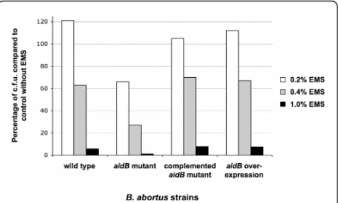
To investigate the putative function of B. abortus AidB protein, we tested the effect of the aidB mutation on the survival during an alkylating stress. A B. abortus 544 strain with a disrupted aidB gene was constructed (XDB1121 strain). An aidB overexpression strain was constructed by inserting a medium-copy plasmid (pDD003) bearing the aidB coding sequence in B. abor-tus, generating the XDB1122 strain. The disruption and overexpression strains (XDB1121 and XDB1122, respec-tively) were analyzed for their sensitivity to the alkylat-ing agent EMS. In summary, the parental strain, the disruption strain (XDB1121), the overexpression strain (XDB1122) and the complemented strain (XDB1127) were incubated in 2YT medium with 0.2, 0.4 and 1.0% EMS for 4 h at 37°C. The alkylating agent was then removed, and serial dilutions of the cultures were plated on 2YT agar. The number of colony forming units (c.f. u.) was determined and the percentage of survival after treatment was expressed by comparison to a culture of these different strains without EMS. A representative result is shown in Figure 1. After exposure to EMS (0.2 to 1%), the disruption strain (XDB1121) was more sensi-tive than the parental strain and the disruption strain
complemented with a low copy plasmid carrying the aidB-yfp fusion (XDB1127). This data was confirmed by reconstructing three independent B. abortus aidB mutants that were more sensitive than the wild-type strain to the presence of 0.4% EMS for 4 h. Indeed, we observed 10.2% ± 2.0 survival for the aidB mutants (n = 3), compared to 62% survival for the wild-type strain. This phenotype was complemented for the three strains, since we observed 61.3% ± 9.1 survival after 4 h in 0.4% EMS for the three aidB mutants complemented with the pDD001 plasmid (Table 1). In order to confirm that aidB mutant was more sensitive to alkylating agents and not just EMS, we also tested the sensitivity of the aidB mutant and wild type strain to methyl methanesulfonate (MMS), another alkylating agent. After 4 h of incubation with 0.02% MMS in rich med-ium, 45% of survival was obtained for the wild type strain, while only 2.1% of the aidB mutants survived, according to c.f.u. counting. Altogether, these experi-ments indicate that the B. abortus aidB gene is probably involved in the repair or the prevention of alkylation damage, as suggested by its homology with E. coli AidB. It also indicates that AidB remains active when it is fused to YFP.
AidB-YFP is localized at the new pole, and at the constriction site in dividing cells
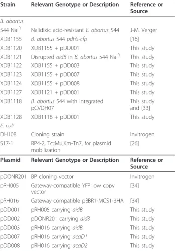
The localization of the AidB-YFP fusion protein was analyzed in a B. abortus strain carrying a chromosomal pdhS-mCherry fusion (XDB1128 strain) during the expo-nential growth phase in liquid cultures, in rich medium (Figure 2A). In these conditions, the localization of the AidB-YFP fusion protein displayed three patterns, Figure 1 The B. abortus aidB mutant is more sensitive to EMS. The sensitivity of B. abortus wild-type, aidB mutant strain,
complemented aidB mutant and aidB overexpression strains was scored by counting the c.f.u. recovered after 4 h of incubation 2YT medium at 37°C, in the presence of 0.2, 0.4 or 1% EMS. The results are expressed as the percentage of c.f.u. compared to a control in which EMS was omitted. Bacteria were obtained from cultures stopped during exponential growth phase.
depending on the presence or the absence of a constric-tion site. In bacteria without detectable constricconstric-tion, AidB-YFP localized at the new pole and PdhS-mCherry at the old pole in 66% of the bacteria (n = 125), with 34% of bacteria labelled only with polar AidB-YFP and not PdhS-mCherry. In the bacteria displaying a constric-tion site, 65% (n = 84) displayed a single AidB-YFP focus at the constriction site, while the remaining 35% have two foci of AidB-YFP, one at the“young” pole and one at the constriction site. Here we define a “young” pole as a new pole that is becoming old, because bac-teria show a detectable constriction, meaning that there is uncertainty about the completion of cytokinesis, and therefore uncertainty about the status of this pole (either new or old). We never observed the PdhS-mCherry and AidB-YFP fusions at the same pole (n = 256) (Figure 2A). Western blots analysis using an anti-GFP antibody on this strain suggested that AidB-YFP fusion was stable when it was produced from the low-copy plasmid pDD001 (data not shown). As proposed in the model depicted in the discussion, the cells labelled with polar AidB-YFP without polar PdhS-mCherry could
correspond to bacteria produced by division of cells car-rying PdhS-mCherry at the old pole and AidB-YFP at the constriction site. Indeed, after cell division, one of the two cells does not inherit PdhS-mCherry from the mother cell, but AidB-YFP at the constriction site is proposed to be transmitted to the new pole of this daughter cell.
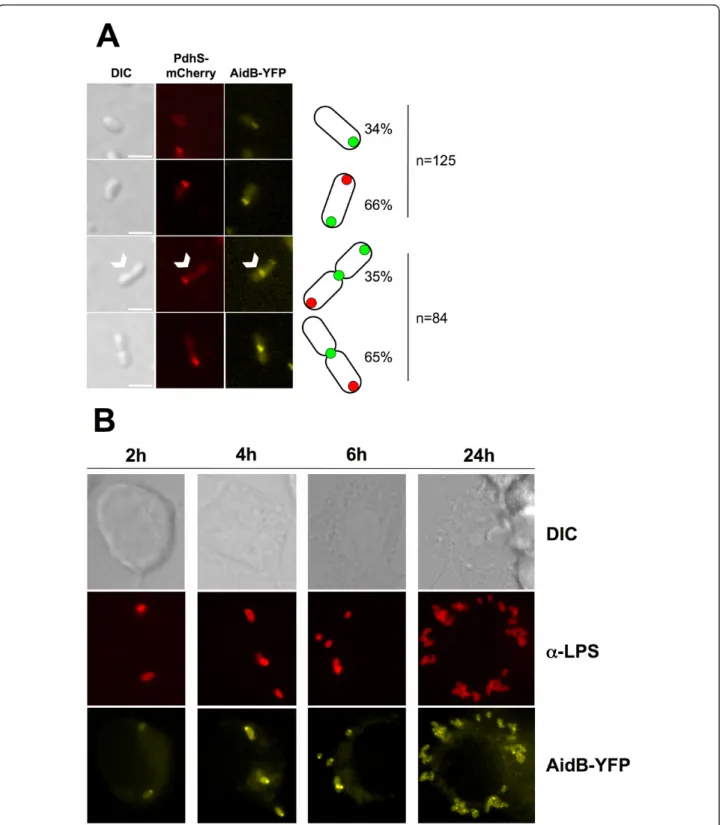
Interestingly, the cells harbouring the two AidB-YFP foci are significantly (p < 0.005) smaller (1.93 μm on average) than the bacteria having a single focus of AidB-YFP at the constriction site (2.08 μm on average), sug-gesting that in the cell cycle, bacteria with 2 foci precede those with a single focus at the constriction site (Figure 3A). This feature of the cell cycle is depicted in the discussion.
Furthermore, the localization of AidB-YFP is still at the new pole after 4 h of exposure with 0.4% EMS (80% of the bacteria exhibited PdhS-mCherry at one pole and AidB-YFP at the opposite pole, n = 237). This observa-tion indicated that AidB-YFP is not released from the new pole in the presence of an alkylating stress with EMS, further suggesting that AidB is active at the new pole, because in these conditions an aidB mutant is killed. Interestingly, bacteria exposed to EMS displayed detectable constriction at the much less frequency (2 constrictions observed among 254 bacteria) compared to the untreated control (44 constrictions observed among 254 bacteria). Moreover, bacteria treated with 0.4% EMS for 4 h and were significantly (p < 0.001) longer on average than unconstricted bacteria that were not exposed to EMS (Figure 3B). This suggests that growth is not arrested by the presence of EMS, while constric-tion is clearly inhibited. This is consistent with a replica-tion arrest caused by alkylareplica-tion of the bacterial genome, as previously reported for E. coli [22].
AidB polar localization persists inside host cells
B. abortus is an intracellular pathogen that encounters various stresses during its life cycle [9]. Since these stresses could result in the alkylation of DNA, e.g. through nitrosative stress [14], we tested the localization pattern of AidB-YFP in B. abortus (XDB1120 strain) during an infection of human epithelial cells (HeLa cells). At 6 h and 24 h post-infection, infected cells were fixed, and bacteria were detected using a monoclonal anti-lipopolysaccharide antibody. The polar foci of AidB-YFP were similar to those observed in bacteriolo-gical culture, suggesting that in these conditions, there is no systematic delocalization of AidB-YFP. Similar results were also obtained with XDB1120 strain in RAW264.7 macrophage infection. At 2 h, 4 h, 6 h and 24 h post-infection, AidB-YFP fusion proteins were still polar (Figure 2B).
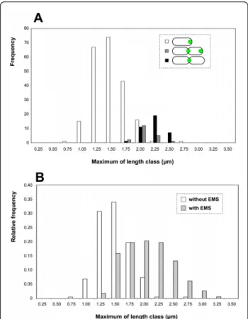
Table 1 Strains and plasmids
Strain Relevant Genotype or Description Reference or Source B. abortus
544 NalR Nalidixic acid-resistant B. abortus 544 J-M. Verger XDB1155 B. abortus 544 pdhS-cfp [16] XDB1120 XDB1155 + pDD001 This study XDB1121 Disrupted aidB in B. abortus 544 NalR This study
XDB1122 XDB1155 + pDD003 This study XDB1123 XDB1155 + pDD007 This study XDB1124 XDB1155 + pDD008 This study XDB1127 XDB1121 + pDD001 This study XDB1118 B. abortus 544 with integrated
pCVDH07
This study and [33] XDB1128 XDB1118 + pDD001 This study E. coli
DH10B Cloning strain Invitrogen S17-1 RP4-2, Tc::Mu,Km-Tn7, for plasmid
mobilization
[26] Plasmid Relevant Genotype or Description Reference or
Source pDONR201 BP cloning vector Invitrogen pRH005 Gateway-compatible YFP low copy
vector
[34] pRH016 Gateway-compatible pBBR1-MCS1-3HA [34] pDD001 pRH005 carrying aidB This study pDD002 pDONR201 carrying aidB This study pDD003 pRH016 carrying aidB This study pDD007 pRH016 carrying acaD1 This study pDD008 pRH016 carrying acaD2 This study
Figure 2 The B. abortus AidB-YFP is localized at new poles and at constriction sites, in culture and in macrophages. The B. abortus XDB1128 strain was carrying an aidB-yfp fusion on a low copy plasmid, and pdhS-mCherry at the pdhS chromosomal locus. (A) Bacteria were grown in rich medium and the pictures were taken in exponential phase. Differential interference contrast (DIC) is shown on the left. The white arrowheads indicate the dividing cell in which two AidB-YFP foci are detectable. Each scale bar represents 2μm. The bacterial types are schematically drawn on the right side of the pictures, as they are represented in figure 6. The two upper panels were made with non-diving bacteria, and counting was made with 125 bacteria. The two lower panels were made with dividing bacteria, and counting was made on 84 dividing bacteria. (B) RAW264.7 macrophages were infected for 2, 4, 6, or 24 h with the B. abortus strain expressing aidB-yfp (XDB1120). The infected cells were fixed and immunostained with 12G12 anti-lipopolysaccharide ("a-LPS”) primary antibody and anti-mouse secondary antibody coupled to Texas Red. A majority of the bacteria present a single focus of AidB-YFP, suggesting that polar localization of this fusion is also occurring during the infection of macrophages.
Morphological analysis of aidB disruption and overexpression mutants
Since AidB-YFP is mainly polar, we tested whether either a disruption or an overexpression of the aidB gene affects growth, bacterial morphology, and virulence in cellular models of infection. The growth curve of an aidB mutant (XDB1121) strain was similar to the wild-type control in 2YT medium (Figure 4). The aidB mutant strain (XDB1121) was morphologically indistin-guishable from the wild-type strain (data not shown and Figure 5). The localization AidB-YFP fusion protein (expressed from pDD001) was similar in the aidB mutant compared to the wild-type strain (data not
shown), suggesting that polar localization of AidB-YFP does not depend on the presence of endogenous AidB, not fused to YFP. The virulence of the aidB mutant in HeLa cells and RAW264.7 macrophages was also similar to the wild-type strain (data not shown and Additional file 3). In summary, the aidB gene seems to be dispensa-ble for growth in bacteriological medium, maintenance of cell shape and for B. abortus virulence in a cellular model of infection.
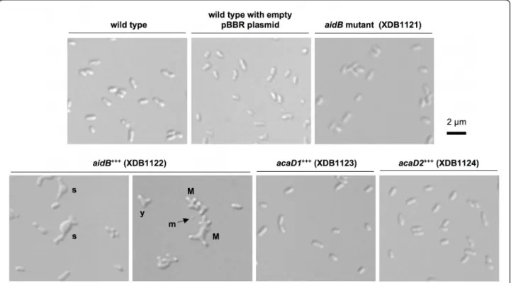
The growth curve of the strain expressing aidB-yfp (XDB1120) in rich media (2YT or tryptic soy broth) is abnormal compared to the wild-type strain (Figure 4). Indeed, the OD during the stationary phase is lower OD with the XDB1120 strain compared to the wild-type control. In stationary phase in 2YT medium, the OD of the aidB overexpression strain is even decreasing while the OD of the wild-type control remained stable. This correlates with a higher frequency of dead cells in the aidB overexpression strain XDB1122 (22.8% in station-ary phase, n = 400) compared to the wild-type strain (5.2% dead cells, n = 400) or the wild-type strain with an empty pBBR1 plasmid (6.7% dead cells, n = 400), the backbone of the aidB overexpression plasmid in XDB1122 strain. This observation suggests that aidB overexpression is partially lethal in stationary phase. In stationary phase cultures of the XDB1120 strain, the bacteria display abnormal morphologies at much higher frequency (22%; n = 200) than the wild-type strain (< 1%; n = 200). This phenotype is probably due to the overproduction of AidB-YFP because the aidB overex-pression strain (XDB1122) displayed similar morphologi-cal defects (61%; n = 200) (Figure 5). Among these abnormal morphologies, bacteria with multipolar shapes were very frequent, swollen cells were often observed, as well as Y-shaped bacteria, elongated cells and minicells. The morphological phenotype of this strain is thus pleiotropic. The analysis of AidB-YFP and PdhS-CFP localization in XDB1120 bacteria with aberrant morphologies, during the exponential growth phase, did not yield a systematic localization pattern, the AidB-YFP and PdhS-CFP fusions being often diffuse in the bacter-ium (data not shown).
Subcellular localization and overproduction effects of AidB are specific to this acyl-CoA dehydrogenase homolog
Since AidB is a member of the 8 ACADs paralogs, we wondered if the particular localization of AidB-YFP and the presence of multipolar forms for the aidB overex-pression mutant were specific characteristics of this ACAD homolog. We chose two B. abortus ACAD homologs that are stably produced at a detectable level using Western blot (data not shown). Both paralogs were annotated (BAB2_0433 and BAB2_0216, Figure 3 Size distribution of B. abortus carrying AidB-YFP, in
the presence or absence of an alkylating agent (EMS). The bacterial lengths were grouped in classes of 0.25μm, and the maximum value for each class is given on the × axis. (A) Size distribution of 276 bacteria (XDB1128 strain) with AidB-YFP either at the new pole (white), the new pole and the constriction site (dark grey), or the constriction site only (black). (B) Size distribution of B. abortus (XDB1128 strain) exposed to 0.4% of EMS for 4 h (light grey, n = 340) or the unexposed control (white, n = 218, bacteria without detectable constriction). (C) DIC and fluorescence pictures of the XDB1128 strain expressing aidB-yfp and pdhS-mCherry fusions, as described in figure 2. The bacteria in the lower panels have been exposed to 0.4% EMS for 4 h in rich (2YT) medium. On the top panels, control bacteria were incubated for 4 h in 2YT in the absence of EMS. Constriction sites are indicated by arrowheads. Each scale bar represents 2μm.
respectively named AcaD1 and AcaD2) as ACADs and would be involved in the fatty acidb-oxidation pathway. We observed that both ACADs homologs had a diffuse localization in the cytoplasm when fused to YFP (XDB1123 and XDB1124 strains, data not shown),
suggesting that the particular localization of AidB-YFP (at young poles and at the constriction site in dividing cells) is not a common characteristic shared by all ACADs homologs in B. abortus. The phenotype of the strains overproducing one of these two ACADs Figure 4 Growth defect of the B. abortus strain expressing the aidB-yfp fusion (XDB1120). The growth of B. abortus wild-type, aidB mutant and XDB1120 (pMR-aidB-yfp) strains was followed by recording OD at 600 nm in a Bioscreen. Duplicates (1) and (2) are shown for each strain, for 2YT (left panel) or tryptic soy broth (right panel) as culture media. In both culture media, the OD600during stationary culture phase of the
XDB1120 strain is lower compared to the wild type control.
Figure 5 Morphological defect of the B. abortus aidB overexpressing strain. Differential interference contrast (DIC) images were taken with bacteria of the aidB (aidB+++), acaD1 (acaD1+++) and acaD2 (acaD2+++) overexpression strains, the aidB disruption strain, and the wild-type strain with or without the control pBBR1MCS plasmid [32], without insert. Two panels are shown for the aidB overexpression strain, the only strain displaying a morphological defect during stationary culture phase. The morphological defects are multiple, with multipolar bacteria (M), Y-shaped cells (y), swollen cells (s) and some minicells (m).
homologs is similar to the B. abortus pdhS-cfp control strain (Figure 5), with a very low frequency (< 1%) of morphological defects. This suggests that overexpression of any ACAD gene does not produce a morphological defect in B. abortus, further supporting a specific -although probably indirect- role of aidB in events related to morphogenesis.
Discussion
The screen of the ORFeome for polar localization allowed the identification of AidB, that shows a clear localization pattern at new poles as well as at constric-tion sites and/or at young pole in cells in division in bacteriological medium. The polar localization of AidB-YFP is preserved in HeLa cells and RAW264.7 macro-phages at different times post-infection. We therefore propose that AidB is a marker of new poles and con-striction sites. To the best of our knowledge, it is the first time that a particular subcellular localization is described for one of the actors involved in the alkylation damage repair. Interestingly, the constriction site corre-sponds to the location of the future new poles just after completion of cell division. We therefore propose a model (Figure 6) in which AidB-YFP is not only loca-lized at the new pole, but also at the constriction site in dividing cells, a mechanism by which AidB-YFP would be ideally localized for a localization at the new pole in
newly formed sibling cells. This model implies that when new poles mature to old poles, after cell division, they are no longer labelled with AidB-YFP (Figure 6).
In the conditions tested, overexpression of aidB leads to bacteria with aberrant morphology (Figure 5). This could be due to defects in cell division, cell growth or coordination between both. One hypothesis would be that AidB could indirectly contribute to the generation of new poles, and overexpression of aidB would result in the generation of additional new poles, forming bac-teria with abnormal morphology, e.g. multipolar shapes (Figure 5). The selective advantage of the polar localiza-tion of AidB is unknown, but it could be related to its role in the adaptative response to alkylating agents, sug-gested here to block cell cycle before cell division (Fig-ure 3B). This would be consistent with a role of AidB in limiting alkylating damage to DNA, which would logi-cally block replication initiation and/or progression.
The B. abortus AidB protein has a high level of iden-tity (42%) to E. coli AidB, suggesting functional conser-vation between the two proteins. This prediction is supported by the increased sensitivity of the B. abortus aidB mutant strain to the alkylating agent EMS com-pared to the wild-type control (Figure 1). Brucella gen-omes contain the ada, alkA and alkB genes necessary for an adaptative response to alkylation damage similar to the one reported for E. coli [11]. We propose that one possible function of AidB would be to help in the detoxification of some alkylating agents, like in E. coli. These alkylating agents could be found at particular stages of the infection in the natural hosts, or generated by the bacteria themselves [14]. The absence of attenua-tion of the aidB mutant in HeLa cells or in RAW264.7 macrophages suggests that such alkylating agents are not crucial for the control of the number of c.f.u. during infection of these cell lines. Our data do not confirm the previous observation that a transpositional aidB mutant was attenuated in THP-1 macrophages [10], unless these specific macrophages have specific features differentiating them from RAW264.7 macrophages for the generation of an alkylating stress. In Salmonella enterica, an aidB mutant was more sensitive than the wild-type strain to several alkylating agents but pre-sented no effect on the virulence in the mouse model. Indeed, the virulence of a S. enterica mutant defective in all genes specifically involved in DNA alkylation damage repair was not affected [23].
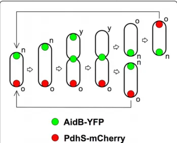
Recently, in C. crescentus, Radhakrishnan et al. reported that KidO, an NAD(P)-binding oxidoreductase homolog with conserved residues in its NAD(P)-binding pocket, acts directly on the FtsZ tubulin [24]. Localiza-tion of KidO to the Z-ring is disrupted by mutaLocaliza-tions in the cofactor-binding pocket that disturb the association with NAD(P), implying that NAD(P) binding is Figure 6 Model for the localization of AidB-YFP along B.
abortus cell cycle. The PdhS-mCherry is labelling the old pole of B. abortus. AidB-YFP is therefore localized at the new pole, as suggested by Figure 2. In dividing cells, we hypothesize that AidB-YFP is first present at the young pole (the new pole that becomes old) and at the constriction site. This localization at the young pole would be lost afterwards, allowing the generation of two sibling cells with a unique pole of AidB-YFP. The new (n), young (y) and old (o) poles are labelled. In this model, the constriction region would be the preparation site for the new poles of the sibling cells.
important for the recruitment of KidO to the Z-ring [24]. In this context, it should be interesting to con-struct a mutated AidB defective for FAD binding and observe the impact of this mutation on the AidB-YFP localization. Finally, the selective advantage of AidB recruitment at the new pole remains to be discovered. One possibility would be that crucial regions of the nucleoid located close to the new pole, such as replica-tion origins, could be more protected from alkylating agents. This would resemble the proposed specific pro-tection of genes by AidB in E. coli [25] that would be dependent on subcellular localization of AidB in B. abortus. The aberrant morphology of the strain overex-pressing aidB indicates that either growth or division are affected, which suggest that AidB could be (indir-ectly) involved in the control of these processes, for example by providing a checkpoint for cell division.
Conclusion
AidB is induced during alkylation damage response in E. coli, however its molecular function is mostly unknown. Here we report that a B. abortus aidB mutant is more sensitive to EMS, suggesting that AidB is playing a func-tional role in the response to alkylation damage. The AidB-YFP fusion is a marker of new poles (Figures 2 and 6). The AidB-YFP fusion is also localized to con-striction sites, which could be considered as preparation sites for new poles in dividing cells. AidB molecular function at the new pole is unknown, but it is expected to be active at this site, since its new pole localization is preserved in B. abortus exposed to EMS. Our data also suggest that exposure to EMS blocks the cell cycle before the constriction event, presumably before or dur-ing genome replication. The characteristic multipolar morphology of the aidB overexpression strain suggests that AidB could (indirectly) play a role in growth or cell division of B. abortus.
Methods
Strains, plasmids and cell growth
All Brucella strains used in this study (Table 1) were derived from B. abortus 544 NalR (a spontaneous nali-dixic acid-resistant mutant of B. abortus 544 strain), and were routinely cultivated in rich medium 2YT (1% yeast extract, 1.5% tryptone and 0.5% NaCl, with 1.5% agar for solid medium). E. coli strains DH10B (Invitro-gen Life-Technologies) and S17-1 [26] were cultivated in LB broth (0.5% yeast extract, 1% tryptone, 0.5% NaCl) with streptomycin. Antibiotics were used at the following concentrations when appropriate: nalidixic acid, 25μg/ml; kanamycin, 20 μg/ml; chloramphenicol, 20 μg/ml. Plasmids were mobilized from E. coli strain S17-1 into B. abortus as previously described [27]. Growth curves were monitored using a Bioscreen
system (Thermo Fisher, ref. 110001-536), allowing con-tinuous monitoring for growth curves in a multiwell format. B. abortus liquid cultures in 2YT medium with the appropriate antibiotic were centrifuged, washed once with PBS and diluted to an OD600 of 0.1 in 2YT
(or tryptic soy broth) to start the culture in the Biosc-reen system. Each culture (200 μl per well) was per-formed at 37°C.
Control of the B. abortus strain used for the localization screen
The fact that the XDB1155 strain is viable and does not present any apparent morphological defects or growth delay suggests that the CFP fusion at the C-terminal of PdhS is not affecting PdhS essential functions. Control immunoblots with anti-GFP antibodies revealed that this fusion protein was stable (data not shown). Obser-vation using fluorescence microscopy showed that PdhS-CFP accumulated at one pole in more than 90% of the cells as previously described [17].
Molecular techniques
DNA manipulations were performed according to stan-dard techniques [28]. All plasmids used in this study (Table 1) were constructed by the Gateway™ technique (Invitrogen). To construct an aidB disruption mutant strain, a central 380-bp portion of the aidB CDS was amplified by PCR using AcoA and AcoB primers, and was subcloned into at the EcoRV site of pSKoriTkan vector [29]. The recombinant plasmid was transformed into the E. coli strain S17-1 and introduced into B. abor-tus 544 NalRstrain by mating. Clones in which the plas-mid integrated in the genome were selected by growing the bacteria in the presence of kanamycin, and were checked by PCR using AcoDHP1 and pGEM-T-aval pri-mers. Since B. abortus and B. melitensis are nearly iden-tical at the genomic level, entry clones were recovered from the B. melitensis ORFeome version 1.1 [15]. LR recombination cloning procedure was performed as recommended by the manufacturer (Invitrogen Life-Technologies). The sequences of primers are available in Table 2.
Western blotting
For every fluorescent observations reported in this study, we carried out Western blot analyses with antibo-dies against YFP and CFP. These results allowed us to rule out the possibility that a particular localization pat-tern could result from protein degradation or from a deficiency in fusion protein production. Western blot analysis was carried out as described previously [8] with monoclonal antibodies against GFP (JL8, BD Bios-ciences) at 1/1000 dilution to check the stability of translational fusions to YFP or CFP.
Microscopy
For fluorescence imaging, cell populations of B. abortus strains were immobilized on a microscope slide that was layered with a pad of 1% agarose containing phosphate-buffered saline (PBS) [30]. These slides were placed on a microscope stage at room temperature. Samples were observed on a Nikon i80 fluorescence microscope through a differential interference contrast (DIC, Nor-marski) 100X objective with a Hamamatsu Orca-ER LCD camera. Images acquisition and processing were done with NIS element (Nikon) software.
The detection of dead cells was performed with the Live/Dead BacLight kit L7007 (Invitrogen), according to manufacturer instructions.
Treatment of B. abortus strains with a DNA-alkylating agent
B. abortus strains were grown in 2YT at 37°C overnight, centrifuged and the pellet was resuspended in PBS to a cell density of 109 c.f.u./ml (optical density of 0.33 at 600 nm). 500 μl of these cell suspensions were diluted into 5 ml of 2YT and exposed to methanesulphonic acid ethyl ester (EMS) at final concentrations of 0, 0.2, 0.4 and 1.0%. These suspensions were incubated at 37°C with shaking for 1 h or 4 h, and aliquots (1 ml) were recovered, washed once in PBS, and serially diluted in PBS. 100 μl of these cell suspensions were spread on individual 2YT agar plates. These plates were incubated for 72 h at 37°C, and the c.f.u. were enumerated.
Cellular infection and immunofluorescence labelling
Infections and immunofluorescence of HeLa cells and RAW264.7 macrophages by the different B. abortus strains were performed as described previously [6]. Anti-Brucella lipopolysaccharide O-chain monoclonal anti-body 12G12 [31] was used. The secondary antianti-body used was Texas red-conjugated anti-rabbit IgG (Molecu-lar Probes) diluted 500 times.
Additional material
Additional file 1: Sequence alignment between E. coli and B. abortus AidB. Alignment of E. coli and B. abortus AidB highlighting the conserved parts of these enzymes, and the absence of high similarity in the C-terminal portion of these proteins.
Additional file 2: 3D structure of E. coli AidB and 3D model of B. abortus AidB. The 3D model of B. abortus AidB suggests that while regions involved in tetramer formation are conserved, the C-terminal domain involved in DNA binding is not conserved.
Additional file 3: Infection of RAW264.7 macrophages with wild-type and aidB mutants strains. c.f.u. countings during macrophages infection show that aidB mutation or overexpression does not dramatically impair intracellular survival and replication of B. abortus.
List of abbreviations
ACAD: Acyl-CoA dehydrogenase; BCV: Brucella-containing vacuole; CDS: coding sequence; CFP: cyan fluorescent protein; DIC: differential interference contrast; EMS: methanesulphonic acid ethyl ester; ER: endoplasmic reticulum; pCDS: predicted coding sequence; ROS: reactive oxygen species; YFP: yellow fluorescent protein
Acknowledgements and funding
We thank M. Deghelt and C. Van der Henst for critical reading of the manuscript. This work was supported by FRFC (Fonds de la Recherche Fondamentale Collective, conventions n°2.4521.04 and 2.4541.08), by ARC fellowship (Actions de Recherche Concertée, conventions 04/09-325 and 08/ 13-015, French-Speaking Community of Belgium) and by the University of Namur (FUNDP). D. Dotreppe and C. Mullier were holding a Ph.D. fellowship from the FRIA (Fonds pour la formation à la Recherche dans l’Industrie et dans l’Agriculture).
Authors’ contributions
DD made all experiments, except macrophages infections reported in Figure 2B, that were performed by CM. JJL and XDB participated to the design of the work. DD and XDB wrote the manuscript. All authors read and approved the final manuscript.
Received: 25 May 2011 Accepted: 23 November 2011 Published: 23 November 2011
References
1. Boschiroli ML, Foulongne V, O’Callaghan D: Brucellosis: a worldwide zoonosis. Curr Opin Microbiol 2001, 4:58-64.
2. Gorvel JP, Moreno E: Brucella intracellular life: from invasion to intracellular replication. Vet Microbiol 2002, 90:281-297.
3. Arenas GN, Staskevich AS, Aballay A, Mayorga LS: Intracellular trafficking of Brucella abortus in J774 macrophages. Infect Immun 2000, 68:4255-4263. 4. Pizarro-Cerda J, Meresse S, Parton RG, van der Goot G, Sola-Landa A,
Lopez-Goni I, Moreno E, Gorvel JP: Brucella abortus transits through the autophagic pathway and replicates in the endoplasmic reticulum of nonprofessional phagocytes. Infect Immun 1998, 66:5711-5724. 5. Pizarro-Cerda J, Moreno E, Sanguedolce V, Mege JL, Gorvel JP: Virulent
Brucella abortus prevents lysosome fusion and is distributed within autophagosome-like compartments. Infect Immun 1998, 66:2387-2392. 6. Delrue RM, Martinez-Lorenzo M, Lestrate P, Danese I, Bielarz V, Mertens P,
De Bolle X, Tibor A, Gorvel JP, Letesson JJ: Identification of Brucella spp. genes involved in intracellular trafficking. Cell Microbiol 2001, 3:487-497. 7. Starr T, Ng TW, Wehrly TD, Knodler LA, Celli J: Brucella intracellular
replication requires trafficking through the late endosomal/lysosomal compartment. Traffic 2008, 9:678-694.
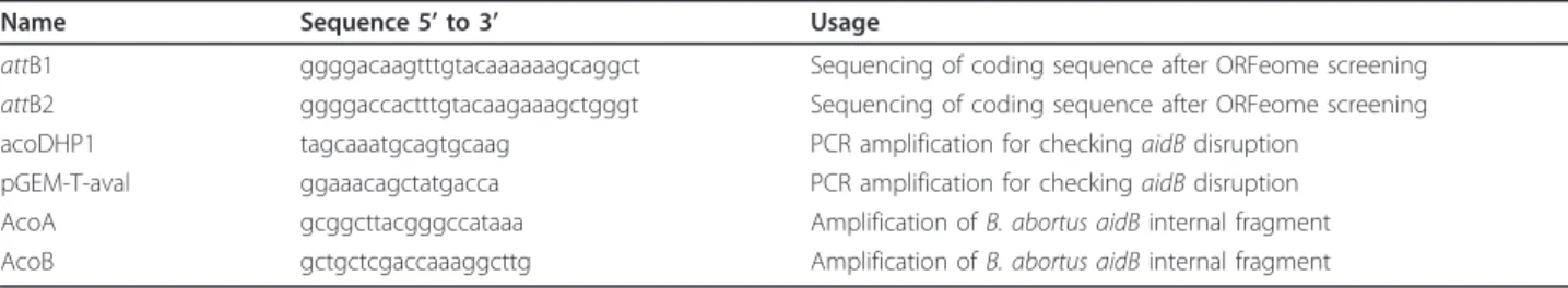
Table 2 Primers
Name Sequence 5’ to 3’ Usage
attB1 ggggacaagtttgtacaaaaaagcaggct Sequencing of coding sequence after ORFeome screening attB2 ggggaccactttgtacaagaaagctgggt Sequencing of coding sequence after ORFeome screening acoDHP1 tagcaaatgcagtgcaag PCR amplification for checking aidB disruption
pGEM-T-aval ggaaacagctatgacca PCR amplification for checking aidB disruption AcoA gcggcttacgggccataaa Amplification of B. abortus aidB internal fragment AcoB gctgctcgaccaaaggcttg Amplification of B. abortus aidB internal fragment
8. Dozot M, Boigegrain RA, Delrue RM, Hallez R, Ouahrani-Bettache S, Danese I, Letesson JJ, De Bolle X, Kohler S: The stringent response mediator Rsh is required for Brucella melitensis and Brucella suis virulence, and for expression of the type IV secretion system virB. Cell Microbiol 2006, 8:1791-1802.
9. Roop RM, Gaines JM, Anderson ES, Caswell CC, Martin DW: Survival of the fittest: how Brucella strains adapt to their intracellular niche in the host. Med Microbiol Immunol 2009, 198:221-238.
10. Kohler S, Foulongne V, Ouahrani-Bettache S, Bourg G, Teyssier J, Ramuz M, Liautard JP: The analysis of the intramacrophagic virulome of Brucella suis deciphers the environment encountered by the pathogen inside the macrophage host cell. Proc Natl Acad Sci USA 2002, 99:15711-15716. 11. Volkert MR, Nguyen DC: Induction of specific Escherichia coli genes by
sublethal treatments with alkylating agents. Proc Natl Acad Sci USA 1984, 81:4110-4114.
12. Nakabeppu Y, Kondo H, Sekiguchi M: Cloning and characterization of the alkA gene of Escherichia coli that encodes 3-methyladenine DNA glycosylase II. J Biol Chem 1984, 259:13723-13729.
13. Yamamoto Y, Katsuki M, Sekiguchi M, Otsuji N: Escherichia coli gene that controls sensitivity to alkylating agents. J Bacteriol 1978, 135:144-152. 14. Taverna P, Sedgwick B: Generation of an endogenous DNA-methylating
agent by nitrosation in Escherichia coli. J Bacteriol 1996, 178:5105-5111. 15. Dricot A, Rual JF, Lamesch P, Bertin N, Dupuy D, Hao T, Lambert C, Hallez R,
Delroisse JM, Vandenhaute J, et al: Generation of the Brucella melitensis ORFeome version 1.1. Genome Res 2004, 14:2201-2206.
16. Mignolet J, Van der Henst C, Nicolas C, Deghelt M, Dotreppe D, Letesson JJ, De Bolle X: PdhS, an old-pole-localized histidine kinase, recruits the fumarase FumC in Brucella abortus. J Bacteriol 2010, 192:3235-3239. 17. Hallez R, Mignolet J, Van Mullem V, Wery M, Vandenhaute J, Letesson JJ,
Jacobs-Wagner C, De Bolle X: The asymmetric distribution of the essential histidine kinase PdhS indicates a differentiation event in Brucella abortus. EMBO J 2007, 26:1444-1455.
18. Bowles T, Metz AH, O’Quin J, Wawrzak Z, Eichman BF: Structure and DNA binding of alkylation response protein AidB. Proc Natl Acad Sci USA 2008, 105:15299-15304.
19. Rippa V, Amoresano A, Esposito C, Landini P, Volkert M, Duilio A: Specific DNA binding and regulation of its own expression by the AidB protein in Escherichia coli. J Bacteriol 2010, 192:6136-6142.
20. Sedgwick B: Repairing DNA-methylation damage. Nat Rev Mol Cell Biol 2004, 5:148-157.
21. Volkert MR: Adaptive response of Escherichia coli to alkylation damage. Environ Mol Mutagen 1988, 11:241-255.
22. Lawley PD, Brookes P: Cytotoxicity of alkylating agents towards sensitive and resistant strains of Escherichia coli in relation to extent and mode of alkylation of cellular macromolecules and repair of alkylation lesions in deoxyribonucleic acids. Biochem J 1968, 109:433-447.
23. Alvarez G, Campoy S, Spricigo DA, Teixido L, Cortes P, Barbe J: Relevance of DNA alkylation damage repair systems in Salmonella enterica virulence. J Bacteriol 2010, 192:2006-2008.
24. Radhakrishnan SK, Pritchard S, Viollier PH: Coupling prokaryotic cell fate and division control with a bifunctional and oscillating oxidoreductase homolog. Dev Cell 2010, 18:90-101.
25. Rippa V, Duilio A, di Pasquale P, Amoresano A, Landini P, Volkert MR: Preferential DNA damage prevention by the E. coli AidB gene: A new mechanism for the protection of specific genes. DNA Repair (Amst) 2011, 10:934-941.
26. Simon R, Priefer U, Pühler A: A broad host range mobilisation system for in vivo genetic engineering: transposon mutagenesis in gram negative bacteria. Biotechnology 1983, 10:783-791.
27. Ely B: Genetics of Caulobacter crescentus. Methods Enzymol 1991, 204:372-384.
28. Ausubel FM: Current Protocols in Molecular Biology New York: John Wiley & Sons.; 1989.
29. Haine V, Sinon A, Van Steen F, Rousseau S, Dozot M, Lestrate P, Lambert C, Letesson JJ, De Bolle X: Systematic targeted mutagenesis of Brucella melitensis 16M reveals a major role for GntR regulators in the control of virulence. Infect Immun 2005, 73:5578-5586.
30. Jacobs C, Domian IJ, Maddock JR, Shapiro L: Cell cycle-dependent polar localization of an essential bacterial histidine kinase that controls DNA replication and cell division. Cell 1999, 97:111-120.
31. Cloeckaert A, Jacques I, Bowden RA, Dubray G, Limet JN: Monoclonal antibodies to Brucella rough lipopolysaccharide: characterization and evaluation of their protective effect against B. abortus. Res Microbiol 1993, 144:475-484.
32. Kovach ME, Phillips RW, Elzer PH, Roop RM, Peterson KM: pBBR1MCS: a broad-host-range cloning vector. Biotechniques 1994, 16:800-802. 33. Van der Henst C, Charlier C, Deghelt M, Wouters J, Matroule JY, Letesson JJ,
De Bolle X: Overproduced Brucella abortus PdhS-mCherry forms soluble aggregates in Escherichia coli, partially associating with mobile foci of IbpA-YFP. BMC Microbiol 2010, 10:248.
34. Hallez R, Letesson JJ, Vandenhaute J, De Bolle X: Gateway-based destination vectors for functional analyses of bacterial ORFeomes: application to the Min system in Brucella abortus. Appl Environ Microbiol 2007, 73:1375-1379.
doi:10.1186/1471-2180-11-257
Cite this article as: Dotreppe et al.: The alkylation response protein AidB is localized at the new poles and constriction sites in Brucella abortus. BMC Microbiology 2011 11:257.
Submit your next manuscript to BioMed Central and take full advantage of:
• Convenient online submission
• Thorough peer review
• No space constraints or color figure charges
• Immediate publication on acceptance
• Inclusion in PubMed, CAS, Scopus and Google Scholar
• Research which is freely available for redistribution
Submit your manuscript at www.biomedcentral.com/submit