RESEARCH OUTPUTS / RÉSULTATS DE RECHERCHE
Author(s) - Auteur(s) :
Publication date - Date de publication :
Permanent link - Permalien :
Rights / License - Licence de droit d’auteur :
Bibliothèque Universitaire Moretus Plantin
Institutional Repository - Research Portal
Dépôt Institutionnel - Portail de la Recherche
researchportal.unamur.be
University of NamurStudy by the sum-frequency generation spectroscopy of a model biosensor : The biocytin-avidin complex
Sartenaer, Yannick; Dreesen, Laurent; Humbert, Christophe; Mani, Alaa Addin; Lemaire, Jean-Jacques; Salmain, Michelle; Pradier, Claire-Marie; Thiry, Paul; Peremans, André Published in:
Physicalia Magazine
Publication date:
2003
Document Version
Peer reviewed version
Link to publication
Citation for pulished version (HARVARD):
Sartenaer, Y, Dreesen, L, Humbert, C, Mani, AA, Lemaire, J-J, Salmain, M, Pradier, C-M, Thiry, P & Peremans, A 2003, 'Study by the sum-frequency generation spectroscopy of a model biosensor : The biocytin-avidin complex', Physicalia Magazine, vol. 25, no. 3, pp. 221-229.
General rights
Copyright and moral rights for the publications made accessible in the public portal are retained by the authors and/or other copyright owners and it is a condition of accessing publications that users recognise and abide by the legal requirements associated with these rights. • Users may download and print one copy of any publication from the public portal for the purpose of private study or research. • You may not further distribute the material or use it for any profit-making activity or commercial gain
• You may freely distribute the URL identifying the publication in the public portal ?
Take down policy
If you believe that this document breaches copyright please contact us providing details, and we will remove access to the work immediately and investigate your claim.
Study by sum-frequency generation spectroscopy
of a model biosensor :
The biocytin-avidin complex
Y. Sartenaer, L. Dreesen, C. Humbert, A. A. Mani, J.-J. Lemaire, M. Salmain*, C.-M. Pradier*, P. A. Thiry and A. Peremans
Laboratoire de Spectroscopie Moléculaire de Surface, FUNDP, B-5000 Namur, Belgium
Abstract
Infrared-visible sum-frequency generation (SFG) spectroscopy is used to study the recognition of a protein (avidin) by a derivated vitamin (biocytin). No interaction is observed when the measurements are performed on metallic substrates in the normal co-propagative configuration. When the biocytin layer is deposited on a CaF2
substrate and the SFG measurements are performed in the total internal reflection configuration, drastic modifications in the C-H and N-H spectral ranges attest the molecular recognition of avidin by biocytin. The specificity of the recognition process is confirmed by replacing avidin by other proteins.
Introduction
The general scheme of a biosensor is a monolayer of probe molecules adsorbed on a substrate and capable of recognizing target molecules in some specific way. Such devices, which become more and more popular in modern biotechnology, need a physical technique in order to measure the recognition process. Up to now, atomic force microscopy (AFM) [1][2], visible-ultraviolet absorption spectroscopy [3][4] and surface plasmon resonance [5][6] have been
used. Recently, Fourier transform infrared reflection absorption spectroscopy (FT-IRRAS) [7][8] allowed to identify the recognized species through their vibrational signature, with the help of IR-markers.
In order to obtain similar vibrational information without the use of markers, we apply for the first time, sum-frequency generation (SFG) spectroscopy [9] to the characterization of such biosensors. This technique has been successfully applied to study the conformational modifications of a biopolymer film due to water [10], to investigate protein adsorption at interfaces involving water [11], to distinguish surface polymer species from bulk ones [12] or to probe the surface melting of ice [13].
In the present work, we choose to study the ligand-receptor biocytin-avidin complex. Avidin is a tetrameric protein (63,200 Da) that presents 4 binding sites specific to biotin, a vitamin (H) whose chemical formula is C5H7N2OS-(CH2)4-COOH.
Biocytin [C5H7N2OS-(CH2)4-CONH-(CH2)4-CHNH2-COOH] is the product of the
bonding of biotin to a lysine residue (C6H14N2O2). This system has been intensively
studied in the litterature [1]-[8][14][15][16] because avidin binds to biotin with one of the most important affinities known in biology (dissociation constant Kd ~10-15 M) [17].
In the following sections, we proceed in three steps. First, we characterize the formation of the biocytin probe layer. Second, we study its interaction with avidin. Third, we check its binding specificity by immersing biocytin in two other protein solutions. Metallic (Ag, Au, Pt) and insulating (CaF2) substrates are used.
Experimental section
Infrared-visible sum-frequency generation (SFG) spectroscopy is an optical spectroscopic technique which uses two laser beams, a visible one and an infrared one, that are focused on the sample surface where a non-linear optical process of the second order generates a third beam at the sum-frequency. Within the electric-dipole approximation, this process is forbidden in the bulk of centrosymmetric media but not at their surface, thus making SFG an interface sensitive technique. The vibrational fingerprint of the surface is obtained by recording the SFG signal as a function of the IR frequency, allowing the identification of chemical species [18]. Moreover, SFG is sensitive to the conformational order of the adsorbed monolayer.
A detailed description of the SFG setup used in this work can be found elsewhere [19]. Briefly, we use two optical parametric oscillators (OPO) to generate the two laser beams. The infrared one is built around a LiNbO3 nonlinear crystal and
is pumped by one third of the intensity generated by an all-solid state Nd:YAG pulsed laser. The visible beam is built around a BBO nonlinear crystal and is pumped by the remaining part of the injection laser after frequency tripling. The pump laser operates at a repetition rate of 25 Hz and generates about 100 single pulses (duration ~ 12 ps) per laser burst (duration ~ 1 µs). This device delivers an IR beam tunable between 2.5 and 4.2 µm with an average power of 25 mW (resolution ~ 2 cm-1) and a visible one tunable between 415 and 700 nm with an average power of 15 mW (resolution ~ 3 cm-1). The visible wavelength used in the present work is 568 nm.
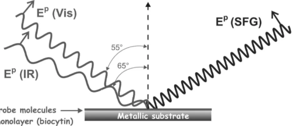
We adopted two different experimental configurations, one for the metallic substrates and the other one for CaF2. In both cases, the laser beams are
p-polarised and focused on the sample with beam diameters of 1 mm and angles versus sample normal of 65° (IR beam) and 55° (visible b eam), respectively. The first configuration is a co-propagative one (Figure 1A) where the two laser beams impinge directly on the metal surface and where the SFG signal is collected after reflection. In the second configuration (Figure 1B) the two beams pass through an equilateral CaF2 prism and are focused at its base. With the same incidence angles mentioned
above, SFG spectroscopy is performed under total internal reflection (TIR) conditions because total reflection at the CaF2/air interface occurs at about 45°. Such a
configuration has been applied previously with success to study the effect of water on a polymer film [10]. In both configurations, the nonlinear signal of a ZnS crystal serves as a reference to normalize the SFG spectra and to compensate the laser power fluctuations and the absorption by the ambient water vapour. The detection chain consists of Raman filters followed by a double-grating monochromator for spatial and spectral filtering and of a photomultiplier tube for the detection of the sum-frequency photons.
As for the sample preparation, all substrates are sonicated in acetone, ethanol and water before use. The metal substrates are Ag(111) and Pt(111) single crystals, and a 200 nm Au film deposited on glass with a 4 nm thick Cr buffer (Arrandee). The CaF2 substrate is a prism of λ/10 flatness (Foctek).
Figure 1 : SFG Experimental configurations used for metallic substrates (A) and for CaF2 (B).
For the Ag(111) and CaF2 substrates, the biocytin films are realized by
immersion into a 10 mM trifluoroethanolic solution of biocytin (Aldrich) during 2h. Biocytinylated thiol is used for Au and Pt(111). All the substrates are intensively rinsed with the solvent and dried under nitrogen flow. After analysis of the biocytin monolayer, the substrates are immersed in a 10-5 M avidin solution (or an other protein for CaF2 experiments) in a HEPES buffer (10-2 M, pH ~ 7,4) for 16 h before
Results and discussion
On one hand, the bonding of biocytin to Ag(111) and CaF2 occurs through the
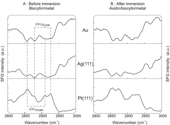
two carboxylic oxygens in a bridging configuration [20]. On the other hand, on Au and Pt(111), the biocytinylated thiol binds to the metals through the thiol group [21]. The SFG spectra of biocytin adsorbed on metallic substrates (Pt, Au, Ag) are shown in figure 2A which displays the C-H spectral range (2800 – 3000 cm-1). The spectrum of biocytin/Pt(111) reveals four resonances at 2860, 2875, 2915 and 2930 cm-1. By comparing with FTIR spectra of biocytin and in keeping with previous SFG works on alkanethiols [21], we assign the two peaks at 2860 and 2915 cm-1 to the symmetric and asymmetric stretching vibrations of methylene groups pertaining to the alkane chain of biocytin. In an “all trans” configuration, those 8 groups are distributed symmetrically along the molecule skeleton and the CH2 vibrations are thus not
expected to be SFG active. Their observation is the indication of a certain number of gauche defects in the biocytin layer. The two peaks at 2875 and 2930 cm-1 are assigned to the stretching vibrations of the single CH2 group pertaining to the ureido
bicycle of the biocytin molecule. In the normal configuration of the adsorbed biocytin, this CH2 group is pointing upward from the surface so that its observation by SFG
attests to the orientation and the self-assembly of the monolayer. In the N-H (3000 - 3500 cm-1) spectral range (data not shown), the SFG spectrum is devoid of any structure.
The same resonances are observed and the same conclusions can be drawn for the Au and Ag(111) substrates. However, as can be seen on Figure 2, SFG resonances become valleys due to interferences with the non-linear response of the metal. This notwithstanding, there exists a difference with respect to Pt(111), i.e. the occurrence of a new spectral feature at 2965 cm-1 which is attributed to a slightly different film order. After immersion of the biocytin films in avidin, the spectra (figure 2) of Au and Ag(111) do not change, which means that there is no significant recognition of the protein. In the case of Pt(111), we observe some slight modifications of the vibrational fingerprint which could be interpreted as a weak interaction between probe and target molecules. However, the observed effects are too weak for claiming molecular recognition between the two biological species. The
probable reason for the absence of change is that the protein layer is destroyed by the laser beam.
Figure 2 : SFG spectra recorded in the C-H vibrational range for Biocytin/Metal interfaces before (A)
and after (B) immersion in an avidin solution.
In order to circumvent this difficulty, a transparent substrate in TIR configuration is preferred because in this case, the interface is probed not by the incident laser beams, but by the evanescent waves resulting from the total reflection. The new setup makes use of CaF2 prisms, optically transparent between 0.2 and 7
µm. Both C-H (2800 - 3000 cm-1) and N-H (3000 - 3400 cm-1) spectral ranges were measured by SFG before and after immersion in avidin. For the biocytin adlayer, the same resonances were observed as on the metallic substrates indicating that a self-assembled oriented layer is formed, with a certain number of gauche defects (figure 3A). After interaction with the avidin solution, significant modifications of the SFG spectrum attest the molecular recognition (figure 3B). First, in the C-H spectral range, the resonances associated to the methylene backbone stretching vibration modes at
2850 and 2915 cm-1 (CH2 chain) disappear which indicates an ordering of the
biocytin layer.
Figure 3 : SFG spectra of (A) Biocytin/CaF2, (B) Avidin/Biocytin/CaF2, (C) Pre-saturated avidin
/Biocytin/CaF2 and (D) BSA/Biocytin/CaF2 interfaces in the C-H and N-H spectral ranges.
Second, two new spectral features are detected in the N-H spectral range : a sharp band at 3055 cm-1 and a broad band centered at 3150 cm-1. These bands are characteristic of amide B and A vibrations respectively. However, they are significantly red shifted from their original values measured in pure avidin at 3070 and 3300 cm-1 respectively. Those modifications attest that the avidin is effectively fixed on the surface and that, as expected, the bonding of those proteins affects the biocytin layer.
In a final step, we checked the biosensor selectivity by substituting avidin with bovine serum albumin (BSA or bovidin) (figure 3D) and with avidin previously saturated with biotin (figure 3C). BSA is a protein with no affinity for biotin but with a molecular weight and a isoelectric point similar to the avidin ones. As for pre-saturated avidin, the four protein binding sites are occupied by biotin making them
insensitive to further interaction. In those cases, no significant modification of the SFG spectra is observed in comparison to the pure biocytin layer, demonstrating the specific binding between biocytin and pure avidin.
Conclusion
By confronting SFG spectroscopy with the biocytin/avidin system as a “model biosensor”, we have shown that the SFG spectroscopy is capable of detecting the specific binding between molecular species, which is the basis of molecular recognition.
Acknowledgment
This work is supported by the Ministry of the Walloon Region (Belgium), by the Belgian Fund for Scientific Research (FRFC) and by the Belgian Interuniversity Research Program on "Quantum Size Effects in Nanostructured Materials" PAI/IUAP 5/1 initiated by the Belgian Office for Scientific, Technical and Cultural Affairs (OSTC). A.P. is senior research associate of the Belgian Fund for Scientific Research.
References
[1] Y.-S. Lo, N. D. Huefner, W. S. Chan, F. Steven, J.M. Harris and T. P. Beebe Jr., Langmuir 15, 1373 (1999)
[2] R. De Paris, T. Struntz, K. Orozlan, H.-J. Güntherodt and M. Hegner, Single Mol.
4, 285 (2000)
[3] N. Lala, A. G. Chittiboyina, S. P. Chavan and M. Sastry, Coll. and Surf. A 205, 15 (2002)
[4] M. Sastry, N. Lala, V. Patil, S. P. Chavan and A. G. Chittiboyina, Langmuir 14, 4138 (1998)
[5] L. Häusling, H. Ringsdorf, F.-J. Schmitt and W. Knoll, Langmuir 7, 1837 (1991) [6] S. Roy, J.-H. Kim, J. T. Kellis Jr., A. J. Poulose, C. H. Robertson and A. P. Gast, Langmuir 18, 6319 (2002)
[7] C.-M. Yam, C.-M. Pradier, M. Salmain, N. Fischer-Durand and G. J. Jaouen, J. Coll. And Int. Sci. 244, 1 (2001)
[8] C.-M. Pradier, M. Salmain, L. Zheng and G. Jaouen, Surf. Sci. 502-503, 193 (2002)
[9] Y. R. Shen, Nature 337, 519 (1989)
[10] L. Dreesen, C. Humbert, P. Hollander, A. A. Mani, K. Ataka, P. A. Thiry and A. Peremans, Chem. Phys. Lett. 333, 327 (2001)
[11] G. Kim, M. Gurau, J. Kim and P. S. Cremer, Langmuir 18, 2807 (2002)
[12] D. Zhang, Y. R. Shen and G. A. Somorjai, Chem. Phys. Lett. 281, 394 (1997) [13] W. Wei, P. B. Miranda and Y. R. Shen, Phys. Rev. Lett. 86, 1554 (2001) [14] Y. Hiller, E. A. Bayer and M. Wilchek, Biochem. J. 278, 573 (1991) [15] R. B. Honzatko and R.W. Williams, Biochem. 21, 6201 (1982)
[16] L. Pugliese, A. Coda, M. Malcovati and M. Bolognesi, J. Mol. Biol. 231, 698 (1993)
[17] P. C. Weber, D. H. Ohlendorf, J. J. Wendoloski and F. R. Salemme, Science
243, 85 (1989)
[18] Y. R. Shen, The principles of Nonlinear Optics, Wiley: New-York (1984)
[19] A. A. Mani, L. Dreesen, C. Humbert, P. Hollander, Y. Caudano, P. A. Thiry and A. Peremans, Surf. Sci. 502-503, 261 (2002).
[20] L. Dreesen, Y. Sartenaer, C. Humbert, A.A. Mani, J.-J. Lemaire, C. Méthivier, C.-M. Pradier, P.A. Thiry, A. Peremans, submitted.
[21] L. Dreesen, C. Humbert, M. Celebi, J.-J. Lemaire, A.A. Mani, P.A. Thiry, A. Peremans, Appl. Phys. B, 74, 621 (2002).