HAL Id: tel-01880504
https://tel.archives-ouvertes.fr/tel-01880504
Submitted on 25 Sep 2018
HAL is a multi-disciplinary open access archive for the deposit and dissemination of sci-entific research documents, whether they are pub-lished or not. The documents may come from teaching and research institutions in France or abroad, or from public or private research centers.
L’archive ouverte pluridisciplinaire HAL, est destinée au dépôt et à la diffusion de documents scientifiques de niveau recherche, publiés ou non, émanant des établissements d’enseignement et de recherche français ou étrangers, des laboratoires publics ou privés.
Molecular guidance of serotonin raphe neurons during
development
Teng Teng
To cite this version:
Teng Teng. Molecular guidance of serotonin raphe neurons during development. Neurons and Cogni-tion [q-bio.NC]. Université Pierre et Marie Curie - Paris VI, 2016. English. �NNT : 2016PA066584�. �tel-01880504�
THESE DE DOCTORAT DE L’UNIVERSITE PIERRE ET MARIE CURIE Spécialité NEUROSCIENCES
Ecole doctorale Cerveau Cognition Comportement
Présentée par Teng TENG Pour obtenir le grade de
DOCTEUR DE L’UNIVERSITE PIERRE ET MARIE CURIE
Molecular guidance of serotonin raphe neurons
during development
Soutenue le 23 septembre 2016 devant le jury compose de:
Dr Michael Reber Rapporteur
Dr Christina Lillesaar Rapporteur
Dr Salah EI Mestikawy Examinateur
Dr Fekrije Selimi Examinateur
Dr Afsaneh Gaillard Examinateur
Dr Patricia Gaspar Directeur de thèse
Acknowledgements
First of all, I would like to thank to Patricia Gapsar for being my supervisor and the teacher of enlightenment in neuroscience. Under her careful supervision, I become more and more interesting in neuroscience. She teaches me how to be a good researcher and a good anatomist. She makes me holding a unbreakable faith to keep on my way in science.
I would like to thank all my jury members for taking time to read and judge my thesis. And I would like to thank Jean-Antoine Girault for applying the opportunity to perform my project in the institute.
I would like to give my great thanks to all the members in Gaspar and Metin’s team. With all of your help, I can enjoy the nice atmosphere on doing research in a foreign country. Thanks to Aude Muzerelle for your excellent logistics work that is the strongest guarantee making my projects going well. And thanks for your help on stereotaxic injection experiment, your amazing injections really improved my findings. Thanks to Mariano Soiza-Reilly, to discuss with you can always solve my doubts and many thanks to your proposals and ideas. Thanks to Sophie Scotto for giving me many suggestions on the study of olfactory bulb. Thanks Alexandra Rebsam who taught me the explants culture and helped a lot on axon guidance studies.
I would like to thank David Godefroy who used to work in the Plateforme Imagerie of Institut de la vision. With your help I can have perfect figures and 3D videos that supports my research. As well as Mythili Savariradjane and Theano Eirinopoulou who work in the Plateforme Imagerie of Institut du fer a Moulin, they also help me on imagining.
I would like to thank all the collaborators. Thanks to Fabrice Ango, with your help I learn more about the cell sorting. Thanks to Afsaneh Gaillard and Filippo Rijli for providing the precious animal models. Thanks to Michael Reber for providing the plasmids for ISH and IUE, these plasmids help me a lot on my studies.
At the end, I would like to thank my wife for supporting my daily life without any grumble. You are the source of strength that support me working hard!
Part 1: Serotonin raphe neurons and topographic mapping of serotonergic projections
1. History p.4
2. Biochemistry of serotonin system p.6 3. Serotonin neuroanatomy - Anatomical
organization of the raphe nuclei and serotonergic
raphe neurons p.8 4. The development of serotonin neurons in raphe nuclei p.20
Part 2: Molecular pathway of serotonin system and Eph family
1. From growth cone to axon guidance p.23
2. Molecular mechanism of serotonin raphe neuronal wiring. p.25
3. Eph receptors and their ligands ephrins p.29 4. Eph/ephrin signaling mechanisms p.30 5. EphA5 and ephrinA5 from past to present p.33
6. The mechanism of EphA5 modulated growth cone collapse p.35
7. The role of EphA5-ephrinA5 interaction within the hypothalamus from glutamine system to
aggressive behavior p.36
Results
Part 1: EphAs-EphrinAs signaling is required for the
distinctive targeting of raphe 5-HT neurons in the telencephalon. p.39
Part 2: Study of serotonin raphe neurons projections
during development with an IDISCO method p.66
Discussion p.80
Abstract
In mice, serotonin (5-HT) midbrain neurons are born from embryonic day 10 to 12, and start extending axons, shortly after neurogenesis, both rostrally to the telencephalon and caudally to the brainstem. These projections are highly collateralized but with some degree of topographic organization. In the telencephalon, the pattern of 5-HT innervation arising from the dorsal (B7, B6) or the medial (B5-B8) nuclei differs. However, there are no systematic detailed developmental studies in mice, which are the most extensively used model, in particular for genetic studies. Such data are important to gather in order to analyze the effects of mouse mutations on defined molecular pathway of serotonin neurons. Moreover the guidance molecules that direct these 5-HT raphe neurons to different targets are not known. We performed several studies of 5-HT innervation aimed at detecting how the dorsal and median raphe nuclei are targeted to different forebrain regions during development.
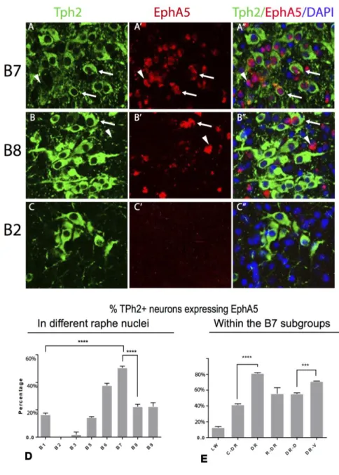
We investigated the role of ephrinA-EphA signalling in selective targeting. Our results demonstrate that EphA5 mRNA is selectively expressed in distinct subpopulation of serotonin raphe neurons. Particularly, EphA5 exhibited the highest level in dorsal raphe serotonin neurons (B7). The results of in vitro explant cultures and in vivo electroporation analyses indicated that the ligands of EphA5 (ephrinA5 and ephrinA3) act as repellent factors for the serotonergic axon growth cones. Anterograde tracing in the ephrinA5 -/- mice showed mistargeting of dorsal raphe neurons projections, including the serotonergic projection. Particularly, our analysis of tracing studies shows that targeting of the dorsal and median raphe axons to different layers of the olfactory bulb is altered in the ephrinA5 KO. However we do not know at what developmental stage these alterations occur, in particular whether this reflects an alteration in the orientation of ascending fibre tracts, or whether this reflects late developmental maturation when raphe axons collateralize and branch in specific target regions.
We have taken advantage a new morphological method, which allows analysing immunocytochemical labelling in 3D. 5-HT immunolabeling, in whole brain serotonergic projection in 3D. Our findings show that serotonergic fibres projecting to olfactory bulb require a special timing to enter the target. The expression pattern of ephrinA5 suggests that ephrinA5 can be one of the factors that modulate this timing.
Overall, our results show for the first time the implication of a guidance molecule for the region-specific and time-specific targeting of serotonin raphe neurons and have implications for the anatomo-functional parsing of raphe cell groups.
Abbreviation list:
5-HT 5-hydroxytryptamine, serotonin
AAV adeno-associated virus
Arc arcuate nucleus
BLA basolateral amygdala nucleus
BNST bed nucleus of stria terminals
DR dorsal raphe nucleus
DRD dorsal raphe nucleus, dorsal part
DRV dorsal raphe nucleus, ventral part
DRL dorsal raphe nucleus, lateral part
EPL external plexiform layer
GCL granular cell layer
GL glomerular cell layer
GPI Glycosylphosphatidylinisotol
ML mitral cell layer
MnR median raphe nucleus
RMS rostral migratory stream
SCN superchiasmatic nucleus
SERT serotonin transporter
VMH ventromedial hypothalamic nucleus
VMHDM ventromedial hypothalamic nucleus, dorsomedial part
Part 1: Serotonin raphe neurons and topographic mapping of
serotonergic projections
Serotonin (5-hydroxytryptamine, 5-HT) neurotransmission is implicated in a large number of physiological functions from the most elemental such as feeding, sleep, and biological rhythms, to more elaborate such as mood, and learning (Lucki, 1998; Fernandez and Gaspar, 2012). As a consequence, 5-HT dysfunction has been implicated in a large number of neurological and psychiatric disorders such as depression, anxiety, autism, and attention deficit disorder. Furthermore, 5‐ HT modulators are indispensable therapeutic tools in depression and anxiety disorder (Hussain, 2016).
1. History
Serotonin was initially isolated from beef serum extracts as an extract that produced peripheral vasoconstriction; because of this vasoconstriction function this substance was named serum vasoconstrictor, serotonin (a serum factor that affected blood vessel tonus) (Rapport et al., 1948). A year later, he identified the chemical composition of this extract as 5-hydroxytryptamine, 5-HT. At about the same time Vially and Erspamer had identified « enteramine » in the enterochromaffin cells of the gastrointestinal mucosa that was increasing gut motility. In 1952, enter amine was identified to be the same substance as serotonin (Erspamer and Asero, 1952). Shortly after that, serotonin was detected for the first time in the mammalian CNS by biochemical analyses of brain tissue extracts initially in dogs. (Amin et al., 1954) Their study indicated that the central nervous system of mammals as well as certain ganglia and peripheral nerves of invertebrates contain detectable quantities of serotonin and that some of the richest areas containing serotonin were in the diencephalon and midbrain. Further studies extended this analysis to a wide range of species from mammals, to birds, reptiles and fish (Correale, 1956). The first visualization of serotonin neurons was achieved with fluorescence histochemistry methods that were based on the endogenous yellow fluorescence of indoleamines when these are fixed with aldehydes. The studies of Kjelle Fuxe and Annita G_Dahlstrom in rat in 1964 described for the first time the localization and the distribution of the neurons containing serotonin in the brain stem, as the B1-B9 cell groups; that were named as such to oppose them to the A1-A15 catecholamine-containing neurons.(Dahlstroem and Fuxe, 1964)
The studies of Dahlström and Fuxe attracted the interest of neuroscientists to the structure of the “raphe” nuclei in the brainstem. Raphe nuclei are an anatomical term for brain nuclei located in the midline of the hindbrain. Raphe nuclei were in particular described by Santiago Ramon y Cajal using the Golgi method. (Figure1) The silver chromate-impregnation method was in these early days the most important method to describe the cellular composition and organization of the nervous system, Ramon y Cajal studied the brains of newborn rabbit and kitten; he designated the raphe nuclei as “intermediate or unpaired nucleus” and “median subaqueductal nucleus of the raphe” which approximates the location of the rostral raphe group. He also mentioned that, in the magnocellular central nucleus of the raphe, a “thin cellular trail extends ventrally penetrating between both longitudinal fascicles”. This description resembles the localization of the interfascicular DRN and, possibly, rostral parts of the MRN. Cajal also observed that the DRN contained four types of neurons, which he described as being voluminous, fusiform, triangular and stellate. (Ramón Cajal, 1906)
Figure1. Cajal's drawing of a transverse section through the caudal region of the superior colliculus of a few-days-old kitten. A, cells of the trochlear nerve nucleus; B, collaterals within the same nucleus; C, medial longitudinal fasciculus; D, fibers of the superior cerebellar peduncles; E, cells of the “subaqueductal nucleus of the raphe”, which probably resembles the DRN. F, ventral cells of the raphe; G, radicular fibers of the trochlear nerve. Image source: the annotated and edited translation of Cajal's “Texture of the Nervous System of Man and the Vertebrates” by Pedro Pasik and Tauba Pasik (Ramón Cajal, 1906), used by permission.
In the early sixties, Elizabeth Taber described the detailed anatomical structure of raphe in the cat brain stem. She described the raphe nucleus as two linear nuclei (nucleus linearis intermedius and
nucleus linearis rostralis), which are approximately the location of rostral raphe group and caudal group, and subdivided these nuclei into 8 groups (Taber et al., 1960). Later on, as mentioned above, this was described more precisely by Dahlström and Fuxe in 1964. They used the formaldehyde-induced fluorescence (FIF) method to study the rat raphe nuclei; this technique had been developed by Falck & Torp, (1962) to visualize monoamines and soon became the most popular tool for visualizing serotonergic neurons. Based on this technique, the morphology and the organization of the raphe nuclei neurons were described in many different species, such as cat (Taber et al., 1960), human (Braak, 1970; Nobin and Bjorklund, 1973; Hornung and Fritschy, 1988), frog (Parent, 1973), rabbit (Felten and Cummings, 1979) and rat (Steinbusch and Nieuwenhuys, 1981) and fish (Pierre et al., 1992). The total number of raphe serotonin neurons differs between species, for instance, in rodents the population of serotonin raphe neurons was estimated to be 17000 (Ishimura et al., 1988), 20000 in rats (Jacobs and Azmitia, 1992), in cat this number increased to 60000 (Jacobs and Azmitia, 1992) and in human approximate 300 000 (Baker et al., 1991). The serotonin raphe neurons are well organized and topographically distributed along the caudal to rostral axis of the hindbrain as different raphe subnuclei. Indeed, Dahlstrom and Fuxe identified 9 serotonin subgroups in the rat hindbrain, from B1caudally to B9. (Dahlstroem and Fuxe, 1964) These cell groups compose 2 main clusters: a rostral raphe group comprising the B9-B4 groups and the caudal raphe group that comprise the B3-B1 cell groups. The rostral raphe group is the
largest cluster of serotonin neurons; it contains approximately 70% of the total serotonin population. This rostral cluster also corresponds to defined cytoarchitectonic entities that are named as follows: caudal linear nucleus (CLi) corresponding to the rostral B7, the supralemniscal cell group, that corresponds to the B9 group, the dorsal raphe nuclei which corresponds to the B7-B6 groups and the median raphe nuclei, which corresponds to the B5-B8 cell groups. The caudal raphe clusters comprises the raphe obscurus, the raphe pallidus and the raphe magnus. (Steinbusch and Nieuwenhuys, 1981; Jacobs and Azmitia, 1992; Michelsen et al., 2007; Hale and Lowry, 2011)
2. Biochemistry of serotonin system
Serotonin neurons share a common neurochemical phenotype. The biosynthetic pathway for serotonin includes
Figure2. The synthesis and metabolism of serotonin. (From NEUROtiker)
tryptophan hydroxylase (TPH), the key enzymes for producing serotonin from amino acid, tryptophan (Grahame-Smith, 1964; Sato et al., 1967) and decarboxylase of aromatic l-amino acids. TPH carries out the first step and is the rate-limiting enzyme in 5-HT biosynthesis. Moreover, it is the only specific enzyme in serotonin metabolism. In contrast, the decarboxylase of l-aromatic amino acids is a widespread non-specific enzyme.
Once 5-HT is produced it is concentrated into synaptic vesicles by the vesicular monoamine transporter, VMAT2, which is required for calcium dependent vesicular release. Once released 5-HT is rapidly cleared away from the extracellular space by the serotonin transporter SERT (Scl6a3) either to be recycled or to be degraded. In addition to the high affinity transporter 5-HT uptake is removed by high affinity amine transporters, OCT3.
After reuptake, 5-HT is degraded essentially by 2 enzymes: monoamine oxidase-A which has the highest affinity for 5-HT, and MAOB which has a lower affinity for 5-HT (Levitt et al., 1982); (Figure2).
TPH
The enzyme TPH belongs to a small family of structurally and functionally related aromatic aminoacid hydroxylases that utilize tetrahydropterins as cofactor. In eukaryotes, these enzymes are composed of a homologous catalytic domain with a highly conserved C-terminal tetramerization regions and sequence-specific N-terminal regulatory domains are attached (Fitzpatrick, 1999). About 15 years ago, the first TPH gene in rat, mouse and human (Darmon et al., 1988; Boularand et al., 1990; Stoll et al., 1990) was described and, most of this time, this gene has been thought to be the only TPH gene in the genome. Later, evidence accumulated indicating different biochemical properties of the TPH enzymes depending on the analyzed tissue. However, efforts undertaken to identify another TPH gene isoforms were unsuccessful until Diego Walther with colleagues generated a TPH knockout mice (Walther and Bader, 2003). They found that these mice lacked 5-HT in the blood, in the periphery tissues and in the pineal gland. However, there was only a minor 5-HT decrease in the brain structures. These surprising results, suggested the existence of another gene not affected by the gene targeting. This lead Diego Walther and colleagues to discover a second TPH gene in the genome of mice, rats and humans, called Tph2. It has been shown that Tph2 is predominantly expressed in the brainstem and located on human chromosome 12 and mouse chromosome 10 (Walther and Bader, 2003; Walther et al., 2003). TPH1 and TPH2 enzymes are
highly homologous proteins exhibiting 71% of amino acid identity in humans; however, the N-terminals containing the regulatory domain are quite different. (Walther and Bader, 2003)
MAO
MAO catalyzes oxidative deamination of monoaminergic neurotransmitters, 5-HT, noradrenaline and dopamine. There are two forms of MAO (A and B) that are involved in 5-HT metabolism. MAO A and MAO B enzymes are encoded by different genes (Bach AW, 1988) localized on the X-chromosome (Lan et al., 1989; Sims et al., 1989). MAO A has a higher affinity to 5-HT than MAO B and is considered as the principal enzyme of 5-HT degradation.
Co-neurotransmission
There is evidence that serotonin can be associated with other neurotransmitters. This was first discovered for neuropeptides. Substance-P was found in the Nucleus Raphe Magnus (Chan-Palay, 1981; Magoul et al., 1986; Halliday et al., 1988; Arvidsson et al., 1994). Glutamate, an excitatory neurotransmitter, is also co-localized with serotonin and substance-P in raphe neurons (Nicolas et al., 1992). Other peptides described within the serotonergic neurons are calretinin (Acsady et al., 1993), galanin (Arvidsson et al., 1991), enkephalin (Millhorn et al., 1989; Henry and Manaker, 1998), N-acetyl-aspartyl-glutamate (Forloni et al., 1987), neuropeptide-Y (Halliday et al., 1988; Krukoff et al., 1992), angiotensin II (Krukoff et al., 1992), and thyrotropin releasing hormone (Ulfhake et al., 1987; Sharif, 1989; Arvidsson et al., 1994). It has been suggested that serotonin is localized to the small clear vesicles and peptides are concentrated in the dense core vesicles (Pelletier and Laflamme, 1977; Johansson et al., 1980; Van Bockstaele and Chan, 1997). There is evidence for the co-localization of peptides and serotonin in the dense core vesicles (Pelletier et al., 1981). It would be interesting to know if glutamate exists within both the small and dense core vesicles with serotonin.
3. Serotonin neuroanatomy - Anatomical organization of the raphe nuclei and
serotonergic raphe neurons
Serotonin neuroanatomy is difficult for the neuroscientists to concretize, since its decentralized structure and complex cell morphological constitution. The serotonergic raphe nucleus forms an inhomogeneous reticular group of neurons. This reticular group is located approximately from the spinal cord caudally to the interpeduncular nucleus rostrally. (Jacobs and Azmitia, 1992) The distribution of the serotonin raphe neurons is irregular, for instance, in the dorsal raphe (DR) and
caudal linear nucleus (CLi), the nuclear boundaries of the serotonin neurons shows the classical assignation, whereas the serotonin neurons in the lateral wing (LW) and B9 group are scattered in unconventional locations. (Detail describe see followed part)
3.1 Serotonin neurons morphology
Histochemical studies of the raphe nucleus showed that the neurons, that contain the serotonin have different morphologies, overall, 4 types were identified, large, small, multipolar and fusiform
perikarya. In electron microscopic studies, the nucleus is seen as being invoginated and the cytoplasm has a well-defined Golgi apparatus and abundant microcanuculi.
Moreover, electron microscopic analysis showed that serotonin terminals contained 2 types of vesicles: small clear vesicles and large dense core vesicles (Leger and Descarries, 1978; Wiklund et al., 1981; Johnson and Yee, 1995)
Serotonin raphe neurons have different morphologies, this determined the variability of their size, generally, and the diameter of serotonin neurons is varies from 15 to 60 um. However this can also depend on the hormonal status of the animal: in adrenalectomized animals, lacking circulating adrenal glucocorticoids, the serotonergic neurons in all the raphe groups appear small with thin processes extending from the soma (Whitaker-Azmitia et al., 1993). If dexamethasone are added to the drinking water, the size of the soma and the processes increases, within an approximately 80%
Figure3. An electron microscopic photograph taken of the Dorsal Raphe Nucleus of the adult rat. (Azmitia, 1999)
increase within 24–72 hours in the volume of the tryptophan hydroxylase immunoreactive neurons (Azmitia, 1999).
3.2 Serotonin dendrites and axons morphology
Cajal (1911) described the serotonergic neurons as large neurons with extensive but untraceable axonal projections. He indicated that they were equipped with “several divergent and strongly spiny dendrites”. Using 3H-proline as a marker, at least five separate tracts were found ascending from the superior group of raphe nuclei (Azmitia and Segal, 1978). Some serotonergic fibers are myelinated, whereas others are unmyelinated, and a variety of fiber diameters can exist within many brain regions (Kohler et al., 1980; Cropper et al., 1984). Some serotonergic neurons form synapses while others engage in non-synaptic interactions (Azmitia and Segal, 1978; Herve et al., 1987; Hornung et al., 1990).
Some researchers described morphological differences between the serotonergic fibers, and divided serotonin neurons into two groups depending on fiber morphology. One fiber type, which is thick, relatively straight and non-varicose, was described as originating from the Median Raphe Nucleus while the other, formed by thin, highly branched and varicose fibers was described as arising from the Dorsal Raphe Nucleus (Kosofsky and Molliver, 1987; Hornung et al., 1990). However, this is controversial, as other researchers reported that the morphology of the fibers may depend on the target region innervated (Azmitia and Segal, 1978). For instance one study showed that the origin morphology of fibers was changed after they arrived the target. In another study it was shown that serotonergic projection tract from raphe to lateral ventricles, the fibers are initially thick, straight, and non-varicose at the first postnatal week in the rat. However, the same fibers become thin, highly branched, and varicose by the third postnatal week (Dinopoulos and Dori, 1995).
3.3 Anatomical organization of the raphe nuclei
To understand function in the central nervous system it is a good idea to start from the anatomy studies which can let you have a spatial overview. Neurons in different region are not scattered or randomly, distributed, they have a very precise cytoarchitectonic organization that corresponds to their developmental origin, and to their particular connectivity patterns that are informative of function. The composition /organization of the different raphe nuclei are listed below. (Figure 4)
Dorsal raphe nucleus (DR) c = B7+ B6
The DR is considered as the one of the most important nucleus in the raphe complex since it contains the largest population of serotonergic neurons and is the focus of the vast majority of the serotonin research. The DR is located in the caudal midbrain and rostral pons, in a part of the brainstem called the tegmentum, just below the cerebral aqueduct. The DR has been further subdivided into several components based of the spatial location and density of cells.as the rostral extension, rostral, dorsal, ventral, ventrolateral, interfascicular, and caudal parts. There are four morphological cell types of neurons located in the DR: round, ovoid, fusiform and triangular (Steinbusch and Nieuwenhuys, 1981; Baker et al., 1991). However these cell morphologies have not been related to any functional characteristics. On the other hand, as will be discussed later there are neurochemical and connectivity differences among the DR subdivisions.
The following DR subdivisions are considered: DR-Caudal linear nucleus (CLi)
The caudal linear nucleus is located in the midbrain just dorsal to the decussating fibers of the superior cerebellar peduncle and merges caudally and dorsally with the DR; it can be considered as the rostral extension of DR. There are two main types of neurons in the CLi: serotonergic neurons and dopaminergic neurons that are intermixed (Steinbusch and Nieuwenhuys, 1981). The morphology of the serotonergic neurons in the CLi is small and spherical with fewer dendrites that are oriented along the rostral-caudal axis (Tork, 1990). The morphology of dopaminergic neurons is small and fusiform which are extends caudally into the ventromedial portion of the rostral DR (Wiklund et al., 1981; Minami et al., 1999). Serotonin neurons in the CLi are considered as the smallest population in the raphe complex, in cats, the CLi contains 2,000 serotonergic neurons, approximately one tenth the number of total DR nucleus (Wiklund et al., 1981).
DR, Rostral part (DRr)
The rostral part of dorsal raphe located on the cerebral aqueduct – CLi axis dorsal-ventrally. Caudally, the rostral DR is bordered by the DRD and the DRV. The specificity of neurons in the rostral DR is like the CLi included both serotonergic neurons and dopaminergic neurons (Descarries et al., 1986; Stratford and Wirtshafter, 1990).
DR, dorsal part (DRD)
The DRD is bordered dorsally by the cerebral aqueduct, ventrally by the DRV, and laterally by the DRVL/VLPAG. The central midline region of the DRD contains the medium size, fusiform or bipolar cells which could be characterized as glutamatergic neurons, serotonergic neurons and GABAergic neurons. The distribution of serotonergic neurons can be grouped into two clusters, which are referred to as the DRD “core” and “shell” depends on the dense or scattered distributed serotonergic neurons respectively (Abrams et al., 2005; Clark et al., 2007; Hioki et al., 2010).
DR ventral part (DRV)
The DRV is bordered dorsally by the DRD, more precisely, a border of glutamatergic neurons which between DRD and DRV, ventrally and laterally by the dorsal tegmental nucleus. The DRV contains small, round serotonin neurons but also a population of glutamatergic neurons that are labeled by Vglut3. (Fremeau et al., 2002) (Gras et al., 2002; Schafer et al., 2002; Takamori et al., 2002) Serotonergic neurons in the DRV are densely packed, and as a consequence, this region shows the highest expression of Tph2, SERT and some 5-HT receptors mRNA raphe complex (Clark et al., 2006).
DR ventrolateral part (DRVL)
The DRVL group also called the lateral wings of raphe. Serotonergic neurons in this region are notable for their large size, relative to other subpopulations of serotonergic neurons, and their multipolar morphology, which is easily discernable with immunostaining for serotonin or tryptophan hydroxylase. The lateral wings do not include the glutamatergic neurons anymore but a large amount of GABAergic neurons (Jolas and Aghajanian, 1997; Day et al., 2004), which appear to play a role in local inhibitory control of serotonergic neurons within the dorsal raphe nucleus (Jolas and Aghajanian, 1997).
Median raphe nucleus (MnR)
The MnR is a midline structure ventral to the DR. The MnR and the DR have different developmental origins. Serotonergic neurons in the DR derive entirely from rhombomere 1 of the developing mouse brain, whereas serotonergic neurons of the MnR derive predominantly from rhombomere1, 2 and 3. (Jensen et al., 2008; Bang et al., 2012).
The B9 serotonergic cell group/supralemniscal serotonergic cell group (B9)
B9 serotonergic neurons extend throughout the rostral and mid-pons and are located within and dorsal to the medial lemniscus. Although it has received considerably less attention than other midbrain and pontine serotonergic cell groups, it contains a substantial number of serotonergic neurons (estimated to be 4,571 cells in the rat brain, roughly equivalent to the number in the MnR (Vertes and Crane, 1997). Substantial numbers of B9 serotonergic neurons also have been described in primates, including humans (Azmitia and Gannon, 1986). Steinbusch (1984) described a high
concentration of serotonergic neurons dorsal to the medial lemniscus and ventral to the nucleus rubber, particularly its lateral parts. A few cells were located in the nucleus rubber itself.
3.4 Serotonin raphe neurons axonal projections: from past to the present
There is hardly no region in the central nervous system that does not receive 5-HT innervation; all this innervation derives from the brain stem raphe nucleus, which are in relatively low numbers compared to the vast amount of neurons that they innervate. This indicates a high degree of collateralization; however anatomical evidence has also shown that there is also a fair amount of topographic organization of the serotonin projections arising from the raphe.
Cajal recognized that the fibers emerging from the raphe nuclei tended to “concentrate into ascending and descending dorsoventral bundles”. However, he was not able to determine how far the fibers continued, because these fibers were not labeled with the Golgi method. The first specific description of raphe nucleus axonal projections was described by Dahlstrom and Fuxe using Formaldehyde induced Fluorescence (FIF) which could visualize the monoamines, this allowed the first description of the distribution pattern of serotonin projections. However, this technique has a big weakness which is the rapid fading of fluorescence (Dahlstroem and Fuxe, 1964). Later in the 70s to 80s, two crucial techniques were developed for serotonin projection histological analysis: specific antibody for immunohistochemistry analysis and anterograde/retrograde tracing. The first specific antibody against serotonin was developed and used by Steinbusch (Steinbusch and Nieuwenhuys, 1981); this allowed a much more precise and detailed analysis of the serotonin axon terminals in different targets. At the same time period axon tracing methods made tremendous progress. Horseradish peroxidase (HRP) was found to be very efficient for tracing fiber pathway as a retrograde tracer and was used for studying serotonin projection since 1975. (Miller et al., 1975) In parallel, anterograde tracing methods were developed such as the autoradiographic detection of [3H]-serotonin uptake and [3H]-labeled reserpine that were used for anterograde tracing analysis of serotonin system. (Calas et al., 1974; Richards, 1979) More precise anterograde tracing were developed such as Biolytic, which allowed a fine cellular of anterogradely labeled dorsal and medial raphe neurons, identifying their full axon projections (Vertes, 1991; Vertes et al., 1999). However these tracing methods are not selective of a defined cell type, thus identification of the serotonin projections relied on a combination of fluorescent retrograde tracers (fast blue, fluoroglo) with immunocytochemistry. More recently, genetic tools are available for selective serotonin tracing as there are many transgenic mouse lines expressing the “Cre” recombinase under the control of specific promoters. For instance, in serotonin system the common promoters are: Pet1, Sert and Tph2
Based on the improvement of tracing methodology; the initial map of raphe serotonin neuron has been extended and refined over the years; Initially serotonin pathways were described as emerging from two cluster bases on the specificity of projection targets- a rostral group (B5-B9) which mainly projects to the forebrain targets whereas the caudal group (B1-B4) mainly project to the brainstem and spinal cord. Serotonin neurons from the rostral group are the essential neurons that release serotonin into the forebrain targets and furthermore play the key role in the modulation of CNS functions. And this is also why researchers who study the serotonin system are more interested in rostral raphe neurons. The rostral raphe group contains the majority or most the population of serotonin neurons and innervated the whole forebrain, it’s not difficult to imagine the complexity of their ascending pathways.
In the beginning of 21st century, neuroanatomists initially clarify the different pathway from rostral raphe which were described characteristic as followed: (a) the dorsal raphe forebrain bundle tract, travelling within the ventrolateral part of the medial forebrain bundle to the basal ganglia, amygdala and piriform cortex; (b) the medial raphe forebrain bundle tract, travelling within the ventromedial part of the medial forebrain bundle to the prefrontal cortex, cingulate cortex, medial septum and hippocampus. Four additional fiber tracts are located outside the medial forebrain bundle; (c) dorsal raphe cortical tract, travelling ventrolateral to the medial longitudinal fasciculus to the caudate putamen and parieto-temporal cortex; (d) dorsal raphe periventricular tract, travelling immediately below the midbrain aqueduct to the periventricular thalamus and hypothalamus; (e) dorsal raphe arcuate tract, travelling through the ventrolateral edge of the midbrain to the ventrolateral geniculate body nuclei and the suprachiasmatic nuclei; (f) raphe medial tract, travelling from the dorsal and median raphe nuclei ventrally between the fasciculi retroflexus to the interpeduncular nucleus and midline mammillary nuclei.
All these pathways can be simplified into three pathways: dorsal ascending pathway, median ascending pathway and ventral ascending pathway.
Dorsal ascending pathway
The dorsal ascending pathway rises from the medial and rostral DRN and innervates the striatum and globus pallidus (GP). The striatum is the most important target for DRN innervation and one of the first to be extensively studied. The earliest anatomical indications for DRN projections to caudate-putamen (CP) of the dorsal striatum (Anden, 1965) were subsequently supported by lesion studies, which showed a drop in striatal tryptophan hydroxylase (TPH) activity (Geyer et al., 1976) as well as a decrease in [3H]5-HT uptake (Kellar et al., 1977) after DRN lesions. Approximately a third of all serotonergic DRN neurons project to the CP. This is, however, region-specific: in a cluster in the dorsomedial DRN, 80–90% of serotonergic neurons were found to project to the CP (Steinbusch and Nieuwenhuys, 1981). Twenty percent of DRN neurons that project to the CP are non-serotonergic. Nucleus accumbens of the ventral striatum, and particularly its core, are also extensively innervated by DRN fibers (Van Bockstaele and Pickel, 1993; Van Bockstaele and Chan, 1997; Brown and Molliver, 2000). DRN efferent target the striatum at caudal to midlevels (Vertes, 1991). Approximately half of the neurons are located in the rostral third of the DRN, fewer in the middle third and very few in the caudal third (Steinbusch and Nieuwenhuys, 1981; Waselus et al., 2006). Pallidal afferents from the DRN have been demonstrated by tracing studies (DeVito et al., 1980; Vertes, 1991). The innervation of the GP is mainly serotonergic, as confirmed by micro-dialysis studies in the rat (McQuade and Sharp, 1997)
Medial ascending pathway
The main target of the medial ascending pathway is the substantia nigra (SN). The projections seem to arise from the rostral DRN (Miller et al., 1975; Imai et al., 1986a) and they target the pars compact division in particular (Fibiger and Miller, 1977; Bobillier et al., 1976). However, a study using the retrograde tracer PHA-L failed to demonstrate DRN innervation of the pars reticulate (Vertes, 1991). To a lesser extent, the pathway also innervates the CP. Some of the fibers branch, and target both the SN and CP (van der Kooy and Hattori, 1980; Imai et al., 1986a). Thus, single DRN neurons exert control over both the SN and the CP.
Ventral ascending pathway
Via the ventral ascending pathway, the DRN innervates many areas. The bilateral pathway ascends ventrolateral and then turns rostral to enter the medial forebrain bundle. The pathway also contains fibres from other raphe nuclei, especially median raphe. The main targets are thalamic and hypothalamic nuclei, habenula, septum, amygdala, cortex, the olfactory bulb, interpeduncular nucleus and geniculate body.
Indeed 5-HT neurons are generally intermingled with larger populations of non 5-HT neurons in each raphe subnuclei. An example of this is the DR cell group, which contains the highest proportion of 5-HT producing neurons, and yet comprises twice as much non-5-HT neurons (Descarries et al., 1982). So in order to identify 5-HT projections from the raphe relied on tracing techniques coupled to the histochemical revelation of 5-HT or combined with lesions. However, although these studies allowed to describe the projections of the raphe cell groups, many limitations effect the reliability of the studies, such as the variability of the injection site, variability of the uptake by passing fibres, the background from histochemical methods and the difference of the morphology after labelled by specific antibodies. Moreover, most of the studies generally focused on a particular target brain region or at most on a combination of few targets (van der Kooy and Hattori, 1980; Kohler et al., 1982; Kohler and Steinbusch, 1982; Waterhouse et al., 1986; Jones and Cuello, 1989; Van Bockstaele and Pickel, 1993; Vasudeva et al., 2011). Summarizing these studies is difficult because they relied on different experimental protocols and different animal models.
This is why, nowadays, neuroanatomists prefer to use genetic tools to study connectivity. Recent genetic studies defined the rhombomeric origin of the different raphe nuclei (Jensen et al., 2008) and provided a useful description of the 5-HT axon projections arising from the different
rhombomeres (Bang et al., 2012). In these studies, they performed multi genetic tools, which cross different recombinase marks promoted Cre recombinase to detect the connectivity relationships characterizing three serotonergic neuron subpopulations. These subpopulations were characterized by developmental gene expression associated with different rhombomeric (r) segments of the embryonic hindbrain. Subpopulation of serotonin neurons from r1 was derived from the combined expression of Pet1 (as a serotonin marker) and En1 as a marker of rhombomeric origin (r1). Likewise, r2 serotonergic neurons were characterized by the combined expression of Pet1 and Hoxa2 (as a marker of r2), while Egr2 was used for identifying the serotonin neurons from r3 and r5. The mature serotonin neurons of these three subgroups are located in the dorsal raphe (DR), median raphe (MR), B9 group and the rostral portion of raphe magnus (RMg) (Jensen et al., 2008). The same intersectional and subtractive genetic approach was used to visualize and selectively map the axon projections associated with each of these three different subpopulations of serotonin neurons. The multi genetic approach of Jensen is a set of Cre recombinase transgenic – En1::cre, Rse2::cre or Egr2::cre – each separately partnered with an ePet1::Flpe transgene, based on that they develop the approach which is with the indicator allele RC::FrePe to selectively identify the intersection neurons with enhanced green fluorescent protein (eGFP) expression. With this approach they separately mapped target brain regions served by each of these three subpopulations. Additionally, they examined how each of these eGFP-containing subgroups of serotonin neurons provided recurrent innervation to the remaining serotonin neurons that lacked eGFP, as a means to explore potential inter-system feedback inhibition. This could greatly enhance the dynamic and differential changes in extracellular serotonin concentrations across brain regions.
Their results suggest that rhombomeric origin is a contributor to organizing not only the location of serotonin cell bodies (Jensen et al., 2008), but also the organization of their projections, and thus genetic and developmental process may provide key insights into resolving aspects of the functional heterogeneity within the serotonin system. However, these approaches do not allow a specific delimitation of the topographic organization of raphe neurons originating from the same rhombomere.
In 2016, Muzerelle et al used conditional tracing methods (AAV, lox-stop-lox-GFP) in mice expressing the Cre-recombinase in 5-HT raphe neurons to obtain a selective anterograde tracing of these neurons. Using this method, they labeled small groups of 5-HT neurons within the different raphe sub-nuclei allowing a comprehensive mapping of their terminal axon fields in the brain. With these studies they provided a description of the main projecting areas of 5-HT axons arising from
different raphe sub-nuclei as well as a systematic semi-quantitative analysis of correlations between the origin of 5-HT axons (raphe sub-nuclei) and their targeted brain regions(Muzerelle et al., 2016).
DR projections (B7)
As I described above, dorsal raphe contain the biggest population of serotonin neurons and spatially divided into several subregions. Aude et al. showed that even in these sub-regions, the targets are different from each serotonin neurons.
Dorsal component of the dorsal raphe, DRD (B7d)
Ascending projections from the DRD were detected as ventrally and bilaterally towards the mlf, then serotonin fibers are spreading laterally to innervate the substantia nigra pars compacta (SNC) and ventral tegmental area (VTA). The most abundant terminal innervation was localized in the hypothalamus, followed by the lateral geniculate nuclei of the thalamus, SNC and parts of the basal ganglia, namely the nucleus accumbens, and caudal parts of the caudate-putamen and pallidum. These projections were mainly but not exclusively ipsilateral.
In the brainstem, DRD axons terminal innervation was dense in the superior colliculus and the periaqueductal gray (PAG). More ventrally it was essentially concentrated in the superior Oliver complex, 7N, and dorsal and ventral cochlear nuclei (DC, VC)
Ventral component of dorsal raphe, DRV (B7v)
Amygdala was strongly innervated by DRV serotonin fibers, where a strong accumulation of axon varicosities was observed with a particularly dense distribution in the central and basolateral components of the amygdala. A somewhat similar dense innervation was visible within the extended amygdala, such as the bed nucleus of the stria terminalis (BST). In contrast, despite the presence of ascending fiber tracts in the medial septum, no terminal innervation was visible in the lateral septal (LS) nuclei.
A dense innervation also reached the cerebral cortex, with a particular concentration in its rostral and ventrolateral parts including the orbital cortex and olfactory-related brain areas, in particular the piriform cortex, the anterior olfactory nuclei (AON) and the mitral and granular layers of the olfactory bulb. In the medial prefrontal cortex, varicose fibers were visible mainly in the infralimbic cortex, whereas the dorsal part and the cingulate cortex were more sparingly innervated.
In the thalamus, innervation was essentially concentrated in the midline thalamic nuclei. The habenula was only moderately innervated, with terminal fibers mainly in the lateral habenula (LHb). Strikingly, and contrasting with the heavy ascending fiber tract in the mfb, the hypothalamic nuclei received hardly no innervation. Similarly the hippocampus contained only few fibers that were essentially localized in the stratum lacunosum-moleculare of both the dorsal and ventral hippocampus. However, hardly any innervation was noted in the dentate gyrus. In the mesencephalon, innervation was particularly conspicuous in the substantia nigra pars reticulata with moderate innervation of the ventral periaqueductal gray (PAG) and the superior colliculus.
Lateral wings of the dorsal raphe, LW (B7l)
Ascending 5-HT innervation to the forebrain in this case was essentially concentrated in the thalamus, predominating in its more lateral components throughout its rostrocaudal extent. Bilaterally in the parasubthalamic nucleus and the LHb. Projections were also visible in the hypothalamus mainly in the ipsilateral mammillary/supramammillary components, and in the lateral hypothalamic cell groups. In the brainstem, terminal axons were essentially found in the superior and inferior colliculi, in the cochlear, motor trigeminal, and facial nerve nuclei.
Dorsal raphe caudal part, DRc (B6)
The common projections in these cases were projections to the dorsal and ventral hippocampus, the septal nuclei, and the preoptic cell groups. Projections to the amygdala were limited to the medial amygdala nuclei. Only few projections were found in the brainstem. Interestingly, however, in all the cases in which the B6 5-HT neurons were targeted, there was a substantial innervation of the subventricular 5-HT terminal network as well as in the SVZ.
Median raphe (B8)
provided a bilateral terminal innervation to a number of midline brain structures from the mesencephalon to the forebrain. Innervation was particularly dense in the interpeduncular nucleus, mammillary nuclei and in their lateral parts, and in anterior hypothalamic areas, including the preoptic area. Very dense terminal axons were noted in the suprachiasmatic nucleus. In the dorsal thalamus, the B8 5-HT innervation was particularly concentrated in the paraventricular thalamic nuclei (PV), whereas moderate innervation was visible in the reuniens and in midline non-specific thalamic nuclei, as well as in the medial habenula cell groups. More rostral, B8 axons provided a very dense innervation to both the medial and lateral septal nuclei. The B8 group provided a massive innervation to all components of the septum and hippocampus. A distinctive terminal-like innervation was also noted all along the subgranular cell zone (SGZ) of the dentate gyrus (DG), where adult neurogenesis is found. In the cerebral cortex, B8 innervation was generally scattered and essentially concentrated in the dorsomedial components of the cortex: in the medial prefrontal cortex, the
Figure6. A summary scheme of the principal targets of the main B5–B9 subgroups in sagittal brain diagrams. (Muzrelle et al. 2016)
anterior cingulate and in the primary motor cortex. In contrast, in the olfactory bulb, a strong and selective B8 axon projection was found in the periglomerular cell layer In addition, few fibers were detected in the rostral migratory stream (RMS), at the OB. In the brainstem, the most remarkable concentration of 5-HT-labeled axons was found in the dorsolateral tegmental nucleus (DTg) also known as the tegmental nucleus of Gudden. This dense 5-HT innervation was visible in the mesopontine tegmental nuclei and the anterior tegmental nucleus (ATg), which are components of the DTg.
Supralemniscal (B9)
Ascending projections from the B9 were relatively sparse, in comparison with the DR and MR groups: they occupied a medial and dorsal position in the mfb providing terminal innervation essentially to parts of the septum and basal ganglia. Terminal-like innervation was observed in the medial septum, and fine terminal innervation of variable abundance according to cases was noted in the caudate-putamen, nucleus accumbens and pallidum. Sparse innervation was also noted in the cerebral cortex and hippocampus, but with no systematic distribution. In the hypothalamus, axon terminal innervation was mainly targeting the anterior and preoptic cell groups and the supramammillary area. Thalamic innervation was scarce and oriented toward midline structures. In the midbrain, terminal-like fibers were visible in the SNC and the VTA. Moreover B9 5-HT axons were the presence of descending projections towards the hindbrain.
Base on those studies of serotonin projections mapping, one can conclude that different subgroups of serotonin raphe neurons have their specific target and similar to other brain circuits, a combination of axon guidance molecules both attractive and repulsive is involved, to guide 5-HT raphe neuron axons to their proper brain targets.
3.5 Afferent connections to the raphe
In addition to sending broad projections to many brain areas, the raphe receives connectivity from many functional regions in the forebrain and brainstem. The following areas have been reported to provide innervation to the raphe nucleus: the medial prefrontal cortex, hypothalamus, hippocampus, habenula, dorsal tegmental nucleus and spinal cord. (Wang and Aghajanian, 1977; Behzadi et al., 1990; Hermann et al., 1997) These innervations determined that the serotonergic neurons itself can receive signals from both serotonin and non-serotonin axons. Moreover, many publications showed amount of neuropeptides could been detected in axonal terminals in the raphe
nucleus: serotonin (Dong and Shen, 1986); norepinephrine (Baraban and Aghajanian, 1981; Takagi et al., 1981; Dong and Shen, 1986; Lee et al., 1987); dopamine (Ferre et al., 1994); acetylcholine (Chen et al., 1992; Honda and Semba, 1994); GABA (Hayashi et al., 1997), substance-P (Magoul et al., 1986); CLIP/ACTH (Leger and Descarries, 1978); or neurotensin (Uhl et al., 1979) Serotonergic dendrites extend into the axonal bundles of the medial longitudinal fasciculus, medial lemniscus, trapezoid body, cerebral peduncles, the superior cerebellar peduncles and other fibre bundles which traverse the brainstem carrying both ascending and descending fibres (Azmitia and Segal, 1978). The spectrum of inputs from numerous anatomical sources having diverse chemical neurotransmitters and neuropeptides is not atypical for a large reticular neuron. But given the number of afferents and efferents of this single chemical system, it can transmit the varied signals to both neuronal and non-neuronal targets.
4. The development of serotonin neurons in raphe nuclei
During development, the serotonin neurons are divided into two groups based on their spatial difference as inferior group and superior group. The two groups of serotonin neurons have different migration patterns (Lidov and Molliver, 1982b; Wallace and Lauder, 1983), and within the superior group, there is evidence the 5-HT neurons form different subsets of cells. (Azmitia and Gannon, 1986)
In many studies, the neuroanatomical development and neuronal organization of 5-HT neurons has been analyzed in many different species, such as rats, primates and zebrafish (Steinbusch and Nieuwenhuys, 1981; Lidov and Molliver, 1982b; Wallace and Lauder, 1983; Azmitia and Gannon, 1986; Lauder, 1990; Lillesaar et al., 2007), and more recently in mice with genetic-based fate mapping (Jensen et al., 2008). Moreover, during embryogenesis, the hindbrain is transiently subdivided into several components called rhombomeres, which correspond to different subpopulations of serotonin neurons (Figure 7). Jensen et al. demonstrated that the raphe rostral group derive entirely from r1-r3, whereas the caudal from r5-r8. Among the rostral group, the DR nucleus (B7, B6) origioned from r1 whereas the median raphe nucleus (B8, B5) from entire r1-r3. (Jensen et al., 2008)
Mouse serotonin neurons are specified and appear initially in the ventral rhombencephalon during a brief period of neurogenesis, embryonic days 9.5–12. The initial contributing of the raphe nuclei is based on the ultimately cluster of newborn serotonin neurons in different disparate regions
of the midbrain, pons and medulla. The 5-HT neuron generation consists in several developmentally recognizable stages.
During the first stage, serotonergic progenitors in the ventral hindbrain are specified in a spatial organization along the dorsoventral and anterior-posterior axes, which are modulated by signaling molecules sonic hedgehog and fibroblast growth factors 4 and 8 respectively (Goridis and Rohrer, 2002; Vitalis et al., 2003; Cordes, 2005). The initial serotonin neurons are generated by progenitors from about E9.5 to E10.5 in rhombomere 1 (r1) co(Pattyn et al., 2003). Then a second wave of serotonin neurons are born about a day later in r2 and r3, as progenitors at these longitudinal levels initially generate visceromotor neurons (VMN) before becoming competent to generate serotonin neurons (Pattyn et al., 2003). Progenitor fate of caudal serotonin groups occurs with similar temporal characteristics in r5–r8, such that caudal serotonin neurons are born virtually simultaneously with those in the rostral domain. Interestingly, the synthesis of 5-HT in these newborn caudal serotonin neurons is delayed for 1–2 days (Lidov and Molliver, 1982a). Moreover, the serotonergic neurogenesis is never launched under normal circumstances in r4 (Pattyn et al., 2003).
During the second stage, progenitors do not exhibit serotonergic characteristic as yet. Serotonergic identity is then acquired through coordinated expression of serotonergic-type gene battery. The newborn serotonin neurons are phenotypically immature and have not been integrated into neural circuitry. Thus, beginning immediately after the birth of the 5-HT neurons and extending to at least the end of the third postnatal week, mouse 5-HT neurons undergo a series of complex maturation events (Lidov and Molliver, 1982b, a; Pattyn et al., 2003). These events include cell migration, dendric growth, expression of serotonin autoregulatory pathways, formation of highly collateralized axonal pathways and synaptogenesis.
Development of serotonergic projections
Lidov and Molliver provided a detailed immunohistochemical study on the development of serotonergic projection in rat. (Lidov and Molliver, 1982b) In their study, the first time described the serotonin containing processes in the immature rodent CNS, but only ascendings were described but not descending. They described the ontogeny of the serotonergic axonal projections in three steps: At E13-E16, the initial axon elongation; E15-E19, the development of selective pathways; E19 to P21, the terminal field development.
Initially, serotonergic ascending axons are fasciculated and enter several well-defined fiber tracts starting from the medial forebrain bundle: specifically, the fasciculus retroflexus, stria medullaris, external capsule, fornix, and supracallosal stria. Depending on the different pathways, the serotonergic axons start to form the terminal arborizations in the thalamus, hypothalamus, basal and limbic forebrain, and cerebral cortex. In reaching the serotonergic terminal innervation targets, Lidov and Molliver observed that serotonergic fibers seem to be guided by some “pre-existing non-serotonergic tracts” which could be guidance cues. In particular, they described the terminal development in the cerebral cortex. From E19 to P21, the serotonergic fibers were starting and terminating the innervation in the cerebral cortex. Axons enter directly into the marginal and intermediate zones of the immature cortex, at the medial, frontal and lateral edges of the hemisphere, and subsequently spread tangentially to cover the hemispheres. Terminal ramifications then arise
During the first postnatal weeks the growth of serotonin axons across the forebrain appeared to be continuing. Interestingly, the development of terminal innervation is highly heterogeneous, occurring at different times and at different rates from region to region, which implies a possible role of chemical gradients produced in the targets to trigger the innervation.
These observations showed that serotonergic axons do not innervate immature, primarily proliferative neuronal populations and the formation of serotonin axon terminals is dependent on maturation of other elements in local neuronal circuitry. Evidences to surport this hypothesis are their findings of the delay in serotonin innervation of the suprachiasmatic nucleus, striatum, and middle cortical layers long after the axons have reached these structures.
Part 2: Molecular pathway of serotonin system and Eph family
1. From growth cone to axon guidance
Growing axons have a specialized structural differentiation at their tip known as the growth cone, which plays a crucial role in integrating the many signals that direct axons to their proper targets. The first detailed described of a growth cone was done by Cajal on spinal commissural axons, on three day old chick embryos. Morphologically, the growth cone is a fan-shaped structure with many long, thin spikes that radiate outward much like fingers on a glove. The prediction of Cajal that this might be a structure permitting the growing neuron to receive and integrate the variety of physico-chemical signals present along its pathway has now been validated by numerous cell biology experiments in vitro, but also with the improved visualizing techniques, in vivo. The signals that are sensed by the growth cones are produced by intermediate targets and the surrounding tissue and involve both chemical and biophysical cues. Such cues guide axons to their final target, where they establish synapses with one or more neurons (Mueller, 1999; Song and Poo, 1999). As predicted by Cajal, the growth cones are also highly motile structures. Video microscopic studies have shown that growth cones undergo continuous expansion and retraction before they reach their final targets. This was initially demonstrated in vitro on cell cultures (Harrison, 1959) and more recently in vivo (Tashiro et al., 2003).
We can explain the growth cone navigation system as driving. This system is similar to an experienced driver who can grasp different environmental changes, such as the traffic lights, street signs or bad road conditions in order to guarantee the right destination. Definitely, as the multiple environmental variables, the navigation system needs a series of molecules to form a signaling pathway in order to introduce spatial bias for steering the growth cone to the right targets.
To study the complex effects on the growth cone cytoskeletal machinery, recently, as interdisciplinary development, biophysical techniques became more and more popular to be used for cell biology assay. By performing the microfluidic devices has allowed the production of stable, precisely controlled gradients in various combinations of both diffusible and substrate-bound factors (Lang et al., 2008; Wang et al., 2008). These types of combined cues can more faithfully recapitulate the complexity of the in vivo environment, and thus, future experiments utilizing this method, in
combination with high-resolution cytoskeletal imaging, hold considerable potential for refining our understanding of the growth cone guidance mechanism.
Besides the growth cone and axon themselves, the extracellular effectors that functionally steer extending neuronal processes are also important. There are probably both mechanical and chemical factors that orient the growing. However only molecular guidance cues will be discussed here. These can be subdivided into two types: long-range and short-range. Adhesion and guidance molecules are also important for the fasciculation of fibres into nerves and brain fascicles that connect one brain area to the other, such as the corpus callosum, or the corticospinal tract. Moreover, molecular signals can mediate interactions between axons and the substrates on which they extend. This is illustrated by the fact that some guidance molecules (such as entrain for instance) can be either chemo-attractive or chemo-repulsive, according to the substratum on which they are presented, which is thought to involve interactions of downstream signalling cascades. In particular,
Figure1. Photomicrographs taken from some of Cajal's original preparations impregnated with the Golgi method. (A) Pyramidal cells in the Ammon horn of the adult rabbit (10×). (B) Pericellular nest formed around a Purkinje cell (empty) by a climbing fiber in the newborn kitten cerebellum (20×)
intermediate or final targets provide information essential for selective guidance of distinct neuronal populations.
Main class of Molecular guidance cues
Molecular guidance cues play a key role in neuronal signalling to establish proper neuronal connectivity during CNS development and in adulthood. And the function of the guidance cue is critically on identifying the molecules and signalling events of neuronal guidance. Three experimental approaches over the past two decades have identified a wide variety of guidance molecules and their receptors: (1) pairing biochemistry and in vitro tissue culture assays to detect proteins with either attractive or repellent properties; (2) using forward genetics to identify mutations that affect axon trajectories in vivo; or (3) using genetic and tissue culture approaches to characterize the functions of molecules with distributions or molecular structures that make them attractive candidate guidance cues. (Kolodkin and Tessier-Lavigne, 2011) Using these different approaches, four major families of guidance cues with well-established roles in neuronal guidance have been identified: the Netrins, the Slits, the Semaphorins, and the Ephrins.
Other classes of molecules best known in different contexts are also now recognized to function as neuronal guidance cues; this includes certain morphogens and growth factors. For instance, cell-adhesion molecules (CAMs) of various classes have been implicated in neuronal guidance, and members of the immunoglobulin (Ig) and cadherin super-families play key roles in regulating distinct aspects of neuronal wiring. The identification and characterization of these cues and their receptors have led to several important generalizations about guidance mechanisms, including the existence of short- and long-range guidance cues, the multifunctional nature of several cues, and the evolutionary conservation of many guidance molecules and the roles they perform in neuronal guidance (Tessier-Lavigne and Goodman, 1996; Dickson, 2002). It is possible that additional classes of guidance cues remain to be discovered. However, the known guidance-cue families illustrate major principles of neuronal wiring mechanisms; it is likely that the combinatorial assembly of these signals ensure the precise encoding of specific neural targeting.
2. Molecular mechanism of serotonin raphe neuronal wiring.
As described in part 1, serotonin raphe neurons provide a massive and diffuse axonal projections to almost all the forebrain, brainstem and spinal cord. Moreover, as the studies of serotonin axonal projections become more complete and more detailed, the question that now arises is to identify the specific signals that allow this specific targeting.
Here I remind the three step of the development of serotonergic projections in rat, for helping to understand the relation of axon guidance in each period. First neurons migrate from their original site of production in the ventricles both ventrally and toward the midline. They initially form 2 bands on either side of the midline, and eventually fuse on the midline. The initial organization and orientation of serotonergic raphe neurons, can influence the spatial orientation of axon growth and the direction in which they grow; Secondly long-range guidance cue can guide serotonin axons grow along the main tracts toward their main targets; Finally . The short-rang effect, which is the terminal innervation of the specific targets of the axons. The molecular mechanisms of these steps during serotonin projection development were unknown until recent years- the genetic tools were developed, many genetic mouse lines were used to study these mechanisms.
Figure2. The diversity of neuronal guidance mechanisms. Neuronal processes are guided by cues that can function at long and short distances to mediate either attractive or repulsive guidance. (Alex L. Kolodkin, 2011)
Wnt—the anterior–posterior (A–P) organizer of serotonin axons in the hindbrain
The A–P projections of 5-HT neurons located in the hindbrain are established during midgestation and are essential for appropriate serotonergic circuit formation during subsequent developmental stages. Wnt family proteins are evolutionary conserved A–P guidance cues (Zou, 2006; Zou and Lyuksyutova, 2007). One of the Wnt signalling pathways, the planar cell polarity (PCP) pathway, is involved in tissue morphogenesis and directed cell migration (Wang and Nathans, 2007; Zallen, 2007; Goodrich, 2008; Simons and Mlodzik, 2008). Core PCP components include Frizzled, Flamingo (Celsrs in vertebrates), Van Gogh, Disheveled, Prickle, and Diego. Some of these factors were presented in serotonin neuron and axons at embryonic day 12, such as Frizzled3 (Fzd3), Celsr3, and Vangl2 (Fenstermaker et al., 2010). Interesting phenotypes were detected in the Frizzled3-/-, Celsr-/- and Vangl2-/- mice. At E12.5, ascending 5-HT neurons were detected with misprojections of R4 descending or lateral projection. In particular, the Frizzled3-/- and Vangl2-/- mice exhibit aberrant anteriorly projects 5-HT projections in the descending population and a marked reduction of proper descending axons. Additionally, the serotonin neuron cell body showed a different orientation in Frizzled3-/- and Vangl2-/- mice.
Slit—long projection pathways modulator of serotonin axon in the forebrain
Slit proteins have been implicated in axon guidance in both vertebrates and invertebrates. In mammals, the three Slit genes are expressed in, floor plate and roof plate of the spinal cord, and the septum area, hypothalamus, and hippocampus (Metin and Godement, 1996; Brose et al., 1999; Yuan et al., 1999). The expression patterns and in vitro observations suggest that Slit proteins are also likely to play important roles as repulsive guidance cues for multiple populations of migrating axons and cells, likely using Robo receptors for their functions as well (Brose et al., 1999; Hu, 1999; Nguyen Ba-Charvet et al., 1999; Piper et al., 2000; Ringstedt et al., 2000; Shu and Richards, 2001). A mammalian Slit protein was also independently identified as a positive regulator of branching and elongation of sensory axons.
Bagri et al. in 2002 studied on the long projection pathways of serotonin in forebrain in Slit1-/-, Slit2-/- and the double knockout mice. Their results indicated that in contrast to control mice serotonin fibers in the medial forebrain bundle of Slit2 mutants were displaced ventrally as they coursed through the diencephalon. In Slit1/Slit2 double mutants the medial forebrain bundle was commonly split in two components and numerous fibers descended ventrally into the hypothalamus,