HAL Id: tel-02160511
https://tel.archives-ouvertes.fr/tel-02160511
Submitted on 19 Jun 2019
HAL is a multi-disciplinary open access
archive for the deposit and dissemination of sci-entific research documents, whether they are pub-lished or not. The documents may come from teaching and research institutions in France or abroad, or from public or private research centers.
L’archive ouverte pluridisciplinaire HAL, est destinée au dépôt et à la diffusion de documents scientifiques de niveau recherche, publiés ou non, émanant des établissements d’enseignement et de recherche français ou étrangers, des laboratoires publics ou privés.
Synthesis of two-photon sensitive pro-fluorescent
photoremovable protecting groups
Elie Abou Nakad
To cite this version:
Elie Abou Nakad. Synthesis of two-photon sensitive pro-fluorescent photoremovable protecting groups. Organic chemistry. Université de Strasbourg, 2018. English. �NNT : 2018STRAF039�. �tel-02160511�
UNIVERSITÉ DE STRASBOURG
ÉCOLE DOCTORALE DES SCIENCES CHIMIQUES
Laboratoire de Conception et Application de Molécules Bioactives (CAMB)
UMR7199
THÈSE
présentée par :Elie ABOU NAKAD
Soutenue le : 16 Novembre 2018
pour obtenir le grade de :
Docteur de l’Université de Strasbourg
Discipline/ Spécialité
: Chimie Organique
Synthèse de groupements protecteurs
photolabiles pro-fluorescents sensibles
à l’excitation bi-photonique
THÈSE dirigée par :
M SPECHT Alexandre Directeur de recherche au CNRS, Université de Strasbourg
RAPPORTEURS :
Mme JOSEPH Delphine Professeur, Université de Paris-Sud
Mme MAHUTEAU-BETZER Florence Directeur de recherche au CNRS, Institut Curie-Orsay AUTRES MEMBRES DU JURY :
“Teach me knowledge and good judgment,
for I trust your commands.”
Psalms 119:66
To my lovely late Aunt Salwa,
You’ve always believed in me
This is my gift to you...
ACKNOWLEDGEMENTS
All the thesis work presented in this manuscript was done in the lab of “Conception et Applications des Molécules Bioactives” in the Chimie et Neurobiologie Moléculaire” group at the Faculty of Pharmacy of Strasbourg under the direction of Dr Alexandre SPECHT.
First of all, I would like to express my sincere gratitude to the jury members: Prof Delphine JOSEPH, Dr Florence MAHUTEAU-BETZER and Dr Aurélien BLANC, for accepting to evaluate this thesis work and also for spending important time in reading this manuscript.
Alexandre, or as you like us to call you, Alex, I have a lot to thank you for. I want to thank you for accepting me to be a PhD student in your lab and putting your trust in me that I can do the best for this project. A big thank you for all the help you gave me since the day of my arrival, with all the paper work and the administrative stuff. I am so grateful to have had a director like you; thank you for all the time you spend with me on “chemical discussions” as well as “polymer chain mass calculations” … Thanks to you, my knowledge on chemistry has grown day after day and my skills have evolved a lot. I am so glad that we were able to get the most out of this project. You will always be the “big brother who’s watching me”. Endless thanks for you!
Fred, I would like to thank you for all the time you put in doing the photophysical analysis and thank
you for the 20 fluorophores you gave me to decorate my bench. Thank you for all the help in understanding the wide world of photophysics and for giving me a lot of your expertise in the domain. It was always a pleasure working with you with all your English-French-Italian sense of humor and even though you are a busy man thank you for making everything “EASY!”.
Thomas, thanks a lot for your wisdom and for sharing with us your highly-respected knowledge.
Thank you for always being there when I had hesitations in biology and thank you for all the advice and because of you I know a lot on the historical part of Strasbourg.
Thierry, Le Jeune, thank you for making every single lab meeting a stress-less time, thank you for
always giving us the push to continue and thank you for all the rich discussions we had.
Juline, my labmate, you were always saying time is going by slowly, now we’ve reached the end
of this 3-year journey, we started together, and we are graduating together…Thank you for being the funniest labmate, thank you for reminding me when it is time to eat. Thank you for being beside me in all the hard times and thanks for helping me organize all the lab activities (including the unforgettable Pancakes Party). I wish you, from the bottom of my heart, all the best in your endeavors.
I would like to thank as well, Bastien and Laurie for making the lab a great working atmosphere and thank you for all the help and fruitful discussion we had. You stood beside me since my arrival to the lab, you will always be the best labmates and the dearest friends. I wish you all the best in your future jobs.
Kate, I wish you also all the best in your PhD, you still have time to change your mind…I guess…
I will miss you correcting my American English with your British one. Thank you for making me memorize all your iTunes library. Wish you all the success and perseverance.
I would like to thank all the lab members who worked with me during these 3 years: Florian, Lucas, Clément and Benoit. A sincere thank you for Philippe and Elodie who worked with me as master students, the project gave amazing results during your presence and with your help. Best of luck in your PhDs.
Issam and Lodi (Les Issas) thank you for being my family in France and thank you for all the help
and care and for making me discover lots of places in Alsace. You made this period the best time of my life, with you I have never felt far away from home. And to my 3 little siblings: Timothé,
Pauline and Elena, I love you!
I would like to thank as well: Dr Halim, Dr Ghina, Dr Eliane, and the future Drs Fatima, Elias,
Naimah, and my friends: Carole, Alexa, Dima, Fadi, Mireille and the long list of friends who stayed
beside me and supported me especially during the writing period. You guys are the best!
To my everlasting support my aunt Hayat and the 2 best cousins: Tony and Rawad, thank you for always believing that I can do the best and for supporting me throughout the 8 years of chemistry. You are the greatest gift in my life!
To my biggest support rock, my brother, Georges, thank you for being there for me all these times, and thank you for making me laugh when I was in need. To my childhood friend, Miray, you are the sister that I never had, you are the one of the most amazing souls I know, thank you for standing by my side at all times. To my nephew, Charbel, thank you for bringing joy to my life and thank you for making me the luckiest uncle in the world. You are my second family, I love you all.
To my parents, Roukos and Hiam, you have waited a lot for this moment, I am sure that as much as I say, it will not be enough. Thank you for the full support, thank you for staying beside me all this time, thank you for believing in me and having faith in my skills. It is because of you that I had this chance and because of your endless prayers I reached this moment. Thank you for standing by my side in the hard times and the good times. It is to you that I owe my success. You’ve sacrificed a lot in order to make my dream a reality and today my dream, and your dream, is coming true. I respectfully love you!
My first, last and forever thanks are to you, my biggest teacher. You gave me a lot since day 1 and now, I am giving you this work to bless it as you blessed my entire life… None of this would have been possible without you…I am grateful for you endlessly…GOD!
Table of Contents
CHAPTERI ... 1
1. Photoremovable Protecting Groups (PPGs) ... 2
1.1. Definition: ... 2
1.2. Requirements: ... 2
1.3. Types of PPGs ... 3
1.3.1. Benzoin PPGs ... 3
1.3.2. Arylcarbonylmethyl or Phenacyls (Pac) ... 4
1.3.3. Coumarin-4-ylmethyl ... 5 1.3.4. O-hydroxycinnamates PPGs ... 11 1.3.5. Nitroindoline groups ... 13 1.3.6. O-nitroaryls ... 14 2. Two-Photon Excitation ... 20 2.1. Overview ... 20 2.2. Design ... 22
3. Applications of two-photon sensitive PPGs in biology ... 26
3.1. Light control release of neurotransmitters ... 26
3.1.1. Two-Photon Uncaging of GABA (the principal inhibitory neurotransmitter) ... 27
3.1.2. Two-Photon Uncaging of Glutamates (the principal excitatory neurotransmitter) ... 29
3.2. Calcium as second messenger in physiological and biochemical processes. ... 31
3.3. Light control of Protein Expression ... 34
3.3.1. DNA Hybridization by Two-Color Two-Photon Uncaging ... 34
3.3.2. Light Induced Gene Expression ... 35
3.4. Photo-triggering of Cell Adhesion ... 37
4. Recently Developed Photoremovable Protecting Groups with New Properties ... 40
4.1. BODIPY Photocages ... 40
4.2. Thiochromone-Type Groups ... 43
4.3. Bimane-Based Photolabile Groups ... 45
5. Fluorescence as an Important Method to Monitor Uncaging ... 47
6. Project ... 48
CHAPTERII ... 50
1. Strategy ... 51
2. Design ... 53
2.1. Exploration of Pro-Fluorescent EANBP Analog Synthesis Via Vicarious Nucleophilic Substitution ... 54
2.2. Exploration of Pro-Fluorescent EANBP Analog Synthesis Via Deprotonation of Methyl/Ethyl Nitrobenzene ... 61
2.3. Exploration of Pro-Fluorescent EANBP Analog Synthesis Via Umpolung Effect ... 65
1. Strategy ... 70 2. Design ... 71 2.1. Retrosynthetic Analysis ... 71 2.2. Epoxide Synthesis ... 72 2.3. Epoxide Opening ... 73 2.4. Epoxide Reduction ... 75 2.5. Epoxide Alkylation ... 77
2.6. Determination of the Two-Photon Absorption (2-PA) Cross-Section da ... 79
2.6.1. Determination of δa using the method of fluorescence ... 79
CHAPTERIV ... 84
1. Strategy ... 85
2. Synthesis of o-nitrobenzyl Photoremovable Protecting Group ... 85
2.1. Synthesis of the bromo derivative of o-nitrobenzyl PPG ... 85
2.1.1. Study of the Photolysis by one-photon Excitation ... 87
2.2. Synthesis of the conjugated derivatives of o-nitrobenzyl PPG ... 91
2.2.1. Study of the photolysis reaction ... 92
2.2.2. Hydrolytical stability ... 99
2.3. Characterization of the nitroso photo-product ... 99
2.3.1. FT-IR Analysis ... 100
2.3.2. 1H NMR Analysis ... 101
2.3.3. HPLC and UV profiles ... 103
3. Fluorescent uncaging report in cells ... 104
3.1. Synthesis of a soluble o-nitrobenzyl derivative ... 104
3.2. Uncaging on cells ... 105
CHAPTERV ... 108
1. State of Art ... 109
2. Design and Synthetic Strategy ... 110
2.1. Retrosynthetic Analysis ... 111
2.2. Design ... 111
2.3. Synthesis of pro-fluorescent BNPET-BB derivative ... 113
2.4. Study of the Photolysis by One-Photon ... 116
2.4.1. Absorption / Emission Profiles ... 117
3. Towards the synthesis of the PEGylated derivative of BNPET ... 119
3.1. Suzuki Cross-Coupling Reaction via Synthesis of Boronic Derivatives ... 119
3.2. Sonogashira Cross-Coupling Reaction via Synthesis of Acetylene Derivatives ... 122
4. Photo-Physical Properties of BNPET Derivative ... 124
CHAPTERVI ... 128
1. State of Art ... 129
2. Design and Synthesis ... 130
2.1. Synthesis of o-nitrophenethyl (o-NPP) caged copolymer ... 130
2.1.1. Evaluation of surface adhesive properties via surface fluorescence loss ... 131
2.1.2. Evaluation of surface adhesive properties via surface mass loss ... 133
2.2. Synthesis of o-nitrobenzyl (o-NB) caged copolymer ... 134
2.2.1. Evaluation of adhesive properties via after cleavage on-surface click reactions ... 135
2.2.2. Evaluation of surface adhesive properties on o-NB containing PLL polymers ... 136
CHAPTERVII ... 140
1. Conclusions ... 141
2. Perspectives ... 144
CHAPTERVIII ... 145
1. Experimental Part ... 146
1.1. Materials and Methods ... 146
1.2. Synthesis ... 149
List of Abbreviations and Symbols
a : Alpha
e : Molar extinction coefficient f : Quantum yield l : Wavelength n : Frequency h : Planck’s constant 6.62607004 × 10⁻³⁴ m2. kg/s p : Pie UV : Ultraviolet Vis : Visible o-NB : ortho-nitrobenzyl o-NPP : ortho-nitrophenethyl
TLC : Thin Layer Chromatography CC : Column Chromatography THF : Tetrahydrofuran
DCM : Dichloromethane IR : Infra-red
GM : Göppert-Mayer PEG : Polyethylene glycol DNA : Deoxyribonucleic acid RNA : Ribonucleic acid
cAMP : cyclic Adenosine Monophosphate ATP : Adenosine triphosphate
DIBAL-H : Di-isobutyl Aluminum Hydride
DIC : N,N’-Diisopropylcarbodiimide
DMF : N,N’-Dimethylformamide
DMSO : Dimethyl sulfoxide
AcOEt : Ethyl Acetate
p-TsOH : para-toluenesulfonic acid p-TsCl : para-toluenesulfonyl chloride
HCl : Hydrochloric acid
DiPEA : N,N-Diisopropylethylamine
GABA : γ-aminobutyric acid
Glu : Glutamic acid
HPLC : High Performance Liquid Chromatography
LC-MS : Liquid Chromatography – Mass Spectrometry
MCM : 7-methoxycoumarinyl-4-methyl
MCM-OH : hydroxy-7-methoxycoumarinyl-4-methyl
PBS : Phosphate Buffer Saline
NMR : Nuclear Magnetic Resonance
tR : Retention time mL : Milliliter min : Minutes Rf : Frontal ratio °C : Degrees Celsius h : hours mmol : millimole mM : millimolar mg : milligram g : gram Zn : Zinc CDNI : 4-carboxymethoxy-5,7-dinitroindolinyl MNI : 4-methoxy-7-nitroindolinyl µs : micro-seconds
AMPA : α-amino-3-hydroxy-5-methyl-4-isoxazolepropionic acid PMNB : p-methoxynitrobiphenyl
BNSMB : 4,4’-bis-{8-[4-nitro-3-(2-propyl)- styryl]}-3,3’-di-methoxybiphenyl,
BNSF : 2,7-bis- {4-nitro-8-[3-(2-propyl)-styryl]}-9,9-bis-[1-(3,6-dioxaheptyl)]- fluorene nm : nanometer
µL : micro-liters µM : micro-molar M : Molar
RESUME DES TRAVAUX DE THÈSE
Les groupements protecteurs photolabiles (GPP) sont des composés chimiques photoactivables, qui lorsqu’ils sont attachés à une biomolécule (comme un neurotransmetteur ou une molécule de signalisation cellulaire) permettent de restaurer la fonctionnalité de la biomolécule sous l’action de la lumière. Ce processus dénommé « uncaging » génère également un sous-produit de photolyse.
Si ce dernier est un rapporteur capable de générer un signal spécifique (par exemple un signal fluorescent), le processus de photolibération pourrait être analyser et ainsi nous devrions pouvoir quantifier le saut de concentration de l’effecteur biologique en particulier lors d’études sur des milieux complexes comme les organes ou les tissus. (Schéma 1). Le développement de rapporteur optiques de la photolibération n'a attiré que très peu d'attention et ce, principalement en raison de la faible différence de fluorescence observée entre le précurseur photolabile de biomolécule (« caged-compound ») et le sous-produit de photolyse.
Schéma 1: Représentation du processus de libération par photoclivage.
Le premier exemple de libération d’un effecteur biologique avec une augmentation de la fluorescence associée à la libération de ce dernier été rapporté avec des nucléotides cycliques photoactivables qui utilisent un GPP de type coumarinique (Schéma 2). Cependant, l'absence d'émission de fluorescence des précurseurs photolabiles de nucléotides a été réalisée par une extinction de la fluorescence (« quenching ») de la coumarine par le substrat nucléotidique.
Schéma 2: Photoclivage de coumarin Adenosine monophosphate cagé.
Biomolécule GPP hν Biomolécule
+
produitUne stratégie plus intéressante et indépendante du substrat a été développée plus récemment par le groupe de L. Jullien, en utilisant des groupes photolabiles de type o-hydroxycinnamate conçus pour libérer un sous-produit présentant une bonne fluorescence. Dans cette série, le groupement photolabile non fluorescent est en mesure de libérer efficacement une biomolécule, masquée via une fonction alcool, avec libération concomitante d'un sous-produit de type coumarinique.
Toutefois, cette plate-forme très efficace du point de vue du relargage d’un rapporteur optique de la photolyse est restée limitée à quelques substrats bien spécifiques.
Dans le laboratoire, des nouveaux groupements photolabiles ont été développé récemment:les
composés ortho-Nitro biphényle divisés en 2 familles majeures : les ortho-nitrophenéthyles ([I], o-NPE) et les ortho-nitrobenzyles ([II], o-NB) (Schéma 3).
Schéma 3: Les 2 familles de GPP de type ortho-Nitro biphényle.
Ces composés présentent une efficacité élevée pour l'absorption à 2 photons en raison d’une structure biphenylique donneur-accepteur (schéma 4-5). Le phénomène d’absorption
bi-photoniquerepose sur la capacité d’un chromophore à absorber simultanément deux photons
afin d’accéder à des états excités de haute énergie. Lorsque cette propriété d’optique non-linéaire est appliquée aux groupements protecteurs photolabiles cela permet d’envisager une photoactivation localisée dans la fenêtre de transparence optique des tissus (comprise le plus souvent entre 600 et 1200 nm).
Pour suivre avec précision la photo-libération, par exemple sur des cellules, nous souhaitons développer des nouveaux groupes photolabiles basés sur le noyau o-nitrobiphenyl en implémentant une propriété de rapporteur de fluorescence au sous-produit de photolyse.
NO2 OH N R R NO2 OH R’ N R R R’ I II R = Methyl, pegyl,… R’ = aryl
Sur la base des mécanismes photolytiques de nos groupements photolabiles sensibles à deux photons, nous souhaitions concevoir des nouveaux groupements photolabiles peu fluorescents qui libèrent un sous-produit fortement fluorescent (A et B) induit par la conjugaison crée par les doubles liaisons (respectivement alcène et énol) lors de la formation des sous-produits de photolyse (Schémas 4 et 5).
Schéme 4: Mécanisme de photoclivage de I.
Schéma 5: Mécanisme de photoclivage de II.
Afin de développer ces nouveaux GPP, un schéma rétrosynthétique (Schéma 6) a été imaginé afin d’accéder aux deux familles de GPP via l’époxyde 3.
Schéma 6: Une stratégie rétrosynthétique pour le développement de ce groupe de cage. N O O X N X HO O hv N X O O NO O N R' R' R' N R' R' NR' R' N R' R R R NO OH + X N R' R' R R B II N O NO2 NO2 N O Br O Br O Br NO2 NO2 N R OH Br NO2 N OH Br + R 3 1 2
Après avoir obtenu l’époxyde 3 en 5 étapes. Plusieurs essais d'ouverture de l'époxyde ont été réalisés à l'aide d'agents de méthylation dérivés de composés organométalliques commerciaux
tels que: MeLi, MeMgBr, Me3Al et d'autres agents de méthylation qui ont été générés in situ
tels que Me2CuLi et Me3ZnLi avec ou sans activateurs (AlCl3, BF3.EtO2, CuI). Tous les essais mentionnés ci-dessus n’ont pas permis d’ouvrir efficacement l’époxyde en raison d’une réaction acido-basique survenante plus rapide que l’ouverture de l’époxyde.
Une nouvelle stratégie a été explorée, cette dernière repose sur l’utilisation de 4 pour effectuer une réaction de Barbier en utilisant un composé organozincique. Cette stratégie fonctionne très bien avec une conversion très élevée et donne exclusivement un accès direct à la famille des GPP de type o-nitrobenzyle II. Nous avons réussi à synthétiser quatre composés tout en variant les substituants entre les groupements électro-donneur (NMe2 et OCH3) et les groupements
électro-attracteur (NO2) (schéma 7) visant à observer une longueur d'onde décalée vers le rouge
du signal fluorescent photoinduit via le mécanisme présenté dans le schéma 5.
Schéma 7: Synthèse de groupement o-nitrobenzyl 7a-c cagés.
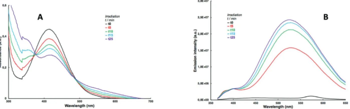
Ces GPP présentent des propriétés photophysiques, photochimiques et optiques très intéressantes lorsqu'elle est couplée à un chromophore. Lors de l'irradiation (libération monophotonique à 405 nm), nous avons observé une diminution de l'absorbance UV (Schéma 8A) et une augmentation considérable de la fluorescence (Schéma 8B) due à la formation du nitroso-énol conjugué (B, schéma 5) après libération du chromophore (utilisé ici pour quantifier, par CLHP, le taux de conversion de la photolyse en fonction du temps d’irradiation).
Schéma 8: (A) Variation de l’absorbance UV pendant l’irradiation et (B) Variation de l’intensité de fluorescence entre le produit de départ et le produit de photolibération de
composé 7c.
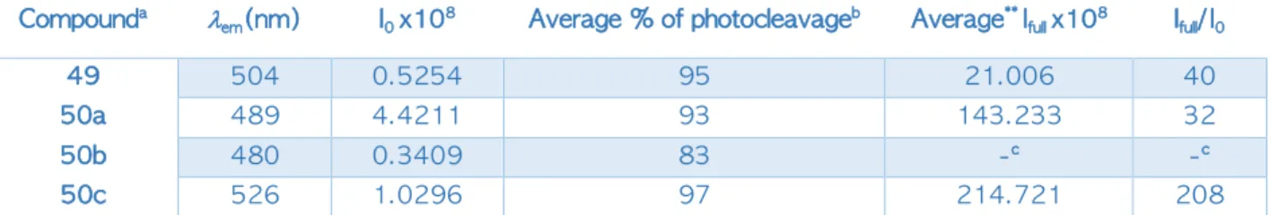
En comparant les propriétés photochimiques et photophysiques de ces composés 7a-c, on a observé une augmentation importante de l’intensité de fluorescence après photolyse complète des composées (100% de libération (Ifull)) comparé à l’intensité avant irradiation I0. De façon
remarquable, l’ajout d’un groupement électro-donneur (OCH3) engendre un décalage vers le
rouge de la longueur d’onde d’émission lemm du fluorophore photolibéré et avec une
augmentation de fluorescence de 200x (Tableau 1).
Composé lem(nm) I0 x108 % de libération
moyenne*
Ifull x108 moyenne* Ifull / I0
6 504 0.5254 95 21.006 40
7a 489 4.4211 93 143.233 32
7b 480 0.3409 83 -** -**
7c 526 1.0296 97 214.721 208
* Moyenne de trois manipes différentes
** le sous-produit subit beaucoup de bleaching, on n’a pas pu calculer les intensités de fluorescence correspondantes.
La formation d’un sous-produit en équilibre céto-enolique a été confirmée à l’aide d’analyses
RMN-1H, cela a permis de vérifier la formation du produit énol (B, Schéma 5), en équilibre
avec un apha-hydroxystibene fortement conjugué responsable du signal fluorescent du sous-produit de photolyse.
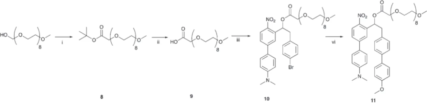
Les excellentes propriétés du composé 7c, qui présente la plus forte augmentation linéaire de fluorescence photo-induite et une émission de fluorescence la plus déplacée vers le rouge, nous a permis d’utiliser ce GPP pour évaluer la possibilité de suivre la photolibération par microscopie de fluorescence sur des cellules en culture. Pour ce faire, nous avons synthétisé le composé 11, une version soluble dans l’eau du dérivé méthoxy-o-nitrobiphenyl 7c (Schéma 9).
Schéma 9: Synthèse de 11: une version soluble du composé méthoxy-o-nitrobiphenyl 7c.
i) NaH, bromoacetate d’éthyl, THF, 0°C, 2h, 87 %; ii) CF3COOH/ dichlorométhane,
3h, température ambiante, 100 %; iii) 5, DMAP, diisopropylcarbodiimide,
dichlorométhane, 0°C, 48 %; vi) 4-méthoxyphényl acide boronique, K2CO3,
Ethanol/Toluène/eau, Pd(PPh3)4, 80°C, 45 min, 30 %.
Des cellules HeLa ont été incubées avec une solution de 11. Les cellules ont été incubées pendant 5 minutes puis irradiées (365 nm) pendant 15 min en continue. Une nette augmentation de l'intensité de fluorescence a été observée lors de l'irradiation. Par contre, les cellules incubées pendant 35 min avec une solution de 11 montraient une très faible intensité de fluorescence qui montre que la fluorescence observée dans les cellules est dû de l’accumulation de sous-produit de photolyse du composé 11. Par conséquent, nous pouvons suivre l'événement de libération de ce nouveau type de GPP par l'augmentation d'un signal de fluorescence dans les cellules. Après avoir testé la photolyse de GPP de série o-nitrobenzyle 7a-c et 11, et ses propriétés de fluorescence très avantageuses, nous avons décidé d’adapter la même stratégie de synthèse sur un GPP récemment développer au sein du laboratoire. Ce composé présente un cœur
hétéroaromatique de type thiophène7 plus sensible à la lumière et permet d’envisager des temps
d’irradiation plus court afin de pouvoir envisager des applications biologiques du GPP (Schéma 10). Nous avons utilisé la même stratégie de synthèse afin d’accéder au GPP capable de former un sous-produit fluorescent après photolyse.
NO2 O Br N O O O 8 HO O O 8 O O O O 8 HO O O O 8 NO2 O O N O O O 8 8 9 10 11 i ii iii vi
La molécule 13 a été synthétisée en 8 étapes et a montré après photolyse un sous-produit avec des propriétés de fluorescence très intéressantes. Un dérivé soluble de 13 est actuellement utilisé au sein du laboratoire afin de développer des précurseurs photolabiles du Glutamate (le principal neurotransmetteur excitateur des cellules neuronales).
Schéma 10: Synthèse de groupement o-nitrobenzyl à cœur hétéroaromatique cagé 13. Il existe un besoin important de GPP capables de générer un sous-produit de photolyse facilement quantifiable, en particulier pour être en mesure d’évaluer le saut de concentration photo-induit d'un effecteur biologique donné couplé. Cela nous a amèné à synthétiser une nouvelle classe de groupements photolabiles de la famille des o-nitrobenzyles capables de générer un sous-produit de photolyse fluorescent. Ces derniers composés ont été conçus afin de produire après photoréaction un sous-produit de type nitrosocétone capable de réaliser une
tautomérie céto-énolique conduisant à un produit conjugué de type a-hydroxystilbene. Chaque
GGP synthétisé a montré un signal de fluorescence très faible avant irradiation. À l'exception du dérivé 7b, une libération presque quantitative (≥93%) a été observée après la photolyse complète de chaque précurseur 7a-c. Tous ces composés ont montré une augmentation intéressante du signal fluorescent induite par la réaction photolytique.
En particulier, le dérivé 7c a montré une augmentation de l’intensité de fluorescence de plus de 200x avec un décalage de la longueur d’onde d’émission vers le rouge (526 nm) après photoclivage complet. Par conséquent, une version soluble dans l'eau de ce composé a été utilisée avec succès dans des expériences d’imagerie sur des cellules et a permis de suivre en temps réel le signal fluorescent induit par la formation du sous-produit de photolyse sur des cellules en culture. S O2N NO2 O O S O2N NO2 O O Br O O O O O O Br 12 13
Nos futures études vont se concentrer sur l'utilisation de cette stratégie pour le développement de groupes photolabiles sensibles à la lumière visible plus efficaces dans la serie o-NB afin de pouvoir corréler la libération d’un effecteur biologique, comme un neurotransmetteur, à la formation d’un signal fluorescence induit par la libération du sous-produit de photolyse.
CHAPTER
I
Introduction
to
Photoremovable
Protecting
Groups:
A
Breakthrough
in
Synthesis
and
Biology
1. Photoremovable Protecting Groups (PPGs)
1.1. Definition:
Photoremovable or so-called photo-cleavable, photo-activatable, photo-releasable Protecting Groups (PPGs) are photolabile chemical compounds that when attached to a biomolecule (for example neurotransmitters or cell signaling molecules) form a stable inactive “caged compound”; this designates that the biomolecule is protected by the PPG. Upon photo-irradiation of the resulting compound, the biomolecule is released and restores its active functionality along with the by-product of the PPG; this process is called uncaging (Klàn et al., 2013) (Scheme I.1).
Scheme I.1: Description of the uncaging process and the release of the biomolecule.
Photoremovable protecting groups provide spatial and temporal control over the release of diverse compounds such as biomolecules (neurotransmitters), organic acids and bases or ions (Ca2+) [Baltrop et al., 1962, Barton et al., 1962; 1965, Ellis-Davies., 2007]
1.2. Requirements:
Several requirements need to be fulfilled by the PPG depending on its application, not all the protecting groups follow the same requirements because they vary depending on the application needed or expected for the system. Many of these properties were originally introduced by Sheehan and Umezawa (Sheehan et al., 1973) and were used to evaluate the potential of a PPG:
i- The PPG should show an absorption wavelength above 300 nm, because irradiations
below this wavelength tend to be absorbed by biological entities like proteins and nucleic acids and thus leading to photo-damage resulting in the formation of initiators for mutations.
ii- The PPG should be stable, clean and pure prior to use because any trace of impurities
iii- The photocleavage reaction should be clean and occur with a remarkable quantum yield of release frel. This quantum yield is described as:
frel = nrel / np where nrel is the amount of substrate released and np is the number of photons absorbed by the caged compound upon irradiation
iv- The PPG should be stable and soluble in the media of examination and if possible
should present an affinity toward the target molecules (binding site on cancer cells, active sites on enzymes…).
v- The by-product released after the release of the bioactive molecule should show no
competitive absorbance with the starting material i.e be transparent to the wavelength of irradiation.
vi- The released by-product should be inert and biocompatible i.e does not react with
the system investigated.
These are the important guidelines and properties for an ideal PPG, but still, a caged compound is considered useful even if it lacks one or two of these properties. The absence of several requirements may be a huge limitation of the PPG in several applications.
1.3. Types of PPGs
Several examples of PPG that fulfill the requirements mentioned above, have been reported and found to have interesting chemical, photophysical and optical properties.
1.3.1. Benzoin PPGs
Sheehan and Wilson (Sheehan et al., 1964, 1971) were the first to explore the photochemical rearrangements of certain benzoin derivatives yielding a 2-phenylbenzofuran by-product. These rearrangements occur by the release of the group attached in position a to the carbonyl function. It was found that 3’,5’-dimethoxybenzoin (DMB) could serve as a PPG for carboxylic acids releasing 2-phenyl-5, 7-dimethoxybenzofuran (DMBF) as a side product, after irradiation in quantitative yields and with a quantum yield of 0.64 ± 0.03 (Scheme I.2).
Scheme I.2: Photocyclization of DMB acetate.
DMB phototriggers have been used for several applications for example in the drug delivery field, in order to study protein folding and muscle relaxation (Rock et al., 1996, Rock et al., 2004).
Another benzoin derivative (R=H) was reported by Givens et al., 2012 this latter showed a quantitative release of phosphates. This study was extended for the release of neucleotides through the synthesis and photolysis of benzoin-cAMP. The same derivative was found to likely
protect and release GABA and glutamates (a- or N- protected Glu).
1.3.2. Arylcarbonylmethyl or Phenacyls (Pac)
Arylcarbonylmethyl groups are aromatic ketones that are thermally stable and easy to access by synthesis. These groups undergo a mechanism of photocleavage that involves hydrogen abstraction from an H-donor by an excited state of the carbonyl function of the phenacyl leading to a ketyl ester intermediate (scheme I.3).
Scheme I.3: H-abstraction mechanism of photocleavage of phenacyls.
A very recent application of phenacyls was reported by Speckmeier and his co-workers (Speckmeier et al., 2018) where in this study, this PPG was used to protect amine function by the formation of Phenacyl (Pac) Urethanes (the amines were linked to Pac by the formation of carbamate linkage). The latter “caged amine” undergoes a photocatalytical cleavage using blue
LEDs (l =455 nm) in the presence of a Ruthenium photocatalyst in order to release the amine
function along with acetophenone (Scheme I.4). This method avoids the drawbacks of using UV irradiation and is carried out in a mixture of acetonitrile and water (4:1), in addition to its
R R X Ph O hv R R O Ph + HX X= acetate R= OMe DMB DMBF O X hv X OH H-donor O H-donor -HX
The presence of the [Ru (bipy)3](PF6)2 is significant to avoid the oxidation of the released amine in combination with ascorbic acid as a reductive quencher in water acetonitrile mixture.
Scheme I.4: Photocatalytical cleavage of phenacyl protected amines.
1.3.3. Coumarin-4-ylmethyl
The photolysis of 7-methoxycoumarinyl-4-methyl (MCM) derivatives was first introduced by Givens and collaborators (Givens et al., 1984) that demonstrated the use of coumarins as photoactivatable phosphate-releasing groups (Entry1, Scheme I.5).
The use of these derivatives was limited until the introduction of coumarin protecting carboxylic acids by Schade and his colleagues (Schade et al., 1999) that shows upon the irradiation of MCM-ester, the release of the free carboxylic acid along with MCM-OH in water (Entry 2, Scheme I.5).
Scheme I.5: 1-Photocleavage of MCM phosphate derivative liberating free phosphate.
2-Photocleavage of MCM-ester derivative liberating free carboxylic acid.
O O N O R2 R1 [Ru] CH3CN/H2O blue LEDs NH R2 R1 O + Pac Urethanes [Ru] = Ru N N N N N N 2 2 PF6 O MeO O O P O OEt OEt O MeO HO O P OEt OEt O + Nu hv 360 nm Nu Nu = OH, OR
1
O MeO O O O R O MeO HO O R O + OH hv 325 nm2
MCM-phosphate MCM-ester H2O MCM-OHCompared to other PPGs the coumarin derivatives doesn’t possess a very high quantum yield
(f = 0.25 for the best derivative), but they possess a remarkable molar extinction coefficient
along with their rapid photolysis that makes these coumarin derivatives great candidates for the photo-liberation of biomolecules.
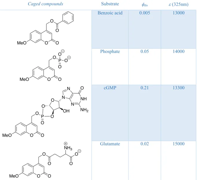
That’s why these derivatives where applied in several research works for the liberation of carboxylic acids (Schade et al., 1999), phosphates (Givens and Matuszewski, 1984), cyclic nucleotides (Furuta and Iwamura, 1998) and glutamates (Furuta et al., 1999). A selection of
coumarin caged compounds with their relative quantum yield of disappearance fdis and their
molar extinction coefficient e is presented in Table 1.
Caged compounds Substrate fdis e (325nm)
Benzoic acid 0.005 13000
Phosphate 0.05 14000
cGMP 0.21 13300
Glutamate 0.02 15000
Table 1: Liberation quantum yield of different caged compounds of the 7-methoxycoumarinyl-4-methyl
family in 1:1 methanol/HEPES buffer
O MeO O O O O MeO O O P O O O O MeO O O P O N NH O NH2 N N O O O OH O MeO O O O O NH3 O
Over the time, several structural modifications were studies in order to modify the photochemical and optical properties of coumarin derivatives.
Certain modifications succeeded in presenting the first coumarin Photoremovable Protecting Group model sensitive to two-photon excitation based on
6-bromo-7-hydroxycoumarin-4-ylmethyl (Bhc) which presents an interesting two-photon efficiency cross section du of 1 GM
(Goeppert-Mayer) at 740 nm for the release of glutamates as described by Furuta and co-workers (Furuta et al., 1999), but this series suffers from low water-solubility. Another interesting and more soluble generation of coumarin was presented by introducing an electron donating amino group in position 7 for example, 7-N,N-diethylamino-coumarin-4-yl (DEACM). This structural modification permitted to observe an important red-shift in the absorbance maximum from 325 nm to around 400 nm which makes these derivatives more adapted for in vivo applications (Shembekar et al., 2005). The performance in two-photon excitation of the DEACM PPG in water was studied by Lin and collaborators (Lin et al., 2012)
and showed an efficiency cross section du = 0.12 GM at 800 nm. These values of cross section
are relatively important and permits the uses of these derivatives for the photo-regulation of various biological events (plasma membrane-specific photoactivation, modulation of calcium ion oscillations).
Recently the group of Ellis-Davies used the DEACM platform to synthesize a new PPG, the DEAC450. This latter presents an extended p-conjugated system that induces a red-shifted
absorbance leading to an absorption maximum lmax at 450 nm. Also, the DEAC450
Photoremovable Protecting Group shows an interesting activity for two-photon excitation with an uncaging cross section du = 0.5 GM at 900 nm for the liberation of glutamates (Olson et al.,
2013). These interesting results make this coumarin derivative an intriguing tool for biological experimentation. The structure of the 3 PPG mentioned above are presented in Scheme I.6.
Scheme I.6: Structures of the coumarin derivatives efficient in two-photon excitation.
O -O O O O H N Br CO2 -CO2 -O N O O O O O N O Glu O R Bhc-Glu λabs = 365 nm δu = 1 GM at 740 nm DEACM-OPMB λabs = 395 nm δu = 0.12 GM at 800 nm DEAC450 λabs = 450 nm δu = 0.5 GM at 900 nm
1.3.3.1. Thio-carbonyl Coumarin PPGs
Most of the presently reported photoremovable protecting groups show strong absorbance below 450 nm, a region where riboflavin and carotenoids absorb light. Research groups are working on adjusting the chemical structure of these groups in order to overcome this limitation of absorbance. One of the explored strategies is to increase the conjugation of the absorbing chromophore and/or to insert heteroatoms in the backbone of the chromophore.
An interesting study on the photophysics and the photochemistry of thiocarbonyls (thiones) was established by Maciejewski and his co-workers (Maciejewski et al., 1993) and emphasizing on the fact that the energy of the C=S bond is weaker than a C=O bond therefore the excited electronic states are found at lower energies in the sulfur-containing species leading to the fact that C=S species absorb light at higher wavelengths compared to that of C=O.
A more recent work on thiones and their use in the field of PPG photoactivation focuses on the use of thiocoumarin (tc) backbone which possesses a red-shifted absorption compared to the carbonyl analogue (Fournier et al., 2013). This derivative was used to cage cyclofen-OH (Ind) in order to photocontrol the activity of a transcription factor (En2) fused into modified estrogen
receptors. The tcInd system shows a maximum of absorbance at 469 nm with etcInd = 270000
M-1 cm-1 and upon irradiation tcInd is converted into tcOH and Ind (Scheme I.7).
Scheme I.7: Synthesis of a caged thicoumarin-cyclofen system.
Another research work on coumarin groups was established recently aiming to increase the absorption wavelength of these derivatives (Lin et al., 2018). This work focused on the development of new coumarin based PPGs by modifying position 3 by adding an electron rich (donor) styryl moieties which give access to red-shifted absorption wavelengths. Several donor groups were used like 4’-N,N-dimethylaminostyryl (1a, Scheme I.8) and julolidinestyryl (1b, Scheme I.8), and these groups were also incorporated in the thiocoumarin platforms (2a and 2b Scheme I.8). O OH S Et2N + H2N O OH DIC CH2Cl2 DMAP O O S Et2N H N O O OH tcOH Ind tcInd
Scheme I.8: Different structural modifications of coumarin and their effect on the absorption
wavelength.
In this study, the incorporation of julolidine rings contributes to a 10 nm shift due to the better conjugation of the amino group with the aromatic core and the incorporation of the sulfur atom induces a 40-50 nm shift due to an intramolecular charge transfer to the empty 3d orbitals of the sulfur atom.
1.3.3.2. Coumarin-Caged Adenosine Cyclic 3’,5’-Monophosphates
Fluorescence is very important for following the liberation of the attached biomolecules after irradiation of the caged compounds and can be used to monitor the “uncaging” event. Coumarin derivatives like MCM and DEAC possess a fluorescence emission between 397 and 491 nm in methanol/HEPES buffer depending on the substitution on the coumarinyl moiety.
One drawback of coumarins is that the starting PPG possesses the same fluorescence as the released by-product (MCM-OH) and in this manner the follow up of the uncaging becomes impossible.
Only one example of the uncaging event quantification by fluorescence using a coumarin PPG was reported by Bendig and co-workers (Bendig et al., 1999) in the case of coumarin-caged Adenosine Cyclic 3’,5’-Monophosphates (MCM-caged cAMP).
O Et2N O X N O Et2N S X N O O X N N O O X N N 1a λmax = 446 nm 1b λmax = 467 nm 2a λmax = 490 nm 2b λmax = 515 nm
This work focuses on the fact that CM-caged nucleotides are weakly fluorescent whereas their by-product MCM-OH presents strong fluorescence which facilitates monitoring the release process (Scheme I.9).
Scheme I.9: Photocleavage of coumarin-caged cAMP
This system is characterized by the difference in fluorescence between the starting caged compound and the uncaging by-product. Normally coumarin-based caged compounds are fluorescent before photocleavage but when attached to adenosine nucleotides they show a very weak fluorescence due to an accelerated internal conversion from the excited state caused by
mixing the pp* and the np* states. This energy transfer between the coumarin moiety and the
adenosine moiety leads to a drop in the fluorescence or so-called fluorescence quenching. This quenching is advantageous in this case because it helps in quantifying the release of cAMP from the fluorescence observed after photocleavage due to the liberation of the coumarin by-product.
As much as this example is interesting it still has an important limitation were this fluorescence quenching is exclusive for coumarin-caged adenosine and is not valid for other biomolecules attached to coumarins.
1.3.3.3. Coumarins for signaling lipids uncaging at the plasma membrane
Another interesting use of coumarin PPGs was demonstrated by Schultz and co-workers (Nadler et al., 2015) in the end of 2015, this study focused on the use of the fluorescence characteristic of coumarin for plasma membrane-specific photoactivation.
+
CM-caged cAMP CM-OH
The principle of this study was to synthesize hydrophilic sulfonated coumarin caged fatty acids that shows before uncaging a fluorescent signal on the cellular membrane, followed by a lipid flip-flop. Upon photocleavage by UV light the fatty acid remains in the plasma membrane whereas the coumarin-OH by-product is released inside the cellular space and a fluorescence is observed at the vesicles level due to the entrance of the highly hydrophilic coumarin-OH to these vesicles by endocytosis. With further irradiations the fluorescence signal starts to decrease due to photobleaching (Scheme I.10, Nadler et al., 2015).
Scheme I.10: The mechanism of plasma membrane photoactivation using sulfonated coumarin caged
fatty acid.
This method, that was recently developed using coumarin derivatives, acts as a tool in plasma membrane marking. But yet this method has its limitations since it doesn’t provide any information about the concentration jump of the biological effector and it remains a qualitative method (fluorescence) more than quantitative (concentration).
1.3.4. O-hydroxycinnamates PPGs
In 2007, inspired by the work of Porter’s group in the late 1980s (Turner et al., 1988), Ludovic Jullien and his coworkers (Gagey et al., 2007) investigated the use of hydroxycinnamates for alcohols uncaging. This work focuses on the release of a coumarin by-product and taking advantage of the fluorescence signal induced by this uncaging by-product after photo-cleavage to quantify the release and report the uncaging event. These cinnamates occur in two stereoisomers, the Z and E isomers, induced by light irradiation.
The transformation of the E isomer to the Z isomer under the action of light leads to the liberation of an alcohol along with a coumarin by-product (Scheme I.11).
Scheme I.11: Mechanism of photo-release of alcohols and coumarin after irradiation of
hydroxycinnamates.
Both isomers show weak fluorescence as long as the alcohol is attached, and whenever the alcohol is released under the action of light, strong fluorescent coumarin by-product is released. This signal is helpful to quantify the release of alcohols from these caged-systems.
Another very recent interesting example for the use of o-hydroxycinnamates is that illustrated by Paul and his collaborators (Paul et al.; 2017). This example introduces excited-state intramolecular proton transfer (ESIPT) induced fluorescent o-hydroxycinnamates. ESIPT is an ultrafast enol-keto phototautomerization exhibited by a proton transfer intramolecularly in the excited state.
In this study, ESIPT induces a fluorescence that help monitoring the uncaging in real time by the release of the benzothiazole-coumarin by-product and this study can extend to diverse bioactive molecules containing a terminal hydroxyl function. The ESIPT has two advantageous effects on these hydroxycinnamates; (1) a huge stokes’ fluorescence shift (orange color) and (2) a distinct fluorescence color change upon photorelease. In other words, the uncaging is monitored by the drastic change in fluorescence color from orange (hydroxycinnamates) to blue (benzothiazole-coumarin) after photocleavage (Scheme I.12).
(E) o-Hydroxycinnamate (Z) o-Hydroxycinnamate
Scheme I.12: Photoinduced uncaging of alcohols from the ESIPT induced fluorescent
o-hydroxycinnamates.
But still this system has its limitations and drawbacks since these cinnamates tend to auto-isomerize even in absence of light leading to a leak in the biomolecule since the transformation of the E isomer to Z isomer leads to the release of the alcohol attached. Another drawback is that this system is only adapted for the release of alcohols and thus it is not valid for a diversity of biomolecules.
1.3.5. Nitroindoline groups
The 7-nitroindoline (NI) derivatives were first introduced as photo-labile protecting groups in organic synthesis for the protection of carboxylic acids in aprotic medium (Amit et al., 1976). After that, the group of Papageorgiou (Papageorgiou et al., 1999) adapted these derivatives in order to adjust their photochemical properties and to render them useful as photoremovable protecting groups that are stable (in aqueous media) and efficient caged compounds for the photo-release of L-glutamates (Scheme I.13).
Scheme I.13: Different photolysis pathways for the nitroindoline derivatives with respect to the medium
used. In aprotic medium, the irradiation releases a nitroindoline by-product along with the carboxylic acid attached (right pathway). In physiological buffer, the irradiation releases a nitrosoindoline by-product of photolysis along with the release of L-Glutamate (left pathway).
O O R S N OH hv ≥ 365 nm ESIPT O O R S N O H S N O O + ROH * o-hydroxycinnamates benzothiazole-coumarin OMe N NO2 R O OMe N H NO2 OMe N H NO hυ physiological buffer aprotic solvent 1% water hυ + RCO2H R= Phe RCO2H + R= L-Glutamate
These nitroindoline derivative present a negligible hydrolysis percentage in physiological conditions in contrary to coumarins, and in addition to that, Kerry and his group (Kerry et al., 1988) has demonstrated that the photochemical reaction leads to the liberation of glutamate from nitroindoline is faster (order of nanoseconds) compared to the kinetics of activation of glutamate receptors.
Inspite of their low quantum yields (f = 0.085), these groups have a very weak basal activity
due to the hydrolytical stability (Canepari et al., 2001) making them a good candidate in investigating the central nervous system using caged neurotransmitters.
1.3.6. O-nitroaryls
1.3.6.1. O-nitrobenzyl groups
The o-nitrobenzyl (o-NB) groups have been introduced by in 1970 and they have been reported as the most used protecting groups due their efficiency in protecting different functional groups: carboxylic acids, imidazoles, phosphates for the synthesis of nucleosides, alcohols and amines (Patchornik et al., 1970). These groups undergo the following mechanism upon irradiation (Scheme I.14): (a) photoactivation of the nitro function leading to the formation of an aci-nitro intermediate (I1), followed by a (b) cyclization to form a 1,3-dihydrobenz-isoxazol-1-ol
intermediate (I2), that upon (c) ring opening releases the attached biomolecule (X) along with
the nitrosocarbonyl by-product (R=H nitrosoaldehyde, R=Ar, Ph, CH3,… nitrosoketone).
Scheme I.14: Mechanism of photocleavage of o-NB protecting groups.
O-nitrobenzylic protecting groups have been the most used PPGs for synthetic and biological
applications. One example is the use of o-NB under mild conditions for automated RNA and DNA synthesis developed by Stutz and Pitsch (Stutz et al., 1999) by protecting the oxygen atom of the four nucleic bases (Adenine, Cytosine, Guanine, and Uracil) for the synthesis of oligoribonucleotides (Scheme I.15).
NO2 X hv H2O X N OH O NO OH X R R R NO O R +HX I1 I2
Scheme I.15: O-NB caged nucleotides as building blocks for the automated RNA synthesis.
Also, o-nitrobenzyl PPGs were used in natural product synthesis like the one reported by Wong in 1993 in the total synthesis of Leukotriene (LTC4). During the final steps of the synthetic route, a deprotection is done by irradiating the o-NB-caged system at 350 nm to afford the final compound (Scheme I.16).
Scheme I.16: Final steps in the total synthesis of N-methyl LTC4.
Over the years, several modifications were applied to this type of PPGs in order to render them more stable, more efficient, and also to increase the quantum yield and/or the wavelength of absorbance (for efficiency in biological applications) together with the rate of release. In order to do that two important modifications were done: (1) substitution in the benzylic position and (2) substitution of the aromatic ring of the o-NB group.
1.3.6.1.1. Substitution in the benzylic position
In the 1970s, Woodward has proposed such modification by adding a substituent in the benzylic position. These substituents provide an electronic effect at the benzylic site and also adds a second hydrogen-abstracting unit that helps in increasing the efficiency of the group.
One synthetic drawback could be faced when having a chiral benzylic center (due to a substituent different from the o-NB core) because it will be more difficult to protect chiral molecules especially in the synthesis of amino acids and oligonucleotides.
O O Base OTMD HN P N O NC O O O NO 2 NO2 Base = C, A, G, U CO2Me OH C6H11 NO2 N CO2Me HN O S N H O CO2Me CO2Me OH C6H11 NH CO2Me HN O S N H O CO2Me 1. hv (350 nm) 2. hydrolysis N-methyl LCT4
When using substituted o-NB core at the benzylic position, a significant increase in the quantum yield of release was observed. This increase could be explained through the mechanism of photocleavage of this family of PPG after the formation of the aci-nitro intermediate, leading to the release of nitrosocarbonyl. In the case of substituted o-NB, the molecule released is a nitrosoketone which shows a higher quantum yield of release compared to that observed for nitrosoaldehyde (originating from no benzylic position substitution) after photocleavage. It is also known that water-solubility is really an important criterion for the use of PPGs in biological applications, this property can be achieved by adding substituents that render the molecule more soluble such as COOH functions. Forming more water-soluble groups tend to increase the yield of release compared to that of the non-soluble analog. This was confirmed by
an example illustrated by Bassani and his group (Bassani et al., 2010), where a
-carboxy-6-nitroveratryl (aCNV) tend to release carboxylic acids with f = 0.17 and a yield of release 3
times more than the parent compound a-carboxynitrobenzyl (aCNB). This proves that adding
a water-solubilizing substituent to the molecule increase the yield of release of the protected compound (Scheme I.17).
Scheme I.17: Structures of aCNB and aCNV.
1.3.6.1.2. Substitution of the aromatic ring of the o-NB group
Due to the drawback of chirality that could be induced by the substitution on the benzylic position and their negligible effect on the absorbance wavelength, numerous modifications have been applied on the aromatic ring of the o-NB chromophore.
These modifications could induce 3 important effects: (1) tuning the absorbance wavelength and/or having a better quantum yield, (2) possibility of attaching the compound to a solid support or on a linker and (3) possibility to modify the solubility properties of the molecule. For example, addition of another electron-withdrawing nitro substituent leads to an increase of the quantum yield from 0.13 for 1 to 0.62 for 2 for the release of amines from carbamates.
NO2 O CO2H NO2 MeO MeO MeO MeO R O O O R ⍺CNB ⍺CNV
Another example of modification is the addition of a carboxylic acid with one carbon spacer to form compound 3 making it more soluble than the parent compound 1. Also, the addition of 2 methoxy group substituents to 1 makes compound 4 absorb at longer wavelength (>350 nm). These examples are illustrated in Scheme I.18.
Scheme I.18: Different modification on the aromatic ring of o-NB.
As a conclusion, the substitution at the benzylic position and the substitution on the aromatic ring of o-NB protecting groups has a significant effect on the photochemical and photophysical properties. Substituent effects are not only of crucial importance for the absorption spectrum of the chromophore but are also prominent for the stability of the C–R bond (where R is the caged function).
By only making minor changes in the substitution pattern of structural derivatives of the ortho-nitrobenzyl protecting group it is possible to create PPGs that can be photocleaved with different wavelengths of light (ldeprotect: 345–420 nm). The most useful way to obtain a bathochromic (red) shift of the absorption band is the addition of an electron-withdrawing group (EWG) at the para-position. Additionally, substituting the ortho-nitrobenzyl core with a moderately electron-donating group (EDG) like alkoxy (–OR) in the meta-position permits cleavage with longer wavelength of light. A significant hypsochromic shift can be obtained by changing the a-substituent with respect to the R-group. Furthermore, extending the linker
between the chromophore and the cleavable C–R bond gives similar results. Scheme I.19 shows the shift in wavelength of absorbance depending on the different substitutions and modifications on the molecules. NO2 X NO2 NO2 X NO2 X O HO2C MeO MeO NO2 X 1 2 3 4
Scheme I.19: Effect of modification on the aromatic ring and the benzylic position of o-NB on the
wavelength of absorption.
Alexander Heckel group has demonstrated very recently (Becker et al., 2018) that the addition of a dimethyl amino (DMA) group to the nitro dibenzofuran (NDBF) shifts the wavelength of absorbance from 312 nm to 424 nm. Another system was developed by Specht and his co-workers in 2012 based on a nitro biphenyl core (NBP) with different R groups, this system shows a maximum of absorbance at 405 nm which is due to the conjugation of the nitrobenzene with the second phenyl ring (Donato et al., 2012).
1.3.6.2. o-Nitrophenethyl groups
Another interesting series of o-nitroaryls is the o-nitrophenethyl (o-NPP), where the biomolecule is attached on carbon 2 of the ethyl substituent at the ortho position to the nitroaryl moiety. These PPGs have been synthesized and tested for their efficiency in photocleavage mainly by Specht’s group (Donato et al., 2012 and Herbivo et al., 2013) for the release of GABA and inorganic phosphate. These groups undergo a complex mechanism that was described in 2001 (Walbert et al., 2001). In fact, two mechanistic photochemical pathways for
these o-NPP groups exist. One of these pathways is a b-elimination based pathway (Pathway
A) which is commonly known as the major pathway leading to the photo-liberation of the biomolecule in aqueous medium. But there exists a second pathway considered as the minor one which leads to the formation of the nitroso-derivative (Pathway B). This latter mechanism doesn’t induce any photolysis leading to photo-liberate biomolecule attached. Upon irradiating the o-NPP PPG, it absorbs an amount of energy that permits the system to pass from a fundamental state to an excited state, in this state an a-exo-cyclic hydrogen transfer towards the
nitro group occurs, that leads to the formation of the aci-nitro intermediate which is kinetically favored (Scheme I.20).
O NO 2 OH O NO 2 OH N λabs=312 nm λabs=424 nm NO2 NO2 O O NO2 R
λabs=260 nm λabs=355 nm λabs=405 nm
The acid-base equilibrium of the aci-nitro intermediate is the determining step of which photochemical pathway is adopted. The weak basic character of water is sufficient for the deprotonation of the aci-nitro intermediate to form a new anionic form which undergoes further a b-elimination liberating the biomolecule along with o-nitrostyrene (Pathway A, Scheme I.20).
In the presence of an acidic medium (millimolar order of hydrochloric acid) the acid base equilibrium is shifted toward the formation of the neutral aci-nitro intermediate which then favors the formation of a cyclic intermediate followed by the formation of the o-nitroso derivative (Pathway B, Scheme I.20).
Scheme I.20: Mechanism of photo-liberation of a biomolecule from the o-NPP series following 2
pathways: (A) which is the favored mechanism in basic medium releasing o-nitrostyrene along with the liberation of the biomolecule and (B) which is favored in “acidic” medium and forms o-nitroso derivative and doesn’t permit the liberation of the biomolecule.
The removal of the organic molecule attached to the protecting groups mentioned previously (among many others) necessitates the use of UV-near visible irradiations. It is well known that these irradiation wavelengths are not biocompatible since these lights are absorbed by tissues mainly oxyhemoglobin which absorbs every light below 650 nm. On the other hand, water molecules become absorbent at wavelengths >950 nm. Depending on this information a “phototherapeutic window” could be defined for the tissue transparency above 650 nm and below 950 nm (Scheme I.21, Juzenas et al., 2002).
N O O OR N OH OR O N HO O OR NO OR OH - H+ + H+ N O OR O N O O β-elimination
Aci-nitro intermediate Cyclic intermediate o-nitroso derivative Pathway B
anionic Aci-nitro intermediate o-nitrostyrene Pathway A
Scheme I.21: Transparency window for living tissue absorbance showing the absorption spectra of
various endogenous relevant molecules.
The majority of chromophores absorb light in the regions below 400 nm and some caged compounds absorb visible light which makes them very complicated to handle. In addition to that, the use of wavelengths between 650-950 helps in reducing photodamage and phototoxicity as well as increasing the depth of tissue penetration. In order to overcome these problems and to use a diversity of PPG in biological applications an important approach is the use of two-photon excitation (2-PE).
2. Two-Photon Excitation
2.1. Overview
The use of mono-photonic excitation in the field of in vivo applications of Photoremovable Protecting Groups has been problematic until the introduction of two-photon excitation was proposed. This technique uses types of irradiations different from those used for the classical mono-photon excitation. The two-photon excitation functions in a non-linear optic process defined by the following equation (1):
𝑃 ∝1
Where P is the molecule excitation probability, da is two-photon absorption cross section. The
efficiency of a molecule to absorb two photons is expressed in GM (1 GM = 10-50 cm4. s. photon -1) in the honor of Maria Goeppert-Mayer that was the first to introduce the theoretical phenomenon of non-linear optics in 1930 (Goeppert-Mayer et al., 1931). Finally, I is the light intensity of the excitation source. P is directly proportional to the square of the light intensity I, this shows that the possibility to excite a molecule will be higher in the zone where the light intensity is at its maximum. In other words, the focal point of the optical system used for irradiation in 2-PE presents the highest light intensity for excitation whereas in the classical confocal microscopy the excitation takes place over the full optical length (Scheme I.22).
Scheme I.22: Difference in fluorescence emission between mono and bi-photonic excitation.
This phenomenon was used in the development of 3D imaging, photonics, photodynamic therapy… The two-photon excitation technique requires light irradiations with very high intensities which can only be achieved by using ultrafast pulsed laser like the titanium:sapphire laser that provides 3 mm bean at 100 fs pulse duration with a frequency of 90 MHz. This laser type provides optimum radiations at 800 nm which is compatible with biological applications and corresponds to minimum mammalian tissue absorbance.
The excitation of a molecule is observed when the molecule absorbs one or more photons with a certain energy in order to pass from the fundamental ground state to the excited state. In order for this to happen, the molecule absorbs either one photon with E = hn (mono-photonic
absorption) or two photons with E = hn/2 (bi-photonic absorption). Using lower energy for
2-PE reduces photodamage and cytotoxicity and using higher excitation wavelengths reduces the risk of tissue proteins absorbance and mutations (Scheme I.23, Benninger and Piston, 2013).
Scheme I.23: Simplified Jablonski (energy-level) diagram of conventional one-photon excitation (left) and nonlinear two-photon excitation (right). In each case, the absorption of photon(s) populates an excited state from which the molecule can relax by emitting a photon.
2.2. Design
In order to increase the efficiency of compounds to increase their two-photon excitation sensitivity, several approaches have been developed on fluorophores and chromophores. Numerous chromophore geometries have been investigated; linear (1D), planar (2D) and tetrahedral (3D) structures, and these investigations led to the construction of the optimal and typical chromophore architecture.
The smallest system to be useful in biology is composed of a donor (D)-acceptor (A) system with p-system cores incorporated in the system as well. The extension of the p-system (increase
in conjugation) improves the 2-PE properties of the molecule (Scheme I.24).
A wide range of electron-donating and electron-accepting terminal groups have been
investigated including many p-deficient heterocycles (Albota et al., 1998, Mongin et al., 2007
and Terenziani et al., 2008) and it was found that D-p-D and D-p-A-p-D structures are more
efficient than A-p-A and A-p-D-p-A systems (Pawlicki et al., 2009) as shown in Scheme I.25.
Scheme I.25: Variation of two-photon action cross section by structural modifications.
It is known that the use of electron donating groups (EDG) like alkylamino increases the
two-photon efficiency more than electron accepting groups (EAG) like SO2CF3. And also, the use
of alkylamino as EDG is more efficient than using alkoxy or other oxygen-based donors. And it is remarkably interesting to use phenoxides which are very strong donors and give very high 2-PE cross sections as in the case of the pink molecule in Scheme I.25 (Pawlicki et al., 2009). Similarly to mono-photon absorption, it is possible to determine the two-photon uncaging
action cross section (du) of a molecule by multiplying the two-photon absorption cross section
(da expressed in GM) by the two-photon quantum yield of uncaging (fu): 𝛿𝑢 = 𝛿𝑎 . 𝜙𝑢 (2)
Not all PPGs present a structure that shows efficiency in two-photon excitation since few PPGs have the donor-acceptor core with an extended p-system. It has been demonstrated that a
photoremovable protecting group with da < 0.1 GM and fu <10% is considered as non-efficient for biological applications (Corrie et al., 2005).
R R R = NHex2 δ = 1200 GM at 705 nm R = SO2CF3 δ = 83 GM at 705 nm R = OMe δ = 1200 GM at 705 nm R = NBu2 δ = 83 GM at 705 nm R R O O N O N O Na Na δ = 4100 GM at 705 nm