HAL Id: tel-03156509
https://tel.archives-ouvertes.fr/tel-03156509
Submitted on 2 Mar 2021
HAL is a multi-disciplinary open access
archive for the deposit and dissemination of sci-entific research documents, whether they are pub-lished or not. The documents may come from teaching and research institutions in France or abroad, or from public or private research centers.
L’archive ouverte pluridisciplinaire HAL, est destinée au dépôt et à la diffusion de documents scientifiques de niveau recherche, publiés ou non, émanant des établissements d’enseignement et de recherche français ou étrangers, des laboratoires publics ou privés.
Regulation of NFATc3 stability by SUMO and E3
ubiquitin-ligases Trim39 and Trim17
Meenakshi Basu Shrivastava
To cite this version:
Meenakshi Basu Shrivastava. Regulation of NFATc3 stability by SUMO and E3 ubiquitin-ligases Trim39 and Trim17. Agricultural sciences. Université Montpellier, 2020. English. �NNT : 2020MONTT043�. �tel-03156509�
RAPPORT DE GESTION
2015
Devant le jury composé de
Dimtris Xirodimas, DR2, CNRS Solange Desagher, CR1, CNRS
Andrea Pichler, Professor, Max Planck Institute of Immunobiology and Epigenetics Germana Meroni, Professor, University of Trieste
Manuel Rodriguez, DR2, CNRS Dimitris Liakopoulos, DR2, CNRS Prèsident Directrice de thèse Rapporteur Rapporteur Examinateur Examinateur
THÈSE POUR OBTENIR LE GRADE DE DOCTEUR
DE L’UNIVERSITÉ DE MONTPELLIER
En Biologie Santè
École doctorale Sciences Chimiques et Biologiques pour la Santé CBS2 168 Unité de recherche IGMM-CNRS UMR5535
Regulation of NFATc3 stability by SUMO and
the E3 ubiquitin-ligases Trim39 and Trim17
Présentée par Meenakshi Basu
Shrivastava
Le 14/12/2020
Dedicated to my loving parents, Mrs. Malika Basu and Mr. Tarun Kanti Basu
&
i
Acknowledgements
My journey to France commenced in 2015 and five years later, I really feel very fortunate for being able to document my experience, both at scientific and human level.
I would first like to thank Université de Montpellier, La Fondation de l’Association pour la Recherche contre le Cancer (ARC), La Ligue contre le Cancer, and La Fondation pour la Recherche Médicale (FRM) for funding my thesis and for their constant research support. I am also grateful to all the facilities at Institute of Molecular Genetics of Montpellier, Centre National de la Recherche Scientifique (CNRS).
I would like to thank my supervisor, Dr. Solange Desagher, whose expertise was invaluable in formulating the research questions and methodology. Thank you so much for your valuable inputs throughout my graduate study. Thank you for encouraging and supporting me to attend conferences, both national and international. Thank you so much for donning many hats. You have been my supervisor, my confidante, my psychologist, my go to person in these four years. I have been blessed to have you as my supervisor and could not have asked for more. I honestly did not even realize where all the time flew. Thank you so much for shaping my academic career.
A big thank you to my thesis committee jury: Dr. Manuel Rodriguez and Dr. Dimitris Liakopoulos for their invaulable inputs and constructive discussions. I cannot thank both of you enough for being super nice and so supportive. I feel really fortunate to have interacted with both of you, both at professional and personal level. A special thank you for the material support given by Dr. Manuel Rodriguez.
Thank you Dr. Guillaume Bossis for really making SUMO manageable for me. When I first started, I had a hate relationship with it and now, over the years, I can say that we have become good friends, all thanks to you. A big thank you for the material support, for discussion, and for critical inputs in developing the SUMO part.
I would like to thank my entire team. I am thankful to Dr. Irina Lassot for her contribution to this work. My nick name for her is the ‘in vitro Queen’. Thank you so much for teaching me and for your scientific inputs throughout my study. Thank you for being so inspiring and for lifting my spirits when the results went all the over the place. Thank you for always being there for me.
Stephan Mora, the lab genie. Thank you so much for teaching me techniques during my stay in the group. Thank you so much for your contribution to this work and for selflessly continuing even during the pandemic. I cannot thank you enough for this. Thank you for teaching me so much not only in research but in life too. I was a lost bird with no idea about the administration and you really helped me kickstart my life here. Thank you for being my go to person.
ii A big thank you to Dr. Barbara Mojsa or as for laying the foundation of my thesis and for your contribution. It was a pleasure meeting you during my stay here. I would also like to acknowledge Alina Kozoriz and it was really nice interacting with you. I wish you good luck for your thesis! Finally, I am so grateful to my parents. I have no words to put in this section to express what both of you mean to me. Simply put. I owe my existence to both of you. Thank you for being my best friends. I cannot write more as I cannot even attempt to formulate my sentences when it comes to both of you. A big thank you to my husband. Thank you for being so understanding and so supportive during my study. I honestly again have no words for what you mean to me. The three of you are my lifeline and I love you so damn much. Thank you to Dr. Anais Dreau, my mentor from the ‘Female in Sciences’ program. You have been so supportive and so encouraging from the very first time we met. Thank you for being a good listener and for being so approachable. Thank you for motivating me. A big thank you to Dr. Eugenia Basyuk. Whenever I think about you, I only say that you were like my second mother here. I am so fortunate to have worked with you during my internship at IGMM and I feel so blessed to have known you and spend time with you. Thank you so much for helping me selflessly with everything and for being so supportive when I was applying for my PhD. The biggest thank you to the little princess whom I have known since the time she was in her mamma’s belly, Devyanshi Chahar aka chutku. You are a cutiepie and so adorable. My little munchkin. Your smile has made my day even on the worst of the days. Thank you to my friends, Dhanvantri and Sanjay, for providing me a home away from home. Thank you to all the people in IGMM for providing a good work environment.
iii
Table of Contents
List of Figures ... vi
List of Tables ... vii
Abbreviations ... viii
Chapter 1 The Ubiquitin-Proteasome System ... 1
1.1 Ubiquitin and the Enzymatic Cascade ... 1
Ubiquitin ... 1
Enzymes at work ... 1
Deubiquitinating enzymes (DUBs) - spoilers alert ... 5
An Insight into Ubiquitin Chain-Signalling ... 6
Complexity of Ubiquitin Chain Assembly ... 7
Mechanisms of the Chain Assembly ... 8
1.2 26S Proteasome ... 10
Components of the 26S Proteasome and its Assembly ... 11
Recognition of the Substrate and its Processing by the Proteasome ... 12
Inhibitors of the Proteasome ... 13
1.3 Crosstalk with other Post Translational Modifications ... 15
Chapter 2 SUMOylation ... 17
2.1 SUMO Paralogs, Pathway and Enzymology ... 17
SUMO Paralogs ... 17
Regulation of SUMO Isoforms ... 18
SUMO Pathway ... 18
SUMO Enzymology ... 20
SUMO Proteases ... 27
2.2 SUMO Consensus Sequence ... 28
2.3 Non-covalent interactions with SUMO ... 29
Types and Features of SIMs influencing their binding to SUMO ... 30
Regulation of SUMO substrates and SUMO-binding proteins by SIMs ... 32
SIMs in the SUMO conjugation machinery ... 33
iv
2.4 Crosstalk with other Post-translational Modifications ... 36
2.5 Molecular and Cellular Consequences of SUMOylation ... 39
Molecular consequences of SUMOylation ... 39
Cellular Consequences of SUMOylation ... 40
2.6 SUMO in Disease ... 42
SUMO in Neurodegenerative Diseases ... 42
Chapter 3 TRIM proteins ... 45
3.1 The Tripartite Motif ... 47
RING domain and its structural aspects ... 47
B-Box domains and its structural aspects ... 48
Coiled-coil domains and their structural aspects ... 49
C-Terminal domains and their structural aspects ... 50
PRY-SPRY domains ... 50
3.2 Functional and Structural aspects of TRIM proteins ... 51
Interactions between ubiquitin E2 enzymes and TRIM proteins ... 51
Homo and hetero-interactions between TRIM proteins ... 52
3.3 Molecular and cellular functions of TRIMs ... 54
Molecular functions of TRIMs ... 54
Cellular functions of TRIM proteins ... 58
3.4 Pathological roles of TRIM proteins ... 64
TRIM proteins in neurodegenerative diseases ... 64
3.5 TRIM17 ... 68
Expression of Trim17 ... 69
Structure of TRIM17 ... 69
Partners and substrates of TRIM17 ... 70
Molecular and cellular functions of Trim17 ... 72
Trim17 and disease ... 73
3.6 TRIM39 ... 74
Expression of TRIM39 ... 74
Structure of TRIM39 ... 76
Partners and substrates of TRIM39 ... 76
Molecular and cellular functions of TRIM39 ... 77
v
Chapter 4 Nuclear Factor of Activated T-cells ... 80
4.1 NFAT family ... 81
4.2 Physiological roles of NFATs ... 82
4.3 NFAT and disease ... 85
4.4 Structure and functional domains of NFATs ... 88
4.5 NFAT and transcription ... 89
4.6 Regulation of NFAT ... 92
Chapter 5 NFAT and neuronal Apoptosis ... 99
5.1 Apoptosis ... 99
5.2 Pathways of Apoptosis ... 99
5.3 Neuronal apoptosis ... 101
5.4 NFATc3 and NFATc4- similar but different too ... 102
Objectives ... 105 Results ... 108 Unpublished Results ... 157 NFATc3 ... 157 Challenges ... 157 Results ... 157 NFATc4 ... 158 Introduction ... 158 Results ... 159 Conclusion ... 160 General Discussion ... 161 Bibliography ... 171 Résumé ... 172 Abstract ... 172
vi
List of Figures
Figure 1 : Structure of ubiquitin ... 2
Figure 2 : Schematic representation of the three steps involved in ubiquitination of a substrate ... 2
Figure 3 : Main role of DUBs ... 5
Figure 4 : List of E2 or E3 enzymes ... 6
Figure 5 : Representation of various combinations of ubiquitin modifications. ... 7
Figure 6 : Schematic representation of sequential addition mechanism of ubiquitin chain assembly. ... 9
Figure 7 : Schematic representation of the 26S proteasome ... 10
Figure 8 : Structure of proteasome inhibitors. ... 14
Figure 9 : Sequence comparison of SUMO paralogs.. ... 18
Figure 10 : Schematic representation of the SUMO pathway. ... 19
Figure 11 : Binding interfaces on a surface structure of Ubc9 ... 21
Figure 12 : Mechanisms depicting E2 dependent substrate interactions. ... 22
Figure 13 : Bona fide classes of E3 ubiquitin ligases. ... 24
Figure 14 : Residue conservation of the three SIM types. ... 30
Figure 15 : Model of the β-grasp fold of SUMO. ... 31
Figure 16 : Schematic representation of possible combination of hydrophobic (h) core of the SIM ... 32
Figure 17 : Hydrophobic core amino acids of bona fide SIM domains ... 32
Figure 18 : Schematic representation of SUMO-targeted ubiquitin ligases (STUbLs). ... 34
Figure 19 : Schematic representation of STUbL in different models ... 35
Figure 20 : Lys post-translational modifications (PTMs) ... 36
Figure 21 : Crosstalk of PTMs such as phosphorylation, and acetylation. ... 37
Figure 22 : Modification of SUMO family members by other PTMs produces complex signaling codes. ... 38
Figure 23 : Molecular consequences of SUMOylation. ... 40
Figure 24 : Schematic representation of potential mechanisms of SUMOylation in neurodegenerative diseases. ... 43
Figure 25 : Protein domain organization of TRIM proteins. ... 45
Figure 26 : Classification of different TRIM protein family members ... 46
Figure 27 : RING finger domain ... 47
Figure 28 : Structure of B-Box domains of MID1 ... 49
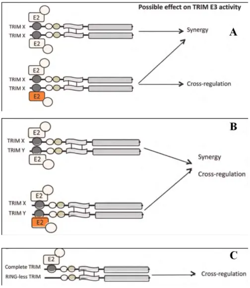
Figure 29 : Different proposed models for possible TRIM homocomplexes on the E3 activity. ... 52
Figure 30 : Different functions of TRIM proteins. ... 58
Figure 31 : Schematic representation of regulation of cell cycle by TRIM proteins. ... 64
Figure 32: Gene expression for TRIM17 ... 69
Figure 33: Predicted homodimeric 3D structure of CC and PRY-SPRY domain ... 70
Figure 34 : GST-pulldown of TRIM17 ... 71
Figure 35 : Gene expression for TRIM39 ... 74
Figure 36 : 3DSolution structure of the RING domain of human TRIM39 ... 75
Figure 37 : 3D Solution structure of the B-Box domain of human TRIM39... 75
Figure 38 : The interaction network of human TRIM39 revealed different interactors ... 77
Figure 39 : Schematic representation of human calcium-regulated NFAT genes and their isoforms. ... 80
vii
Figure 41 : Primary structure of NFAT composed of different domains ... 87
Figure 42 : SUMOylation consensus motifs of murine NFATc3 and corresponding sequences. ... 89
Figure 43 : Representation of NFAT/AP-1 binding site. ... 90
Figure 44 : Alignment of the genomic sequences around the transcription start site of mouse Trim17.. ... 90
Figure 45 : Schematic representation of the activation and regulation of the calcineurin-NFAT pathway. ... 93
Figure 46: The experimentally validated SUMOylation sites ... 98
Figure 47 : Schematic representation of the main players of the intrinsic and extrinsic pathways of apoptosis. ... 100
Figure 48 : CGNs deprived of serum and KCl as a model of transcription-dependent apoptosis. ... 102
Figure 49 : Trim17 is not an E3 ubiquitin-ligase of NFATc3 ... 105
Figure 50 : Neuro2A cells transfected with HA-NFATc3 together with different TRIMs. ... 106
Figure 51 : Schematic representation of the questions raised on the regulation of NFATc3. ... 107
Figure 52 : Trim39 binds to 4xSUMO2. ... 158
Figure 53 : Trim39 and NFATc4 interact with each other.. ... 159
Figure 54 : Trim17 reduces the interaction between Trim39 and NFATc4. ... 160
Figure 55 : Three putative SIM sites in Trim39 obtained by using GPS-SUMO ... 165
Figure 56 : Schematic representation of the effects of Trim17 on the regulation of NFATc3. ... 169
Figure 57 : Mechanisms by which Trim17, Trim39, and SUMO regulate the stability of NFATc3 ... 170
List of Tables
Table I : Properties of human SUMO proteases. ... 28Table II : SUMO acceptor sites. ... 29
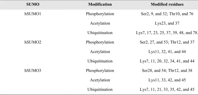
Table III: List of modified residues in human SUMO1, SUMO2, and SUMO3 ... 38
Table IV : Deregulations in gene expression and locations of SUMO conjugation system in cancer. ... 44
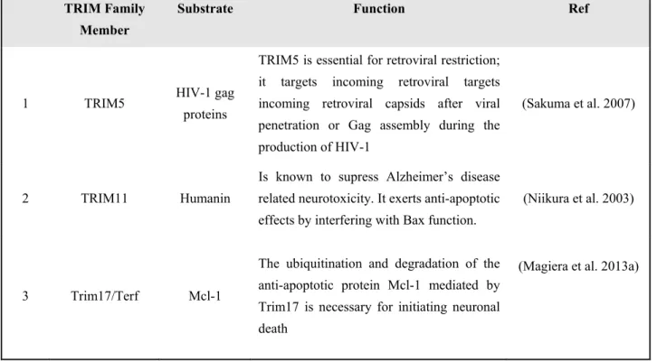
Table V : List of few E3 ubiquitin ligases in TRIM proteins and their function ... 54
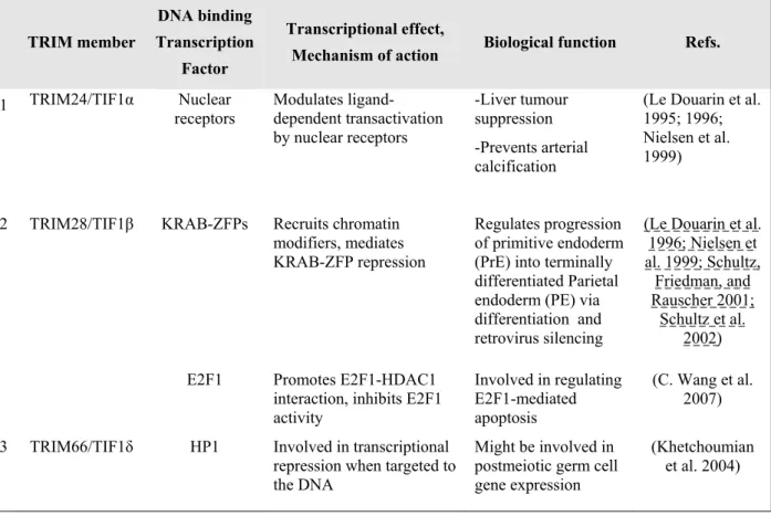
Table VI : Role of different TRIM proteins in transcriptional regulation. ... 57
Table VII : Interactions between some of the autophagy-related proteins and TRIM proteins. ... 61
Table VIII : Role of different TRIMs in the regulation of kinetochore. ... 65
Table IX : List of NFAT family members. ... 81
Table X : Phenotype in the immune system in NFAT-deficient mice. ... 83
Table XI : Summary of regulation of NFAT members in cancer. ... 86
Table XII Partners of NFAT in transcription. ... 91
Table XIII : Different cellular functions, genes and effect on transcriptional regulation by NFAT... 92
Table XIV : List of NFAT kinases involved in the signaling. ... 95
viii
Abbreviations
AAD Alzheimer’s Disease
ADAR Adenosine Deaminase that acts on RNA AL Autophagy Lysosome
AP Activator Protein
Apaf-1 Apoptotic Protease Activating Factor 1 APL Acute Promyelocytic Leukaemia APP Amyloid Precursor Protein APR Acute Promyelocytic Leukaemia ARF ADP-ribosylation Factor
ATG Autophagy-related Genes/Proteins ATO Arsenic Trioxide
Az Antizyme Aβ Amyloid beta
B
BCL B-cell Lymphoma BD Behcet’s Disease
BDNF Brain-derived Neurotrophic Factor Bdnf Brain Derived Nerve Growth Factor BHK Baby Hamster Kidney
BR Bromodomain BRAT BRAin Tumour
bZIP Basic Regiion-leucine Zipper
C Cactin Cactus-interactor CaN Calcineurin CASP Caspase CBP CREB-binding Protein CC Coiled-coil domain Cdc34 Cell Division Cycle 34 CDK Cyclin Dependent Kinases CGN Cerebellar Granule Neurons
CHIP C-terminus of HSP70-interaction Protein CHX Cycloheximide
CK Casein Kinase CK2 Casein Kinase2
CKI Cyclin-dependent Kinase Inhibitor C-L Caspase-like
ix CLE Cutaneous Lupus Erythematosus
CNRS Center National de la Recherche COS C-terminal subgroup One Signature
COST European Cooperation in Science and Technology CP Core Particle
CREBBP CREB-binding Protein CTD C-Terminal Domains CT-L Chymotrypsin-like Cyt-c Cytochrome-c
D
DAG Diacylglycerol
DBD DNA Binding Domain DD Death Domains DE3 ArcticExpress
DESI deSUMOylating isopeptidases Dgrn Degringolade
DISC Death-inducing Signal Complex DIV Days in Vitro
DR Death Receptor Pathway DR Death Receptor
DUB Deubiquitinase
DYRK1a Dual-specificity Tyrosine-phosphorylation Regulated Kinase 1a
E
EFP Oestrogen-responsive Finger Protein ER Endoplasmic Reticulum
ERK Extracellular Signal Related Kinase
F
FADD FAS-associated Death Domain FIL Filamin-type Immunoglobulin FN Fibronectin type
G
GABA Gamma-aminobutyric Acid GAP Growth Associated Protein Gene
GHRH Hypothalamic Growth-hormone Releasing-hormone GPCR G-protein Coupled Receptors
GSK3β Glycogen-synthase Kinase 3β
x
H
HCSM Hydrophobic Cluster SUMOylation Motif HNSCC Human Head and Neck Squamous cell Carcinoma
I
IL Interleukin
INSERM Institut National de la Sante et de la Recherche Medicale IP3 Inositol-1,4,5-triphosphate
J
JAMM JAB1/MPN/MOV34 JNK c-JUN Kinase
K
KBTBD11 Kelch Repeat and BTB Domain-containing Protein 11
L
LGMD Limb-girdle Muscular Dystrophies LIR LC3-interacting Region
LPS Lipopolysaccharides
M
MAPK Mitogen Activated Protein Kinases MATH Meprin and TRAF-homology Domain MCL Mantle Cell Lymphoma
MCL Myeloid cell Leukemia mDA mouse Dopaminergic Neurons MDM2 Murine Double Minute 2 mESC Mouse Embryonic Stem Cell Meth Methamphetamine
MHC I Major Histocompatibility Complex Class I MINDY Motif Interacting with Ub-containing Novel DUB miR microRNA
miRNA microRNA MM Multiple Myeloma MMP Matrix Metalloproteinases MOAP Modulator of Apoptosis
MOMP Mitochondrial Outer Membrane Permeabilization MPN Mpr1-Pad1 N Terminal
MS Multiple Sclerosis MT Mitochondria
xi
N
NB Nuclear Bodies
NCoR Nuclear Receptor co-Repressor
NDSM Negatively Charged Amino-acid-dependent SUMOylation Motif Nedd8 neural precursor cell expressed, developmentally downregulated gene 8 NEM N-ethylmaleimide
NFAT Nuclear Factor of Activated T-cells NHR NFAT Homology Region
NLS Nuclear Localization Signal NMDA N-methyl-D-aspartate Receptor NMR Nuclear Magnetic Spectroscopy NPC Nuclear Pore Complexes
NQO1 NAD(P)H quinone Oxidoreductase NS Nonstructral Protein
Nur77 Nuclear Receptor77
O
ODC Ornithine Decarboxylase OS Opitz Syndrome
OTUs Ovarian Tumor Protease
P
PBL Peripheral Blood Lymphocyte PCD Programmed Cell Death
PCNA Proliferating Cell Nuclear Antigen PD Parkinson’s Disease
PHD Plant Homeodomain PIAS Proteins Inhibitor of STAT PINIT Pro-Ile-Asn-Ile-Thr
PIP2 Phosphatidylinositol 4,5-Bisphosphate PKA Protein Kinase A
PKB Protein Kinase B
PLA Proximity Ligation Assay PLCγ Phospholipase Cγ
PMA Phorbol12-myristate 13-acetate PMA Phorbol-myristate Acetate ProCASP9 Procaspase-9
PSDM Phosphorylation-dependent SUMOylation Motif PTM Post-translational Modification
xii
R
RA Rheumatoid Arthritis RanBP2 Ran-binding protein 2 RanGAP RanGTPase-activating protein
RANKL Receptor Activator of Nuclear factor κB Ligand RARα Retinoic Acid Receptor
RBP RNA Binding Proteins RFP RET Finger Protein RHR Rel-Homology Region RIG Retinoic Acid-inducible Gene RING Really Interesting New Gene RNF111 RING finger protein Arkadia ROS Reactive Oxygen Species RP Regulatory Protein
Rpn Regulatory particle non-ATPase RPP21 Ribonuclease P/MRP 21kDa
Rpt Regulatory particle triple AAA-ATPase
RT-PCR Reverse-transcription Polymerase Chain Reaction
S
S/T Serine/threonine-rich C-terminal
SAP Scaffold attachment factor-A/B, Acinus, and PIAS SCM SUMO Consensus Motif
SIMs SUMO Interacting Motifs SLE Systemic Lupus Erythematosus SN Substantia Nigra
SNP Single Nucleotide Polymorphisms SNP Single Nucleotide Polymorphisms SNURF Small Nuclear RING Finger Protein SOC Store-operated Calcium Channels SOCE Store-operated Ca2+ Entry SP-CTD Unique C-terminal Domain SS Sjogren’s Syndrome
STUbLs SUMO-targeted Ubiquitin Ligases SUMO Small Ubiquitin-like Modifier
T
TCR T-Cell Receptor
terf testis RING Finger Protein TF Transcription Factors
TFP Testis Abundant Finger Protein Th T-helper
xiii TIF Transcriptional Intermediary Factor
T-L Trypsin-like TM Transmembrane TNF Tumour Necrosis Factor TRIM TRIpartite Motif tRNA Transfer RNA
U
UBA Ubiquitin-Associated Domain
Uba1 Ubiquitin-like Modifier-activating Enzyme Ubc1 Ubiquitin-conjugating Enzyme E21 UBD Ubiquitin Binding Domains Ube2S Ubiquitin-conjugating Enzyme E2S Ubl Ubiquitin like modifiers
UBL Ubiquitin-like Domain UCH Ubiquitin C-terminal Hydrolase UIM Ubiquitin-interacting Motifs ULP Ubiquitin-like-specific Protease UPS Ubiquitin Proteasome System USP Ubiquitin-specific Proteases USPL Ubiquitin-specific Protease-like
V
VHL Von Hippel-Lindau tumor suppressor VWA Von Willebrand factor A
W
WBS Williams-Beuren Syndrome
Z
1
Chapter 1 The Ubiquitin-Proteasome System
The ubiquitin-proteasome system is a major protein degradation system after the lysosomal system in eukaryotes (Grune et al. 2001; Berke and Paulson 2003; Goldberg 2003; Levine and Klionsky 2004; Glickman and Ciechanover 2002; Schwartz and Ciechanover 2009). Once the proteins are conjugated to ubiquitin chains, they are directed to a macromolecular protease known as the 26S proteasome where they are eventually degraded (Kleiger and Mayor 2014; Glickman and Ciechanover 2002; Bard et al. 2018; Collins and Goldberg 2017). The entire journey from transfer of ubiquitin onto the protein to the final degradation of the protein into peptides involves lot of complex processes. Indeed, it is safe to say that we have come a long way in the field of ubiquitin research since the discovery of ubiquitin in 1975 (Goldstein et al. 1975).
1.1 Ubiquitin and the Enzymatic Cascade
Ubiquitin
Ubiquitin is a 76 amino acid protein with a molecular weight of 8.5kDa. It is highly conserved, from yeast to man, implying that many of its surfaces are recognized by the UBDs (Ubiquitin binding domains). In mammals, ubiquitin is encoded by four different genes. The genes, UBA52 and RPS27A, code for a single copy of ubiquitin fused to the ribosomal proteins L40 and S27A, respectively. The other two genes namely, UBB and UBC genes code for poly-ubiquitin precursor proteins (Kimura and Tanaka 2010).
The hydrophobic surface comprising of Isoleucine44 (Ile44), Leucine8 (Leu8), Valine70 (Val70), and Histidine68 (His68) facilitates the recognition of ubiquitin by the proteasome (Dikic, Wakatsuki, and Walters 2009). Interestingly, the HECT E3’s (a class of E3 ubiquitin ligases), DUBs (deubiquitinases), and UBDs can recognize another hydrophobic patch, Ileucine36 (Ile36) which involves Leu77 and Leu73 of the ubiquitin tail and can mediate interactions between ubiquitin molecules in chains (Komander and Rape 2012). Ubiquitin can be further modified by either ubiquitin itself or other post-translational modifications such as small ubiquitin-like modifier (SUMO), phosphorylation and acetylation (Swatek and Komander 2016).
Enzymes at work
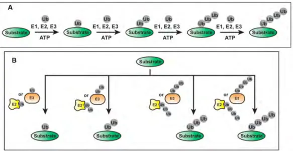
The ubiquitination of proteins involves sequential action of three ubiquitin enzymes namely E1 activating enzyme, E2 conjugating enzyme, and E3 ubiquitin ligase. An E1 activating enzyme
2 activates ubiquitin in an ATP-dependent manner by forming a covalent bond between the C-terminal end of ubiquitin and a cysteine residue in its active site (Figure 1).
The thioesterified ubiquitin is then passed to the E2 conjugating enzyme (Figure 2A). The final step involves the E3 ubiquitin ligase that binds to both the E2-bound ubiquitin and the substrate, therefore promoting the transfer of ubiquitin onto the substrate (Figure 2C) (Bence, Sampat, and Kopito 2001; Adams 2003; B. Gong et al. 2016). The mammalian ubiquitin cascade consists of two members of the E1 family which tags all the E2s with ubiquitin whereas there are 40 E2s that help facilitate the delivery of ubiquitin to more than 600 E3s (Deshaies and Joazeiro 2009).
Proteins, in general, are ubiquitinated on the lysine residues which results in the formation of an isopeptide bond between the carboxyl end of ubiquitin and the lysine primary amine. Proteins can be Figure 1 : Structure of ubiquitin: a. The C-terminal tail and residues of Ile44 patch are indicated b. Structure displaying
the seven Lys residues and Met1. The amino groups used in ubiquitin chain formation are indicated by blue spheres. From (Komander and Rape 2012)
Figure 2 : Schematic representation of the three steps involved in ubiquitination of a substrate. A. E1 activating enzyme forms a thioester bond with ubiquitin in an ATP-dependent manner; B. The ubiquitin is then transferred to the E2 conjugating enzyme; C. E3 ubiquitin ligase helps in the recognition of the substrate and catalyses the covalent attachment of ubiquitin to the target substrate via an isopeptide bond. The polyubiquitinated substrate is then rendered to degradation by the 26S proteasome. From (B. Gong et al. 2016)
3 either mono-ubiquitinated (conjugated to one ubiquitin monomer) or polyubiquitinated (conjugated to several ubiquitins). The state of protein ubiquitination (mono or poly-ubiquitinated) and the type of ubiquitin chain attached on a protein finally decides its fate i.e. whether it undergoes degradation or participates in cellular responses (Komander and Rape 2012; P. Xu et al. 2009; Tokunaga et al. 2009). To make matters more interesting, ubiquitin can crosstalk with other ubiquitin-like modifiers (Ubl’s) such as SUMO, Nedd8 (neural precursor cell expressed, developmentally downregulated gene 8), phosphorylation (Schulman and Harper 2009).
E1 Activating Enzyme
The multidomain E1 enzymes aid in the activation of ubiquitin and thus facilitate its transfer to the active site of E2. This is a crucial function of E1s to maintain cellular homeostasis because failure to do so results in the entire UPS being shutdown (Yili Yang et al. 2007). Several structural studies have contributed to our understanding of how E1 activates and transfers ubiquitin to the E2. The E1-Uba1 (ubiquitin-like modifier-activating enzyme) for ubiquitin is a monomeric protein of 110-120kDa (I. Lee and Schindelin 2008; Lake et al. 2001; S. K. Olsen and Lima 2013). The E1-mediated reaction involves ATP-dependent adenylation of the C-terminal carboxyl group of ubiquitin, the formation of a thioester bond between the catalytic cysteine residue of E1 and the C-terminus of ubiquitin, and ultimately the transfer of ubiquitin onto the catalytic cysteine of E2. An elegant study presented the crystal structure of a UbE1-E2 (Ubc4)/Ub/ATP.Mg complex stabilised by inducing
disulfide bond between the E1 and E2 active sites. It revealed that the conformational changes in the E1 brings E1 and E2 active sites together (S. K. Olsen and Lima 2013).
E2 conjugating enzymes
The transfer of ubiquitin to the substrates is catalyzed by the E2 conjugating enzymes (Pickart 2001; Ye and Rape 2009; Varshavsky 2012; Ciechanover 2015; Stewart et al. 2016). All E2s share an approximately 150 amino acid conserved core domain. The thioester bond between the C-terminal end of ubiquitin and the conserved cysteine residue of the E2 forms the core of the E2 active site. The positioning of the thioester bond in an optimal conformation governs the rate of ubiquitin transfer and this could be achieved by the E2, the E3 or their combination. Initial study had shown that E2 Ubc1 (ubiquitin-conjugating enzyme E21) forms a non-covalent interface with a ubiquitin whose active site is thioesterified (Hamilton et al. 2001). On the other hand, it was also demonstrated that in addition to the covalent thioester bond, E2s Cdc34 (cell division cycle 34) and Ube2S (ubiquitin-conjugating enzyme E2S), also form noncovalent interfaces with ubiquitin (Saha et al. 2011; Wickliffe et al. 2011). This suggested that E2s catalyze ubiquitin transfer (in part) by holding ubiquitin against an interface on the surface of E2 which optimizes the thioester bond position in the active site. Despite the fact that E2s have similarity, different E2s exert different biological functions. For example, many substrates associated with cell cycle regulation (such as cyclin-dependent-kinase inhibitor Sic1 and the G1 cyclins) were stabilised with mutations in the Cdc34 gene alone. The stabilization of these two substrates reflects the specificity of Cdc34 when it partners with two different SCF E3s (Pickart 2001).
4
E3 ubiquitin-ligases
Currently there are more than 600 E3s in humans which participate in the ubiquitination pathway by mediating substrate specificity. Based on the presence of characteristic domains and the mechanism by which they transfer ubiquitin on to the substrate, E3 ubiquitin-ligases can be classified into three types: RING E3s, HECT E3s, and RBR E3s (Morreale and Walden 2016).
RING E3s: They are the most abundant type of ubiquitin ligases which are characterized by the presence of a zinc-binding domain called Really Interesting New gene or by a U-box domain, which has RING domain but lacks the presence of zinc. The RING and U-Box domains are responsible for binding the ubiquitin-charged E2 and stimulating ubiquitin transfer. The RING E3s mediate a direct transfer of ubiquitin to the substrate thereby acting as a scaffold to orient the ubiquitin-charged E2 with respect to substrate protein. The RING E3s could exist as either monomers, homodimers or heterodimers while the U-Box domains can function as monomers or homodimers. Examples of RING E3s include Cullin-RING ligases (CRLs) and Anaphase promoting complex/cyclosome (APC/C) (Deshaies and Joazeiro 2009). Interestingly, RING E3s can be regulated by neddylation, phosphorylation, and interaction with small molecules.
HECT E3s: HECT (Homologous to the E6 AP carboxyl terminus) domain family of E3 ligases catalyze the ubiquitin transfer to the substrate protein in a two-step reaction: ubiquitin is transferred from a catalytic cysteine to the E3 and then from E3 to the substrate. Located at the C terminus of the proteins, the conserved HECT domain is characterized by a bi-lobar architecture where the N-terminal lobe interacts with the ubiquitin charged E2 whereas the C-terminus contains the catalytic cysteine. Human HECTs can further be classified into three sub-families: (i) Nedd4 family which contains tryptophan-tryptophan (WW) motifs (ii) HERC (HECT and RCC-1-like domain) family which contains one or more regulators of chromosome condensation1(RCC1)-like domains (RCDs), and (iii) other HECTs that contain various domains (Metzger, Hristova, and Weissman 2012; Morreale and Walden 2016). Furthermore, the intramolecular interactions facilitate the regulation of catalytic activity of HECT E3s that keep the protein in an autoinhibited state and this autoinhibitory state is released in response to various signals.
RBR E3s: The RING-between RING-RING (RBR) E3s catalyze the ubiquitin transfer via a two-step reaction where ubiquitin is transferred to a catalytic cysteine on the E3 first and then to the substrate. These E3s contain two predicted RING domains (RING1- and RING-2) separated by an in-between-RING domain (IBR). in-between-RING1 recruits ubiquitin charged E2 while in-between-RING domain possesses the catalytic cysteine and is referred to as Rcat (required-for-catalysis) domain. Similar to the RING2 domain, the IBR domain lacks the catalytic cysteine and is referred to as BRcat (benign-catalytic) domain. Several domains in RBRs are involved in intramolecular interactions that keep the protein in an auto inhibited state and again, this state is released through mechanisms that include phosphorylation or protein-protein interactions (Dove and Klevit 2017; Morreale and Walden 2016).
5
Deubiquitinating enzymes (DUBs) - spoilers alert
The recycling of ubiquitins, once the substrate has been committed to the degradation pathway, is critical for maintaining ubiquitin homeostasis. As a result, both the proteasomes and the lysosomal sorting machinery are associated with deubiquitinating enzymes (DUBs) (Metzger, Hristova, and Weissman 2012; Morreale and Walden 2016). DUBs greatly influence many biological processes and cellular pathways such as tumorigenesis, DNA damage response, and DNA repair pathway to name a few. DUBs catalytically cleave single Ub or polyubiquitin chains from proteins and thus help in maintaining the ubiquitin pool. The human genome encodes approximately 100 DUBs and can be classified into six families. Ubiquitin-specific proteases (USP) family is the largest family boasting approximately 55 members. Other DUBs belong to ubiquitin C-terminal hydrolases (UCHs), ovarian tumor proteases (OTUs), Josephin, JAB1/MPN/MOV34 (JAMMs), and motif interacting with Ub-containing novel DUBs (MINDY)s families (Komander, Clague, and Urbé 2009; Nijman et al. 2005; Turcu, Ventii, and Wilkinson 2009; He et al. 2016).
The main roles of DUBs include modulating the activity of E2s, counteracting E3s, assisting degradation machinery, maintaining ubiquitin homeostasis, etc. (Error! Reference source not found.). DUBs are capable of inhibiting ubiquitination by interfering with the reactivity and the
formation of the E2-Ub intermediates. For example, USP7 (DUB found in all eukaryotes) removes the ubiquitin from target proteins such as Mdm2, ICPO, and p53 (Nicholson and Suresh Kumar 2011). USP7 binds to the N-terminal ASTS sequence of UbE2E1 (an E2) and forms a complex. This
Figure 3 : The figure represents the main role of DUBs from counteracting E2/E3s, recycling of ubiquitins to editing of ubiquitin chains to change non-degradative ubiquitin signals. From (He et al. 2016).
6 binding results in the attenuation of UbE2E1-mediated ubiquitination via the ASTS motif within its N-terminal extension and the catalytic domain of USP7. Interestingly, the disruption or inactivation of the interaction between USP7 and UbE2E1 leads to the destabilization of UbE2E1 (Sarkari et al. 2013). DUBs often co-regulate with E3 ligase partner to orchestrate the ubiquitin loading and removal in target proteins. For example, the stability of p53 is regulated by the DUB, USP10, under the physiological conditions as well as in DNA damage response with its E3 partner Mdm2. USP10 is responsible for maintaining stable levels of p53 in the cytosol, however post DNA damage, part of USP10 translocates to the nucleus to deubiquitinate p53. This translocation ultimately results in a boost of p53 activation (J. Yuan et al. 2010).
DUBs assist the degradation machinery (Figure 4). The hydrolysis of ub-chains before proteins are unfolded and degraded (Verma et al. 2002) is carried out by the DUB, POH1. It is known that two other DUBs, Ubp6/USP14 and UCH37, oppose protein degradation as they trim ubiquitin chains from the distal end of the chain. This results in a decreased affinity of the protein for the proteasome (Jacobson et al. 2014; Bashore et al. 2015). DUBs maintain the ubiquitin homeostasis by generating Ub precursors from encoded genes, trimming the Ub precursors to free Ubs, disassembling polyubiquitin chains from proteins, and by recovering ubiquitin from chains. The major DUB, USP5, releases ubiquitin from unanchored isopeptide-linked ubiquitin chains (Reyes-Turcu et al. 2006).
An Insight into Ubiquitin Chain-Signalling
The nature of the ubiquitin modification governs the fate of the ubiquitinated substrate. Ubiquitin plays an integral role in modulating functions of various proteins and its dysregulation has a deep
Figure 4 : The figure displays a small list of E2 or E3 enzymes that help in the assembly and the DUBs that support the disassembly of different ubiquitin chains. The biological processes associated with different chain types are also depicted by a cartoon below. From (Swatek and Komander 2016)
7 impact in the development of numerous human diseases. It is well established that each ubiquitin chain type has a different function (Akutsu, Dikic, and Bremm 2016a). The covalent attachment of ubiquitin on the protein substrates can lead to either the elimination of the substrate by the proteasome or it might affect its localisation, substrate activity, binding with partners and can also have non-proteolytic consequences (Oh, Akopian, and Rape 2018).
A single ubiquitin molecule (mono or multi-mono) can modify one or multiple Lys (K) residues in proteins. In a ubiquitin chain, ubiquitin moieties can be conjugated through one of their K residues (K6, K11, K27, K29, K33, K48, K63) or N-terminal Met residue (M1) (Meyer and Rape 2014). There are two types of ubiquitin conjugates: the homotypic type where the same K or methionine residue connects all the building blocks of chains and heterotypic type which contains mixed ubiquitin chain linkages within the same polymer. The different polymers of ubiquitin on a substrate (homo and heterotypic) lead to distinct cellular responses. Additionally, there might be a possibility to form mixed and branched chains where the ubiquitin molecule is ubiquitinated at two or more than two sites (Figure 4) (Swatek and Komander 2016). Recent studies have also added new layers of complexity wherein ubiquitin can be modified by several ubiquitin-like modifications such as SUMOylation, acetylation, and phosphorylation (Swatek and Komander 2016; Akutsu, Dikic, and Bremm 2016b; Cuijpers, Willemstein, and Vertegaal 2017; Kane et al. 2014; Kazlauskaite et al. 2014; Koyano et al. 2014).
Complexity of Ubiquitin Chain Assembly
The study of ubiquitin chains is evolving at a rapid pace and varied approaches exist (Figure 5). The chain types that regulate tremendous amounts of biological processes can be like a spider’s web.
8 Monoubiquitination was thought to be involved in subcellular localization of proteins (van der Horst et al. 2006) rather than promoting degradation (Hicke 2001). The requirement of a minimum of four or more ubiquitin moieties for degradation is being challenged. For instance, Lu et al. showed that used single- molecule kinetics studies to demonstrate that monoubiquitination of cyclinB1 was able to stimulate degradation by human proteasomes (Lu et al. 2015). In another study by Dimova et al., it was shown in Xenopus extracts that multi monoubiquitination of CyclinB1 was an efficient signal for its degradation (Dimova et al. 2012).
If we talk about homotypic ubiquitin chains, they are formed when a substrate is modified by the same type of ubiquitin chain. It was identified that homotypic K11-polyubiquitin chains did not bind to the proteasome or to the ubiquitin receptors associated with the proteasome. Considering an example of Cyclin B as the substrate, it was shown that the homotypic K11 linkages did not signal its degradation. In contrast, heterotypic K11/K48-polyubiquitin chains were able to signal degradation by the proteasome (Swatek and Komander 2016).
The heterotypic (mixed) chains can be categorized into two groups: a. Tandem mixture of different linkages (mixed or hybrid chains) or b. A ubiquitin moiety within a polyubiquitinated chain can be attached to different linkages (branched or forked chains) (Figure 5). An example of cellular roles of mixed chains is the use of K11/K63 mixed linkages in intracellular trafficking. These are formed the process of endocytosis of major histocompatibility complex class I (MHC I) membrane proteins. The role of branched ubiquitin chains emerged with APC/C, an E3 ubiquitin ligase that regulates mitosis. A study highlighted that in-vitro branched chains comprising of K11/K48 (and also K11/K63) enhanced the proteasomal degradation of the substrates. They suggested that the density of the ubiquitin signal could be increased when K11-branched from already formed short K48-linked chains that may enable multiple K11-linked chains from a single K48-linked chain (Meyer and Rape 2014).
Mechanisms of the Chain Assembly
Over the years of the ubiquitin study, several models for chain assembly have been proposed (Hochstrasser 2006). The two basic mechanisms that are followed are the sequential addition and the en bloc transfer. The former involves the transfer of individual ubiquitin moiety on to the substrate. In the case of the latter, the pre-formed chains are transferred to the active cysteine site of an E2 or HECT/RBR E3 to a substrate (Deol, Lorenz, and Strieter 2019a).
Sequential Addition
According to this model, each ubiquitinated protein acts as a substrate to form long substrate-linked chains (Figure 6A). It, therefore, becomes necessary to perform kinetic studies in order to validate this mechanism as we should be able to detect individual products on rapid timescales ranging from milliseconds to seconds. This mechanism was supported by a study that employed a millisecond kinetics measurement approach (Pierce et al. 2009). Using Cdc34 as E2, SCFCDC4 and SCFβ-TRCP as
9 each round of chain elongation used each reaction intermediate as a substrate. The study concluded that most enzyme-substrate encounters were not productive as the first ubiquitin transfer is the slowest step in the assembly of ubiquitin chains. After the first ubiquitin is successfully transferred, it becomes easier to sequentially add the ubiquitins on the growing chain before the dissociation of the enzyme-substrate complex. The rate of this sequential addition of ubiquitins (kub-n) is faster than
substrate dissociation rate (koff) resulting in processive poly-ubiquitination of the substrate. The
consequence of sequential addition is that the fate of the ubiquitinated substrate would depend on the rate with which ubiquitin chains can be formed on it (Deol, Lorenz, and Strieter 2019a).
En-Bloc Transfer
In contrast to the above-mentioned model, there are studies indicating that, pre-formed chains are assembled on the active site of cysteine of an E2 or E3 before the transfer on the substrate (Figure 6B). En bloc transfer mechanism would be impeccable if we consider the efficiency with which a substrate could be poly-ubiquitinated. The advantage of this mechanism over the sequential addition is that in order for a substrate to be tagged with ubiquitin chains of sufficient length, the complex of E3- substrate need not be long-lived. On the other hand, one of the cons of this model is that the mechanism of these pre-formed chains should be faster than the substrate transfer.
Kinetic studies supporting this model were done using UBE2G2 (Fang et al. 2001; B. Chen et al. 2006), a human E2 and Uch7 (Biederer, Volkwein, and Sommer 1996; Hiller et al. 1996; Bays et al. 2001; Deak and Wolf 2001), its yeast ortholog and GP78, an endoplasmic reticulum residing RING E3 ubiquitin-ligase (W. Li et al. 2007). The mentioned E2, UBE2G2, is known to be associated with the ER membrane and it forms K48 linked-ubiquitin chains on the misfolded proteins which are exported from the ER during ERAD. UBE2G2 together with GP78 catalyzed the assembly of ubiquitin chains on the cysteine active site. The driving factor behind this pre-assembly process is the ability of GP78 to oligomerize. The hetero-oligomerization of UBE2G2 and GP78 can bring the Figure 6 : Schematic representation of sequential addition mechanism of ubiquitin chain assembly. From (Deol, Lorenz, and Strieter 2019b)
10 active sites of several UBE2G2 molecules into close proximity (W. Liu et al. 2014). Therefore, the en bloc assembly of ubiquitin chains by GP78 implies that the ubiquitin conjugated to the active site of GP78 provides the acceptor lysine residue (K48) to attack the C-terminus of another thioester-linked ubiquitin attached to either a GP78 subunit or an associated E2 (Deol, Lorenz, and Strieter 2019b).
1.2 26S Proteasome
Protein degradation is an indispensable process to assess the level of proteins in a cell and the maintenance of protein homeostasis. In mammalian cells, the major part of non-lysosomal protein degradation is the proteasome. The proteasome is a large complex protease that is responsible for the degradation of intracellular proteins. The 26S proteasome consists of two subcomplexes: 20S core particle, which is responsible for the proteolytic cleavage of the protein substrates and a 19S regulatory particle, which serves as a site for initiation of substrate recognition (Murata, Yashiroda, and Tanaka 2009; Collins and Goldberg 2017; Budenholzer et al. 2017). The polymerization of ubiquitin, the main molecule that works with the proteasome, serves as a signal for the degradation of numerous target proteins. Once the target proteins are tagged with polyubiquitin chains, they are shuttled to the proteasome where they are proteolytically broken down. Almost all basic cellular processes such as cell cycle progression, apoptosis, signal transduction, immune responses are controlled by the ubiquitin-proteasome system (UPS) (Groothuis et al. 2006; Sujashvili 2016; Grumati and Dikic 2018; Michael James Emanuele and Enrico 2019; Kliza and Husnjak 2020; Q. Fan et al. 2020).
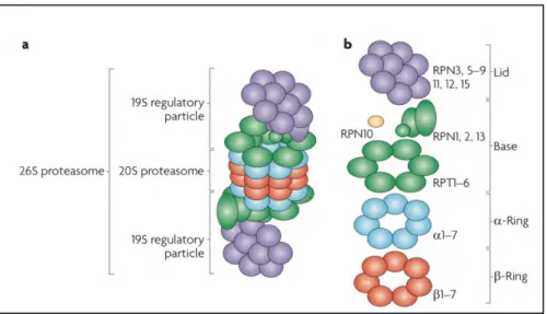
Figure 7 : Schematic representation of the 26S proteasome. a. The 26S consists of the catalytic 20S proteasome (CP) and the 19S regulatory particle (RP); b. Composition of the 26S proteasome: 19S RP (composed of lid and base subunits) and the 20S CP (composed of two outer α-rings and two inner β-rings). From (Murata, Yashiroda, and Tanaka 2009).
11
Components of the 26S Proteasome and its Assembly
20S Core Particle (CP)
In eukaryotes, the 20S core particle (CP) forms a cylinder-like structure composed of 2 outer α rings and 2 inner β rings. Indeed, it is a packed particle constituting 7 structurally similar α and β subunits arranged in the order: α1-7 β1-7 β1-7 α1-7 (Figure 7). The alpha rings form the substrate entry channel
whereas proteolytic cleavage takes place in the beta rings. The α subunits in the CP include conserved N terminal peptide extensions. These are important as they form a gate controlling the substrate entry via the central α ring channels. The crystal structure of the ring suggests that the center of the α-ring is completely closed. This closed conformation prevents the proteins from enteα-ring into the β ring (Arendt and Hochstrasser 1997; W. Heinemeyer, Ramos, and Dohmen 2004; Murata, Yashiroda, and Tanaka 2009).
The three main proteolytic active sites in the two β-rings (β1, β2, β5) contain catalytically active threonine residues at its N terminus indicating that proteasome is a threonine protease. The β1 is associated with caspase-like, β2 with trypsin-like and β5 with chymotrypsin-like activities that have the ability to cleave peptide bonds at the C terminus of acidic, basic and hydrophobic amino acid residues, respectively (Groll et al. 1997; Unno et al. 2002; Tanaka 2009). The protein substrates are cleaved into oligopeptides that range from 3 to 15 amino acid residues by the 20S proteasome. They are then subsequently hydrolyzed to amino acids by oligo-peptidases or amino-carboxyl peptidases.
19S Regulatory Protein (RP)
The 19S regulatory protein (RP), also known as proteasome activator (PA700), generally caps the proteasome on either or both the ends of the central 20S CP. The 19S RP recognizes the protein substrates followed by removal of ubiquitin chains, unfolds the protein substrates and then translocates them to the 20S CP for proteolytic cleavage. The process of translocation requires the hydrolysis of ATP, which is achieved by a heterohexameric ring of ATPases present in the RP. The 19S RP is composed of two conformationally dynamic sub-complexes: a subunit lid and a 9-subunit base (Figure 7) (Tanaka 2009; Finley 2009; Im and Chung 2016; Saeki 2017).
The Lid Subcomplex
The lid subcomplex is composed of at least 9 Regulatory particle non-ATPase (Rpn) subunits: Rpn3, 5, 6, 7, 8, 9, 11, 12 and 15 (Figure 7) (Tanaka 2009). The key function of the lid is to deubiquitinate the captured substrates. Indeed, it is the Rpn11 subunit of the lid that functions to recycle the ubiquitin and is the principal component of the lid subcomplex. Rpn11 is a zinc (Zn2+) dependent
deubiquitinase (DUB) of the Mpr1-Pad1 N terminal (MPN) family and is in charge of removing the ubiquitin chains attached to the substrates before they enter the AAA+ATPase ring (located at the center of the base). Additionally, in mammalian cells, two other DUBs: Usp14 (associated with Rpn1 of the base subcomplex) and Uch37 (associated with Rpn13 of the base subcomplex) cleave the
12 ubiquitin at a distal site (Finley 2009; de Poot, Tian, and Finley 2017). The function of other lid subunits needs to be further elucidated.
The Base Subcomplex
The base subcomplex contains 6 homologous regulatory particle triple AAA-ATPase subunits (Rpt1-6) and 4 non-ATPase subunits (Rpn1, 2, 10 and 13) (Figure 7) (Tanaka 2009). The three functions of the base include recognition of proteins tagged with ubiquitin, unfolding the substrate and finally to open the channels in the α-ring. Rpn10 and Rpn13 capture poly-ubiquitinated substrates and function as integral ubiquitin receptors. It should be noted that the 6 ATPases (Rpt1-6) helps to open the gate in the α-ring and allows the substrate to reach the catalytic sites. Further, these ATPases assist in substrate unfolding. The opening of the gate by the proteasome activators PA700 and PA28 activates the 20S proteasomes (P. Chen and Hochstrasser 1996; Tanaka 2009; Finley 2009; Budenholzer et al. 2017).
Recognition of the Substrate and its Processing by the Proteasome
The process of degradation of proteins by the proteasome is quite tangled. It involves the participation of different complexes that regulate this process. As mentioned earlier, the polyubiquitin chains on a substrate serves as a signal for degradation. This tagging of proteins by ubiquitin plays an integral role in the recognition of the protein for degradation and ultimately decides its fate (Finley 2009).
Recognition of Ubiquitin by the Proteasome
Ubiquitin receptors play an important role in the recognition of ubiquitin chains. The two intrinsic ubiquitin receptors are Rpn10 and Rpn13 (subunits of 19S RP). Indeed, the first ubiquitin receptor described was Rpn10 (van Nocker et al. 1996; Deveraux et al. 1994). It is made up of N-terminal von Willebrand factor A (VWA) domain and C-terminal ubiquitin-interacting motifs (UIMs) that facilitates its binding to ubiquitin. On the other hand, the Pru (pleckstrin-like receptor for ubiquitin) domain in the Rpn13 helps in the recognition of the ubiquitin.
The electron cryomicroscopic studies show that Rpn10 is seated near the ATPases and Rpn11 whereas Rpn13 is at the apical location in the RP (Sakata et al. 2012). These proteins have several ubiquitin-binding domains (UBDs) which have different affinities for distinct ubiquitin linkages. Most UBDs have micromolar affinities for tetra ubiquitin chains but lower affinities for a single ubiquitin molecule. Studies in yeast helped in the identification of other UBDs within other intrinsic components of the proteasome namely, Rpt5, and Sem1 (Dss1), however, their contribution is not clear yet (Peth, Uchiki, and Goldberg 2010; Schreiner et al. 2008; Hamazaki et al. 2007).
Other extrinsic receptors such as Rad23, Dsk2, and Ddi1 act as “shuttle factors” as they may capture ubiquitinated substrates remotely from the proteasome and then escort them to the complex (Finley 2009). These receptors use a Ubiquitin-like domain (UBL) and a ubiquitin-associated domain (UBA) domain to bind ubiquitin chains.
13
Ubiquitin Chain Structure and Targeting of the Ubiquitinated Proteins to the Proteasome
As mentioned above, the K48 linked ubiquitin chains target proteins for degradation. It is known that for a protein to be effectively degraded by the proteasome, it should be tagged with at least four or more K48-linked ubiquitins (Thrower et al. 2000). Biochemical analysis revealed that binding of K48-polyubiquitin chains to the proteasome is facilitated by Rpn10 and Rpn13. Interestingly, the binding of K48-polyubiquitin chains might bind to lower affinity ubiquitin receptors in the absence of Rpn10 and Rpn13. Notably, Rad23 (an Ubl-UBA protein) showed selectivity towards K48-polyubiquitin conjugates and delivered them to the proteasome (Grice and Nathan 2016).
Ubiquitin-independent Protein Degradation
It has been well established that only ubiquitinated proteins are recognized by the 26S proteasome, however, there are some exceptions. An example of a protein that is degraded by the proteasome without prior ubiquitination is Ornithine Decarboxylase (ODC). Its recognition by the 26S proteasome is mediated by a polyamine-induced protein called antizyme (Az). It was also reported that the 20S proteasome degrades ODC by a process regulated by NAD(P)H quinone oxidoreductase 1(NQO1) (Murakami et al. 1992; Tanaka 2009).
Degradation of p21, a cyclin-dependent kinase inhibitor, garnered an interesting discussion because it’s cell cycle dependent degradation by the 26S proteasome required prior ubiquitination. In contrast, an unregulated, basal degradation of p21 is carried out in a ubiquitin-independent manner (Ciechanover and Stanhill 2014).
Inhibitors of the Proteasome
Proteasome inhibitors have proven themselves as significant tools and have helped in identifying novel substrates of the ubiquitin-proteasome pathway (Kisselev and Goldberg 2001). Owing to the importance of proteasome in various cellular and biological processes, its inhibitors have made an enormous contribution to our understanding of the cellular and biological functions governed by the proteasome (Goldberg 2012). The inhibitors are short peptides which are linked to a pharmacophore. As mentioned earlier, there are six active sites located in the two central β-rings in the 20S core particle and they differ in their specificities. The two ‘caspase-like (C-L)’ located in the β1 subunit preferentially cleave peptide bonds after acidic residues, the two ‘trypsin-like (T-L)’ located in the β2 subunit cleave after basic amino acids and the remaining two ‘chymotrypsin-like (CT-L)’ located in the β5 subunit have a preference for hydrophobic residues (Nussbaum et al. 1998; Dick et al. 1998). The specific interactions of the catalytic subunit with one of the adjacent β subunits result in the formation of substrate binding sites for each catalytic β subunits (Lowe et al. 1995; Kisselev and Goldberg 2001). It was demonstrated in different studies that there was a large reduction in the protein breakdown rate when CT-L site alone was inhibited or mutated (Rock et al. 1994; P. Chen and Hochstrasser 1996; Wolfgang Heinemeyer et al. 1997) while inhibition of the other two sites (C-L and T-(C-L) had only little effect (Wolfgang Heinemeyer et al. 1997; Kisselev et al. 1999; Arendt and Hochstrasser 1997).
14 The proteasome inhibitors can be classified into several groups such as peptide aldehydes, peptide boronates, peptide vinyl sulfone, and peptide epoxyketone to name a few. Peptide aldehydes are the
most widely used inhibitors and were the first proteasome inhibitors that were developed (Rock et al. 1994; Vinitsky et al. 1992). They are characterised with fast dissociation rates, rapid oxidation into inactive acids by cells, and a reversible nature. Cysteine and serine proteases are the known targets of these inhibitors. MG132 (Z-Leu-Leu-Leu-al, also known as Cbz-LLL or z-LLL) is a proteasome inhibitor that falls into this category. It has high selectivity and potency against the proteasome. Interestingly, MG132 remains the first choice to study proteasome involvement in a process in cell cultures or tissues owing to its low cost and reversibility of its action (Kisselev and Goldberg 2001). Other peptide aldehyde inhibitors include ALLN, MG115 and PSI, however, they are less potent and selective compared to MG132 (Figure 8).
Compared to peptide aldehydes, peptide boronates are much more potent and selective inhibitors
of the proteasome(Adams et al. 1998). Bortezomib (Velcade) is an elegant example of peptide boronate inhibitors which has paved its way from basic research to the clinic (Goldberg 2012). Bortezomib has been successfully used in the treatment of cancers such as multiple myeloma (MM) and mantle cell lymphoma (MCL) (Thibaudeau and Smith 2019; Kisselev, van der Linden, and Overkleeft 2012; Schmidt and Finley 2014; Cromm and Crews 2017). It results in a potent inhibition of the CT-L activity with an IC50 (half maximal inhibitory concentration- a quantitative measure of
how much of a particular inhibitory substance is needed to inhibit a given biological process by 50%) of 7nM (S. D. Demo et al. 2007). At high concentrations, it also inhibits T-L and C-L activities, however, again its much less potent when compared to CT-L inhibition.
Peptide vinyl sulfones are irreversible inhibitors of the proteasome (Bogyo et al. 1997). They do not inhibit serine proteases, however, they have been described as cysteine protease inhibitors. The
15 advantage of using these irreversible inhibitors is that they can be used for proteasome mechanistic studies in different cells and tissues where they act as the sensitive active site probes. An example of vinyl sulfones is MG412. Epoxyketones, such as epoxomicin and eponemycin, have been shown to
exert biological effects by channelling proteasome inhibition. Epoxomicin displays a preference for CT-L activity while eponemycin reacts with C-L and CT-L sites. These compounds are known to act with both amino and hydroxyl groups of the catalytic N-terminal threonine of the proteasome due to which they are the most selective proteasome inhibitors (de Bettignies and Coux 2010). Carfilzomib is an example of such inhibitors which is in a clinical trial stage (Susan D. Demo et al. 2007).
1.3 Crosstalk with other Post Translational Modifications
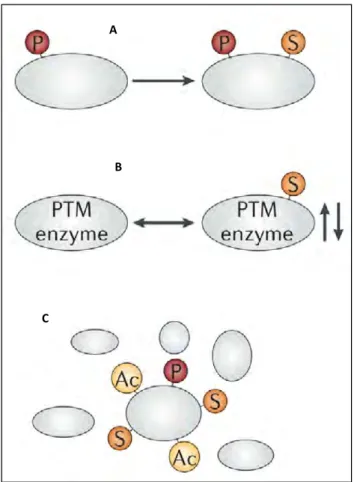
Post-translational modification (PTM) plays a crucial role in the functional regulation of proteins. Interestingly, different PTMs on multiple residues of one protein may co-ordinate with each other to determine a functional outcome resulting in a PTM crosstalk. Strikingly, Huang et al., had collected 193 PTM cross-talk pairs in 77 human proteins from the literature, tested the location preference and co-evolution at the residue. Their interesting data revealed that cross-talk events occurred preferentially among nearby PTM sites, and the cross-talk pairs had a tendency to co-evolve (Y. Huang et al. 2015). A crosstalk between ubiquitination, phosphorylation, SUMOylation, acetylation has been observed for different proteins. However, these modifications may either work in synergy or may have antagonistic roles. For instance, an impressive number of proteins (498) have been identified to be co-modified by SUMO and ubiquitin. The targets include many enzymatic components of PTM machinery, involved not only in ubiquitination and SUMOylation but also phosphorylation, acetylation, and methylation, suggesting an intricate system of crosstalk between different PTMs (Cuijpers, Willemstein, and Vertegaal 2017). For example, SUMOylation and ubiquitination have been shown to have antagonistic roles pertaining to the regulation of the transcription factor NFκB. NFκB is activated by ubiquitination and proteasome-mediated degradation of IκBα (an inhibitor of NFκB). However, the modification of K21 (which is also the site of ubiquitination) of IκBα by SUMO-1 stabilizes it by blocking its ubiquitination and proteasome-mediated degradation (J. M. Desterro, Rodriguez, and Hay 1998).
Phosphorylation is another post-translational modification which is the primary mechanism for regulating cellular signalling whereas ubiquitination is critically involved in protein degradation. Furthermore, mammalian cells express more than 500 protein kinases, and together with protein phosphatases regulate different cellular processes (J. V. Olsen and Mann 2013). Ubiquitination machinery, on the other hand, consists of ~40 E2 enzymes and more than 600 E3 ligases that facilitate the ubiquitination of proteins in the cell. Therefore, the number of proteins modified by both phosphorylation and ubiquitination in the cell is quite large (Yau and Rape 2016). One such example of cross-regulation between phosphorylation and ubiquitination is that of phosphodegrons (short linear motif that is inert until phosphorylated but generates a binding surface that interacts with a ubiquitin ligase upon phosphorylation), in which one or more phosphorylation sites function in a cis-regulatory manner subsequently promoting ubiquitination of a substrate. Interestingly, PTMs might involve regulation of the machinery of other modification type, for example, phosphorylation activating E3 ubiquitin ligase activity (Holt 2012; Ichimura et al. 2005; Khosravi et al. 1999; Michael
16 J. Emanuele et al. 2011; Swaney et al. 2013). Taken together, such communications between different modifications have had significant roles in expanding the ubiquitin code and in understanding the regulation of different proteins controlling key cellular functions (Herhaus and Dikic 2015; Swatek and Komander 2016; L. Song and Luo 2019).
17
Chapter 2 SUMOylation
SUMO (Small Ubiquitin-like MOdifier) is covalently linked to various proteins and is deconjugated by SUMO-specific proteases. Due to the opposing activities of SUMO conjugation and deconjugation, SUMOylation can be highly dynamic. SUMO proteins are ~10kDa in size and are
distantly related to ubiquitin (20% identity). SUMO was first identified in mammals where it was found to be covalently linked to the GTPase activating protein RanGAP1 (Rohit Mahajan et al. 1997; Matunis, Coutavas, and Blobel 1996). The long-term fate of the modified protein can be altered by SUMO conjugation, thereby, hugely increasing the complexity of the proteome in eukaryotic cells. SUMO conjugation plays a critical role in most cellular processes such as DNA replication, transcription, cell cycle regulation, chromatin organization, ribosome biogenesis, pre-mRNA splicing, nuclear trafficking, protein degradation, etc. (Droescher, Chaugule, and Pichler 2013; Chymkowitch, Nguéa P, and Enserink 2015; Cubeñas-Potts and Matunis 2013; N. García-Rodríguez, Wong, and Ulrich 2016; Eifler and Vertegaal 2015). Furthermore, defects in the SUMOylation pathway are associated with various diseases such as neurodegenerative diseases, cancer, and heart-failure (Yanfang Yang et al. 2017).
2.1 SUMO Paralogs, Pathway and Enzymology
SUMO Paralogs
In lower eukaryotes (yeast, insects, and nematodes), only one SUMO gene is expressed, however, up to eight versions of SUMO are expressed in plants. In vertebrates four different paralogs namely, SUMO1, SUMO2, SUMO3, and SUMO4 are expressed. In humans, SUMO1-3 are ubiquitously expressed whereas SUMO4 is limited to immune cells, pancreatic islands, and kidneys (Bohren et al. 2004; D. Guo et al. 2004; C.-Y. Wang and She 2008). SUMO2 and SUMO3 are ~ 96% identical to
each other (often referred to as SUMO2/3) whereas SUMO1 shares ~45% sequence identity with
SUMO2/3. SUMO4 shares 86% identity with SUMO2 (Figure 9), however, the conjugation of SUMO4 to cellular proteins is not known yet.
SUMO2/3 contains Lys residues near the amino terminus which are used as SUMO acceptor sites. SUMO2/3 are able to form chains efficiently, in vitro and in vivo through Lys11 in the consensus SUMOylation site but also via non-consensus site Lys5 in vitro (Vertegaal 2010). The SUMO2/3 chains were found to accumulate during very high levels of cellular stress such as acute heat shock which suggests their involvement in stress responses (Golebiowski et al. 2009). On the other hand, SUMO1 lacks the SUMO consensus site but can form SUMO chains in the presence of a short E3 ligase fragment in vitro (Pichler et al. 2002a). SUMO1 can SUMOylate SUMO2/3 and rather functions as a chain terminator. Interestingly, it was found that SUMO1 is able to multimerize in
18 vitro via non-consensus Lys residues at the N-terminal, however, the physiological relevance of these findings remains unclear (Pedrioli et al. 2006).
SUMO2/3 is predominantly localized in the nucleus and PML bodies whereas SUMO1 is localized in the nucleoli, nuclear envelope, and cytoplasmic foci. Further, the dynamics of SUMO1 is slower than SUMO2/3 and the distribution of two paralogs changes rapidly throughout the cell-cycle (Boddy et al. 1996; Matunis, Coutavas, and Blobel 1996; Rohit Mahajan et al. 1997; Ayaydin and Dasso 2004).
Regulation of SUMO Isoforms
The regulation of SUMO isoforms is poorly understood, however, several studies indicate regulation at the transcriptional and post-transcriptional levels. Despite having a ubiquitous expression of SUMO1-3, their level differs across tissues and during development (Loriol et al. 2013; Z. Xu and Au 2005). In cells, free form of SUMO1 is less abundant and it is found mostly conjugated to substrates. In contrast, free forms of SUMO2/3 are found in high levels, however, they get conjugated upon stimuli such as heat shock and arsenic treatment (Lallemand-Breitenbach et al. 2008a; Saitoh and Hinchey 2000; Michael H. Tatham et al. 2008; Weisshaar et al. 2008).
Post translational modifications are important regulators of SUMO proteins. Both acetylation and phosphorylation have been demonstrated to modify SUMO1 (Lallemand-Breitenbach et al. 2008a; Matic et al. 2008). For example, acetylation at Lys37 in SUMO1 and Lys33 in SUMO2 affected specific non-covalent SUMO interactions by neutralising the basic charge of SUMO (Ullmann et al. 2012). Importantly, ubiquitin also modifies SUMO variants such as SUMO3 at Lys20 and 32 (Lamoliatte et al. 2013) raising the possibility that abundance of SUMO proteins is regulated by ubiquitination of SUMO (Droescher, Chaugule, and Pichler 2013). The identification of SUMO2 chains assembly upon stress serves as the best example of how SUMO pathway is regulated by ubiquitin-dependent degradation. These chains are recognized by SUMO-targeted ubiquitin E3 ligases (STUbLs- discussed later) which contain multiple SUMO interaction motifs (SIMs- discussed later). Indeed, SUMO chains themselves have been shown to be substrates for ubiquitination (Tatham et al. 2008).
SUMO Pathway
SUMO is expressed as a precursor protein and maturation is achieved by specific SUMO proteases which expose the C-terminal di-glycine that is critical for conjugation (Hickey, Wilson, and Hochstrasser 2012a). The enzymatic cascade facilitates the conjugation of matured SUMO to the substrates (Figure 10) (Droescher, Chaugule, and Pichler 2013). SUMO is attached to an internal
19 cysteine of the heterodimeric E1 activating enzyme Aos1/Uba2 in an ATP-dependent step, forming a thioester bond (J. M. P. Desterro, Thomson, and Hay 1997; Erica S. Johnson and Blobel 1997). SUMO is then transferred to the catalytic cysteine of Ubc9, an E2 conjugating enzyme, again resulting in a thioester bond (J. M. P. Desterro, Thomson, and Hay 1997; L. Gong et al. 1997; Erica S. Johnson and Blobel 1997). Ubc9 is able to directly recognize and conjugate SUMO to its substrates through the formation of an isopeptide bond between the C-terminal glycine of SUMO and the ε-amino group of the target lysine (Okuma et al. 1999; R. Mahajan, Gerace, and Melchior 1998). SUMO E3 ligases greatly enhance the efficiency of SUMO conjugation as this reaction is very weak (E. S. Johnson and Gupta 2001; Sachdev et al. 2001; Pichler et al. 2002a). The substrate specificity is ensured by SUMO E3 ligases, however, only a few SUMO E3 ligases are known (Gareau and Lima 2010; Geiss-Friedlander and Melchior 2007; Flotho and Melchior 2013). Substrates can be modified with a single SUMO, with multiple SUMOs or, with SUMO chains. SUMO proteases are able to deconjugate substrates thereby circulating free SUMO back to the conjugation cycle. The consequence of SUMOylation result in changed binding interfaces that can be implicated in diverse protein functions such as intracellular localization, activity, stability and, conformational changes (Droescher, Chaugule, and Pichler 2013).
SUMOylation is a reversible process and the maintenance of balanced SUMO conjugation/ deconjugation is critical for the survival of the cell. In knockout experiments in mice, abolition of SUMO conjugation by targeting Ubc9 (Nacerddine et al. 2005) or by preventing deconjugation of