HAL Id: tel-01147174
https://tel.archives-ouvertes.fr/tel-01147174
Submitted on 29 Apr 2015
HAL is a multi-disciplinary open access
archive for the deposit and dissemination of
sci-entific research documents, whether they are
pub-lished or not. The documents may come from
teaching and research institutions in France or
abroad, or from public or private research centers.
L’archive ouverte pluridisciplinaire HAL, est
destinée au dépôt et à la diffusion de documents
scientifiques de niveau recherche, publiés ou non,
émanant des établissements d’enseignement et de
recherche français ou étrangers, des laboratoires
publics ou privés.
switch recombination
Ebe Schiavo
To cite this version:
Ebe Schiavo. Molecular mechanisms controlling immunoglobulin class switch recombination.
Ge-nomics [q-bio.GN]. Université de Strasbourg, 2013. English. �NNT : 2013STRAJ084�. �tel-01147174�
UNIVERSITE DE STRASBOURG
ECOLE DOCTORALE DES SCIENCES DE LA VIE ET DE LA SANTE
INSTITUT DE GENETIQUE ET DE BIOLOGIE MOLECULAIRE ET CELLULAIRE
Thèse présentée par :
Ebe SCHIAVO
soutenue le : 30 Septembre 2013
pour obtenir le grade de
Docteur de l’Université de Strasbourg
Discipline : Sciences du vivant
Spécialité : Aspects moléculaires et cellulaires de la Biologie
Molecular mechanisms controlling
immunoglobulin class switch recombination
Dr. Bernardo REINA-SAN-MARTIN
Directeur de thèse
Pr. Qiang PAN-HAMMARSTRÖM
Rapporteur externe
!…though my soul may set in darkness, it will rise in perfect light; I have loved the stars too truly to be fearful of the night." Sarah Williams
!Amo il tempo avvenuto, buono se # stato buono, cattivo perch$ non pu% tornare." Erri De Luca
The following pages represent a kind of !sum" of these last four years, but this thesis, as well as my work and everything it came along, it wouldn#t have been possible without all the people I wish to acknowledge in these pages.
First of all, I have to say thank you to Bernardo: for accepting me in your lab in the far October 2009, for your trust and your support, for not allowing me to give up when things were turning from bad to worst; you have been my mentor and my guide and I have learnt so many things during all this time $although you still
have to show me how to get 1010 competence for bacteria!%, and I will try to keep the enthusiasm you
transmitted me as well as the positive attitude, which makes the impossible to became possible.
To Pan, Fran&oise, Bertrand and Silvo, for accepting to evaluate the work I am presenting in this dissertation.
To H'l(ne, for being part of my mid-thesis committee ) as well as Fran&oise ) and for the advices you provided me with.
To Susan and Philippe, for making me feel as part of a !huge lab", for the lab meetings, the journal clubs and the barbecues!
To the Reina lab, my !second family": to Anne-Sophie, with whom I shared the good and the bad time, in particular in this last year made of endless days, crazy sorts, super-high-throughput experiments: we went through all this together, by supporting each other and discovering that two brains are better than one! To L'a, who brought a ray of light in the lab: for bringing me to see Radiohead again, for our !elective affinity" and your infinite sensibility, for all the time we spent together close to or far from the bench. To Vincent, the only man able to tolerate the Reina girls!!! For struggling together with the Necker cells which didn#t want to grow, for your !Sicilian accent", the chocolate you gave me during coffee time to get some energy for the afternoon, and all the galettes de roi you baked $sometimes not really spontaneously!%. To M'lanie, the new
!cloning queen" of the lab, for her kindness, her Nutella muffins and for our !French lunches" in the CROUS.
To Lila, for the good time we had together when she was around, and also to the !unforgettable seniors"!!! Thanks to Beena, for allowing me to discover the wonderful person you are, because since you left anything changed, and also for your help during these last weeks of !writing marathon"; and to Sara, !my wife", who initiated me to cloning and also to the SPB: for the dances in the lab, the night chats, the !zen sessions" and Christmas decorations in the lab!
as an amazing scientist; you know that if you are reading these words is also because of you.
To the Chan/Kastner lab, for the good time we had together and the reagents I borrowed! To Attila, for bugging me while waiting for the coffee and also for his advices $and for always appreciating my cakes!%, Apos for sharing those problems we finally managed to overcame and for letting me skating on ice again; to the dear Deepika, for everything we shared $and share%, for your support and your friendship; to Beate, for her kindness and her advices on B cells development, to Peggy, Rose, Kate, Marie, Isma, Marie-Pierre, Arnaud, for the "pots#, the birthdays and the good atmosphere you created.
To the Soutoglou lab: Evi, Tibor, Anne-Sophie, Zita, Audrey, Charl&ne, C'line, Alkmini and Anastazja: for the time we shared while spending whole days in tissue culture room, for the cakes, the advices, the NCS and HU! And for Nespresso orders $thanks Tibor!!!:%.
To Shankar, who once told me something such as "if you share the problems, you divide them by two; if you share your happiness, you multiply it by two#. This is what you did $and do%: share feelings, thought, experience. Thanks so much for being my "living Maniatis#, for the night chats in the lab about Fellini movies or Italian football players, for being the friend I can rely on.
To Patricia, for her enthusiasm and her participation to thesis movies, and for her smiles! And to Claudine, for her patience with our impossible sorts, for explaining me 100 times that, at the LSR, I need to "replicate without data# an existing folder to keep the setting, for her amazing cakes and biscuits and for our chats in Italian!
To Marco, for our discussions about DGE, and to Bernard, Serge, Stephanie, C'line, Doulaye and Laurent, for all their help with the DGE and data analysis, and for the interesting discussions we had.
To Christophe and Marie-Laure, for letting me discover the wonderful world of protein co-expression, and for all the attempts we made to make AID soluble!
To Betty and the "angels# of the culture facility: for your kindness, for the last minute orders of medium which saved many experiments of mine and for caring about the Necker cells stock.
To all the people of the common IGBMC services: for making this institute a very nice place to work in; to B'n'dicte and Armelle, who cared about the administrative "details# and to Eveline and Mait' for their kindness.
for the time we shared, and for the activities; for showing me that is still possible to make science and have fun! To Ben, J!r"me, Thomas #Tommaso!$, David and Adrien, for reaching the end of this path together.
To David, because when he sent me the link for the PhD program call while we were in Cambridge I couldn%t imagine that it would have such an impact on my future. To Mr. Grey, because his &sunglassed smile' turned from bad to good a few days of mine. To the volunteers of the SPA, for our nice chats, and especially to the cats and the dogs, hoping they will find a home.
To the &Italian family in Strasbourg': Claudia, Serena, Maria Vittoria, Manuela and Floriana, for being my &safety net' in those tough moments, for the dinners, parties, trips, movies, for the grappa and Cannonau, pan di stelle and aperol spritz. To Manu, my &older sister', and Floriana, because when we met for the interview here in 2009 we couldn%t imagine that it was the beginning of a wonderful friendship #and Sicily is waiting for us!!!:$. To Monica, Pietro and the sweet Edoardo; to Rocco for her enthusiasm and support, to Goffredo, for his &technical advices', and to Angelo, for being able to understand my thoughts without the need of words.
To my friends #and cousins!$: Sonia, Emi, Antonia, Raf, Alejandro, Gianlorenzo, Laura, Francesca, Vera, Ale: for their support and their friendship.
To my family: my cousins, aunts and uncles, because no matters where I am or what I do, as I know that you will be there for me, and me for you as well.
To my grandmothers, Ebe and Italia, for their support and their love #and prayers! Thanks!!!$ and to my grandfathers, Vincenzo and Domenico, who would have been happy to know that I am writing these pages. To my beloved sister Irene, for being the &good part of me', for her advices, her support and for her love; to my mum and my dad, for believing in me and reminding me that &everything is going to be ok', for the freedom they always gave me although the distance is hard to accept sometimes. And for knowing that, in the end, I always come back home. To Bizet, Pisolo and Asia, for proving that tolerance between cats and dogs is possible!
TABLE OF CONTENTS
!
LIST OF FIGURES ... 3
LIST OF TABLES ... 5
LIST OF ABBREVIATIONS ... 6
INTRODUCTION ... 9
I. B cell receptor diversification ... 10
1. The B cell receptor ... 10
2. Antigen-independent Ig diversification: V(D)J recombination ... 11
3. Antigen-dependent Ig diversification ... 16
3.1. Gene conversion ... 16
3.2. Somatic hypermutation ... 17
3.3. Class switch recombination ... 17
II. Activation-induced cytidine deaminase (AID) ... 19
1. The RNA editing model ... 20
2. The DNA deamination model ... 20
3. Role of AID in somatic hypermutation ... 21
4. Role of AID in class switch recombination ... 23
4.1. Transcription at the IgH locus ... 23
4.2. Sequence specificity and IgH locus regulatory elements ... 25
4.3. Formation of double stranded DNA breaks at the IgH locus ... 26
4.4. Processing of double stranded DNA breaks: DNA damage response and repair ... 27
4.4.1. “Sensing” the lesions: the DNA damage response (DDR) ... 27
4.4.2. The DSBs repair through non-homologous end joining pathway ... 28
4.4.3. The alternative non-homologous end joining pathway ... 29
5. Role of AID outside the immune system ... 30
6. AID and pathogenesis: B cell lymphomas ... 31
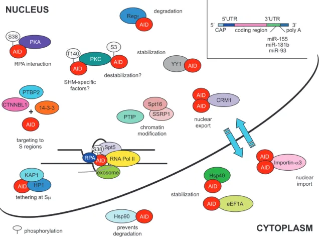
7. AID regulation: how B cells limit AID-mediated DNA damage ... 33
7.1. Transcriptional regulation ... 33
7.2. Post-translational modifications ... 34
7.3. Subcellular localization ... 34
7.4. AID and its cofactors ... 35
III. Class switch recombination-immunodeficiencies ... 39
1. CSR-ID due to a CD40 signaling defect ... 39
1.1. CD40 ligand deficiency ... 40
1.2. Loss of CD40 ... 40
1.3. Impaired activation of NF-kB pathway ... 40
2. CSR-ID due to an intrinsic B cell defect ... 41
2.1. AID deficiency ... 41
2.1.1. AID C-terminal mutations ... 41
2.1.2. AID N-terminal mutations ... 42
2.2. Loss of UNG ... 42
2.3. Deficiency of PMS2 ... 43
2.4. CSR-ID due to a known DNA repair defect ... 43
2.5. CSR-ID due to an unknown defect and associated to normal SHM ... 43
2.5.1. Defect upstream of DSBs: AID CSR-specific cofactor hypothesis ... 44
IV. Spt5 and RNA polymerase II: the breakthrough ... 45
1. Spt5: the missing link between AID and transcription ... 46
V. Transcription and chromatin-regulating factors ... 47
1. Spt6: more than a chaperone ... 47
2. The PAF complex: the “transcription platform” ... 48
2.1. Role of the PAF complex in histone modifications and transcription ... 49
VI. The Structural maintenance of chromosomes (Smc) complexes in genome
regulation ... 51
1. The cohesin complex ... 52
1.1. Cohesins and Ig loci reorganization ... 54
1.2. Cohesin deficiency and pathological consequences ... 54
2. The Smc5/6 complex ... 55
VII. Working hypothesis ... 58
RESULTS ... 59
I. Overview of thesis work ... 60
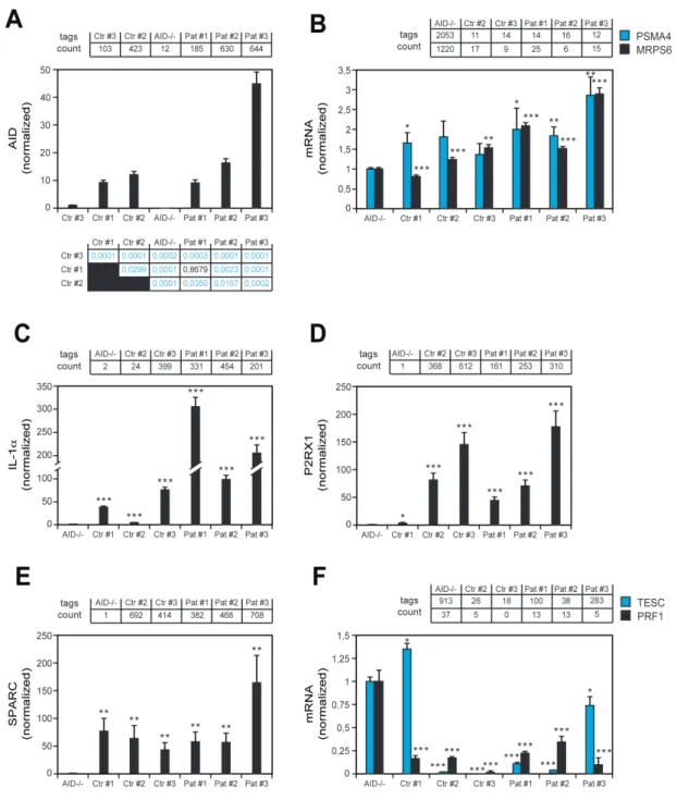
II. Identification of CSR-specific factors ... 63
1. Transcriptome profiling of human B cell lines derived from healthy donors, CSR-ID
and AID deficient-patients ... 63
2. Comparative proteomic analysis between human and mouse B cell lines to identify
CSR-specific AID cofactors ... 74
III. Spt6: the “missing factor” in CSR-ID patients? ... 75
IV. Role of the PAF complex in CSR ... 80
Publication - A role for the RNA pol II-associated PAF complex in AID-induced immune
diversification ... 81
V. Smc5: a new potential regulator of CSR ... 98
VI. Smc5/6 complex: is Smc6 required for CSR? ... 103
VII. Role of the cohesin complex in CSR ... 106
Manuscript - The cohesin complex regulates class switch recombination ... 108
DISCUSSION ... 133
1. Patient analysis and transcriptome profiling: the dark side of the approach ... 134
2. Validated or not validated: that is the question ... 135
3. Missing factor: is it really downregulated? ... 136
4. The importance of being within the nucleus ... 137
5. Does AID size and domains matter? ... 138
6. The short story of Spt6 ... 139
7. The PAF complex, AID and transcription-associated factors: a complicated
relationship ... 141
8. AID targeting: what does it mean? ... 142
9. The Smc5/6 complex: a functional “divorce”? ... 143
10. The cohesin complex in CSR regulation: long-range interactions, repair or both? 145
Working model for CSR ... 147
General conclusions ... 149
LITERATURE CITED ... 150
ANNEX I - MATERIALS AND METHODS………...I
LIST OF FIGURES
INTRODUCTION
Figure 1. Structure of the B cell receptor (BCR)……….10
Figure 2. Antibody diversification mechanisms………...11
Figure 3. Organization of the mouse Ig loci……….12
Figure 4. The steps of V(D)J recombination………14
Figure 5. Model of immunoglobulin gene conversion (IGC) at the chicken Igλ locus………..…………16
Figure 6. Somatic hypermutation (SHM)………...17
Figure 7. Class switch recombination (CSR)………..18
Figure 8. AID domains organization………...19
Figure 9. The DNA deamination model………21
Figure 10. AID-mediated mutagenesis during SHM………..22
Figure 11. Class switch recombination reaction……….23
Figure 12. Formation and repair of DSBs during CSR………..27
Figure 13. Cellular regulation of AID………....38
Figure 14. Main roles of the PAF complex………..49
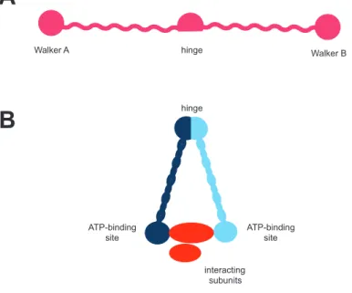
Figure 15. Domain organization of the Smc protein and Smc complex. ………51
Figure 16. Cohesin complex.……….52
Figure 17. Smc5/6 complex.………..57
RESULTS
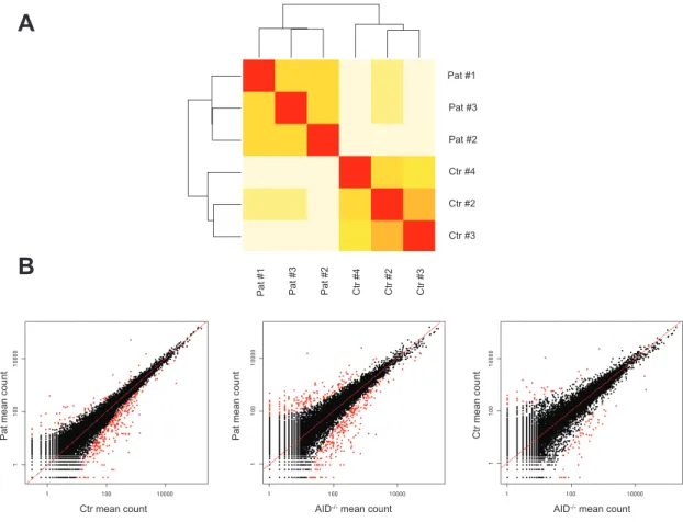
Figure 18. Workflow of identification and functional characterization of CSR-specific regulators…….62Figure 19. Comparison of transcriptomic data obtained from healthy donors, CSR-ID and AID-/- EBV-immortalized B cell lines.………64
Figure 20. DGE data validation by RT-qPCR: genes upregulated and downregulated in patients and controls when compared to AID-/-.………70
Figure 21. DGE data validation by RT-qPCR: genes upregulated and downregulated in patients when compared to controls.……….71
Figure 22. Comparative analysis of deregulated genes in CSR-ID patients.………73
Figure 23. Strategy for proteome identification of CSR-specific AID partners on human and mouse B cell lines.………...74
Figure 24. Spt6 and Spt5 expression in human B cell lines.………75
Figure 25. Retrovirus-mediated Spt6 knockdown on CH12 B cells.………77
Figure 26. Lentivirus-mediated Spt6 knockdown on CH12 B cells might have an effect on CSR.…….78
Figure 28. Lentivirus-mediated Smc5 knockdown induces a partial CSR reduction upon 48h
stimulation.………..101 Figure 29. Lentivirus-mediated Smc5 knockdown induces opposite effects on CSR upon 72h
stimulation.………..102 Figure 30. Lentivirus-mediated Smc6 knockdown has an impact on CSR upon 48h stimulation……104 Figure 31. Lentivirus-mediated Smc6 knockdown affects CSR upon 72h stimulation………...105
DISCUSSION
Figure 32. AID N-terminal and C-terminal truncations……….139 Figure 33. Working model for CSR……….148
LIST OF TABLES
!
INTRODUCTION
Table 1. Class switch recombination immunodeficiencies (CSR-ID) .………39
Table 2. Cohesin subunits and regulatory factors.……….53
Table 3. Smc5/6 complex subunits and associated non-Smc elements (Nse) .………57
RESULTS
Table 4. Characteristics of the human B cell lines analyzed in this study.……….60Table 5. Deregulated genes in patients when compared to AID-/-.………..65
Table 6. Deregulated genes in controls when compared to AID-/-.………..66
Table 7. Deregulated genes in patients when compared to controls.……….67
Table 8. List of top ten Ingenuity networks………..72
!
!
LIST OF ABBREVIATIONS
3’RR IgH locus 3’ regulatory region 53BP1 p53 binding protein 1
A-NHEJ Alternative non-homologous end joining pathway
Aicda Activation-induced cytidine deaminase gene (mouse)
AICDA Activation-induced cytidine deaminase gene (human)
AID Activation-induced cytidine deaminase APE Apurinic/apyrimidinic endonuclease APLF Aprataxin and PNK-like factor APRIL A proliferation-inducing ligand ATM Ataxia telangiectasia mutated
BATF Basic leucine zipper transcriptional factor ATF-like bp Base pairs
BCR B cell antigen receptor BER Base excision repair pathway
C-NHEJ Classical non-homologous end joining pathway C/EBP CCAAT/enhancer-binding protein
CD40L CD40 ligand
CDR Complementarity-determining region CE Coding end
CFSE Carboxyfluorescein succinimidyl ester ChIP Chromatin immunoprecipitation CSR Class switch recombination
CSR-ID Class switch recombination immunodeficiency CTNNBL1 Catenin beta like 1
DDR DNA damage response
DGE Digital gene expression-tag profiling
DNA-PKcs DNA-dependent protein kinase (catalytic subunit) DSB Double stranded DNA break
dsDNA double stranded DNA
DSIF 5,6-dichloro-1-β-d-ribofurano-sylbenzimidazole (DRB) sensitivity-inducing factor complex
EDA-ID Ectodermal dysplasia associated with immunodeficiency eEF1A Elongation factor 1 alpha
ES Embryonic stem cell Exo1 Exonuclease 1
FACS Fluorescence activated cell sorting FACT Facilitates chromatin transcription complex GADD45 Growth arrest and DNA damage-inducible 45 GANP Germinal center-associated nuclear protein GC Germinal centers
GLT Germline transcription HIGM Hyper-IgM syndrome HoxC4 Homeobox C4
HP1 Heterochromatin protein 1 HR Homologous recombination hs DNAse I hypersensitive sites
IgL Immunoglobulin light chain locus IL-4 Interleukin-4
IL-7R Interleukin-7 receptor IP Immunoprecipitation iPSC Induced primordial stem cell Iws1 Interacting with Spt6 homolog 1 KAP1 KRAB associated protein 1
Kb Kilobases
Mb Megabases
MBD4 Methyl-domain binding protein 4
MDC1 Mediator of DNA damage checkpoint protein 1 MEF Mouse embryonic fibroblast
MLH1 MutL homolog 1
MMSET multiple myeloma SET domain-containing protein MMR Mismatch repair pathway
MS Mass spectrometry MSH2 MutS protein homolog 2 MSH3 MutS protein homolog 3 MSH6 MutS protein homolog 6
MudPIT Multi-dimensional protein identification technology Nbs1 Nijmegen breakage syndrome protein 1
NELF Negative elongation factor NEMO NF-κB essential modulator NES Nuclear export signal
NHEJ Non-homologous end joining pathway Nipbl Nipped-B-like protein
NLS Nuclear localization signal nt Nucleotide(s)
PAF RNA polymerase II-associated factor complex PARP1 Poly(ADP-rybose) polymerase 1
PARP3 Poly(ADP-rybose) polymerase 3 PCNA Proliferating cell nuclear antigen PGC Primordial germ cell
PKA Protein kinase A
PMS2 Postmeiotic segregation increased (S. cerevisiae) 2 PP2A Protein phosphatase type 2A
PTBP2 Polypyrimidine tract binding protein 2
PTIP Pax interaction with transcription-activation domain protein 1 RAG Recombination-activated gene
REGγ Proteasome activator complex subunit 3 RPA Replication protein A
RPC Replication factor C
RSS Recombination signal sequences SE Signal end
SHM Somatic hypermutation
Smc1 Structural maintenance of chromosomes 1 Smc2 Structural maintenance of chromosomes 2 Smc3 Structural maintenance of chromosomes 3 Smc4 Structural maintenance of chromosomes 4 Smc5 Structural maintenance of chromosomes 5 Smc6 Structural maintenance of chromosomes 6 Spt4 Suppressor of Ty homolog 4
Spt5 Suppressor of Ty homolog 5 Spt6 Suppressor of Ty homolog 6 Spt16 Suppressor of Ty homolog 16 SSB Single stranded DNA break ssDNA Single stranded DNA
STAT6 Signal transducer and transcription activator 6 TCR T cell antigen receptor
TGFβ Transforming growth factor β Top I Topoisomerase I
TSS Transcription start site UNG Uracil-DNA glycosylase Wapal Wings apart-like homolog
WT Wild-type
XLF XRCC4-like factor (Cernunnos)
XRCC4 X-ray repair cross-complementing protein 4 YY1 Transcriptional repressor protein YY1
I.
B cell receptor diversification
1. The B cell receptor
The B lymphocytes originate in the bone marrow from hematopoietic precursors and, throughout their development, the important role these cells play within the immune response is dependent on the repertoire of B cell antigen receptors (BCRs) expressed in their membrane-bound form or in their soluble form, also known as antibodies (Abs) or immunoglobulins (Igs).
The BCR is a transmembrane protein composed of two identical heavy chains (IgH), two light chains (IgL) and additional subunits Ig alpha (Igα) and Ig beta (Igβ, Figure 1). Each IgH chain is covalently bound to an IgL chain. In both IgH and IgL chains, the amino-terminal portion represents the variable (V) region of the receptor, responsible for the recognition of the antigen through the complementarity-determining region (CDR), which dictates the affinity and the clonal selection for the cognate antigen. On the other hand, the IgH carboxy-terminal portion represents the constant (C) or invariant region, which defines the isotype expressed (IgM, IgD, IgG, IgA or IgE) and the effector function of the Ig in terms of downstream pathways and responses activated. In mammals, the light chain can be either kappa (κ) or lambda (λ), whereas the heavy chain can be µ (IgM), δ (IgD), γ (IgG), α (IgA) or ε (IgE).
Figure 1. Structure of the B cell receptor (BCR)
The B cell receptor is composed of two heavy chains and two light chains, each one harboring a variable (V) region and a constant (C) region. V regions of the heavy and light chains represent the antigen binding site, whereas the C regions represent the isotype expressed and consequently exert a different effector function. The BCR is the membrane-bound form of the antibody (or immunoglobulin) and, upon antigen recognition, the
HEAVY CHAIN Igα Igβ LIGHT CHAIN
antigen binding site antigen binding site
variable constant signaling subunits IgM, IgD IgG3 IgG1 IgG2b IgG2a IgE IgA
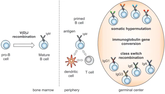
The Ab repertoire produced by B lymphocytes is estimated to be higher than 1011 and is acquired through genomic rearrangements (recombination and mutation) at the IgH and IgL loci. In particular, four mechanisms have been identified to contribute to Ig diversification: V(D)J recombination, which occurs during the early stages of B cells development prior to antigen encounter; and antigen-dependent mechanisms such as class switch recombination (CSR), somatic hypermutation (SHM) and, in species such as chicken and rabbit, immunoglobulin gene conversion (IGC, Figure 2).
Figure 2. Antibody diversification mechanisms
B cells originate in the bone marrow from a hematopoietic precursor and, through V(D)J recombination, they rearrange the V, D and J genes on the Ig heavy and light chains in order to express a functional receptor on their cell surface. Mature B cells migrate to the periphery and, upon cognate antigen recognition and T cell-mediated activation, they start proliferating in the secondary lymphoid organs giving rise to structures named germinal centers. Within germinal centers, B cells further diversify their antibody repertoire through somatic hypermutation, which modifies the affinity for the antigen; class switch recombination, which modifies the antibody isotype expressed and, in some species, through gene conversion.
2. Antigen-independent Ig diversification: V(D)J recombination
V(D)J recombination is a recombination reaction which assembles the variable region exons of B and T cells antigen receptors (BCRs and TCRs) by providing a high variability in the antigen recognition domain from a limited number of exons. This process assembles the variable (V), diverse (D) and joining (J) gene segments at the Ig loci (IgH, Igκ and Igλ) and at the TCR loci (α, β, γ, and δ). V(D)J recombination reaction is strictly controlled: it is tissue-specific, occurs in primary lymphoid tissues (bone marrow and thymus); it is lineage-specific as Ig and TCR loci are rearranged only in B and T cells respectively and, furthermore, it is stage-specific as the IgH locus is rearranged before the IgL,
IgM primed B cell dendritic cell T cell germinal center IgG1 IgE IgA
antigen somatic hypermutation
class switch recombination IgG3 IgM V(D)J recombination pro-B cell Mature B cell
bone marrow periphery
immunoglobulin gene conversion
as well as occurs for the TCRβ before TCRα. However, as my work focused on B cells, this dissertation will be centered on this lineage.
The mouse IgH locus is located on chromosome 12 in proximity of the telomere and it spans for almost 3 megabases (Mb) (Chevillard et al., 2002). The VH genes are approximately 150 – depending
on mouse strain – and classified in 16 families (Johnston et al., 2006); they are located at the 5’ of the locus, upstream of 10-15 DH gene segments (Retter et al., 2007; Ye, 2004). Then, four JH genes
precede the constant (C) region exons (Cµ, Cδ, Cγ3, Cγ1, Cγ2b, Cγ2a, Cε and Cα), coding for the different Ab isotypes. Within the IgH locus, three main cis-regulating elements have been identified: the promoter/enhancer PDQ52, located in proximity of the most 3’ DH gene; the enhancer Eµ located
in the intron between JH and Cµ and the 3’ regulatory region (3’RR), at the 3’ of the IgH locus, which
harbors several DNAse I hypersensitive sites.
The Ig light chain loci (IgL) display a slightly different organization by presenting only V, J and C gene segments. The κ locus spans over 3 Mb on mouse chromosome 6 and is composed by 140 Vκ gene segments and 4 functional Jκ exons, followed by a single Cκ exon. The light chain λ locus, instead, spans about 200 kilobases (Kb) on chromosome 16 and harbors 3 distinct units composed by Vλ/Jλ segments and Cλ exons (Figure 3).
Figure 3. Organization of the mouse Ig loci
Schematic representation of the mouse IgH, Igκ and Igλ loci. The IgH locus harbors about 150 variable (V) genes, up to 15 diverse (D) genes, 4 joining (J) genes and 8 constant (C) genes; the Igκ locus displays 140 V genes and 4 J genes, which precede one C exon; the Igλ locus, instead, has a limited number of V exons, organized in distinct cassettes. Diagram not drawn in scale; adapted from Cobb et al., 2006.
V(D)J recombination allows the expression of the rearranged V coding region and of the downstream C region and depends on recombination-activating genes 1 and 2 (RAG1 and RAG2) (Oettinger et al.,
Vλ2 Vλ2X Jλ2 Cλ2 Vλ1 Jλ3 Cλ3 Jλ1 Cλ1
Igλ
IgH
Cµ Cδ Cγ3 Cγ1 Cγ2b Cγ2a Cε Cα VH genes (150) DH genes (10-15) JH genes CH genes
Eµ 3’RR
PDQ52
Igκ
nonamer (Sakano et al., 1980). RAGs mediate the DNA cleavage, whereas RSSs dictate the order of the reaction, as the recombination occurs only between exons flanked by RSS harboring a 12 bp and a 23 bp spacer (also known as the 12/23 rule) (Tonegawa, 1983). The presence of 23 bp spacers flanking VH and JH genes at their 3’ and 5’ respectively, and of 12 bp spacers located at both ends of
the DH genes, allows the sequential recombination between D and J exons followed by the V and DJ
rearrangement at the IgH locus (Alt et al., 1984).
The recombination is a multistep process and it starts with the expression of RAG1/2 in pre-pro B cells and the recognition of the RSS through the contact between RAG1 and the nonamer sequence (Figure 4) (Swanson and Desiderio, 1998). Then the RAG complex interacts with the heptamer (Swanson and Desiderio, 1999) where it introduces a ssDNA nick in the 12 bp RSS and, followed by synapsis with the 23 bp RSS, it generates a second nick resulting in a double stranded DNA break (McBlane et al., 1995; Schatz and Swanson, 2011). The hydroxyl groups free on both ends interact with the phosphate on the opposite end by generating on one side blunt ends, called signal ends (SEs), and on the other side DNA hairpins, named coding ends (CEs) as they lack RSSs (Gellert, 2002; Roth et al., 1993; Schlissel et al., 1993). The RAG complex is released with the SEs and both the hairpins at the CEs and the blunt ends at SEs are processed by the non-homologous end joining (NHEJ) repair pathway (Taccioli et al., 1994). The first molecular players which act in the repair step are Ku70 and Ku80, which recruit the catalytic subunit of the DNA-dependent protein kinase (DNA-PKcs), whose activation results in phosphorylation of target proteins such as Artemis and the histone variant H2AX. Artemis phosphorylation leads to the opening of the hairpins at CEs and the generation of palindromic sequences (named P elements), whereas its nuclease activity is responsible for the random deletion of nucleotides from the opened ends (Lafaille et al., 1989; Ma et al., 2002). This event, as well as the addition of nucleotides at the CEs mediated by the terminal-deoxynucleotidyl transferase (TdT) enzyme (Alt and Baltimore, 1982), further contributes to generate a pool of antibodies harboring a high variability in the V region. The final resolution of the DSBs generated at CEs and SEs is mediated by X-ray repair cross-complementing protein 4 (XRCC4) and the DNA ligase IV (Grawunder et al., 1997; Li et al., 1995), and allows the expression of the upstream V region and of the C exon in IgH and IgL loci. During the early stages of B cell development, pre-pro B cells undergo the first recombination event which occurs at the IgH locus, leading to the expression of the antibody heavy chains which will be combined with the surrogate light chains in order to express the pre-BCR on the cell surface (Hombach et al., 1990). This step is crucial to inhibit the rearrangement of the second allele (allelic exclusion) and in activating the signaling pathway leading to cell proliferation (Jung et al., 2006). The following re-expression of RAGs, the recombination occurring at the light chain loci, κ and λ, and the association of the light chain with the pre-assembled heavy chain will finally lead to the expression of the BCR harboring the µ heavy chain on naïve B cells.
Figure 4. The steps of V(D)J recombination
The substrates (red and blue rectangles) are flanked by a 12-RSS (red triangle) and by a 23-RSS (blue triangle). The RAG proteins (green ovals) are the central players in the reaction which leads to the cleaved signal complex, and also cooperate with the non-homologous end joining (NHEJ) DNA repair factors in the end processing and joining steps. The final coding joints often contain non-templated nucleotides (pink rectangle) introduced by the TdT. Adapted from Schatz, 2004.
The order and specificity observed during V(D)J recombination raises many questions relative to the tight control of this reaction and, almost thirty years ago, Yancoupoulos and Alt proposed the differential DNA accessibility within the locus as crucial feature to direct the RAGs-mediated lesions (Yancopoulos and Alt, 1985). The “accessibility hypothesis” is supported by evidence showing how transcription, 3D relocation within the nucleus and chromatin status influence the Ig and TCR loci during the early stages of the B and T cell development. Germline (non coding) transcription occurs at the Ig and TCR loci prior D-J and V-DJ rearrangements (Corcoran, 2010; Hesslein and Schatz, 2001). At the IgH locus, germline transcripts have been detected before DH-JH recombination, dependent on
the activity of the promoter/enhancer PDQ52 and on the intronic enhancer Eµ, giving rise respectively to the Iµ and µ0 transcripts. At the same extent, transcription starting from the promoter interspersed within the V region precedes VH-DHJH recombination, and is dependent on interleukin-7 receptor
(IL-7R) signaling (Bertolino et al., 2005). Additionally, antisense transcription has been detected throughout the VH genes prior V-DJ rearrangement and at the DH and JH gene segments before DH-JH
joining, in this latter case mediated by Eµ (Bolland et al., 2007; Bolland et al., 2004; Chakraborty et al., 2007; Perlot et al., 2008). The controversial role of antisense transcription as process which could support the locus availability to RAG-mediated recombination or which could lead to gene silencing by hybridization with the corrisponding sense sequences, still has to be fully elucidated.
RAG1/2 binding paired complex synapsis cleavage cleaved signal complex signal end complex signal joint hairpin opening,
coding end processing
coding joint
NHEJ and TdT
gene and spread over the JH genes in early pro-B cells undergoing DH to JH rearrangements
(Chakraborty et al., 2007; Morshead et al., 2003). After this first step of rearrangement, hyperacetylation is detected at V genes as limited to the promoter, the V segment and the RSS (Johnson et al., 2003), and is lost after productive VH-DHJH recombination in order to make the locus
inaccessible in pro-B cells (Chowdhury and Sen, 2003). Moreover, RAG2 has been shown to bind, through its PHD finger domain, H3K4me3 mark present at the IgH locus and the tryptophan in position 453 (W453) appears to be critical for the binding of the modified H3 histone as well as for efficient V(D)J recombination, thus providing the first direct link between epigenetic modulation of V(D)J recombination and RAGs accessibility (Liu et al., 2007; Matthews et al., 2007). Another feature displayed by the Ig loci undergoing recombination is the differential nucleosome packaging, and DNA sensitivity to restriction enzymes is an indication of accessibility or nucleosome free status. Before DH
to JH rearrangements, the region between PDQ52 and Eµ as well as the JH RSS are DNAse I
sensitive, whereas the accessibility of the VH regions is limited to the V-DJH rearrangement
(Chowdhury and Sen, 2003; Maes et al., 2006). Methylation of cytosines at the CpG dinucleotides is an additional mechanism of gene silencing in mammals (Stein et al., 1982; Vardimon et al., 1982) and, at the Ig loci, modulates V(D)J recombination as well as allelic exclusion. Additionally, locus relocalization and contraction are a hallmark of active recombination. In pro-B cells, the IgH locus is actively relocated towards the center of the nucleus (Kosak et al., 2002), and is regulated by Pax5, a transcription factor required for B cell commitment (Busslinger, 2004), which mediates locus contraction and distal VH-DJH rearrangements (Fuxa et al., 2004).
The productive V(D)J rearrangement at the IgH locus allows the expression of the V gene “chosen” during the recombination and of the downstream C exon; in primary B cells, the expression of the rearranged heavy chain and light chain (κ or λ) leads to the exposure on cell surface of a functional IgM antibody which defines the primary repertoire. Although, the recombination being a random process, self-reactive antibodies can be generated and B cells expressing those antibodies are negatively selected in the bone marrow before entering the peripheral lymphoid organs (Wardemann et al., 2003). By migrating to the periphery, primary B cells will undergo further Ig genes diversification which is dictated by the antigen recognition and T cells-mediated activation, and which will adapt the response in order to provide a faster and more efficient antigen clearance.
3. Antigen-dependent Ig diversification
Naïve B cells that exit the bone marrow display a huge repertoire of antibodies that are potentially able to recognize every antigen. However, being the antigen binding of low affinity, upon antigen recognition and T cell-mediated activation, B cells undergo further Ig diversification mechanisms in order to modulate the immune response according to the pathogen and to the stimuli received. They actively proliferate in the secondary lymphoid organs and form structures named germinal centers (GC). Three additional Ig diversification mechanisms have been identified: immunoglobulin gene conversion, which occurs in some species such as chicken and rabbit, and is characterized by the use of pseudogenes to diversify the V(D)J sequence; somatic hypermutation, which introduces mutations and deletions in the V region of the Ig heavy and light chains and class switch recombination, which replaces the antibody isotype expressed while preserving the antigen specificity of the antibody. Overall, these mechanisms contribute to a more specific and adapted immune response.
3.1. Gene conversion
The antibody repertoire generated upon V(D)J recombination is dependent on the choice of V, D and J gene segments assembled during the reaction. But whereas mammals produce a highly diversified repertoire, some species such as the chicken do not. The chicken IgL locus displays unique VL and JL
exons, as well as the IgH locus and in consequence B cell precursors provide a limited specificity. Thus, antibody diversity occurs through a mechanism called immunoglobulin gene conversion (IGC), based on the usage of pseudogenes located upstream of the VL and VDH exons to replace the coding
sequence of rearranged loci (McCormack et al., 1991; Reynaud et al., 1985; Reynaud et al., 1987; Reynaud et al., 1991; Reynaud et al., 1989; Thompson and Neiman, 1987). The lesions introduced in the V genes are repaired through homologous recombination (HR) by using the upstream pseudogenes as template.
φVλ φVλ φVλ φVλ VλJλ Cλ
3.2. Somatic hypermutation
Somatic hypermutation introduces point mutations, and occasionally insertions and deletions, in the V genes of the Ig heavy and light chain loci and, by modifying the CDR, this mechanism gives rise to a higher affinity antibody repertoire (Saribasak and Gearhart, 2012). Mutations occur at a frequency of 10-5-10-3 mutations/bp/generation, definitely higher than the basal level of mutations in the genome which is estimated being approximately 10-9 (Peled et al., 2008), and the mutations extend about 1-2 Kb downstream of the promoter (Saribasak and Gearhart, 2012). B cells expressing high affinity antibodies which do not recognize self-antigens undergo proliferation and further differentiate into plasma cells or memory B cells (LeBien and Tedder, 2008).
Figure 6. Somatic hypermutation (SHM)
Somatic hypermutation modifies the rearranged V genes of the Ig heavy and light chain in order to express antibodies with higher affinity for the cognate antigen. The V genes are diversified by the introduction of non-templated point mutations, insertions and deletions. Mutation frequency is higher closer to the promoter spanning up to 2 Kb downstream and gradually decreases, as shown by the red peak. Diagram not in scale.
3.3. Class switch recombination
Class switch recombination is a region-specific recombination reaction that occurs at the IgH locus and joins two switch (S) regions, by deleting the intervening sequence and replacing the isotype of the antibody expressed (from IgM/IgD to IgG, IgE or IgA; Figure 7). This mechanism allows a different effector function of the antibodies while preserving their antigen specificity (Chaudhuri et al., 2007). The recombination takes place at the switch regions (S regions), which are repetitive and non-homologous regions of 3-12 Kb located upstream of each C exon (with the exception of Cδ, whose expression occurs by alternative RNA splicing). Each switch region is preceded by a promoter, and transcription at the donor (Sµ) and acceptor (Sγ, Sε or Sα) S regions is activated prior to recombination and mediated by helper T cells and cytokine stimuli (Chaudhuri et al., 2007; Stavnezer et al., 2008b). The introduction of lesions in the DNA and the intermediate DSBs generated are then repaired and allow the expression of the downstream CH gene after the excision of the intervening
Eµ Cµ Cδ V(D)J Iµ Sµ
Eµ Cµ Cδ
region (Chaudhuri et al., 2007). As the S regions are located far apart within the IgH locus, has been proposed that long-range interactions could contribute to class switching, as the generation of a loop could place in close proximity the donor and acceptor S regions allowing the recombination (Kenter et al., 2012).
Figure 7. Class switch recombination (CSR)
Class switch recombination is a region-specific recombination reaction that takes place at the IgH locus. The recombination involves the S regions, repetitive and non-homologous sequences located upstream of each CH
exon. As depicted in the figure, the donor (Sµ) and acceptor (Sγ1) S regions are involved in the recombination reaction, which finally results in the expression of a different antibody isotype (IgG1) and consequently a different effector function exerted, while preserving the antigen specificity.
More than ten years ago, the work performed by Anne Durandy and Tasuku Honjo led to the identification of activation-induced cytidine deaminase (AID) and represented a breakthrough in the understanding of the CSR, SHM and IGC mechanisms. AID was identified in humans and mice, respectively, as the factor able to mediate CSR and SHM (Muramatsu et al., 2000; Revy et al., 2000) and, from further studies, IGC (Arakawa et al., 2002). These findings allowed a completely new point of view on the regulation of these physiological mechanisms and, most importantly, on their misregulation. mouse IgH locus Cµ Cδ Cγ3 Cγ1 Cγ2b Cγ2a Cε Cα V(D)J Eµ 3’RR
IgM
AAAA Sµ Sγ3 Sγ1 Sγ2b Sγ2a Sε Sα+
excised episome Cγ1 V(D)J Sµ/γ1 Cγ2b Cγ2a Cε Cα 3’RRIgG1
G1
AAAA Sγ2b Sγ2a Sε Sα Cµ Cδ Eµ Sµ Sγ1II. Activation-induced cytidine deaminase (AID)
Activation-induced cytidine deaminase (AID) was identified more than a decade ago through a substractive cDNA screen performed on mouse CH12 B cells unstimulated and stimulated to undergo CSR (Muramatsu et al., 1999). Furthermore, the characterization of patients affected by class switch recombination-immunodeficiency due to a loss of AID (CSR-ID, also known as type 2 hyper-IgM syndrome), as well as of mice deficient of AID, clarified its role as master regulator of CSR and SHM in B cells (Muramatsu et al., 2000; Revy et al., 2000). Moreover, two years later, further studies described AID as required for immunoglobulin gene conversion in the chicken DT40 B cell line (Arakawa et al., 2002; Harris et al., 2002). AID is a small protein of 198 amino acids, which harbors a nuclear localization signal (NLS) at the N-terminus, a nuclear export signal (NES) at the C-terminus and a cytidine deaminase motif (Figure 8).
AID belongs to the APOBEC family of deaminases, which includes also APOBEC1, APOBEC2 and APOBEC3 subgroups. APOBECs deaminate cytidine in RNA and/or cytosine residues in DNA and regulate different mechanisms. APOBEC1 deaminates the cytidine 6666 on apolipoprotein B RNA, by creating an in frame stop codon which leads to the expression of a shorter RNA (Navaratnam et al., 1993; Teng et al., 1993). APOBEC2 function has still not been clarified, whereas APOBEC3G and APOBEC3F act in the innate immunity protection by retroviruses (Rosenberg and Papavasiliou, 2007). Although AID displays the highest homology to APOBEC1, phylogenic sequence analysis showed that AID and APOBEC2 are the most ancient members of the family, and that APOBEC1 and APOBEC3 appeared later and are restricted to mammals (Conticello et al., 2005).
Studies based on sequence analysis contributed to clarify the functional domains of AID whereas mutagenic analysis gave insights into AID’s function. In particular, its NLS and NES not only determine the subcellular localization (Ito et al., 2004; Patenaude et al., 2009) but also play a pivotal role in SHM and CSR. While loss of the AID N-terminal domain specifically impairs SHM (Shinkura et al., 2004), the C-terminal domain has been shown to be specifically required for CSR (Barreto et al., 2003; Durandy et al., 2007; Geisberger et al., 2009; Imai et al., 2005; McBride et al., 2004; Ta et al., 2003).
Figure 8. AID domains organization
Schematic representation of the domain structure of AID. The protein harbors a nuclear localization signal (NLS) at the N-terminus (NH2), a nuclear export signal (NES) at the C-terminus (COOH) and a cytidine deaminase motif
(CDM). Many studies have shown that the N-terminus (13-23) and the C-terminus (182-198) are specifically required for SHM and CSR, respectively. Adapted from Muramatsu et al., 2007.
NES CDM NH2 COOH 198 1 9 26 56 94 190 NLS SHM (13-23) CSR (182-198)
1. The RNA editing model
The sequence homology between AID and APOBEC1 allowed a better understanding of the functionality of its domains but, somehow, also contributed to a misleading interpretation of its mechanism of action. As APOBECs are RNA editing enzymes, AID was initially proposed to be an RNA editing enzyme, in light of its ability to regulate two independent mechanisms, such as CSR and SHM. As AID shares 34% homology with APOBEC1 (Muramatsu et al., 1999), it was hypothesized – according to the RNA editing model - that it could modify a putative mRNA precursor coding for a recombinase, which could cleave the DNA on Ig genes to lead to isotype switching and mutations on the V regions resulting in somatic hypermutation; moreover, the specificity of the editing would be dependent on an unknown AID cofactor (Muramatsu et al., 2000). This hypothesis was supported by similarities between AID and APOBEC1: a) APOBEC1 requires APOBEC1 complementation factor (ACF) for its function (Mehta et al., 2000); b) the subcellular localization, with APOBEC1 able to shuttle between nucleus and cytoplasm (Chester et al., 2003); c) APOBEC1 homodimerization (Chester et al., 2003; Teng et al., 1993) which has been predicted for AID by sequence analysis and d) the evidence that de novo protein synthesis is required for CSR (Begum et al., 2004; Doi et al., 2003; Muramatsu et al., 1999). However, as it will be presented in the next section, genetic studies supported an alternative model based on DNA deamination, and Fritz et al. recently showed by RNA-Seq analysis that AID is not editing polyadenilated RNA in activated B cells (Fritz et al., 2013).
2. The DNA deamination model
The DNA deamination model suggests that AID is able to act on the DNA, and proofs in support of this model come from the experiments performed by Petersen-Mahrt and Neuberger in E. coli, where they found that the target nucleic acid of the enzymatic reaction mediated by AID was indeed the DNA and not the RNA. Furthermore, the deamination and mutation profile associated to a loss of uracil-DNA glycosylase (UNG) contributed to dissect the mutagenesis and consequent DNA repair mechanisms that lead to recombination and mutation (Petersen-Mahrt et al., 2002). The DNA deamination model obtained further support from the work performed by other laboratories, which showed that AID is able to deaminate cytosines on ssDNA in vitro (Bransteitter et al., 2003; Chaudhuri et al., 2003; Dickerson et al., 2003; Morgan et al., 2004; Pham et al., 2003; Yu et al., 2004) and that it associates with S regions in cells undergoing CSR through interaction with replication protein A (RPA) (Chaudhuri et al., 2004; Nambu et al., 2003).
AID-mediated deamination converts cytosines to uracil residues on DNA, thus introducing a dU:dG mismatch. At this initial step of the reaction, the different options to resolve these lesions can result in SHM and CSR (Figure 9).
Figure 9. The DNA deamination model
AID deaminates the cytosine to uracil at the V genes of the Ig heavy and light chain and at the S regions of the heavy chain, introducing a mismatch in the primary sequence. The lesion can be processed by the base excision repair pathway (BER): UNG generates an abasic site by removing the uracil whereas APE induces a nick into the DNA, allowing the DNA polymerase β to successfully repair the lesion. If replication occurs after deamination over the dU:dG mismatch or after UNG base removal, a mutagenic profile can be identified, as occurring during SHM. Additionally, the dU:dG mismatch can be processed by the mismatch repair pathway (MMR) which introduces biased mutations at the A:T pairs. If deamination occurs at cytosines located in close proximity, double stranded DNA breaks, intermediates of the CSR reaction, can be generated and repaired by the non-homologous end joining (NHEJ) pathway. Adapted from Petersen-Mahrt et al., 2002.
3. Role of AID in somatic hypermutation
Somatic hypermutation occurs in germinal center B cells upon cognate antigen recognition. According to the deamination model, cytosine deamination mediated by AID introduces the mismatch dU:dG at the Ig genes; although no consensus sequences have been described for AID targeting, deamination occurs at “hot spots” which correspond to the sequence WRCY (W=A/T; R=A/G; Y=C/T) which have been identified in the CDR of the antigen binding site (Betz et al., 1993; Rogozin and Kolchanov, 1992; Sharpe et al., 1991). Furthermore, has been observed that at the Ig V genes mutations are more frequent about 100-200 bp downstream of the transcription start site (TSS) and span up to 1.5-2 Kb downstream of the promoter (Lebecque and Gearhart, 1990; Rada and Milstein, 2001) and that AID is able to mutate both DNA strands (Rada et al., 2004; Shen, 2007; Xue et al., 2006).
AID UNG APE SHM (phase 2) MMR replication replication SHM (phase 1) C G G C polymerase NHEJ CSR G G C G G C U G G U G G
Figure 10. AID-mediated mutagenesis during SHM
During SHM, AID deaminates the cytosine to uracil at the V genes of the Ig heavy and light chain and introduces a dU:dG mismatch. If replication occurs over the deaminated DNA through a high fidelity polymerase, transitions are introduced in the DNA sequence. Alternatively, replication occurring over the abasic site generated by UNG will introduce transitions and transversions. On the other hand, the processing of the dU:dG mismatches mediated by MSH2, MSH6 and Exo1, components of the MMR pathway, leads to the removal of the strand harboring the mismatch which will be resynthesized by the DNA polymerase η, resulting in the introduction of biased transitions and transversions. Adapted from Petersen-Mahrt et al., 2002.
The uracil introduced by AID deamination can lead to a non-mutagenic profile if the base excision repair (BER) is involved in the resolution of the lesion. Upon uracil excision by uracil-DNA glycosylase (UNG), the apurinic/apyrimidinic endonuclease (APE) introduces a single stranded DNA break (SSB) in the corresponding abasic site. Successful repair is mediated by the DNA polymerase β which is able to resynthesize the DNA strand (Figure 10).
Otherwise, the dU:dG mismatches can be processed through two different pathways, defined as phase 1 and phase 2, according to the mutation profile displayed by the targeted sequence (Petersen-Mahrt et al., 2002; Rada et al., 1998). In the phase 1, replication over the deaminated DNA sequence through a high fidelity polymerase leads to a biased mutation towards transitions C!T or G!A (Figure 10) (Petersen-Mahrt et al., 2002); alternatively, replication can occur over the abasic site generated by UNG and will introduce transitions as well as transversions (replacement of a purine with a pyrimidine and vice versa), which can be mediated by Rev1 polymerase (Figure 10) (Jansen et al., 2006; Petersen-Mahrt et al., 2002; Zan et al., 2012).
The phase 2 of mutagenesis, instead, is mediated by MutS protein homolog 2 and 6 (MSH2/MSH6) and processed through the mismatch repair pathway (MMR), which introduces a biased mutagenic
AID UNG APE MSH2 MSH6 Exo1 polymerase Rev1 C G A T DNA polymerase β C G A T U G A T C G A T T A A T C G G C T A A T G C C G A T T A G A T G A T G T C G N T Polη replication transitions transitions & transversions transitions & transversions (A:T bias)
1998; Rada et al., 1998; Wiesendanger et al., 2000). The MSH2/MSH6 heterodimer recruits the exonuclease 1 (Exo1), which removes the strand harboring the mismatch, and then the DNA will be resynthesized by the DNA polymerase η (Di Noia and Neuberger, 2007). In conclusion, SHM modifies the coding regions located at the antigen binding site and B cells expressing high affinity antibodies will be positively selected for cognate antigen recognition.
4. Role of AID in class switch recombination
Class switch recombination is a multi-step process which takes place in germinal centers upon B cell activation, which replaces the antibody isotype expressed and thus the effector functions according to the stimuli received. In this section I am going to dissect the CSR mechanism and to discuss each step of the reaction (Figure 11).
Figure 11. Class switch recombination reaction
Class switch recombination is a multistep process which starts with (A) germline transcription at the donor and acceptor S regions (black dotted lines). (B) Upon AID expression and targeting to the DNA, the deamination of the cytosines in the DNA sequence will introduce a mismatch, which will be (C) processed into DSBs through the BER or MMR pathway. (D) The DSBs will be repaired through the NHEJ pathway, resulting in the expression of a different antibody isotype (from IgM to IgG1).
4.1. Transcription at the IgH locus
The IgH locus displays transcription units, composed of an I exon, a S region and a downstream C exon coding for the different isotypes. S regions are repetitive and non-homologous regions, located upstream of each C exon, with the exception of Cδ; they contain GC-rich sequences which display different length and sequence similarity between each other. Primary transcripts generated along the
Cµ Cδ Cγ1 V(D)J Eµ AAAA Sµ Sγ1 Cγ1 V(D)J Sµ/γ1 IgG1 G1 AAAA Eµ AAAA Iµ Iγ1 Cµ Cδ Cγ1 V(D)J Eµ Iµ Sµ Iγ1 Sγ1
Iµ
switch region transcription
Cµ Cδ Cγ1 V(D)J Eµ AAAA Sµ Sγ1 AAAA Iµ Iγ1 AID AID expression AID targeting DSBs formation
joining through NHEJ dC ! dU IgM
A
B
C
D
locus are spliced to remove the sequence corresponding to the S region and polyadenylated (Chaudhuri and Alt, 2004). Why transcription should be important for CSR? Three roles have been proposed so far: first, it provides the substrate for AID-mediated cytosine deamination, as predicted by the “accessibility model” (Stavnezer-Nordgren and Sirlin, 1986; Yancopoulos et al., 1986). Strikingly, ssDNA exposure would occur during mRNA elongation and through the formation of R-loops, structures represented by an RNA:DNA hybrid and the displaced non-template ssDNA (G-rich), which is identical to the newly-synthesized RNA (Aguilera and Garcia-Muse, 2012). Although original hypotheses described AID as able to target only the non-template strand, further studies showed that deamination occurs instead on both strands of DNA (Rada et al., 2004; Shen, 2007; Xue et al., 2006), and this is supported by the recent involvement of the RNA exosome in the process of switching (Basu et al., 2011).
A second role proposed for germline transcription requirement during CSR involves the targeting of AID. Initial studies both in vitro or aimed to characterize the phenotype of patients affected by CSR-ID have emphasized the specific requirement of the C-terminal domain for CSR (Barreto et al., 2003; Durandy et al., 2007; Geisberger et al., 2009; Imai et al., 2005; McBride et al., 2004; Ta et al., 2003), giving rise to the “CSR cofactor(s) hunt”. Furthermore, the study from Nambu and co-workers showed that AID co-immunoprecipitates with the RNA polymerase II in splenocytes (Nambu et al., 2003) and the identification of the transcription elongation factors Spt5 and Spt6 as CSR regulators (Okazaki et al., 2011; Pavri et al., 2010) supported the importance of transcription as process which involves the recruitment of AID cofactors; however, as the identification of CSR-specific AID interactors is one of the main points of my dissertation, I am going to discuss it in detail later.
The third and last consequence of germline transcription at the IgH locus is represented by chromatin remodeling through histone post-translational modifications. The donor Sµ region presents numerous activating histone marks, such as H3K9ac/K14ac, H3K27ac, H4ac, and H3K4me3 and H3K36me3 detected even in naïve B cells (Chowdhury et al., 2008; Daniel et al., 2010; Kuang et al., 2009; Nambu et al., 2003; Stanlie et al., 2010; Wang et al., 2006a; Wang et al., 2009; Yamane et al., 2011) and which suggest that Sµ is in a state which constitutively allows recombination. Upon activation, the acceptor S region is made accessible by removal of repressive marks such as H3K27me3 (Chowdhury et al., 2008) and active transcription allows the recruitment of factors which modify the histones to make the DNA accessible for recombination, as occurs with the facilitates chromatin transcription complex (FACT) complex (SSRP1/Spt16) (Stanlie et al., 2010). The discovery that stalled RNA polymerase II and Spt5 association with AID is required for CSR further supports this model, leading the histone modifying enzymes to be “carried” by the RNA polymerase II and to exert their function (Li et al., 2013; Pavri et al., 2010). Moreover, H3ac and H3K4me3 are enriched at the acceptor S regions (Wang et al., 2006a; Wang et al., 2009), while H3K9me3 is still present at Sµ region, and recruits KRAB domain associated protein 1 (KAP1)/ heterochromatin protein 1 (HP1) complex to the IgH locus, which will tether AID to the donor S region (Jeevan-Raj et al., 2011). Furthermore, histone modifications are also important to recruit DNA repair proteins such as 53BP1,
transferase multiple myeloma SET domain-containing protein (MMSET) impairs 53BP1 recruitment and results in defective CSR (Pei et al., 2013).
4.2. Sequence specificity and IgH locus regulatory elements
As transcription per se has been discussed being indispensable for CSR, the sequences of the S regions and of the IgH locus regulatory elements have been extensively investigated in order to identify any consensus which would justify why AID is extensively deaminating cytosines at the Ig loci and not at all the transcribed genes in the cell at that particular developmental stage.
The I exon promoters integrity is required for efficient CSR (Bottaro et al., 1994; Harriman et al., 1996; Jung et al., 1993; Seidl et al., 1998; Zhang et al., 1993) as well as the presence of S regions (Daniels and Lieber, 1995b; Kinoshita et al., 1998; Lepse et al., 1994; Leung and Maizels, 1992; Ott and Marcu, 1989; Petry et al., 1999; Stavnezer et al., 1999). On the other hand, replacement of the Sα sequence with the Sγ1 or Sε in the CH12 B cells which switch to IgA does not impair CSR efficiency (Kinoshita et al., 1998). Moreover, the observation that in Xenopus laevis AT-rich S regions support CSR suggests that the G content within these sequences is not a limiting factor, whereas the fact that they are palindromic it can be, by supporting the secondary structures generated during transcription (Tashiro et al., 2001).
The IgH locus contains two enhancer elements: the intronic enhancer Eµ and the 3’RR. Eµ is located between the JH4 exon and the 5’ of Sµ and its targeted deletion reduces CSR (Sakai et al., 1999),
although a debate concerning the system used (knockout of the enhancer core, which contains the Iµ promoter) questions whether or not the enhancer itself or more specifically the promoter deletion in this experimental system leads to a reduced recombination efficiency. The 3’RR, instead, is located downstream of the Cα exon, it spans for approximately 40 Kb and is composed of DNAse I hypersensitive sites (hs): hs3A, hs1,2, hs3B, hs4, hs5, hs6 and hs7. While deletion of hs1,2 and hs3A does not affect CSR, the loss of hs3B and hs4 reduces GLT levels and consequently recombination to all the isotypes except IgG1 (Manis et al., 1998b; Pinaud et al., 2001). Furthermore, the hs3B and hs4 have been involved in the regulation of the locus rearrangement required to bring into close proximity the donor and acceptor S regions involved in the recombination (Wuerffel et al., 2007). Additionally, the proper splicing of germline transcripts has also been shown to be required for efficient CSR, as deletion of splicing donor and acceptor sites impairs efficient recombination (Hein et al., 1998; Lorenz et al., 1995).
4.3. Formation of double stranded DNA breaks at the IgH locus
According to the DNA deamination model, the uracils introduced into the DNA upon AID-mediated deamination can be processed by either the base-excision repair or the mismatch repair pathway, which will lead to a break into the DNA. The involvement of the BER components has been evidenced by loss-of-function experiments and from the study of patients harboring mutations in the UNG gene, pointing out the importance of the uracil excision into the mutagenic profile of CSR and SHM (Imai et al., 2003b; Rada et al., 2002b; Schrader et al., 2005). The single nucleotide gap created by APE is then filled by the DNA polymerase β (Stavnezer et al., 2008a) with efficient repair, as deficiency of the polymerase has been shown to increase CSR (Wu and Stavnezer, 2007).
The result of the BER processing is usually a high fidelity repair of the lesion, although the single strand DNA breaks generated by APE, if occurring on both strands and in close proximity, may lead to DSBs and favor switching. However, this might not occur at a frequency that could sustain massive recombination at the S regions, implying a parallel pathway in the formation of DSBs at S regions. The mismatch repair pathway has been implicated in inducing DSBs at the IgH locus as well as a biased mutagenic profile on V genes at the IgH and IgL loci (Chahwan et al., 2012). The MSH2/MSH6 heterodimer recognizes dU:dG mismatches into the DNA and recruits MutL homolog 1 (MLH1) and postmeiotic segregation increased 2 (PMS2); the complex then recruits replication factor C (RPC), the proliferating cell nuclear antigen (PCNA) and Exo1 for excision of the single stranded DNA patch containing the mismatch, which will be then resynthesized by low-fidelity polymerases such as DNA polymerase η (Chahwan et al., 2012). Deficiency of any of the MMR molecular players (MSH2, MSH6, MLH1, PMS2 or Exo1) leads to a reduction in CSR and/or SHM, with the exception of MSH3 which does not seem to be involved in dU:dG mismatches processing at the Ig loci (Bardwell et al., 2004; Ehrenstein and Neuberger, 1999; Ehrenstein et al., 2001; Li et al., 2004; Martin et al., 2003; Martomo et al., 2004; Schrader et al., 1999).
Figure 12. Formation and repair of DSBs during CSR
(A) During CSR germline transcription at the donor (Sµ) and acceptor (Sγ1) S regions leads to the exposure of
ssDNA; (B) AID targeting and cytosine deamination introduces a dU:dG mismatch that can be processed by the BER or MMR pathway and lead to the formation of DSBs. (C) The breaks are recognized by the MRN complex (Mre11, Rad50 and Nbs1) which binds the breaks and allows the recruitment of ATM which phosphorylates Nbs1, 53BP1, MDC1, KAP1 and histone H2AX, resulting in the further recruitment of effectors and formation of protein foci at the breaks. (D) DSBs can be repaired through the classical NHEJ pathway (C-NHEJ), which results in the presence of short microhomology (red rectangle) or blunt ends at the S junctions, or through the alternative NHEJ pathway (A-NHEJ), whose signature is the presence of junctions displaying longer microhomologies.
!
!
4.4. Processing of double stranded DNA breaks: DNA damage response and
repair
4.4.1. “Sensing” the lesions: the DNA damage response (DDR)
After formation of DSBs at the donor and acceptor S regions, the recombination occurs through a signaling cascade that starts with the sensing of the lesion. This step is crucial to activate protein kinases and signal transduction cascade which overall is defined as DNA damage response (DDR, Figure 12, (Harper and Elledge, 2007). The sensor complex for DNA DSBs is the MRN complex, composed by Mre11, Rad50 and Nbs1 (Lee and Paull, 2005). Its binding at the DNA breaks allows the recruitment of ataxia telangiectasia mutated (ATM) (Shiloh, 2003), a serine/threonine kinase which,
BER/MMR DDR MRN complex ATM 53BP1 MDC1 RNF8 RNF168 PARP1 CtIP Mre11 XRCC1? Ku70/80 DNA-PKcs XRCC4 Ligase IV XLF Artemis APLF C-NHEJ A-NHEJ Cγ1 V(D)J Sµ/γ1 AAAA Eµ Iµ V(D)J Sµ/γ1 Cγ1 AAAA Eµ Iµ Cµ Cδ Cγ1
V(D)J Eµ Iµ Sµ Iγ1 Sγ1
AAAA AAAA AID