HAL Id: tel-03228121
https://tel.archives-ouvertes.fr/tel-03228121
Submitted on 17 May 2021
HAL is a multi-disciplinary open access
archive for the deposit and dissemination of
sci-entific research documents, whether they are
pub-lished or not. The documents may come from
teaching and research institutions in France or
abroad, or from public or private research centers.
L’archive ouverte pluridisciplinaire HAL, est
destinée au dépôt et à la diffusion de documents
scientifiques de niveau recherche, publiés ou non,
émanant des établissements d’enseignement et de
recherche français ou étrangers, des laboratoires
publics ou privés.
Eucalyptus hairy roots and functional characterization
of auxin-dependent transcription factors involved in
wood formation
Ying Dai
To cite this version:
Ying Dai. Implementing the CRISPR-Cas9 technology in Eucalyptus hairy roots and functional
characterization of auxin-dependent transcription factors involved in wood formation. Vegetal Biology.
Université Paul Sabatier - Toulouse III, 2020. English. �NNT : 2020TOU30220�. �tel-03228121�
THÈSE
En vue de l’obtention du
DOCTORAT DE L’UNIVERSITÉ DE TOULOUSE
Délivré par l'Université Toulouse 3 - Paul Sabatier
Présentée et soutenue par
Ying DAI
Le 3 décembre 2020
Implementing the CRISPR/Cas9 technology in Eucalyptus hairy
roots and functional characterization of auxin-dependent
transcription factors involved in wood formation
Ecole doctorale : SEVAB - Sciences Ecologiques, Vétérinaires, Agronomiques et
Bioingenieries
Spécialité : Développement des plantes
Unité de recherche :
LRSV - Laboratoire de Recherche en Sciences Végétales
Thèse dirigée par
Jacqueline GRIMA-PETTENATI et Hua CASSAN-WANG
Jury
Mme Virginie LAUVERGEAT, Rapporteure
M. Richard SIBOUT, Rapporteur
M. Christian DUBOS, Examinateur
M. Vincent BURLAT, Examinateur
Mme Jacqueline GRIMA-PETTENATI, Directrice de thèse
Mme Hua CASSAN-WANG, Co-directrice de thèse
I
Acknowledgement
First and foremost, I would like to sincere thanks to my supervisors Dr. Jacqueline Grima-Pettenati and Dr. Hua Cassan-Wang, who gave me a lot of suggestions and constant support during my whole PhD study and life. Dr. Jacqueline gave me a lot of energy and courage to face difficulties, and also the precious criticism and guidance to push me make progresses and learn more knowledge of science, life philosophies, and logical mode of thinking and speaking. Thanks to Dr. Hua Cassan-Wang for her help all the time not only for study but also in everyday life. These experiences will be my valuable lifetime wealth.
I’m very grateful to my committee members: Dr. Christian Dubos, Dr. Andreas Niebel, and Dr. Nicolas Frei Dit Frey, for providing advice and patient guidance for my PhD research work.
I would also like to thank all members of GFE team, like a family to make me progress. Fabien MOUNET taught me how to present my work logically and confidently, and gave me encouragement. Nathalie LADOUCE and Annabelle DUPAS taught me technics that were important for my research, and also to have a rigorous and patient attitude to scientific stuffs. Thanks to Nathalie LADOUCE for sequencing service and DNA/RNA extraction teaching. Thanks to Annabelle DUPAS patient guidance and illustration of hairy root transformation, she helped me a lot to take care of my in vitro/hydroponic culture plant babies. Ines HADJ-BACHIR gave me the warm care in my daily life and be nice and patient for my demand. All these optimistic and active members really provided me help and support in both scientific area and emotions. Thanks to Guojian HU (in GBF lab) for his experience sharing and guides to my research and thesis writing. Dr.Guojian HU helped me to troubleshooting especially in CRISPR parts during my experiments. Thanks to Hélène San Clemente for the support of bioinformatics. Thanks to Yves Martinez (FR3450) for assistance with microscopy (Plateforme Imagerie TRI). Thanks to Luciano Medina and Nils Blandels for their help of my research work, such as plants harvesting and FT-IR analysis. Thanks to Myriam Badawi, Guillermina Hernandez-Raquet for their contributions of the paper. Thanks to Dr. Zhangsheng ZHU to help me the 1st year experiment and gave me advice during processes.
Thanks to my friends accompany, encouragement and help (Jing AN, Luyang HU, Jie SHAO, Biao CAO, Tongming WANG), in both studies and life.
Thanks to China Scholarship Council (Beijing) provided this opportunity to me for my advanced study and live independently in France. Thanks to my country to provide this favorable chance to me for broaden my horzions and learnt more French culture.
III
Abstract
Eucalyptus is the most planted hardwood worldwide for many industrial end-uses such as pulp and paper and emerging
biofuel production. The analysis of the Eucalyptus grandis genome led to many candidate genes involved in wood formation including key mediators of auxin signaling (Auxin/Indole-3-Acetic Acid (Aux/IAA) and Auxin Response Factor (ARF). The functional characterization of these candidate genes was hampered by the difficulty to general stable transgenic Eucalyptus and to knock out these genes. Taking advantage of rapid and efficient hairy root transformation mediated by
A.rhizogenes, recently implemented by our team, the objectives of my work were to implement the powerful CRISPR/Cas9
gene editing tool and to use it to investigate the potential roles of three Eucalyptus auxin-dependent transcription factors (IAA9A, IAA20 and ARF5) in regulating wood formation.
First, as a proof-of-concept for implementing CRISPR/Cas9, We targeted Cinnamoyl-CoA Reductase1 (CCR1), a key lignin biosynthetic gene whose down-regulation effects are well described in several plants. Almost all transgenic lines were edited but the allele-editing rates and profiles varied greatly depending on the genes targeted. Most edition events generated truncated proteins. The prevalent edition types were small deletions but large deletions were alsoobserved. By using a combination of Fourier Transformed InfraRed (FT-IR) spectroscopy and multivariate analysis (partial least square analysis (PLS-DA), we showed that the CCR1-edited lines, which were clearly separated from the controls. The most discriminant wave-numbers were attributed to lignin. Histochemical analyses further confirmed the decreased lignification and the presence of collapsed vessels in CCR1-edited lines, which are characteristics of CCR1 deficiency. Although the efficiency of editing could be improved, the method described here is already a useful tool to functionally characterize eucalypts genes.
In the second part of my work, we used this genome editing method to knock out two Aux/IAAs (IAA9A and IAA20) and one Auxin Response Factor (ARF5) in order to get more insights into the role of auxin in the regulation of wood formation in Eucalyptus. We generated transgenic Eucalyptus hairy root to overexpress and to knock out these genes. Unfortunately, all the transgenic plants overexpressing IAA9A and IAA20 (under the control of 35S promoter) died during the Covid19 lockdown period and only three IAA20-CRISPR lines survived. Therefore, we could only analyze CRISPR/Cas9 edited transgenic plants for two candidates (IAA9A and ARF5). Editing events were detected either by subcloning and/or web-based tools (DSDecode and ICE synthego). CRISPR/Cas9 generated IAA9A_lines had high knockout rates of 92.3% with 58.3% of biallelic mutations. In contrast, ARF5 lines had quite low editing rates (43%) showing monoallelic and chimera mutations. In IAA9A_edited lines we observed precocious xylem development and increased xylem vessel diameters, while no obvious phenotype was detected in ARF5_edited lines. Finally, we screened a Eucalyptus developing xylem Yeast Two-Hybrid (Y2H) library to find potential partners of IAA9A and IAA20. For IAA9A, we found some potentially promising candidates such as Histone Linker (EgH1.3), CCoAOMT2 previously reported to be involved in xylem formation; for IAA20 the main interactor revealed was IAA9A, suggesting that IAA20 and IAA9A form dimers in developing xylem to regulate wood formation. In addition, we used the yeast two hybrid method to confirm protein-protein interactions of EgrIAA9A and EgrIAA20 with EgrARF5 and other candidates preferentially expressed in Eucalyptus wood-forming tissue.
IV
Résumé
Les Eucalyptus sont les feuillus les plus plantés au monde pour les nombreuses utilisations industrielles de leurs bois telles que la pâte à papier et la production émergente de biocarburants. L'analyse du génome d'Eucalyptus grandis a conduit à l’identification de nombreux candidats impliqués dans la formation du bois, tells que des médiateurs clés de la signalisation de l’auxine (Aux/IAA et Auxin Response Factor (ARF). La caractérisation fonctionnelle de ces gènes candidats a été retardée jusqu’à présent par la difficulté de supprimer leurs fonctions dans un système homologue. Pour pallier à cela, le premier objectif de mon travail a été de mettre au point le puissant outil d'édition de gènes “CRISPR / Cas9” en profitant de la transformation de “hairy roots” transgéniques médiée par A. rhizogenes, récemment développée dans l’équipe. Dans un deuxième temps, mon objectif était d’utiliser cette méthode d’édition de génome pour étudier les rôles potentiels de trois facteurs de transcription dépendant de l'auxine (IAA9A, IAA20 et ARF5) dans la formation du bois d'Eucalyptus. Premièrement, comme preuve de concept pour la mise en œuvre de la technologie CRISPR / Cas9, nous avons ciblé la
Cinnamoyl-CoA réductase1 (CCR1), un gène clé de la biosynthèse de la lignine dont les effets de “down-regulation” sont
bien connus. Nous avons également utilisé le gene IAA9A comme cible. Presque toutes les lignées transgéniques ont été éditées, mais les taux et les profils d'édition alléliques variaient considérablement selon le gène ciblé. La plupart des événements d'édition ont généré des protéines tronquées. Les types d'édition les plus courants étaient de petites délétions, bien que de grandes délétions aient également été observées. En utilisant une combinaison de spectroscopie à infrarouge transformée de Fourier (FT-IR) et d'analyse multivariée (PLS-DA), j’ai pu montrer que les lignées éditées pour CCR1, étaient clairement séparées des témoins. Les nombres d’onde discriminant les deux groupes ont été attribués à la lignine. Les analyses histochimiques ont confirmé la diminution de la lignification et la présence de vaisseaux écrasés dans les lignées éditées pour CCR1, qui sont des caractéristiques de la déficience de ce gène. Bien que l'efficacité de l'édition puisse être améliorée, la méthode décrite ici est déjà un outil utile pour caractériser fonctionnellement des gènes chez l’Eucalyptus. Dans la deuxième partie de mon travail, j'ai utilisé cette méthode d'édition du génome pour muter deux Aux/IAAs (IAA9A et IAA20) ainsi que ARF5 afin de mieux appréhender le rôle de l'auxine dans la régulation de la formation du bois chez l’Eucalyptus. J'ai généré des “hairy roots” soit pour surexprimer ces gènes, soit pour les muter par CRISPR/Cas9. Malheureusement, toutes les plantes transgéniques surexprimant IAA9A et IAA20 (sous le contrôle du promoteur CaMV35S) sont mortes pendant la période de confinement liée au Covid19 et seules trois lignées CRISPR-IAA20 ont survécu. Par conséquent, je n'ai pu analyser que des plantes transgéniques éditées pour deux candidats. Les lignées IAA9A générées par CRISPR / Cas9 présentaient des taux de knock-out élevés de 92,3% avec 58,3% de mutations bialléliques. En revanche, les lignées ARF5 avaient des taux d'édition assez faibles (43%) et des mutations monoalléliques et/ou chimériques. Dans les lignées éditées pour IAA9A, nous avons observé un développement précoce du xylème et une augmentation du diamètre des vaisseaux du xylème, alors qu'aucun phénotype évident n'a été détecté dans les lignées éditées pour ARF5. Enfin, j’ai participé au criblage d’une banque double hybride de xylème d’Eucalyptus (Y2H) pour trouver des partenaires potentiels de IAA9A et IAA20. Pour IAA9A, des candidats prometteurs ont été obtenus tels que Histone Linker (EgH1.3) et CCoAOMT2, connus comme étant impliqués dans la formation du xylème; pour IAA20, l'interacteur principal est IAA9A, ce qui suggère que IAA20 et IAA9A forment des dimères pour réguler la formation du bois. Nous avons également utilisé la méthode double hybride ciblée pour confirmer les interactions protéine-protéine d' IAA9A et d’IAA20 avec ARF5 ainsi qu’avec d'autres candidats préférentiellement exprimés dans le xylème d'Eucalyptus
V
Table of Contents
ACKNOWLEDGEMENTS - - - -I ABSTRACT- - - III Résumé - - - IV TABLE OF CONTENTS - - - V LIST OF FIGURES - - - IX LIST OF TABLES - - - XI ABBREVIATIONS - - - XIIIOBJECTIVES AND ORGANIZATION OF THE MANUSCRIPT - - - 1
CHAPTER I: BIBILOGRAPHIC REVIEW - - - -3
PART 1 EUCALYPTUS AND WOOD FORMATION - - - -5
1. Eucalyptus - - - -- - - -5
2. The vascular system - - - 6
2.1 The vascular system during primary growth - - - 6
2.2 The vascular system during secondary growth - - - -8
3. Wood formation (xylogenesis) - - - 10
3.1 Secondary cell wall (SCW) structure and composition - - - 12
Cellulose - - - 13
Hemicellulose - - - -13
Lignin - - - 15
3.2 The transcriptional regulation of secondary cell wall formation - - - 17
Top-level NAC master regulators - - - 17
SCW-associated MYB transcriptional factors - - - 19
Lignin specific MYBs - - - 20
MYB Negative regulators- - - -- - - -- - - 20
3.3 Hormone control of xylogenesis - - - -21
PART 2 AUXIN REGULATES PLANT GROWTH AND DEVELOPMENT ESPECIALLY IN WOOD FORMATION- - - 24
1. Auxin - - - -24
2. Auxin metabolism - - - 24
2.1 Auxin biosynthesis - - - -24
2.2 Auxin homeostasis - - - -26
3. Auxin signaling - - - 27
VI
SKP2A signaling pathway- - - 29
ABP1 system - - - -- - - 30
3.2 Early (Primary) auxin responsive genes induction - - - -30
SAUR and GH3- - - 30
AuxREs cis-element- - - 31
4. Key mediators in auxin signaling - - - 31
4.1 Aux/IAA family (co-receptor, repressor) - - - 31
4.2 TOPLESS (TPL) family corepressors - - - 33
4.3 Auxin responsive factors (ARFs) family - - - 34
4.4 Protein-protein interactions in auxin signaling - - - 34
5. Roles of auxin and its cross-talk with other hormones in the control of wood formation - - - 35
5.1 Auxin influx- and efflux-carriers control vascular pattern and wood formation - - - 35
5.2 Reading the auxin gradient - - - 37
5.3 Identifying new Aux/IAAs and ARFs as mediators of auxin signaling in wood formation - - - 37
5.4 The cross-talk between auxin and cytokinins stimulates cambium activity - - - 38
5.5 The cross-talk between auxin and gibberellins prmoters expansion of cambial derivatives - - - -39
5.6 Interaction between auxin and other hormones (brassinosteroids and strigolactone)- - - -40
PART 3 CRISPR/CAS9 CURRENT ADVANCES AND APPLICATIONS - - - 41
1. Introduction - - - 41
2. CRISPR/Cas system origin and mechanism - - - 44
3. CRISPR/Cas9 vector systems - - - 46
3.1 sgRNA expression cassettes- - - 51
3.2 Cas9 expression cassettes - - - 51
3.3 CRISPR/Cas9 constructs assembly - - - -52
3.4 Delivery of sgRNA and Cas9 expression cassettes- - - 52
3.5 Methods to identify targeted mutations - - - 55
Non-sequencing methods to detect mutations - - - 56
Sequencing methods to detect editing events - - - -56
4. Factors affecting the editing efficiency and off-targets in tree - - - 57
4.1 Delivery methods and Cas9 optimization - - - -57
4.2 sgRNA parameters affecting editing efficiency - - - -57
sgRNA components - - - 57
sgRNA positions and secondary structure - - - 57
sgRNA specificity and design - - - -- - - 57
VII
5.2 CRISPR-Cas9 to enhance resistance to biotic and abiotic stresses - - - 60
5.3 CRISPR-Cas9 applications for gene functions in trees - - - 60
Objectives of PhD thesis - - - 62
CHAPTER II: IMPLEMENTING THE CRISPR/CAS9 TECHNOLOGY IN EUCALYPTUS HAIRY ROOTS USING WOOD-RELATED GENES - - - -- - - 63
Abstract - - - -65
1. Introduction - - - 65
2. Results - - - -67
2.1 Genotyping Revealed High Knock-Down Rate in CCR1 but High Knock-out Rate in IAA9A- - - 67
High Knock-Down Rate for CCR1 Editing- - - 67
High Knock-Out Rate in IAA9A Lines- - - 70
Mutation Spectra Vary Among sgRNA Targets - - - 70
2.2 Phenotyping Revealed Expected Alterations of Lignification in CCR1-Edited Lines - - - 73
Combination of FTIR Spectroscopy and Multivariate Analyses of CCR1-Edited Hairy Root Lines - - - 73
Histochemical Characterization of CCR1-Edited Hairy Root Lines- - - 75
3. Discussion - - - 75
4. Materials and Methods - - - -77
4.1 Plant Material - - - 77
4.2 CRISPR/Cas9 Targeted Mutagenesis System Selection and Pipeline - - - 77
4.3 CRISPR/Cas9 Target Site Selection and sgRNAs Design - - - -78
4.4 CRISPR/Cas9 Constructs Assembly - - - 79
4.5 Agrobacterium Rhizogenes-Mediated Transformation - - - 81
4.6 DNA Isolation, PCR Amplification and Mutation Identification - - - 81
4.7 FTIR Analyses - - - 81
4.8 Histochemical Analysis - - - 82
Reference- - - 82
CHAPTER III: WOOD FORMATION ASSOCIATED FUNCTIONAL CHARACTERIZATIONS OF EGRIAA9A, EGRIAA20 AND EGRARF5 IN HOMOLOGOUS ORGANISM - - - 89
1. Introduction - - - 91
1.1 EgrIAA genes functional characterizations in wood formation - - - 91
1.2 ARF5 (MONOPTEROS) functional characterizations related to vascular tissues - - - 93
1.3 Objective of Chapter III - - - 94
2. Materials and Methods - - - 94
VIII
2.3 Overexpression constructions of EgrIAA9A and EgrIAA20- - - 97
2.4 CRISPR/Cas9 editing events detection - - - 99
2.5 FT-IR and histology analyses of CRISPR/Cas9 mediated IAA9A, IAA20 and ARF5 lines - - - 100
2.6 Library screening and protein-protein interaction analysis by yeast two-hybrid (Y2H) system - - - 100
3. Results - - - -100
3.1 Mutations detection using web-based tools (ICE, DSDecode)- - - -100
Mutations detection were only achieved by ICE for four ARF5 CRISPR-transgenic lines and one IAA20 line - - - -103
Comparison of web-based tools taking results of two lines ARF5_43, ARF5_44 as examples - - - 104
ICE analysis for IAA9A transgenic roots (compared with DSDecode and subcloning) - - - -105
3.2 FTIR_PLSDA analysis for chemotypes discrimination - - - -105
FT-IR_PLSDA analysis for CRISPR/Cas9 edited transgenic ARF5, IAA9A lines and controls- - - 106
Identification of wavelength numbers associated polymers - - - 108
3.3 Histology analysis of IAA9A_edited lines and ARF5_edited lines - - - 110
3.4 Y2H results - - - 113
4. Discussion - - - -118
GENERAL DISCUSSIONS AND PERSPECTIVES- - - -121
REFERENCES- - - -127
SUPPLEMENTAL DATA- - - -157
SUPPLEMENTAL FIGURES AND TABLES IN CHAPTER I - - - -159
SUPPLEMENTAL FIGURES AND TABLES IN CHAPTER II - - - 163
IX
List of Figures
Figure I-1 Vascular development in dicot angiosperms. 8
Figure I-2 Xylem developing patterns in stems (endarch) and roots (exarch): xylem in green. 8 Figure I-3 Schematic representation of the radial growth of vascular cambium. 9 Figure I-4 Overview of procambial/cambial cell specification and xylem/phloem cell differentiation 11 Figure I-5 Transversal sections of poplar stem (stained with Calcofluor, auramine O, propidium iodide) showing wood
cell formation processes. 12
Figure I-6 Chemical structures of cellulose (A), and building blocks of hemicelluloses (B) 14 Figure I-7 Lignin model structures of three major plant classes: gymnosperm/softwood (a), angiosperm/dicot/hardwood
(b), and monocot (c) 15
Figure I-8 The lignin biosynthesis pathway in Eucalyptus adapted from (Carocha et al., 2015). 17 Figure I-9 Transcriptional network regulation of wood cell formation and SCW formation, involving in a battery of
transcription factors such as MYB46, MYB83 master switches. 21
Figure I-10 Tryptophan dependent auxin biosynthesis pathway 25
Figure I-11 Homeostasis of IAA in plant cell (provided by PhD thesis of Hua Wang). 27 Figure I-12 The key components in auxin perception and signaling in Arabidopsis 29
Figure I-13 Protein structures of canonical Aux/IAA and ARF 31
Figure I-14 Different hormones distributions across cambial developmental zones 36 Figure I-15 Mechanisms of ZFN, TALEN, and CRISPR/Cas9 (A), generated double strand breaks (DSBs) (B) and
possible types of mutations (C). 43
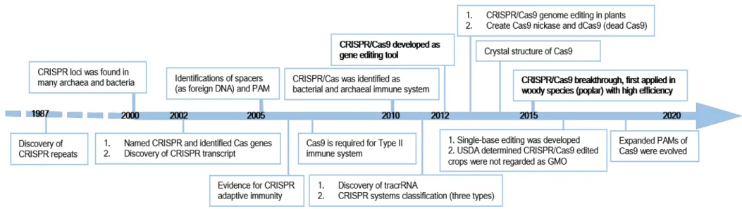
Figure I-16 Timeline of CRISPR/Cas9 genome editing key developments and progress in plants 45 Figure I-17 Pipelines of common-used Cloning methods for CRISPR/Cas9 applications in plants. 54 Figure 1 Genotyping of CCR1 and IAA9A transformants and their corresponding prevalent edition types 69 Figure 2 Compsrison between FT-IR spectra obtained from controls and CCR1 edited lines 74 Figure S1 Wave numbers from FT-IR absorption spectra discriminating between controls and CCR1-edited lines. 165 Figure 3 Comparison of xylem development and lignification of xylem cells between control and CCR1 edited lines’
roots. 75
Figure S2 Comparison of xylem development and lignification of xylem cells between control and CCR1-edited lines’
roots. 165
Figure 4 Pipeline of CRISPR/Cas9 implementation in Eucalyptus grandis hairy roots. 78 Figure 5 CRISPR/Cas9 sgRNA design and mutation detection in CCR1 and IAA9A. 79 Figure 6 CRISPR/Cas9 binary vector targeting two loci simultaneously. 80 Figure III-1A Histochemical analysis of cross sections of OE_EgrIAA9Am transgenic and wild-type Arabidopsis
hypocotyls. 92
Figure III-1B Histochemical analysis of cross sections of OE_EgrIAA20 transgenic and wild-type Arabidopsis
inflorescence stems. 93
X
Figure III-S2 The procedule used in Chapter III 171
Figure III-2 CRISPR/Cas9 sgRNA design and mutation detection in IAA9A, ARF5 and IAA20. 96 Figure III-3 Scheme illustrating the assembling of Cas9/sgRNA expressing constructs using Golden Gate Cloning. 96 Figure III-4 Scheme illustrating the assembling of overexpression constructs with CDS sequences of EgrIAA9Amm using
Golden Gate Cloning (including three levels). 98
Figure III-5 Scheme illustrating the assembling of overexpression constructs with CDS sequences of EgrIAA20m using
Golden Gate Cloning (including two levels). 99
Figure III-S3 Editing events of ARF5_17, ARF5_29, ARF5_33, ARF5_43, ARF5_44 by ICE analysis 172 Figure III-6 Genotyping of ARF5_and IAA20_edited lines and their corresponding prevalent edition types and related
amino acids changes deleted by ICE. 102
Figure III-7 Different edition types (A) and Indel contributions (B) of ARF5_10 analyzed by ICE Synthego. 103 Figure III-S4 Allele mutations of ARF5_43 and ARF5_44 detected by DSDecode. 173 Figure III-8 Different inferred different edited types of ARF5_40 (A) and IAA20_4 (B) obtained by ICE Synthego. 104 Figure III-S5 Combination of FT-IR and PCA (A) /PLS-DA (B) analysis for all ARF5_lines 174 Figure III-S6 Combination of FT-IR and PCA (A) /PLS-DA (B) analysis for all IAA9A_lines 175 Figure III-9 PLSDA analyses were performed using normalized values of FT-IR absorption spectrum (800-4000cm
-1) obtained from controls and ARF5_lines (A), IAA9A_edited lines (B) transgenic hairy roots. 107 Figure III-10 Wavelength numbers (WN) from FT-IR absorption spectra discriminating between controls and ARF5_lines
(A)/IAA9A_edited lines (B). 109
Figure III-11 Comparison of xylem development and lignification of xylem cells especially vessels between transgenic
roots of control and IAA9A_edited lines. 111
Figure III-S7 The comparison of xylem vessel cells diameters between IAA9A_edited lines and control in medium and old
developing stages 176
Figure III-12 The median value comparison of xylem vessel cells diameters between all IAA9A_edited lines and control 112 Figure III-13 Comparison of xylem development and lignification of xylem cells, especially vessels between transgenic
roots of control and ARF5_edited lines. 112
Figure III-S8 Comparison of ARF5_lines and control under 40x magnification using UV light 177 Figure III-14 Protein-protein interactions of EgrIAA9A (A) and EgrIAA20 (B) with potential candidates. 118
XI
List of Tables
Table I-S1 ZFNs, TALENs, CRISPR/Cas9 pros and cons comparison 161
Table I-1 A summary of different optimized CRISPR/Cas9 systems applied in various tree species 47 Table I-S2 CRISPR/Cas9 applications in trees using different targets, associated phnotypes and mutated efficiency 162 Table S1 Details of mutations detected in 3 batches of EgrCCR1 transgenic roots and control 166 Table S4 Details of mutations detected in EgrIAA9A transgenic roots and control 166 Table 1 Numbers of edited plants and type of mutations according to target genes 68 Table 2 Editing frequency and position (identified by PCR cloning and subsequent sequencing) 68 Table 3 Majority of altered alleles introduced significant modifications at the protein level 69
Table S2 The edition rate decrease along the transgenic plants age 166
Table S3 Mortality during the transfert from in vitro culture to hydroponic culture 166 Table S5 The edition type comparison of KOEgrIAA9A transgenic plants between DSDecode and sub-cloning
methods
167
Table 4 Mutation types 72
Table S6 Wave numbers from FT-IR absorption spectra discriminating between controls and CCR1-edited lines and their related compounds
168
Table S7 The selected sgRNAs sequences designed using CRISPOR (http://crispor.tefor.net/) 169 Table S8 The primers and sgRNA scaffold used for generating sgRNA intermediary vectors. 170 Table S9 The primers used in CRISPR/Cas9 system, the primer names, sequences and the specific uses 171 Table III-S1A Information of CRISPR/Cas9 mediated alived ARF5 (30 fluorescenced roots / 50 in total), IAA20
transgenic roots (three alive), and controls
179
Table III-S1B Information of CRISPR/Cas9 mediated alived IAA9A, and controls 181 Table III-S2 The selected sgRNAs sequences of EgrARF5 and EgrIAA20 designed using CRISPOR
(http://crispor.tefor.net/)
183
Table III-S3A The primers used in CRISPR/Cas9 system, the primer names, sequences and the specific uses 184 Table III-S3B The primers used in overexpressed constructions, the primer names, sequences and the specific uses 185 Table III-S4 Mutation types, occurrence, total Indel percentage and presumed knockout score of 11
ARF5_edited lines and one IAA20_edited line
186
Table III-1 Sequencing results of PCR products from Y2H library (secondary xylem) screening colonies using EgrIAA20 as a bait
114
Table III-2 Sequencing results of PCR products from Y2H library (secondary xylem) screening colonies using EgrIAA9 as a bait
XIII
ABREVIATION
2,4-D: 2,4-dichlorophenoxyacetic acid
4CL: 4-hydroxycinnamoyl-CoA ligase
4-Cl-IAA: 4-chloroindole-3-acetic acid
ABC: ATP-binding cassette
ABCB/D/G: ATP-binding cassette subfamily B/D/G
ABP1: AUXIN BINDING PROTEIN1
ACC: 1-amino-cyclopropane-1-carboxylicacid
AD: Activation domain
ARF: Auxin response factor
AuxREs: Auxin response elements
Aux/IAA: Auxin/Indole-3-Acetic Acid
AUX1/LAX: AUXIN RESISTANT 1/LIKE-AUX1 BD: Binding domain
BDL: BODENLOS
BR: Brassinosteroids
BSA: Bovine serum albumin
C4H: Cinnamate 4-hydroxylase
C3H: p-coumarate 3-hydroxylase
CAD: Cinnamyl alcohol dehydrogenase
CaMV: Cauliflower Mosaic Virus
CCoAOMT: Caffeoyl-CoA O-methyltransferase
CCR: Cinnamoyl CoA reductase
cDNA: Complementary deoxyribonucleic acid
CDS: Coding sequence
CesA: Cellulose synthases
CK: Cytokinin
CLE: CLAVATA3/EMBRYO SURROUNDING REGION
CML: Compound middle lamella
COMT: Caffeic acid O-methyltransferase
CRISPR: Clustered Regularly Interspaced Short Palindromic Repeats
crRNA: CRISPR RNA
CSC: Cellulose synthase complex
CSE: Caffeoyl shikimate easterase
CTAB: Cetyl trimethylammonium bromide
CTD: Carboxy-terminal dimerization domain
XIV
DSDecode: Degenerate sequence decoding
EAR: ERF-associated amphiphilic repression
EIN3D: Ethylene insensitive 3D
EMNs: Engineered meganuclease
ER: Endoplasmic reticulum
ERF: Ethylene response factor
F5H: Ferulate 5-hydroxylase
FT: FLOWERING LOCUS T
FT-IR: Fourier Transformed Infra-red spectroscopy
G: Guaiacyl GA: Gibberellin GH3: GRETCHEN HAGEN 3 GT: Glycosyltransferase H: p-hydroxyphenyl HDR: Homology-directed repair
HD-ZIP III: Class III homeodomain-leucine zipper HRM: High-resolution melting assay
IAA: Indole-3-acetic acid
IAM: Indole-3-acetamide
IAOX: Indole-3-acetaldoxime
IBA: Indole-3-butyric acid
ICE: Inference of CRISPR Edits
IFL1/REV: INTERFASCICULAR FIBERLESS/REVOLUTA IPA: Indole-3-pyruvic acid
LBD: Lateral organ boundaries domain
MCTU: Multi-component transcriptional unit system
MP: ARF5/MONOPTEROS
NAA: Naphthalene-1-acetic acid
NAC: NAM/ATAF/CUC transcription factors
nCas9: Nickase Cas9
NHEJ: Non-homologous end joining
NLS: Nuclear localization signal
NST: NAC SECONDARY WALL THICKNING PROMOTING FACTOR
ORF: Open reading frame
PAA: Phenylacetic acid
PAL: Phenylalanine ammonia lyase
XV
PCA: Principal component analysis
PCD: Programmed cell death
PCR: Polymerase chain reaction
PDS: Phytoene desaturase
PIN: PIN-FORMED
PILS: PIN-Like transporters
PGP: P-GLYCOPROTEIN
PLS-DA: Partial Least Square Discriminant Analysis
PXY: PHLOEM INTERCALATED WITH XYLEM
RAM: Root Apical Meristem
RD: Repression domain
S: Syringyl
SAUR: SMALL AUXIN UPREGULATED RNA
SAM: Shoot Apical Meristem
SCF: SKP1-Cullin-F-box
SCW: Secondary cell wall
sgRNA: Single guide RNA
SNP: Single Nucleotide Polymorphism
SL: Strigolactone
SSNs: Sequence-specific nucleases
TAM: Tryptamine
TALENs: Transcription activator-like effector nucleases
TE: Tracheary Elements
TF: Transcription factor
TFL: TERMINAL FLOWER 1
TIR1/AFBs: TRANSPORT INHIBITOR RESPONSE1/AUXIN SIGNALING F-BOX PROTEINS
TPL: TOPLESS
tracrRNA: Trans-encoded small RNA
tRNA: Transfer RNA
VND: Vascular-related NAC-domain
WAT1: WALL ARE THIN1
Y2H: Yeast two-hybrid
1
Objectives and Organization of the manuscript
The overall objective of my PhD was to implement the CRISPR genome editing tool to generate loss-of-function
Eucalyptus mutants in transgenic hairy roots and to use this technology to functionally characterize auxin-signalling
transcription factors Aux/IAAs and ARFs potentially involved in the regulatation of wood (secondary xylem) formation. The thesis is organized in three Chapters. Chapter I consists in a bibliographic review divided in three main parts. The first one is dedicated to Eucalytpus and wood formation, as well as secondary cell walls and its transcriptional regulation. In the second part, the main featuresA of auxin biology (synthesis, homeostasis and signaling), and its roles in the regulation of plant growth and development, with a special focus on wood formation are presented. The third part consists in an overview of the CRISPR/Cas9 technology, with a special emphasis of its implementation and applications in trees.
Chapter II is presented in the form of an article (Implementing the CRISPR/Cas9 Technology in Eucalyptus Hairy Roots Using Wood-Related Genes) published in “International Journal of Molecular Science” April 2020. As
proofs-of-concept, we chose as target genes Cinnamoyl-CoA Reductase1 (CCR1), a key lignin biosynthetic gene and IAA9A an auxin dependent transcription factor of Aux/IAA family. We designed two guide RNAs for each gene to generate CRISPR/Cas9-mediated mutations. Editions were detected in almost all transgenic lines with different allele-editing rates. The transgenic lines of CCR1 were further analyzed by spectroscopic methods combined to multivariate analyses (FT-IR_PLSDA) as well as histochemical analyses, confirming the phenotypes induced by CCR1-deficiency, i.e. decreased lignification and irregular xylem vessels. Although the efficiency of editing could be improved, the method described here is already a useful tool to functionally characterize Eucalyptus genes.
Chapter III focus on the functional characterization of auxin-related candidate genes EgrIAA9A, EgrIAA20 and
EgrARF5 in E. grandis transgenic hairy roots, using gain-of-function (overexpression under the constitutive 35S CamV promoter) and loss-of-function of mutants (CRISPR-cas9). We obtained CRISPR/Cas9 edited plants for all three targeted candidate genes. A fast-chemical screening by FT-IR_PLSDA analysis showed that the transgenic lines separated clearly from the wild-type control based on the global chemical composition analysis. Among them, bigger vessel cells and accerelated xylem development were identified in CRISPR/Cas9 generated EgrIAA9A_edited lines. However, no obvious phenotypes were detected for xylem formation in EgrARF5 edited lines in our experimental conditions. We also generated overexpressing lines for IAA9A and IAA20, unfortunately we lost all of them during the covid lockdown, as well as most plants of CRISPR/Cas9 generated EgrIAA20 lines. Finally, the potential partners of EgrIAA9A and EgrIAA20 (as a bait) were screened out from Eucalyptus developing xylem cDNA library using Yeast Two Hybrid (Y2H). Interestingly, we found that EgrIAA9A is the main protein partner of EgrIAA20, suggesting that EgrIAA20 forms complex with EgrIAA9A to regulate the transcriptional regulation during wood formation. We further using Y2H to validate the protein-protein interaction among our Eucalyptus Aux/IAA and ARF members potentially involved in wood formation.
3
Chapter I
Bibliographic review
5
Part 1 Eucalyptus and wood formation
1. Eucalyptus
The genus Eucalyptus belongs to a basal Rosid lineage (Myrtales order, Myrtaceae family) which evolved mostly in the isolation of the Australian continent, Tasmania and nearby islands. Therefore, they represent independent evolutionary experiments over 22 million years for studies of the woody perennial lifestyle (Myburg et al., 2014). The Eucalyptus genus is highly diverse and displays significant adaptability and phenotypic plasticity. These long-lived, sclerophyllous evergreen and flowering hardwood trees are well adapted to diverse climates from tropical rainfall to temperate semiarid zones, though only very few species are tolerant to coldness (Wiltshire, 2004) mainly due to the absence of dormancy. They can also grow on lean soils. Eucalypts come in a great range of shapes and sizes – from tall trees (Up to 100m for the tallest) to small multi-stemmed shrubs. Among its thirteen subgenera, the most important is Symphyomyrtus, comprising about 474 species including the ones of commercial importance (Grattapaglia et al., 2015). Only 20 or so of those Eucalyptus species have been extensively used in commercial forests plantation given they are fast and easy to grow, provide high forest productivities and deliver wood with high density, durability and interesting fibre properties (Grattapaglia et al., 2015). Because of their wide adaptability, fast growth and multipurpose uses of their wood, eucalypts are the most widely planted forest trees worldwide (Myburg et al., 2007), growing in over 100 countries such as Brazil, China, India, South Africa, Portugal, and covering more than 20 million hectares. China’s eucalypt plantations make around 27% substantial contribution of a total of annual domestic timber output.
Global carbon cycle of forest contains carbon sequestration in tree biomass and soil through photosynthesis and respiration, and forestry timber sustainable harvesting contributes a significant pathway to carbon sequestration. In Brazil, along with the increased Eucalyptus plantation area and productivity, carbon contribution of Eucalyptus increased 13% (from 58% to 71%) in 25 years (Sanquetta et al., 2018). Because of the highly efficient C3 photosynthesis occurring in eucalypts, carbon sequestration is massive and will likely increase along with elevated ambient CO2 and rising temperature (Ghannoum et al., 2010). This strong response to CO2 contributes Eucalyptus to become a crucial tree for carbon sink and an environmental protector in the context of global warming.
Forests cover a third of world’s total land area, and over a half of our forest conservation contributes to human activities, such as wood production, food and other forest products. The trend of global wood sustainable harvests will be up to 80% by planted forests expansion in 2030 (Jim Carle and Peter Homgren, 2008), comprised of softwood (60% supply by Pinus) and hardwood (prefer Eucalyptus and Acacia) (Ramage et al., 2017). Eucalypt native and domesticated forests are an important source of high-quality woody biomass for many wood products (pulp and paper, sawn timber, composites, fuelwood), which are dependent on variable key wood properties (wood density, hardness, color and shape, chemical composition, heartwood, and so on). Pulp and paper production is in nowadays the main purpose of industrial plantations (Greaves et al., 1997). Recently, the urgent need for fuels resources and applications gave rise to emerging biofuel as an alternative choice, hence bioethanol obtained from eucalyptus is a crucial feedstock (Shepherd et al., 2011; Verma et al., 2016). Eucalyptus have extending rotations such as high-value sawlogs with maximum biomass of clear wood, and bark
6
residues of E.grandis and E.grandis x urophylla for biofuel production (Lima et al., 2014). Mechanisms of lignin deposition and lignin changes along with tree maturation have been studied in E.globulus young and mature wood (Rencoret et al., 2011). In addition, non-destructive technics for wood properties assessment (e.g., FT-IR spectroscopy) are required for solid wood products and researches of traits heritability (Raymond, 2000).
The Eucalyptus grandis genome ‘BRASUZI’ sequence has been released (Myburg et al., 2014), which provided insights into candidate genes and regulators involved in wood formation. Genome-wide analysis of lignin biosynthetic genes identified 17 candidate genes associated with E. grandis xylem lignification (Carocha et al., 2015). Also, members of several families of transcription factors, such as MYB, NAC, AFR and Aux/IAA, were identified to be involved in secondary cell wall (SCW) formation and wood formation in eucalypts (Yu et al., 2014, 2015; Soler et al., 2015; Hussey et al., 2015; Shinya et al., 2016). Further investigations of biosynthesis genes and transcription factors associated with regulation of wood formation in Eucalyptus will be described in the corresponding paragraphs, later in this chapter. Transformation using Agrobacterium tumefaciens has been implemented for several Eucalyptus species or hybrids (Tournier et al., 2003; Girijashankar, 2011; de la Torre et al., 2014; Plasencia et al., 2016) and recently freeze-resistant transgenic eucalyptus have been recently obtained in our lab (Cao et al., 2020). However, this is a very long and tedious process that do not allow easy functional characterization of candidate genes. To overcome this difficulty, a protocol using
Agrobacterium rhizogenes has been implemented in our lab (Plasencia et al, 2016) and transgenic hairy roots are currently
being used to functionally characterize candidate genes (Soler et al., 2017; Dai et al., 2020). For my PhD study, we utilize Eucalyptus as our experimental species to investigate the wood formation in tree.
2. The vascular system
From the numerous adaptations that land plants have developed during evolution, the acquisition of the vascular system some 400 million years ago have been a crucial event ensuring their successful earth colonization. The vascular system is thus an evolutionary innovation which enables delivering water and nutrients, as well as mechanical support to ensure plants growing tall to get more access of sun light for their photosynthesis and growth. It is composed of two main tissues, xylem and phloem. Xylem cells play crucial roles in water transport and mechanical support of the entire plant. Phloem is essential to the transport of photosynthate from photosynthetic tissues (leaves, source tissues) to developing tissues (sink tissues).
2.1 The vascular system during primary growth
During embryogenesis, at the globular stage, protoderm cell division generates vascular stem cells. Provascular tissue presents a vascular patterning geometry at the heart stage, and it is specified into Shoot Apical Meristem (SAM) and Root Apical Meristem (RAM) (Scheres et al., 1994; De Rybel et al., 2014) respectively, while no differentiation occurs yet (shown in Figure I-1a). The SAM and RAM are responsible for primary growth, i.e. extension of the shoots and roots, respectively. Later on, but still on embryo stage, vascular plants develop a lateral meristem called procambium. During a first phase of growth in the acropetal direction, these meristems produce the primary plant body including the primary vasculature composed of primary xylem and phloem. Procambium cells undergo two types of division: the periclinal
7
divisions parallel to the plant axis/surface, give rise to phloem and xylem precursor cells, whereas the anticlinal divisions perpetuate the procambium tissue along the plant axis giving rise the enlargement of procambium tissues themself. In some species, primary vasculature, begin to differentiate from procambium in mature embryos, but in many others, vascular tissue differentiation only starts after the seed germinates (Evert, 2006).
In leaves, phloem and xylem generates towards abaxial and adaxial surfaces, respectively. In stems, primary vascular tissues are organized in discrete collateral vascular bundles separated by parenchyma cells, while in roots, the vascular tissue is organized in a bi- or multi-symetric pattern (Figure I-1).
In both root and shoot vasculature, the most common organisation is, from the outside to the inside of the organ: phloem, procambium and primary xylem cells. Primary xylem is formed by differentiation of the procambium into protoxylem and metaxylem. During primary growth, strands of primary xylem are found in stems and roots, presenting narrow, small vessel cells of protoxylem and subsequent extended metaxylem which has larger size (Figure I-2). Among four patterns of protoxylem and metaxylem distribution, exarch development pattern (xylem develops centripetally) is found in vascular plant roots, showing protoxylem near pericycle and metaxylem close to pith. Another development pattern (xylem develops centrifugally) is the endarch one that exists in stems of seed plants, showing opposite distribution of exarch pattern of protoxylem and metaxylem (Figure I-2). Notably, secondary cell wall (SCW) is deposited in protoxylem in elongating organs and metaxylem in non-elongating organs by different patterns to provide mechanical support to xylem conduits (Ye and Zhong, 2015).
Roots differ from stems by the absence of pith, and the presence of two additional single-cell ring-shape layers, the endodermis and the pericycle. Endodermis will further form the Casparian strip, which is essential for impairing the diffusion of solutes from the inside to the outside of roots. The pericyle is a single layer of meristematic cells, the origin of lateral roots and have a role in secondary growth of roots. The vascular tissue in young roots presents a bisymetric pattern, the primary xylem forms a central axis, and two flanking primary phloem poles (Figure I-1) (Nieminen et al., 2015).
8
Figure I-1. Vascular development in dicot angiosperms (Adopted from (Ruonala et al., 2017)). (a) The provascular tissue (orange) is formed during embryogenesis. (b) The vasculature developing into different structures in organs and tissues (shoots, roots, leaves), starting from shoot apical meristem (SAM) and root apical meristem (RAM), and later develops into xylem (inner) and phloem (outer), throughout whole plant organs and tissues.
Figure I-2. Xylem developing patterns in stems (endarch) and roots (exarch): xylem in green.
2.2 The vascular system during secondary growth
During (primary growth), plants grow in the acropetal direction thanks to the activity of root and shoot apical meristems. With the notable exception of the monocotyledons, many vascular plants undergo a second phase of growth (secondary growth), which implies growth in diameter or radial growth. This secondary growth can be limited to the hypocotyls as for instance in Arabidopsis or can be particularly important like in trees where it produces large amount of secondary xylem (wood).
During secondary growth in angiosperm stems, procambium cells will give rise to the fascicular cambium whereas the interfascicular cambium is thought to arise through the de novo recruitment of interfascicular parenchyma cells (Schuetz
9
et al., 2013). In roots, the interfascicular cambium differentiates from pericyle cells. The junction of these two populations of cambial cells, makes the entire circular vascular cambium of mature tree trunks and roots (Figure I-1). The vascular cambium is a cylindrical meristem which contains both division and differentiation zones. Two types of divisions exist in cambial stem cells: anticlinal division (perpendicular to surface and axis) which extend the cambial rings laterally, and periclinal division (parallel to surface) which increases radial cell files for differentiation to seconcary xylem inwards and to phloem outwards. So the vascular cambium not only proliferates to form new cambium cells to renew meristematic cells, but also differentiates to form secondary xylem inwards, or secondary phloem outwards (Groover and Robischon, 2006) in stems and roots/hypocotyls. There are two types of cambium stem cells: the larger fusiform initials and the small isodiametric ray initials. Fusiform initials are responsible for longitudinally aligned cells (such as vessels, fibres, sieve elements etc) with vacuolated cytoplasm generated by periclinal division, and ray initials are responsible for transversely aligned cells (ray parenchyma cells) connecting secondary phloem and xylem in radial cell system (Evert, 2006). The cambial cells undergo producing destined mother cells in differentiation zone, to form either xylem mother cell or phloem mother cell, and xylem and phloem elements afterwards (Figure I-3). Using lineage tracing analysis and sector analysis, vascular cambium was featured as a single layer of true cambial initials (being able to divide both anti- and periclinally) in Arabidopsis and poplar (Bossinger and Spokevicius, 2018; Shi et al., 2019).
Figure I-3. Schematic representation of the radial growth of vascular cambium (Tonn and Greb, 2017). The secondary vasculature is produced by vascular cambium initials in central cambial zone. The cork cambium produces phelloderm inwards and cork outwards. Periclinal division of vascular cambium gives rise to differentiation into xylem or phloem. Anticlinal division produces more initials to increase the circumference of cambial zone.
The stem cell fates are determined by phytohormones and meristematic capacity, that will be maintained by xylem identity cells (initial xylem) directing adjacent vascular cambial cells to act as stem cells (Smetana et al., 2019), and will be complemented by pericycle division to form new vascular cambium (Chiatante et al., 2018). Except for peptide signaling pathways such as TDIF-PXY signaling which promotes cambium proliferation and inhibits xylem differentiation, the long-distance transported hormone signaling molecules involved in cambial cell proliferation and differentiation have been revealed in many researches, for example the pesipetal transported auxin. These genetic controls of vascular cambium
10
activity have been reviewed in (Nieminen et al., 2015; Ruonala et al., 2017). Vascular cambium activity is also affected by environmental factors (Fischer et al., 2019). In addition, hormone signal molecules are crucial for communication between primary and secondary meristems (Wang, 2020).
3. Wood formation (xylogenesis)
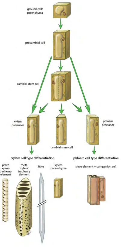
Wood (secondary xylem) is a sequential complex developmental process which starts in vascular cambium. Secondary xylem is produced after phloem, while has the larger quantity than phloem at the end of season in perennial tree stems. Fusiform initials of cambial cells divide periclinally to give rise to long narrow cells which differentiate into conducting cells, comprising tracheid elements and secondary xylem fibers towards inside and sieve elements (in phloem) towards outside (Figure I-4). Ray initials differentiate into small short ray parenchyma cells which provide radial transport of water and minerals between xylem and phloem.
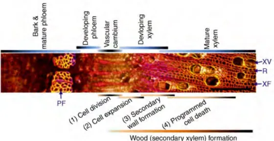
Cambial cells periclinal division towards pith can be activated and subsequently undergo cell expansion to form specialized secondary xylem with final size, along with secondary cell wall deposition and programmed cell death (Figure I-5). There are three types of wood cells: tracheary elements (TEs, tracheids or vessels), xylem parenchyma cells and xylem fiber cells (Figure I-5). Of these, TEs (dead cells when matured) are the main type of secondary xylem acting as well-conducting tubes for transport throughout plants (Turner et al., 2007). The cell expansion of vessels is rapid to give large diameter and is limited in axial orientation as compared to fibers which have moderate expansion. Once vessels and fibers final size is reached, secondary cell wall (SCW) is deposited and then lignified, becoming inflexible and impermeable. Though SCW deposition is the dominant step of wood formation, programmed cell death (PCD) is also essential followed or simultaneously occurring with SCW lignification (Derbyshire et al., 2015). Programmed vessel cells death occurs rapidly starting by vacuolar integrity loss, while fiber cells death is a gradual degradative process in nucleus and cytoplasm (Courtois-Moreau et al., 2009). Parenchyma cells are dead at the end along with sapwood converts into heartwood. Due to complex tissues where TEs forming, TEs in vitro provides advantages of high percentage, easy accessibility, and molecular information about cell wall such as biochemical and gene expression changes (Devillard and Walter, 2014). Lignification is the biological process of lignin deposition in cell wall, which mainly includes cell-autonomous lignification such as vessel and fiber cells, where monolignols are produced and deposited by differentiating cells. Non-cell-autonomous lignification occurs in parenchyma cells, where monolignols produced by neighbored cells are transferred to non-autonomous-cells (Smith et al., 2017).
11
Figure I-4. Overview of procambial/cambial cell specification and xylem/phloem cell differentiation (Extracted from (Schuetz et al., 2013)).
12
Figure I-5. Transversal sections of poplar stem (stained with Calcofluor, auramine O, propidium iodide) showing wood cell formation processes (Ko et al., 2016). The bars above show sequential wood formation stages. PF, phloem fiber; XV, xylem vessel; XF, xylary fiber; R, ray cell. Scale bars represent 200μm.
3.1 Secondary cell wall (SCW) structure and composition
After expansion of differentiated specialized cell types, secondary cell wall (SCW) is deposited between primary cell wall (PCW) and plasma membrane. Primary cell wall is thin, flexible and extensible layer of the cell wall composed of cellulose (50%), pectin (20-30%) and hemicellulose (20-30%). The PCW is a unique fabric that is strong but usually thin, flexible, and capable of both plastic and elastic extension.The primary wall is the cellulose-containing layer laid down by cells that are dividing and growing. To allow for cell wall expansion during growth, primary walls are thinner and less rigid than those of cells that have stopped growing. PCWs have functions of giving cells stability, determining shapes, protection, etc. Different from primary cell walls, SCWs function are to provide mechanical strength, water-proofing for water conduction, and barrier for protection against biotic and abiotic stresses. SCWs contain three major components: cellulose (40-50%), hemicelluloses (xylan and glucomannan (25%)), and lignin (25-35%). SCWs are mainly found in tracheary elements (TEs) and fibers, as well as in some other specialized tissues (Mauseth, 1988). The proportion of three components in SCWs is variable among different vascular plants in different developmental stages, in different cell types, and even changes when facing environmental stresses.
SCWs contain three distinct layers S1, S2 and S3 showing different cellulose content, polymerization, crystallinity and orientation (Timell, 1967; Müller et al., 2006; Mellerowicz and Sundberg, 2008). The S layers are abundant with xylan and cellulose while has less lignin than in compound middle lamella (CML) (Donaldson et al., 2001). The middle lamella serves as a cementing layer between the primary walls and adjacent cells. It is mainly composed of pectic polysaccharides, lignin, and a small amount of proteins. It is the first layer that is formed, which is deposited at the time of cytokinesis. Each S layer has special cellulose microfibril either be aligned irregularly, or in particular angle. In Eucalyptus blenched fibres, reproducible xylan (hemicellulose component) distribution pattern across SCW showed more xylan quantities in S1 and S3 than in S2 (Lekha et al., 2017).
13 Cellulose
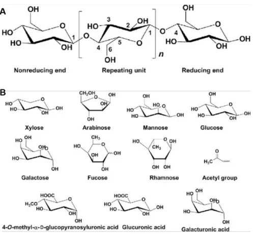
Cellulose microfibril is the load bearing unit in both primary and secondary cell walls consisting of several β-1,4 linked chains of D-glucopyranosyl (Glu) residues (Doblin et al., 2002). It is a polysaccharide which is the most abundant component in SCWs and the first abundant biopolymer on earth, accounting for 40-50% of wood (structure in Figure I-6). In Arabidopsis, SCW cellulose synthases (CESAs) include three non-redundant proteins (CESA4/IRREGULAR XYLEM5b (IRX5), CESA7/IRX3 and CESA8/IRX1) belonging to the glycosyltransferase (GT) family 2 (Taylor et al., 1999, 2000, 2003). They are located at the plasma membrane, colocalize with cortical microtubules bands in older vessels and are functionally conserved in many vascular plants. Loss of function mutants of a single CesA resulted in xylem morphology defects, showing collapsed xylem cells (Taylor et al., 2000). Cellulose synthases are integral plasma membrane proteins organized in hexameric rosette complexes which contain six-fold catalytic subunit trimers (Nixon et al., 2016) with different contributions of each CESA for cellulose synthesis (Kumar et al., 2018). Hampered cellulose synthesis genes led to little effect in the other two polymers (xylan and lignin) contents in SCWs (Turner and Somerville, 1997; Zhong et al., 2003). CESAs belong to multigene families, three PtrCesA1, PtrCesA2, PtrCesA3 in aspen (Populus tremuloides) are specifically highly expressed in xylem tissue during SCW deposition (Joshi et al., 2004), and more than 12 CesA transcripts were identified in differentiated xylem undergoing SCW (Suzuki et al., 2006). Two types of cellulose synthase complexes (PtrCesA7A, PtrCesA3D) were further identified to influence crystalline multilaminar cellulose structure in wood SCW (Xi et al., 2017). Overexpression of PmCesA2 in hybrid poplar resulted in thickening SCW and increased cellulose and lignin content (Maleki et al., 2020). In addition, the membrane-bound β-1,4-endoglucanase KORRIGAN (KOR) is also required for proper cellulose-hemicellulose network synthesis, and the kor mutants of Arabidopsis exhibited decreased cellulose content in primary cell wall (Nicol, 1998; Sato et al., 2001), incomplete cell walls, defect of cell plate formation (Zuo et al., 2000), and irregular xylem vessel development (Szyjanowicz et al., 2004). The downregulation of PdKOR in poplar displayed increased crystalline cellulose but decreased polymerization of cellulose, reduced plant growth, and altered carbon allocation and biomass composition (Kalluri et al., 2016; Bali et al., 2016).
Hemicelluloses
Hemicelluloses account for around 20-35% of dry biomass and has amorphous structure with little strength (Figure I-6). Hemicelluloses consist of xylans, mannans, xyloglucans, and β-1,3;1,4-glucans, and have various structural features (as reviewed in (Zhou et al., 2017)), which are different from cellulose but prevents its flocculation. The β-(1,4)-linked xylose homopolymer xylan is the main hemicellulose of SCW in angiosperms and is one of the most abundant naturally occurring polymers (Ebringerová and Heinze, 2000; Scheller and Ulvskov, 2010). Using genetic approaches, genes associated with biosynthesis and structure of hemicellulose have influence growth and development in mutants as reviewed in (Pauly et al., 2013). Many glycosyltransferase (GT) genes located in Golgi membrane were identified to participate in xylan biosynthesis. A series of IRX, PARVUS, F8H genes in Arabidopsis and functional orthologs GT genes in poplar (Zhou et al., 2006; Lee et al., 2011; Ratke et al., 2018; Busse-Wicher et al., 2016; Ratke et al., 2018) were thought to play roles during SCW not only in linear β-(1,4)-linked xylose backbone formation, elongation and decoration pattern, but also
14
affected growth and lignocellulose saccharification. Moreover GT43B promoter was utilized specifically for secondary wall modification.
15 Lignin
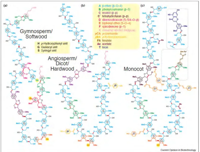
The characteristic of xylem SCW is massive lignin deposition which provides rigidity and impervious to thickened cell walls. Lignin is a complex tridimensional aromatic polymer composed of phenyl propane units. It plays roles in water conduction, mechanical support and protect cell wall polysaccharides from pathogens. Lignin is mainly made of hydroxycinnamyl alcohols (or monolignols), coniferyl alcohol, sinapyl alcohol, and p-coumaryl alcohol, and this cross-linked complex gives rise to guaiacyl (G), syringyl (S) and p-hydroxyphenyl (H) units, as well as other aromatic monomers (Vanholme et al., 2019). Lignin model structures of gymnosperm, angiosperm (some monocots included) were shown in (Ralph et al., 2019) (Figure I-7). In different species and different wood cells, the proportions of G, H and S units are distinct, for instance, S lignin is prevalent in angiosperms but absent in gymnosperms. Lignin containing S units are less condensed compared to H and G lignin due to the absence of very strong [ β-5’, 5’-5’, 4’-O-5’] C-C linkages (Ralph et al., 2004). Dicots lignin mainly contains G and S units (Weng and Chapple, 2010), and wood vessel cells are rich in G lignin while fibres contain S-G lignin (Lourenço et al., 2016). G lignin acts for tracheary elements (TEs) cell wall strength and conduction, while S lignin plays roles for derived traits such as defense (Renault et al., 2019). Lignin biosynthesis, monolignol contents and composition are affected by various factors such as plant growth and development, metabolic stresses, cell wall perturbation, wounding, and a series of biotic and abiotic stresses, (Cano-Delgado et al., 2003; Tronchet et al., 2010; Vanholme et al., 2019). Regulation of lignin biosynthesis genes results in altered H:G:S distribution.
Figure I-7. Lignin model structures of three major plant classes: gymnosperm/softwood (a), angiosperm/dicot/hardwood (b), and monocot (c) (Ralph et al., 2019).
16
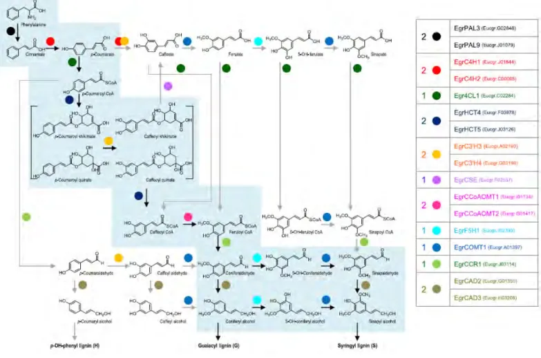
Lignin resistance to degradation is a major obstacle for industrial processing of wood such as during pulp and paper manufacturing where it requires harsh and costly chemical treatments. The huge economic importance of the pulp industry has been a driving force to decipher the lignin biosynthetic pathway, which has proven more complex and reticulate than initially thought. The topology of the pathway has been revised several times in the last decades [reviewed in (Humphreys and Chapple, 2002; Boerjan et al., 2003; Ralph et al., 2004; Vanholme et al., 2010)] and new alternative routes are still being discovered such as that involving the recently described caffeoyl shikimate esterase [CSE; (Vanholme et al., 2013)]. Altogether eleven enzymatic reactions (Figure I-8) are implicated in the synthesis of monolignols that involves the general phenylpropanoid pathway starting with the deamination of phenylalanine and leading to the production of hydroxycinnamoyl CoA esters. The enzymes involved in this short sequence of reactions are phenylalanine ammonia-lyase (PAL); cinnamate 4-hydroxylase (C4H) and 4-coumarate:CoA ligase (4CL). Hydroxycinnamoyl-CoA esters undergo successive hydroxylation and O- methylation of their aromatic rings (Boerjan et al., 2003) involving the following enzymatic activities: shikimate O-hydroxycinnamoyltransferase (HCT); caffeoyl shikimate esterase (CSE); p-coumarate 3-hydroxylase (C3’H); caffeoyl CoA 3-O-methyltransferase (CCoAOMT); ferulate 5-hydroxylase (F5H) and caffeate/5-hydroxyferulate O- methyltransferase (COMT). The conversion of the side-chain carboxyl to an alcohol group is catalyzed successively by cinnamoyl CoA reductase (CCR) and cinnamyl alcohol dehydrogenase (CAD), two enzymes considered to be the most specific of the monolignol biosynthesis pathway.
Notably, the CCR gene, was first cloned in Eucalyptus gunnii (EguCCR) and its identity was proven unambiguously by the enzymatic activity of the corresponding recombinant protein (Lacombe et al., 1997). Since the release of the E. grandis genome (Myburg et al., 2014), a genome-wide survey of the putative lignin biosynthesis genes, followed by comparative phylogenetic analyses, led to the identification of 38 genes of which 17 exhibit strong and preferential expression in highly lignified tissues. These 17 genes constitute the core set of a Eucalyptus lignification toolbox (Figure I-8).
The transport of monolignols from cytoplasm or near endoplasmic reticulum to developing cell wall occurs through mechanisms not yet elucidated. Polymerization is performed by oxidases such as laccases (LAC) and peroxidases (PRX) which can catalyze monolignol oxidation and polymerization (Bryan et al., 2016). In Arabidopsis stem, PRX64 localized in cell corners and fibers middle lamella, and LAC4 was in xylan-rich SCW layers of vessels and fibers, and both of them were highly expressed in lignifying tissue (Yi Chou et al., 2018).
17
Figure I-8. The lignin biosynthesis pathway in Eucalyptus adapted from (Carocha et al., 2015).The 17 E. grandis genes encoding enzymes located in the bona fide clades constitute the core set of a Eucalyptus lignification toolbox. Enzymatic reactions thought to be key steps are indicated with balck arrows.
3.2 The transcriptional regulation of secondary cell wall formation
The transcriptional network underlying SCW formation has been first elucidated (Figure I-9) in Arabidopsis and later in poplar as reviewed in (Hussey et al., 2013; Zhang et al., 2018a; Camargo et al., 2018). It involves mainly members of two transcription factors families: the NACs (NAM/ATAF/CUC) and the MYBs and (MYeloBlastosis) acting as first- and second-level master switches, respectively, to regulate a battery of downstream transcription factors and secondary cell wall biosynthesis genes (Wang et al., 2011; Schuetz et al., 2013). A recent comparison of the regulation of wood formation in angiosperm trees species versus Arabidopsis highlighted conserved and distinct mechanisms (Camargo et al., 2018). Top-level NAC master regulators
Among the NACs, the VASCULAR-RELATED NAC-DOMAIN (VND) 1-7 and NAC SECONDARY WALL THICKNING PROMOTING FACTOR (NST) 1-3 are master switches of the entire SCW transcriptional regulation to
18
control xylem differentiation and lignification by regulating downstream partial overlapped target genes (Zhong et al., 2010d; Ohashi-Ito et al., 2010). In Arabidopsis, VNDs induce differentiation of vessels whereas NSTs/SNDs induce differentiation to be fibers (Mitsuda et al., 2005, 2007; Zhong et al., 2006; Yamaguchi et al., 2008).
Cellulose synthase complexes (CSC) are transcriptionally regulated during the initial of SCW synthesis by VNDs (VND1-7), especially VND6 and VND7 that were preferentially expressed in secondary xylem and have core functions for vessel formation in metaxylem and protoxylem (Kubo, 2005). In suspension culture cells system, VND6 directly regulated expression of cellulose synthesis genes CESA4/IRX5, CESA7/IRX3, CESA8/IRX1 (Ohashi-Ito et al., 2010; Yamaguchi et al., 2010), whereas, VND6 and VND7 controlled lignin biosynthetic genes CCoAOMT7, LAC4 and related PCD genes, XCP1, XCP2, BFN1, RNS3 (Zhong et al., 2010d; Zhong and Ye, 2014). Overexpression of AtVND6 or VND7 was shown to induce xylem vessel transdifferentiation both in Arabidopsis and in poplar (Kubo et al., 2005; Yamaguchi et al., 2010), suggesting that the molecular mechanism of xylem vessel differentiation is, at least partially, conserved between these two species. VND6/7 were shown to be controlled by upstream regulators such as Lateral organ boundaries domain 18 (LBD18) and LBD30 that control VND6/VND7, but are also targets of VND6/VND7 showing a feedback regulation (Soyano et al., 2008; Zhong et al., 2010d). E2Fc is also a regulator located upstream of VND6 and VND7 but has dual-function since it activates/repress by dose effect. E2Fc is also able to bind directly promoter regions of SCW components biosynthesis genes (C4H, CCoAOMT, CAD, LAC4) (Taylor-Teeples et al., 2015). VND-INTERACTING 2 (VNI2) is the negative regulator of VND, showing thickened SCW in xylem vessels of young overexpressing mutants (Yamaguchi et al., 2010). NST1-3 control fiber cells SCW formation and regulation; NST1 and NST3/SND1 are functionally redundant. Double
nst1/(snd1/nst3) Arabidopsis mutant showed no thickness of SCW in fibres whereas SCW was normal in vessels. In this
double mutant, the cellulose biosynthesis related genes IRX3 and IRX5, lignin biosynthesis related IRX4, IRX12 and AtOMT1 were all down-regulated (Mitsuda et al., 2007). NST3 was shown to induce PAL1, CCoAOMT, 4CL3, cellulose synthase-like B02 (CSLB02) and fasciclin-like arabinogalactan-protein 12 (FLA12) expression (Ohashi-Ito et al., 2010) The poplar orthologs of the VNDs and/or NSTs/SNDs genes are called WNDs (for wood-associated NAC domain transcription factors (Zhong et al., 2010b), or VNSs (for VND, NST/SND-, SOMBRERO-related proteins; (Ohtani et al., 2011) or PtrSNDs/PtrVNDs (Li et al., 2012; Johnsson et al., 2018). They could complement the SCW defects of the fibers in the double nst1/(snd1/nst3) Arabidopsis mutant (Zhong and Ye, 2010). However, surprisingly only PtrWND2B and
PtrWND6B were able to induce ectopic deposition of SW when overexpressed in Arabidopsis (Zhong et al., 2010b). To
explain the fact that other VNDs or NST/SNDs members could complement the nst1/snd1 mutant but are not capable of inducing ectopic expression of SCWs, a likely hypothesis is that they need to cooperate with co-factors or other TFs which are only present in cells programmed to be sclerified.
It was initially reported that the fiber- or vessel-specific expression occurring in Arabidopsis was not occurring in poplar where all WNDs/VNS (both VND and NST/SND) were expressed in both developing vessels and fibers as well as in xylem ray parenchyma cells. The clear separation of the expression patterns of VND and NST/SND groups in Arabidopsis did not seem to be extensively shared with other plant species including poplar, rice, and maize (Zhong et al., 2010a, 2011a; Ohtani et al., 2011; Nakano et al., 2015). However, the recent high spatial-resolution RNA sequencing data spanning the secondary phloem, vascular cambium, and wood-forming tissues of Populus tremula (Sundell et al., 2017; Johnsson et al., 2018) provided new clues about the expression patterns of the genes of the VNDs and NSTs/SNDs clades which are more