Negative microbiological results are not mandatory in deep sternal
wound infections before wound closure
†
Hector Rodriguez Cetina Biefer*, Simon H. Sündermann, Maximilian Y. Emmert, Zoran Rancic,
Sacha P. Salzberg, Jürg Grünenfelder, Volkmar Falk and André R. Plass
Clinic for Cardiovascular Surgery, University Hospital Zurich, Zurich, Switzerland
* Corresponding author: Department for Cardiovascular Surgery, University Hospital Zurich, Rämistrasse 100, 8091 Zurich, Switzerland. Tel: +41-44-2552039; fax: +41-44-2554467; e-mail: [email protected] (H. Rodriguez Cetina Biefer).
Received 1 September 2011; received in revised form 20 November 2011; accepted 22 November 2011
Abstract
OBJECTIVES: To define the outcome of treatment for deep sternal wound infections (DSWIs) using direct wound closure (DC) or vacuum-assisted therapy (VAT) based on negative vs. positive microbiological results.
METHODS: Between 1999 and 2008, 7746 patients underwent median sternotomy for cardiac surgery at our institution. Patients were screened for DSWI and out of the cohort 159 were identified (2%). These patients were treated, either using DC or VAT with delayed wound closure. Outcomes were retrospectively analysed to determine the effect of negative cultures at the time of closure.
RESULTS: The indication for sternotomy was CABG 51%, isolated valve 18%, CABG/valve 18% and other related cardiovascular proce-dures 14%. Sixty-five percent of the wound infections was diagnosed during rehabilitation period. One hundred and five (66%) patients were treated with VAT vs. 54 (34%) patients with direct closure. Coagulase negative staphylococci were found in 48% of bacterial cul-tures. In 75% of the patients, the microbiological results were positive at time of wound closure (69.2% VAT vs. 87.0% direct closure, P = 0.014). Out of 159 patients, 5.0% were with positive microbiological results at the time of closure readmitted vs. 5.1% with negative microbiological results (P = 1.0). Patients with VAT stayed significantly longer in the hospital (mean 21 ± 16 vs. 13 ± 12, P = 0.002). CONCLUSIONS: Negative microbiological results are not mandatory before wound closure, as the rate of readmissions for recurrence of infection showed no difference between groups. Our results also suggest that shortening of VAT despite positive microbiological results may be feasible.
Keywords:Vacuum-assisted therapy• Deep sternal wound infection • Microbiological findings • Wound closure
INTRODUCTION
Median sternotomy, either partial or total, is the most common surgical access to the heart in cardiac surgery.
Deep sternal wound infection (DSWI) is one of the most fearful complications in cardiac surgery after a median sternot-omy [1,2]. The frequency of DSWI varies among cardiac centres ranging from 0.2 to 8% [2–8].
The management of DSWI includes several approaches. The most common treatments for DSWI are debridement with direct wound closure (DC) and a combined therapy, which include vacuum-assisted therapy (VAT) with delayed wound closure [9–12]. More invasive approaches have been also described, which are: partial sternal resection and coverage with omentum majus or muscleflaps as well as sternal osteosynthesis among others [2,8]. Although these approaches are well established, the use of VAT in DSWI is the most common practice among centres
[9–13] and has been also the standard approach at our institution over the past years.
Wound closure after DSWI depends on several factors. One of the important factors is negative microbiologicalfindings as well as the surgeon’s assessment of wound granulation [12,14,15]. There is still controversy if wound closure should be done only in case of negative microbiologicalfindings. One way to achieve this is through VAT [12,15–17].
Our hypothesis was that negative microbiologicalfindings are not mandatory before wound closure in DSWI.
MATERIALS AND METHODS
Data from 7746 patients who underwent cardiac-surgical proce-dures at our institution between January 1999 and December 2008 were prospectively collected and retrospectively analysed. Patients with DSWI were selected according to the classification of El Oakley and Wright [18]; however, no further differentiation to the specific subtypes was performed. Patients with partial as well as total sternotomy were included. Before surgery, no †Presented at the 25th Annual Meeting of the European Association for
Cardio-Thoracic Surgery, Lisbon, Portugal, 1–5 October 2011.
© The Author 2012. Published by Oxford University Press on behalf of the European Association for Cardio-Thoracic Surgery. All rights reserved.
European Journal of Cardio-Thoracic Surgery 42 (2012) 306–310
ORIGINAL ARTICLE
pre- and/or postoperative bacterial eradication was performed. A total of 159 patients (2%) developed DSWI postoperatively. Patients were treated either with DC, which included debride-ment, wound irrigation, rewiring, if necessary, and wound closure using a monofilament suture; no additional chest/sternum stabil-ization was used after wound closure; or with VAT, which included debridement, wound irrigation and the application of VAT with a negative suction pressure ranging between 50 and 100 mmHg. The surgeon took thefinal decision, which procedure should be applied to each patient. No standardized criteria were used; however, before 2002, VAT was not in use at our clinic. Therefore, wounds were directly closed, except in cases where pectoralis plasty was needed. VAT changes were performed every 4 to 5 days (median 5 days; inter-quartile range (IQR) 4 to 6 days). A median of two VAT changes (IQR one to three times) were per-formed per patient. VAT therapy was perper-formed until the wound showed an acceptable granulation (assessment performed by the surgeon) or bacterial clearance was achieved; once the decision for closure was made, the wound was closed either with a mono-filament suture in conventional technique or in cases of extensive sternum involvement a sternectomy with pectoralis plasty was performed by the Department of Plastic Surgery at our institu-tion. No further differentiation was made in regard the type of secondary wound closure performed.
DSWI patients were grouped into two groups as follows:
•
VAT group: VAT with secondary/delayed wound closure;•
DC group: primary rewiring, if necessary, and primary woundclosure.
Microbiological samples were obtained intra-operatively at the time of the first wound revision, as well as after each de-bridement/vacuum change. Three tissue samples were taken (deep and superficial sternal wound, respectively, sternal bone) before each debridement. No blood samples were assayed unless body temperature exceeded 38.5°C. However, blood samples are not presented in this study.
An antibiotic therapy was initiated at the time of diagnosis. The therapy was usually started with a double or triple therapy according to our hospital standards. The antibiotics used were: vancomycin, rifampicin, ciprofloxacin or dalacin. At least two or more were applied based on the results of primary as well as enriched cultures and continued for 4 to 6 weeks after diagnosis of DSWI.
The baseline characteristics of VAT patients were compared with DC patients; common outcome parameters were analysed to evaluate the success of each method (Table1).
The data analysis was performed with SPSS software, version 19 (SPSS, Inc., Chicago, IL, USA). Categorical variables were pre-sented as numbers and percents and compared between groups using Fisher’s exact test. Continuous variables are presented as mean ± standard deviation or median with IQR and compared using the Mann–Whitney test. The effect of microbiological find-ings on readmission in both groups was analysed using the Mantel–Haenszel chi-square test. A P-value < 0.05 was considered statistically significant. Several other parameters are also shown.
RESULTS
From 159 patients with DSWI (2%), 51% underwent CABG, 18% underwent isolated valve, 18% underwent CABG/valve
and 14% underwent other procedures which include thoracic aneurysms, aortic dissections as well as congenital repair pro-cedures (Fig.1).
A total of 65% of the infections were detected after patients discharge to the rehabilitation centres. One hundred and five patients (66%) were treated with VAC vs. 54 (34%) patients who underwent DC. According to the microbiologicalfindings made at the time of DSWI diagnosis, coagulase negative staphylococci (53.5%) were the most frequently detected bacteria in both groups together (Fig.2). Besides that, Staphylococcus aureus was found in 21.8% of all cases; other bacteria were present in 24.6% of all cases. No further classification of other bacteria was per-formed, since they were diversified.
Wound closure with positive microbiological results was per-formed in a total of 119 patients (75%); in 40 patients (25%), wound closure was performed with negative microbiological findings. Wound closure with positive microbiological findings was performed in significantly higher frequencies in the DC group (87% DC,n = 47/54 vs. 69% VAT n = 72/105, P = 0.012). The readmission rate of patients who received a wound closure, either DC or VAT, with positive microbiological findings was comparable between groups (5.0%,n = 6/119 vs. 5.1%, n = 2/40, P = 1.0). No effect of positive microbiological findings on re-admission in a stratified analysis by DC was identified (Mantel– Haenszel P = 0.69, common odds ratio 0.71 for reduced risk of readmission for positive microbiological findings (95% CI 0.13–3.9)).
In addition, patients receiving a VAT had a significant longer hospital stay (mean 21 ± 16 vs. 13 ± 12,P = 0.002). No significant difference was detected between groups with regards to the length of ICU stay (VAC mean 4 ± 5 vs. DC mean 3 ± 2,P = 0.353). The overall 90-day mortality was 3.1% (5/159) (DC 5.5%, 3/54 vs. VAC 1.9%, 2/105, P = 0.338). All patients died of multiorgan failure due to severe sepsis.
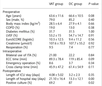
Table 1: Patient characteristics in the VAT and DC groups
VAT group DC group P-value Preoperative
Age (years) 63.6 ± 11.6 66.6 ± 10.5 0.08 Sex (male, %) 79.0 85.2 0.40 Body mass index (kg/m2) 28.5 ± 6.4 27.9 ± 4.1 0.66 COPD (%) 19.0 13.0 0.38 Diabetes mellitus (%) 31.7 31.5 1.00 LVEF (%) 53.2 ± 15 54.7 ± 14.7 0.91 EuroSCORE (logistic) 10.3 ± 12.5 9.4 ± 11.2 0.56 Creatinine (µmol/l) 107.6 ± 70.3 107.7 ± 55.2 0.92 Reoperation (%) 9.5 7.4 0.77 Intraoperative
Bilateral use of ITA (%) 21.00 22.2 0.84 ECC time (min) 89.3 ± 78.4 119 ± 85.4 0.09 Emergency operation (%) 8.6 3.7 0.34 Cross clamp time (min) 45.9 ± 47.2 61.4 ± 54.9 0.11 Postoperative
Length of ICU stay (days) 4.08 ± 5.02 3.2 ± 2.3 0.35 Length of hospital stay (days) 21.10 ± 16.4 13.3 ± 12.1 0.00 Positive culture (%) 69.2 87 0.02 COPD: chronic obstructive pulmonary disease; LVEF: left ventricular ejection fraction; ITA: internal thoracic artery; ECC: extracorporeal
circulation; ICU: intensive care unit. ADUL
T
C
ARD
IA
DISCUSSION
This study demonstrates that negative microbiological results are not mandatory before wound closure, as these do not influence the rate of readmissions for recurrent infection. Furthermore, the results presented here suggest that shortening of VAT despite positive microbiological results may be feasible.
Since the study published by Fleischmann et al. in 1993 [10], VAC therapy established itself as a standard therapy for infected wounds. In 1997, Morykwaset al. [13] highlighted some of the mechanisms of wound healing caused by VAT. According to this and several other works, VAC therapy induces wound healing through the following mechanisms: wound contraction, neo-vascularization, wound granulation, secretion removal and bac-terial clearance [9–11,13].
Wound closure should be performed in the granulation phase while using VAT [14]. The time of wound closure has not been well established and relies vastly on surgeon’s experience. Several attempts have been made to try to establish a standard protocol, although all of these are based on subjective para-meters [9, 12, 14, 15]. This study showed that total bacterial clearance might not be mandatory for wound closure after DSWI.
At our institution, microbiological samples were taken on a regular basis at the time of diagnosis of DSWI and, if a VAC therapy was performed, after each VAT system change. The idea behind microbiological samples is based on the appropriate se-lection of antibiotics as well as a help to establish the appropri-ate timing for wound closure after DSWI [12,19]. According to our clinic experience, wound closure after DSWI, either primary or delayed, will be performed most of the time based on the optical assessment performed by the surgeon and not solely based on microbiologicalfindings.
This study showed that although 119 (75%) patients had posi-tive microbiologicalfinding at the time of wound closure, the re-infection rate based on readmissions after wound closure showed no difference between the DC and VAT groups. Even though this analysis did not show a statistical significant differ-ence, the tendency appears to be clear and correlate to other studies already published [2,12].
One recent study presented by Diefenbecket al. [12] showed a total of 24 patients with deep wound infections on upper and lower extremities. Fourteen from 24 patients showed bacterial growth at the time of wound closure. According to their results, 18 out of 24 patients showed no signs of reinfection. In line with the results presented here, the authors concluded that even
Figure 1:Operations performed in the complete cohort (n = 159) as well as within groups. Values are presented in percentages. VAT: vacuum-assisted therapy; DC: direct closure; CABG: coronary artery bypass graft; C/V: combined surgery (CABG and Valve); other: include congenital surgery and thoracic aorta surgery.
Figure 2:Bacteria found in the microbiologicalfindings presented for the complete cohort (n = 159) as well as within groups. Values are presented in percentages. VAT: vacuum-assisted-therapy; DC, direct closure; CoNS: coagulase negative staphylococci. Other bacteria included a diversified group of bacteria and fungi; the amounts of cases were too small to be presented as a single parameter.
though VAT increased granulation without necrosis, positive microbiologicalfindings were showed by more than 50% of the patients at the time of wound closure without having an in flu-ence on wound healing and clinical outcome. Other studies already published have even shown a no-consistent effect of bacterial clearance with even increase in bacterial loading and no difference in overall healing an reduction in wound surface area [15–17,20]. Therefore, microbiological results are not useful as an indicator for wound closure and a total bacterial clearance may not be necessary. Despite these studies, the beneficial effects of VAT on wound healing have been showed in several other studies [8,21,22].
Our results are in contrast to earlier studies [8,23,24], suggest-ing that early application of sternal wires or direct closure favours reinfection as well as duration of hospital stay. According to the results shown here, there was no difference between our groups in terms of reinfection based on readmission rate or length of ICU stay. The use of VAT did prolong the duration of hospital stay.
Even though the 90-day mortality in our study was higher in the DC group (3/54 vs. 2/105 VAT group), these results were not significant (P = 0.338). Interestingly, these results differ from results presented in earlier studies showing a mortality rate ranging from 14 to 26% [8,21]. In our series, patients in both groups died of multi-organ failure caused by severe sepsis.
Although our findings question microbiological findings as an indicator to determine the time of wound closure, we do believe that microbiologicalfindings are necessary to establish the correct antibiotic therapy. Furthermore, there was no significant benefit using VAT in terms of reinfection rates. The baseline characteristics as well as risk factors presented in this work are comparable to those presented in several publications regarding DSWI and outcome after therapy [2,4,5,7,8,22,25]. At our institution, VAT is still the standard therapy for DSWI and is performed in most DSWI cases, but our results suggest that shortening of VAT might be feasible and even beneficial for some patients.
In conclusion, this study shows that negative microbiological results are not mandatory before wound closure, and do not in-fluence the rate of readmissions for recurrent infection. Furthermore, shortening of VAT despite positive microbiological results may be feasible.
LIMITATIONS
All disadvantages of a non-randomized, retrospective design apply. It cannot be excluded that the decision for primary closure vs. delayed closure and VAT was biased and therefore may have impacted the results. Finally, to confirm the findings presented here, large randomized trials are necessary.
Conflict of interest: none declared.
REFERENCES
[1] Loop FD, Lytle BW, Cosgrove DM, Mahfood S, McHenry MC, Goormastic Met al. Sternal wound complications after isolated coronary artery bypass grafting: early and late mortality, morbidity, and cost of care. Ann Thorac Surg 1990;49:179–87.
[2] Borger MA, Rao V, Weisel RD, Ivanov J, Cohen G, Scully HEet al. Deep sternal wound infection: risk factors and outcomes. Ann Thorac Surg 1998;65:1050–6.
[3] Milano CA, Kesler K, Archibald N, Sexton DJ, Jones RH. Mediastinitis after coronary artery bypass graft surgery. Risk factors and long-term survival. Circulation 1995;92:2245–51.
[4] Olsen MA, Lock-Buckley P, Hopkins D, Polish LB, Sundt TM, Fraser VJ. The risk factors for deep and superficial chest surgical-site infections after coronary artery bypass graft surgery are different. Thorac Cardiovasc Surg 2002;124:136–45.
[5] Lu JCY, Grayson AD, Jha P, Srinivasan AK, Fabri BM. Risk factors for sternal wound infection and mid-term survival following coronary artery bypass surgery. Eur J Cardiothorac Surg 2003;23:943–9.
[6] Sjögren J, Malmsjö M, Gustafsson R, Ingemansson R. Poststernotomy mediastinitis: a review of conventional surgical treatments, vacuum-assisted closure therapy and presentation of the Lund University Hospital mediastinitis algorithm. Eur J Cardiothorac Surg 2006;30: 898–905.
[7] Nakano J, Okabayashi H, Hanyu M, Soga Y, Nomoto T, Arai Yet al. Risk factors for wound infection after off-pump coronary artery bypass graft-ing: should bilateral internal thoracic arteries be harvested in patients with diabetes? Thorac Cardiovasc Surg 2008;135:540–5.
[8] Assmann A, Boeken U, Feindt P, Schurr P, Akhyari P, Lichtenberg A. Vacuum-assisted wound closure is superior to primary rewiring in patients with deep sternal wound infection. Thorac Cardiovasc Surg 2011;59:25–9.
[9] Argenta LC, Morykwas MJ. Vacuum-assisted closure: a new method for wound control and treatment: clinical experience. Ann Plast Surg 1997; 38:563–76.
[10] Fleischmann W, Strecker W, Bombelli M, Kinzl L. Vacuum sealing as treatment of soft tissue damage in open fractures. Der Unfallchirurg 1993;96:488–92.
[11] Fleischmann W, Lang E, Russ M. Infektbehandlung durch Vakuum-versiegelung. Der Unfallchirurg 1997;100:301–4.
[12] Diefenbeck M, Mennenga U, Gückel P, Tiemann AH, Mückley T, Hofmann GO. Vacuum-assisted closure therapy for the treatment of skin and soft-tissue infections. Are wound specimens of use in planning sec-ondary wound closure?. Z Orthop Unfall 2011;149:324–9.
[13] Morykwas MJ, Argenta LC, Shelton-Brown EI, McGuirt W. Vacuum-assisted closure: a new method for wound control and treat-ment: animal studies and basic foundation. Ann Plast Surg 1997;38: 553–62.
[14] Kujath P, Eckmann C, Bouchard R, Esnaashari H. Complicated skin and soft tissue infections. Zentralbl Chir 2007;132:411–8.
[15] Mouës CM, Vos MC, van den Bemd GJ, Stijnen T, Hovius SER. Bacterial load in relation to vacuum-assisted closure wound therapy: a prospective randomized trial. Wound Repair Regen 2004;12:11–7.
[16] Weed T, Ratliff C, Drake DB. Quantifying bacterial bioburden during negative pressure wound therapy: does the wound VAC enhance bacter-ial clearance? Ann Plast Surg 2004;52:276–9.
[17] Braakenburg A, Obdeijn MC, Feitz R, van Rooij IA, van Griethuysen AJ, Klinkenbijl JHG. The clinical efficacy and cost effectiveness of the vacuum-assisted closure technique in the management of acute and chronic wounds: a randomized controlled trial. Plast Reconstr Surg 2006; 118:390–7.
[18] El Oakley RM, Wright JE. Postoperative mediastinitis: classification and management. Ann Thorac Surg 1996;61:1030–6.
[19] Sjögren J, Gustafsson R, Nilsson J, Lindstedt S, Nozohoor S, Ingemansson R. Negative-pressure wound therapy following cardiac surgery: bleeding complications and 30-days mortality in 176 patients with deep sternal wound infection. Interact Cardiovasc Thorac Surg 2011;12:117–20. [20] Wanner MB, Schwarzl F, Strub B, Zaech GA, Pierer G. Vacuum-assisted
wound closure for cheaper and more comfortable healing of pressure sores: a prospective study. Scand J Plast Reconstr Surg Hand Surg 2003; 37:28–33.
[21] Sjögren J, Gustafsson R, Nilsson J, Malmsjö M, Ingemansson R. Clinical outcome after poststernotomy mediastinitis: vacuum-assisted closure versus conventional treatment. Ann Thorac Surg 2005;79:2049–55. [22] Boeken U, Feindt P, Schurr P, Assmann A, Akhyari P, Lichtenberg A.
Delayed sternal closure (DSC) after cardiac surgery: outcome and prog-nostic markers. J Card Surg 2010;26:22–7.
[23] Segers P, de Jong AP, Kloek JJ, de Mol BAJM. Poststernotomy mediastini-tis: comparison of two treatment modalities. Interact Cardiovasc Thorac Surg 2005;4:555–60.
[24] Doss M, Martens S, Wood JP, Wolff JD, Baier C, Moritz A. Vacuum-assisted suction drainage versus conventional treatment in the management of poststernotomy osteomyelitis. Eur J Cardiothorac Surg 2002;22:934–8. ADUL T C ARD IA C
[25] Al-Zaru IM, Ammouri AA, Al-Hassan MA, Amr AA. Risk factors for deep sternal wound infections after cardiac surgery in Jordan. J Clin Nurs 2010;19:1873–81.
APPENDIX. CONFERENCE DISCUSSION
Dr T. Elenbaas(Eindhoven, Netherlands): Your paper tries to answer a very im-portant question. You only have 2% deep sternal wound infections, which I think is very good, and even better is your outcome after treatment, the overall mortality being only 3.1%.
Now, back to the topic of this presentation. Although I agree with the con-clusion, I was wondering why you combined those two groups. As you men-tioned yourself, a limitation of the study is that there is a selection bias based on the decision of the surgeon to go for direct closure or secondary closure after VAC therapy.
I think that these groups are not quite comparable, although the patient characteristics, as you presented them, show more or less the same data. I think if a patient is deep septic and the sternum is fractured in several places, and he is haemodynamically unstable, he would probably be better off if treatment were focused on his septicaemia and good wound drainage rather than on direct reclosure.
First question, how is the decision made in your centre between these two treatment modalities? The second question is about the timing of wound closure. You stated that VAC therapy could be shorter. In your references, you mentioned a paper from the Lund group, by Sjögren, where the C-reactive protein level is one of the guiding factors for timing of sternal closure. My question is, what is the value of the CRP level in your clinic in the treatment of mediastinitis or the timing of reclosure?
And the last question concerns the readmission rate which was the same in both groups, 5%. And the question is, what did you do when the patients were readmitted? Was it just a week of additional antibiotic therapy or did you have to drain the substernal space again? And how were these two add-itional therapies divided between the two groups?
Dr Rodriguez: In regard to the first question, why use direct closure or vacuum-assisted therapy, that’s one of the limitations of this study. The VAT therapy started in our clinic around 2001–02. So we knew that most of the direct closure patients in this series were before 2001. Patients who came with an infected wound were opened and then closed immediately after-wards. As mentioned previously, that was between the time frame of 1999 and late 2001/2002. At this moment in our clinic we are performing mainly VAT therapy. That’s also why the patient numbers are quite differ-ent, 105 VAT therapy and 54 from the direct closure group. So there was
nothing behind the decision, it was just the time frame in which surgery was performed.
We did not analyse the CRP levels with our patients. We were just analysing the microbiology. This is the answer to your second question. And the third question, could you repeat it?
Dr Elenbaas: What did you have to do on readmission?
Dr Rodriguez: All the patients that were readmitted came with the diagno-sis of sternal wound infection. So all the patients that were analysed in this paper were directly taken to the OR and were opened. Debridement was performed and either direct closure or VAT therapy was performed.
Dr T. Gudbjartsson (Reykjavik, Iceland): I was wondering what was the length of the follow-up? And do you have any information on chronicfistulas in the groups? And my other question is about the cultures: how were they performed, how were they verified, was it a tissue sample or was it just a swab?
Dr Rodriguez: It was a swab probe. This is why I said we are changing right now to biopsies to see if that has an influence. And in regard to your first question, could you repeat it, sir.
Dr Gudbjartsson: Did you analyse the late or the chronicfistulas, are they included in the number of patients readmitted?
Dr Rodriguez: I cannot bring to mind the number of current fistulas we had, but the fistulas were treated as deep sternal wound infections so we included all these patients. Because this is a retrospective study, we don’t have a very long-term follow-up. Patients from 1999 or 2002 are perhaps already dead, so we did not make a complete follow-up.
Dr Gudbjartsson: Because it could be that you had positive cultures and you closed the patients, and they can turn up many months or years later with a reinfection.
Dr Rodriguez: We do know that some patients with positive cultures had a readmission rate. However, we did not analyse further. We agree that it would be important.
Dr P. Suwalski(Warsaw, Poland): Your courage in touching upon such a dif-ficult and sensitive issue is commendable. I just wanted to ask you, in both groups of direct closure and VAC therapy, do you leave the flow drainage once you close the chest?
Dr Rodriguez: No.
Dr Suwalski: No? It was just the direct? Dr Rodriguez: Yes.
Dr J. Gummert(Bad Oeynhausen, Germany): Is there any time frame where there were more direct closures done and more VAC therapy?
Dr Rodriguez: Yes, as previously mentioned, direct closure was performed in 54 patients, 34% of patients I believe, who were closed within the time frame from 1999 to late 2001. Afterwards, there were only very rare cases where direct closure was performed, because of vacuum-assisted therapy.
European Journal of Cardio-Thoracic Surgery 42 (2012) 310–311
EDITORIAL COMMENT
doi:10.1093/ejcts/ezs113 Advance Access publication 16 March 2012
Re: Negative microbiological results are not mandatory
in deep sternal wound infections before wound closure
Martin Misfeld*
Department of Cardiac Surgery, Heart Center, University of Leipzig, Leipzig, Germany
* Corresponding author. Leipzig Heart Center, Struempellstrasse 39, 04289 Leipzig, Germany. Tel: +49-341-8650; fax: +49-341-8651452; e-mail: [email protected] (M. Misfeld).
Keywords:Vacuum-assisted therapy• Deep sternal wound infection • Microbiological findings • Wound closure
Deep sternal wound infection (DSWI) is a serious complication of cardiac surgery with high additional morbidity and mortality. The incidence is less than 1%, but associated with mortality rates between 14 and 47% [1]. There are multiple predisposing factors
ranging from patient-risk factors (i.e. obesity, chronic obstructive pulmonary disease, advanced age, male sex), perioperative patient management (i.e. antibiotic prophylaxis, hair removal, blood transfusion, ventilation time) and the surgical procedure