KRIT1 is mutated in hyperkeratotic cutaneous capillary-venous malformation associated with cerebral capillary malformation
6
0
0
Texte intégral
(2) 1352 Human Molecular Genetics, 2000, Vol. 9, No. 9. receptor tyrosine kinase TIE-2 (8). We have also found that the causative gene for inherited cutaneous venous malformations with glomus cells (‘glomangiomas’) maps to 1p21–22 (9). Moreover, we have discovered that a loss-of-function mutation in vascular endothelial growth factor receptor-3 (VEGFR-3) causes congenital hereditary lymphoedema (A. Irrthum, M. Kärkkäinen, K. Alitalo, K. Devrient and M. Vikkula, manuscript in preparation). In addition, three different chromosomal loci have been found to link to CCMs, 3q25.2–27, 7p13–15 and 7q21–22 (10,11), and one of the causative genes, CCM1, has been identified (12,13). This gene encodes KRIT1 (KREV1 interaction trapped 1), a protein interacting with KREV1/RAP1A, an evolutionarily conserved Ras-family GTPase (14). KRIT1 mutations most likely cause loss-of-function effects on the protein. We report that patients with HCCVM, associated with CCMs, also have a mutation in the KRIT1 gene. This suggests that KRIT1 is important for both cutaneous and cerebral vascular morphogenesis. Interestingly, the mutation causing HCCVM is in the first exon, and thus could result in an earlier truncation of KRIT1 than the other 19 identified mutations, which only cause CCMs. We also describe another novel mutation in the CCM1 gene that causes CCMs without cutaneous lesions. RESULTS Five families and two sporadic patients with CCM participated in the study (Fig. 1A). In family A, two members manifested concurrent CCMs and multiple HCCVM (Fig. 1B). The other four families exhibited only CCMs. By linkage and haplotypic analyses, none of the five families showed exclusion of linkage to the CCM1 locus on chromosome 7q21–22 (data not shown), implying that the causative defect could be in the CCM1 gene. Thus, two affected individuals from each of the families and two sporadic cases were examined for the 12 known exons of the CCM1 gene, using single stranded conformation polymorphism (SSCP) and heteroduplex analysis. An abnormal migration pattern was observed in three fragments containing exon 1 in family A, exon 2 in family B and exon 10 in families B and C. Direct sequencing of the abnormally migrating PCR fragment including exon 1, revealed a deletion of guanine (nt 103) (KRIT1∆G103). This would result in a frameshift after the 26th amino acid and premature stop at the 37th codon (Fig. 2A). Co-segregation of the mutation with the affected members of family A was studied by amplifying the fragment from genomic DNA from all family members and running the radioactively labelled PCR products on a 6% denaturing acrylamide gel. A shorter fragment, which migrated slightly more quickly than the wild-type product, was detected in all affected individuals (Fig. 3A). This shorter fragment was found in neither unaffected family members (Fig. 3A), nor 50 healthy controls (data not shown). These findings strongly suggest that in family A, the disorder expressed as CCMs associated with HCCVM is caused by an early truncating mutation in the CCM1 gene. Direct sequencing of the PCR product of exon 2, amplified from genomic DNA of an affected member of family B, demonstrated a T→C transition, which would disrupt the conserved GT dinucleotide in the splice donor site of intron 2 [KRIT1IVS2+2(T→C)] (Fig. 2B). As this mutation destroys a ScaI. Figure 1. (A) Pedigrees of five families with inherited CCMs. Individuals in family A marked by asterisks also exhibit HCCVM. (B) HCCVM of forearm in individual III-1 (family A).. restriction enzyme cutting site, the mutation was screened by restriction enzyme digestion of exon 2 genomic amplification products in all individuals of family B (Fig. 3B). This ScaI site was present in both alleles of unaffected individuals in family B and in 50 controls (data not shown), whereas affected individuals of family B were heterozygous for the site (Fig. 3B). The effect of this mutation on transcription and splicing of premRNA was tested by amplifying—with a primer pair from exon 1 and exon 5—a partial cDNA of the CCM1 gene from total RNA extracted from Epstein–Barr virus (EBV) transfected cell lines of affected individuals. This amplification yielded two extra bands of 451 and 307 bp for which the intensity was clearly lower than for the wild-type band of 567 bp (data not shown). Sequencing of the extracted smaller bands revealed that either exon 2 (the 451 bp fragment) or both exons.
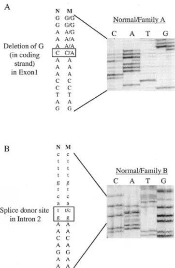
(3) Human Molecular Genetics, 2000, Vol. 9, No. 9 1353. Figure 2. Autoradiographs of sequencing gels. (A) Heterozygous sequence from area with KRIT1∆G103 mutation in family A. Note, sequence shown from non-coding strand. (B) Splice donor site mutation KRIT1IVS2+2(T→C) in family B. Exonic sequence shown in capital letters. (M, mutant; N, normal.). 2 and 3 (the 307 bp fragment) were missing. Thus, the splice donor site mutation causes a skip of either exon 2 or both exons 2 and 3 during splicing of pre-mRNA in EBV-transformed lymphoblasts. The low quantity of these cDNAs suggests that mutated mRNAs have lower stability than normal transcripts. The third shift, present in families B and C and in one control individual in the SSCP and heteroduplex analyses, turned out to be a neutral polymorphism of the coding sequence (A→G at nt 1384). This coding sequence SNP has a frequency of ~17% (three out of nine unrelated individuals carried the polymorphism), and thus may be useful in KRIT1 association analysis. No additional mutations were detected in the three families and two sporadic cases without mutations by amplifying overlapping genomic fragments of 4–15 kb, using the intronic primers listed in Materials and Methods. DISCUSSION A subset of patients with intracranial capillary–venous anomalies are known to manifest peculiar vascular malformations on. Figure 3. Co-segregation of mutations with disorder in families. Individuals participating in study marked with dot. (A) Autoradiography of radioactively labelled PCR products comprising exon 1 in family A. Appearance of a shorter band due to KRIT1∆G103 deletion present in all affected individuals. (B) Agarose gel picture of ScaI-digested PCR fragments containing exon 2 in family B. Appearance of longer fragment indicates presence of KRIT1IVS2+2(T→C) mutation, found only in affected individuals.. the skin. Interestingly, the cerebral and the cutaneous lesions are histologically similar as both are composed of malformed capillary and venous-like channels. In one study, four families out of 57 exhibited hyperkeratotic cutaneous capillary–venous lesions (4). In another report, CCMs were found in a patient with numerous cutaneous lesions called ‘angiokeratomas’ (5). We present an additional family with CCMs with two affected boys exhibiting HCCVM on the limbs. Thus, it seems clear that the intracranial and cutaneous vascular lesions are associated pathogenically. Penetrance of KRIT1 mutations for CCMs is estimated to be 88% (11); however, the penetrance of HCCVMs is lower. In the four published CCM+HCCVM families, eight out of 21 affected individuals had HCCVM (4). Thus, the penetrance for the cutaneous lesions co-occurring with CCMs can be calculated to be ~38%. In the pedigree presented for family A two out of four affected individuals have cutaneous lesions in the extremities. Thus, in a subset of families in which CCMs are caused by KRIT1 mutations with high penetrance, there is an additional risk of ~40% for cutaneous vascular lesions. Prior to this study, 19 different mutations in the CCM1 gene encoding KRIT1 have been identified in patients with CCM.
(4) 1354 Human Molecular Genetics, 2000, Vol. 9, No. 9. (12,13). These mutations are evenly distributed in the gene and comprise nonsense mutations, deletions and insertions, all presumed to result in a premature stop codon. Whether the mutated mRNA is subsequently degraded or translated to a truncated protein containing functional domains, is not known. The two mutations introduced here, deletion of guanine in exon 1 (KRIT1∆G103) and T→C substitution in the splice donor site of intron 2 [KRIT1IVS2+2(T→C)], are similar in this fashion. However, the deletional mutation found in the family manifesting HCCVM results in disruption of the reading frame already after the 26th amino acid, thus causing a premature stop codon at the 37th amino acid. This is the earliest truncation among the 21 mutations reported. As this is the first mutation found in a family in which CCMs co-segregate with cutaneous vascular lesions, more studies are required to determine if similar mutations occur in other patients with this phenotypic subset of CCM. If this were the case, it would suggest that an additional change in function of KRIT1 is linked to the appearance of cutaneous vascular lesions, and that more C-terminal mutations, which only cause CCMs, could possibly produce a protein with some residual activity. The N-terminus of KRIT1 contains three or four ankyrin repeats (amino acids 83–215), which can mediate interactions with other proteins (15). As most mutations occur in the middle of the KRIT1 gene, the ankyrin repeats could be in the truncated proteins. However, in addition to our KRIT1∆G103 mutation, two other mutations have been found that disrupt the reading frame before ankyrin-encoding sequences (after the 74th and the 78th codons) (pedigrees 18 and 19 in ref. 12). As these two families have not, as yet, manifested cutaneous lesions, this different phenotype does not appear to be linked to the presence of ankyrin repeats. The C-terminus of KRIT1 includes the domains for interaction with KREV1/RAP1A, a suppressor of Ras signalling pathway (14). Mutant forms identified so far in CCM patients, whether causing mRNA decay or shortened polypeptides, would nullify this interaction. In addition to the KREV1/RAP1A-interacting region and ankyrin domains, KRIT1 is predicted to contain a FERM domain (209– 433), present in a group of proteins that are involved in the linkage of cytoplasmic proteins to plasma membranes (16). If mutated KRIT1 transcripts are stable and translated, most of the 21 mutations would cause KRIT1 to lack this domain. As reported by Laberge-le Couteulx et al. (12), the northern blot results show a larger mRNA than predicted on the basis of cDNA sequence. Furthermore, no mutations have been found in several families presumed to be linked to CCM1. Thus, it is possible that the CCM1 gene contains other functional domains and genotypic–phenotypic analysis will only be possible when the complete structure of KRIT1 and the stability of the mutant mRNAs are known. It is interesting that KRIT1 may be involved in the Ras signalling pathway. Inactivation of p102-rasGAP, a negative regulator of Ras signalling, results in vascular defects in genetically engineered mice (17). Loss-of-function of another Rassignalling pathway component, b-raf, also causes severe aberrations in vascular development (18). A TIE-2 activating mutation, which may be associated with the Ras signalling pathway, causes venous malformations in humans (8). Thus endothelial cell signalling with convergence to Ras signalling may be a common pathway in the aetiopathogenesis of the various types of vascular anomalies.. In conclusion, we report two novel mutations in the CCM1 gene [KRIT1∆G103 and KRIT1IVS2+2(T→C)] that cause CCMs by introducing premature stop codons. One of the mutations, KRIT1∆G103 in exon 1, causes a rare variant of the disorder in which there are both cerebral and cutaneous vascular malformations. Thus, KRIT1 is important for both cerebral and cutaneous vascular integrity. Interestingly, the mutation causing CCM+HCCVM is the earliest stop codon in the gene among the 21 identified mutations, suggesting a molecular-phenotypic correlation. MATERIALS AND METHODS Families This study comprised five families with inherited CCMs, altogether 15 affected and 16 unaffected individuals and two sporadic cases (Fig. 1A). In family A, individual III-1 had congenital HCCVM on the forearm and hypothenar region (Fig. 1B), and a similar lesion, 2 cm in diameter, appeared on the ankle of individual III-2 when he was 12 years old. Each participating individual gave informed consent, approved by the ethical committee of the Medical Faculty of Université Catholique de Louvain, Brussels, Belgium. Clinical examination and/or magnetic resonance imaging (MRI) were used to determine whether an individual was affected. Genetic analysis Genomic DNA was extracted from the buffy coat of blood samples using QIAamp DNA blood mini kit. For linkage analysis, six polymorphic markers from the CCM1 locus (D7S1813, D7S689, D7S2409, D7S646, D7S652 and D7S657) on chromosome 7q21–22 were genotyped from genomic DNA of the patients, as described earlier (19). The genomic structure of the CCM1 gene was determined by comparing the published KRIT1 cDNA sequence with the reported bacterial artificial chromosome (BAC) sequence containing the gene (GenBank accession nos U90268 and HSAC000120). Twelve primer pairs, synthesized by Life Technologies (Paisley, UK), were designed for the amplification of the 12 exons of the CCM1 gene by PCR using genomic DNA as template. The primers were: exon 1, 5′-AAATCCTGCATATGCTACTG-3′ and 5′-ATTACACTTGAGATAAAACGTC-3′; exon 2, 5′-AACAGAGCAAGACTCATTCTC-3′ and 5′-AGCAATGTGGAGTAAAACCG-3′; exon 3, 5′-AGCTTTCATAGATGTGTCAC-3′ and 5′-TTGGAATGAGAACAGTCTTG-3′; exon 4, 5′-CATTTCAGATGATCTTTTTAGG-3′ and 5′-TCATTACTTGTTATTCACTGC-3′; exon 5, 5′GACATTTTCCCTTAGAATACC-3′ and 5′-GCCTGGCTCTAACTATGAAG-3′; exon 6, 5′-AAAGCACATGAAGTTGAAGG-3′ and 5′-TTCTACCAACCCACTCCC-3′; exon 7, 5′CAGTACAGAAGTGCAGACAG-3′ and 5′-GAAACTCAACAGATTTTGTGC-3′; exon 8, 5′-GCTCAAAACAGTAACAGCTC-3′ and 5′-GCATAGCACAAGACCATGC-3′; exon 9, 5′-AAAGCCATTTGTAACAGAATG-3′ and 5′-CTAACAAAGTTTCAACTAGCC-3′; exon 10, 5′-GATTATCAATGGTACATTTTCC-3′ and 5′-CATGTAGGTTGGTACTGTTG-3′, exon 11, 5′-GAGCAGACAACATAAATGTAG-3′ and 5′-AACACAATAGTTTATGAAGTCC-3′; exon 12, 5′ACTGCCCAATGTCATGAATG-3′ and 5′-ACCATGCTCGGCCAAAAG-3′. The primers were end-labelled with γ-32P.
(5) Human Molecular Genetics, 2000, Vol. 9, No. 9 1355. with polynucleotide kinase (Takara, Kyoto, Japan) before amplification by PCR [95°C for 4 min; (94°C for 30 s; 57°C for 30 s; 72°C for 30 s) ×30; 72°C for 10 min]. The PCR products were analyzed on Sequagel MD SSCP and Heteroduplex gels (National Diagnostics, Atlanta, GA) as well as on 6% denaturing acrylamide gels. The fragments showing abnormal migration pattern were further characterized by direct cycle sequencing, as previously described (20). ScaI digestions of PCR products were performed in 2× One-Phor-All buffer (Amersham Pharmacia, Uppsala, Sweden) overnight to ensure complete digestion. cDNA was synthesized with Superscript Preamplification System (Life Technologies, Merelbeke, Belgium) from total RNA extracted from EBV-transformed lymphoblasts, as described elsewhere (20). A primer pair: 5′-AGAAAACTCACTACATATGG-3′ from exon 1 and 5′-GATCTTCCTTGTTGGTCTG-3′ from exon 5 was used for the amplification of a partial cDNA for KRIT1. ACKNOWLEDGEMENTS We are grateful to our patients and their families for their participation in this study. The authors also thank Ms Ana Gutierrez for her superb technical assistance. This work was supported by the Fonds de Développement Scientifique— Université Catholique de Louvain, the Belgian Federal Service for Scientific, Technical and Cultural Affairs, the FNRS (Fonds National de la Recherche Scientifique) and European Union (all to M.V.). I.E. is supported by grants from the Alfred Kordelin Foundation and the Academy of Finland. REFERENCES 1. Mulliken, J.B. and Glowacki, J. (1982) Hemangiomas and vascular malformations in infants and children: A classification based on endothelial characteristics. Plast. Reconstr. Surg., 69, 412–420. 2. Enjolras, O., Riché, M.C. and Merland, J.J. (1985) Facial port-wine stains and Sturge-Weber syndrome. Pediatrics, 76, 48–51. 3. Bean, W.B. (1958) Vascular Spiders and Related Lesions of the Skin. Charles C. Thomas, Springfield, IL, p. 178. 4. Labauge, P., Enjolras, O., Bonerandi, J.J., Laberge, S., Dandurand, M., Joujoux, J.M. and Tournier-Lasserve, E. (1999) An association between autosomal dominant cerebral cavernomas and a distinctive hyperkeratotic cutaneous vascular malformation in 4 families. Ann. Neurol., 4, 250–254. 5. Ostlere, L., Hart, Y. and Misch, K.J. (1996) Cutaneous and cerebral haemangiomas associated with eruptive angiokeratomas. Br. J. Dermatol., 135, 98–101.. 6. Rigamonti, D., Johnson, P.C., Spetzler, R.F., Hadley, M.N. and Drayer, B.P. (1991) Cavernous malformations and capillary telangiectasia: a spectrum within a single pathological entity. Neurosurgery, 28, 60–64. 7. Vikkula, M., Boon, L.M., Mulliken, J.B. and Olsen, B.R. (1998) Molecular basis of vascular anomalies. [Review] Trends Cardiovasc. Med., 8, 281–292. 8. Vikkula, M., Boon, L.M., Carraway, K.L.R., Calvert, J.T., Diamonti, A.J., Goumnerov, B., Pasyk, K.A., Marchuk, D.A., Warman, M.L., Cantley, L.C. et al. (1996) Vascular dysmorphogenesis caused by an activating mutation in the receptor tyrosine kinase TIE2. Cell, 87, 1181–1190. 9. Boon, L.M., Brouillard, P., Irrthum, A., Karttunen, L., Warman, M.L., Rudolph, R., Mulliken, J.B., Olsen, B.R. and Vikkula, M. (1999) A gene for inherited cutaneous venous anomalies (‘glomangiomas’) localizes to chromosome 1p21–22. Am. J. Hum. Genet., 65, 125–133. 10. Dubovsky, J., Zabramski, J.M., Kurth, J., Spetzler, R.F., Rich, S.S., Orr, H.T. and Weber, J.L. (1995) A gene responsible for cavernous malformations of the brain maps to chromosome 7q. Hum. Mol. Genet., 4, 453–458. 11. Craig, H.D., Gunel, M., Cepeda, O., Johnson, E.W., Ptacek, L., Steinberg, G.K., Ogilvy, C.S., Berg, M.J., Crawford, S.C., Scott, R.M. et al. (1998) Multilocus linkage identifies two new loci for a mendelian form of stroke, cerebral cavernous malformation, at 7p15–13 and 3q25.2–27. Hum. Mol. Genet., 7, 1851–1858. 12. Laberge-le Couteulx, S., Jung, H.H., Labauge, P., Houtteville, J.P., Lescoat, C., Cecillon, M., Marechal, E., Joutel, A., Bach, J.F. and Tournier-Lasserve, E. (1999) Truncating mutations in CCM1, encoding KRIT1, cause hereditary cavernous angiomas. Nature Genet., 23, 189–193. 13. Sahoo, T., Johnson, E.W., Thomas, J.W., Kuehl, P.M., Jones, T.L., Dokken, C.G., Touchman, J.W., Gallione, C.J., Lee-Lin, S.Q., Kosofsky, B. et al. (1999) Mutations in the gene encoding KRIT1, a Krev-1/rap1a binding protein, cause cerebral cavernous malformations (CCM1). Hum. Mol. Genet., 8, 2325–2333. 14. Serebriiskii, I., Estojak, J., Sonoda, G., Testa, J.R. and Golemis, E.A. (1997) Association of Krev-1/rap1a with Krit1, a novel ankyrin repeatcontaining protein encoded by a gene mapping to 7q21–22. Oncogene, 15, 1043–1049. 15. Sedgwick, S.G. and Smerdon, S.J. (1999) The ankyrin repeat: a diversity of interactions on a common structural framework. Trends Biochem. Sci., 24, 311–316. 16. Tsukita, S., Yonemura, S. and Tsukita, S. (1997) ERM proteins: head-totail regulation of actin-plasma membrane interaction. Trends Biochem. Sci., 22, 53–58. 17. Henkemeyer, M., Rossi, D.J., Holmyard, D.P., Puri, M.C., Mbamalu, G., Harpal, K., Shih, T.S., Jacks, T. and Pawson, T. (1995) Vascular system defects and neuronal apoptosis in mice lacking ras GTPase-activating protein. Nature, 377, 695–701. 18. Wojnowski, L., Zimmer, A.M., Beck, T.W., Hahn, H., Bernal, R., Rapp, U.R. and Zimmer, A. (1997) Endothelial apoptosis in Braf-deficient mice. Nature Genet., 16, 293–297. 19. Boon, L.M., Mulliken, J.B., Vikkula, M., Watkins, H., Seidman, J., Olsen, B.R. and Warman, M.L. (1994) Assignment of a locus for dominantly inherited venous malformations to chromosome 9p. Hum. Mol. Genet., 3, 1583–1587. 20. Vikkula, M., Mariman, E.C., Lui, V.C., Zhidkova, N.I., Tiller, G.E., Goldring, M.B., van Beersum, S.E., de Waal Malefijt, M.C., van den Hoogen, F.H., Ropers, H.H. et al. (1995) Autosomal dominant and recessive osteochondrodysplasias associated with the COL11A2 locus. Cell, 80, 431–437..
(6) 1356 Human Molecular Genetics, 2000, Vol. 9, No. 9.
(7)
Figure
Documents relatifs
