B R I E F R E P O R T
Hepatitis B Virus e Antigen
Physically Associates With
Receptor-Interacting Serine/
Threonine Protein Kinase 2 and
Regulates
IL-6 Gene Expression
Shuang Wu,1Tatsuo Kanda,1Fumio Imazeki,1Shingo Nakamoto,1,2 Takeshi Tanaka,1,3Makoto Arai,1Thierry Roger,5Hiroshi Shirasawa,2 Fumio Nomura,4and Osamu Yokosuka1
1Department of Medicine and Clinical Oncology,2Department of Molecular
Virology,3Department of Environment Biochemistry, and4Department of
Molecular Diagnosis, Graduate School of Medicine, Chiba University, Japan; and
5Infectious Diseases Service, Department of Medicine, Centre Hospitalier
Universitaire Vaudois and University of Lausanne, Lausanne, Switzerland
We previously reported that hepatitis B virus (HBV) e antigen (HBeAg) inhibits production of interleukin 6 by suppressing NF-κB activation. NF-κB is known to be acti-vated through receptor-interacting serine/threonine protein kinase 2 (RIPK2), and we examined the mechanisms of in-terleukin 6 regulation by HBeAg. HBeAg inhibits RIPK2 expression and interacts with RIPK2, which may represent 2 mechanisms through which HBeAg blocks nucleotide-binding oligomerization domain-containing protein 1 ligand–induced NF-κB activation in HepG2 cells. Our find-ings identified novel molecular mechanisms whereby HBeAg modulates intracellular signaling pathways by tar-geting RIPK2, supporting the concept that HBeAg could impair both innate and adaptive immune responses to promote chronic HBV infection.
Hepatitis B virus (HBV) nucleoprotein exists in 2 forms [1,2]. Nucleocapsid, designated HBV core antigen (HBcAg), is an intracellular, 21-kDa protein that self-assembles into particles that encapsidate viral genome and polymerase and is essential for function and maturation of virion. HBV also secretes a nonparticle second form of the nucleoprotein, designated
precore or HBV e antigen (HBeAg) [1,2]. Precore and core proteins are translated from 2 RNA species that have different 5′ initiation sites. Precore messenger RNA (mRNA) encodes a hydrophobic signal sequence that directs precore protein to the endoplasmic reticulum, where it undergoes N- and C-terminal cleavage within the secretory pathway and is secret-ed as an 18-kDa monomeric protein [3–5].
Nucleotide-binding oligomerization domain–containing protein 1 (NOD1) and NOD2 are cytosolic pattern-recognition receptors involved in the sensing of bacterial peptidoglycan subcomponents [6]. NOD1 and NOD2 stimulation activates NF-κB through receptor-interacting serine/threonine protein kinase 2 (RIPK2; also known as RIP2, RICK, or CARDIAK), a caspase-recruitment domain-containing kinase. RIPK2 is also involved in Toll-like receptor (TLR)–signaling pathway and plays an important role in the production of inflammatory cy-tokines through NF-κB activation [6,7].
We previously reported that HBeAg inhibits the production of interleukin 6 (IL-6) through suppression of NF-κB activa-tion [4]. In the present study, we investigated the molecular mechanism of HBeAg functions for the requirement of RIPK2 in NF-κB transcriptional regulation.
METHODS
Cell Culture and Plasmids
HepG2, Huh7, HT1080, COS7, and HEK293T cells were used in the present study. Stable cell lines were obtained as previ-ously described [4]. Briefly, HepG2, Huh7, and HT1080 were
transfected with pCXN2-HBeAg(+) or pCXN2-HBeAg(−) in Effectene (Qiagen). After G418 screening, HBeAg-positive and -negative HepG2/Huh7/HT1080 cell lines were collected for further analysis [4]. The plasmid pCXN2-HBeAg(+), which can produce both HBeAg and core peptides, and the plasmid pCXN2-HBeAg(−), which can produce only core peptides, were obtained as described previously [4]. pNF-κB–luc, which expresses luciferase upon promoter activation by NF-κB, was purchased from Stratagene [4]. pGFP–human RIPK2 (kindly provided by Prof John C. Reed, Sanford-Burnham Institute for Medical Research) can express GFP-human RIP2WT[8].
HepG2 cells were transfected with plasmid control–small hairpin RNA (shRNA) or with RIPK2-shRNA (Santa Cruz). After puromycin screening, individual colonies were picked up and examined for expression of endogenous RIPK2, and clones HepG2-shC and HepG2-shRIPK2-3 were selected for subsequent studies.
Received 1 January 2012; accepted 3 February 2012; electronically published 21 May 2012. Correspondence: Tatsuo Kanda, MD, PhD, Department of Medicine and Clinical Oncology, Chiba University, Graduate School of Medicine, 1-8-1 Inohana, Chuo-ku, Chiba 260-8670, Japan ([email protected]).
The Journal of Infectious Diseases 2012;206:415–20
© The Author 2012. Published by Oxford University Press on behalf of the Infectious Diseases Society of America. All rights reserved. For Permissions, please e-mail: journals.permissions@ oup.com.
Luciferase Assays and Treatment of Cells With NOD Ligands
Around 1.0 × 10⁵HepG2 and Huh7 cells were plated in 6-well plates (Iwaki Glass, Tokyo, Japan) for 24 hours and transfect-ed with 0.4μg of pNF-κB–luc. For luciferase assay of NF-κB activation, cells were treated for 4 hours with or without NOD1 ligand (C12-iEDAP, 2.5μg/mL) and NOD2 ligand (muramyl dipeptide [MDP], 10μg/mL) (InvivoGen) at 44 hours after transfection [9]. After 48 hours, cells were lysed with reporter lysis buffer (Promega), and luciferase activity was determined as described previously [4].
RNA Extraction, Complementary DNA (cDNA) Synthesis, Real-Time Polymerase Chain Reaction (PCR) Analysis, and PCR Array
Total RNA was isolated by RNeasy Mini Kit (Qiagen). A total of 5μg of RNA was reverse transcribed using the First Strand cDNA Synthesis Kit (Qiagen) [4]. Quantitative amplification of cDNA was monitored with SYBR Green by real-time PCR in a 7300 Real-Time PCR system (Applied Biosystems). Gene expression profiling of 84 TLR-related genes was performed using RT2 profiler PCR arrays (Qiagen) in accordance with the manufacturer’s instructions [4].
Gene expression was normalized to 2 internal controls (GAPDH and/orβ-actin) to determine the fold-change in gene expression between the test sample (HBeAg-positive HepG2/ Huh7/HT1080) and the control sample (HBeAg-negative HepG2/Huh7/HT1080) by the 2−ddCT (comparative cycle threshold) method [4]. Three sets of real-time PCR arrays were performed. Some results of HepG2 cells were previously reported [4].
Coimmunoprecipitation
Cells were cotransfected with 2.5μg pCXN2-HBeAg(+) or 2.5μg pCXN2-HBeAg(−), as well as with 2.5 μg pGFP– human RIPK2, and cell lysates were prepared after 48 hours, using lysis buffer containing a cocktail of protease inhibitors. Cell lysates were incubated with anti-GFP rabbit polyclonal antibody (Santa Cruz) or anti-HBe mouse monoclonal antibody (Institute of Immunology, Tokyo, Japan) for 3 hours at 4°C, followed by overnight incubation with protein G–Sepharose beads (Santa Cruz). Immunoprecipitates were separated by sodium dodecyl sulfate–polyacrylamide gel electrophoresis and electroblotted onto a nitrocellulose membrane. Immunoblotting was performed by incubating the membrane for 1 hour with anti-HBe antibody. Proteins were detected by enhanced chemilu-minescence (GE Healthcare), using an image analyzer (LAS-4000, Fuji Film). The membrane was reprobed with a monoclo-nal antibody to GFP or RIPK2 (Cell Sigmonoclo-naling).
Transfection of pGFP–Human RIPK2 and Confocal Microscopy
Formaldehyde (3.7%)–fixed cells were incubated with anti-HBe antibody and stained withfluorochrome-conjugated sec-ondary antibody (Alexa Fluor 555 conjugate, Cell Signaling).
Cells were mounted for confocal microscopy (ECLIPSE TE 2000-U, Nikon). Whenever necessary, images were merged digitally to monitor colocalization. Cotransfection of 0.1μg pCXN2-HBeAg(+) or 0.1μg pCXN2-HBeAg(−) with 0.3 μg pGFP–human RIPK2 into the cells was performed. After 48 hours, intracellular localization of RIPK2 was visualized by confocal microscopy.
Enzyme-Linked Immunosorbent Assay (ELISA) for IL-6
Cell culture fluid was analyzed for IL-6 by ELISA (KOMA-BIOTECH, Seoul, Korea), in accordance with the manufactur-er’s protocol [4].
Small Interfering RNA (siRNA) Transfection and Wound-Healing Assay
Control siRNA (siC) and siRNA specific for RIPK2 (siRIPK2) were purchased from Thermo Fisher Scientific. Cells were transfected with siRNA by electroporation. After 48 hours, cells were treated with 10 ng/mL tumor necrosis factor α (TNF-α) (Wako Pure Chemical, Osaka, Japan), while the wound-healing (ie, scratch) assay was performed using a p-200 pipette tip to induce RIPK2 [10]. Up to 12 hours after scratching, the cells were observed by microscopy. Cell migra-tion was measured using Scion Images (SAS). Migramigra-tion by siC-transfected cells was set at 1.
Statistical Analysis
Results are expressed as mean values ± SD. The Studentt test was used to determine statistical significance.
RESULTS
HBeAg Downregulates RIPK2 Expression
To explore the effect of HBeAg on TLR-related gene expres-sion, we generated HepG2, Huh7, and HT1080 cell lines that stably expressed HBV core region with or without precore region. HT1080, a primatefibrosarcoma cell line, is useful for the study of interferon signaling. HBeAg and HBV core– related antigen (HBcrAg) levels of these cell lines demonstrat-ed that expression of HBV core region without HBV precore region did not allow HBeAg secretion by cells (data are cited elsewhere [4] or not shown). First, we performed real-time RT-PCR analysis of these cell lines, using focused gene arrays (Figure1A). We observed that, in 3 cell lines, 5 genes (RIPK2,
TLR9, TNF, CD180, and IL1A) were downregulated ≥1.3-fold in HBeAg-positive cells than in HBeAg-negative cells. We chose to focus our investigation on RIPK2 because HBeAg inhibits the production of IL-6 through the suppression of NF-κB acti-vation [4], and NF-κB is known to be activated through
RIPK2 [4]. RIPK2 expression was >100-, 1.41-, and 1.45-fold lower in HBeAg-positive HepG2, Huh7, and HT1080 cells, re-spectively, compared with their HBeAg-negative counterparts
(Figure 1A). Western blot analyses confirmed lower levels of
RIPK2 in HBeAg-positive HepG2 than in HBe-negative HepG2 or parental HepG2 (Figure1B). The fact that RIPK2 is one of
the targets for the ubiquitin proteasome system and uses a ubiquitin-dependent mechanism to achieve NF-κB activation [6] might be a reason for the differences between RIPK2 mRNA and protein expression status. We also observed lower levels of RIPK2 mRNA expression (0.18-fold) in HepG2.2.15
cells, which secrete complete HBV virion and HBeAg, com-pared with expression in HepG2 cells (data not shown).
Knockdown of RIPK2 and HBeAg Impairs Hepatic Cell Migration
It has recently been reported that RIPK2 expression is induced by TNF-α plus scratch wounding in keratinocytes [10]. There-fore, we next examined whether RIPK2 affected hepatic
Figure 1. Receptor-interacting serine/threonine protein kinase 2 (RIPK2) expression is downregulated by hepatitis B virus e antigen (HBeAg), and
knockdown of RIPK2 and HBeAg impairs hepatic wound repair. A, Venn diagram representing Toll-like receptor (TLR)–related genes downregulated
≥1.3-fold in HBeAg-positive HepG2/Huh7/HT1080 cells, compared with HBeAg-negative cells. Cellular RNA was extracted and analyzed with focused
array, quantifying 84 genes. Gene expression levels were normalized to actin and GAPDH expression levels.B, HBeAg downregulates RIPK2 expression
in HepG2 cells. Western blot analysis of RIPK2 and tubulin expression in HepG2, HBeAg(+) HepG2, and HBeAg(−) HepG2. C, Experimental protocol of
electroporation of control (siC) and RIPK2 (siRIPK2) small interfering RNA (siRNA) into HepG2 cells.D and E, Real-time polymerase chain reaction (PCR;
D) and Western blot (E) analyses of RIPK2 expression in siC- or siRIPK2-expressing HepG2 cells. RIPK2 messenger RNA (mRNA) levels were normalized
to GAPDH levels.F–H, siC- and siRIPK2-transfected HepG2 cells were scratch wounded and incubated with 10 ng/mL tumor necrosis factor α (TNF-α),
and cell migration was analyzed after 12 hours and quantified using Scion Image (F). Interleukin 6 (IL-6; G) and interleukin 8 (IL-8; H) mRNA expression
are quantified by real-time reverse transcription–PCR (RT-PCR) and expressed relative to GAPDH mRNA expression. I, Protocol of wound-healing (ie,
scratch) assay in HBeAg(+) and HBeAg(−) HepG2 cells. TNF-α was used at 10 ng/mL. J, Cell migration was analyzed using Scion Image. K, RIPK2
mRNA expression was quantified by real-time RT-PCR and expressed relative to GAPDH mRNA expression. Primers specific for RIPK2 were
5′-AGACAC-TACTGACATCCAAG-3′ (sense) and 5′-CACAAGTATTTCCGGGTAAG-3′ (antisense), and primers for other genes were as described previously [4]. Data are
wound healing in the presence of TNF-α in vitro (Figure1C).
As shown in Figure 1D and 1E, RIPK2 mRNA and protein
expression were efficiently decreased in HepG2 cells transfect-ed with RIPK2 siRNA (siRIPK2), but not control (siC). RIPK2 silencing reduced hepatic wound closure 1.8-fold, which was associated with a 2-fold decrease in IL-6 production, known to be an important cytokine for the regeneration of liver [11],
and a 2.3-fold decrease in interleukin 8 production (Figure 1F–H). Importantly, RIPK2 silencing did not affect
cell viability (data not shown).
Given that HBeAg downregulates RIPK2 expression (Figure 1A and 1B), we examined whether HBeAg has an
effect on hepatic wound healing in the presence of TNF-α (Figure1I). As expected, we observed that both cell migration
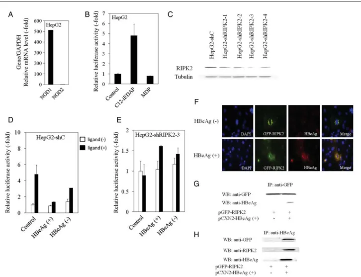
Figure 2. The nucleotide-binding oligomerization domain–containing protein 1 (NOD1) ligand C12-iEDAP induces NF-κB activation, knockdown of
receptor-interacting serine/threonine protein kinase 2 (RIPK2) inhibits NOD1 ligand–induced NF-κB activation in HepG2 cells, and hepatitis B virus e
antigen (HBeAg) interacts with RIPK2. A, Real-time reverse transcription–polymerase chain reaction analysis of NOD1 and NOD2 messenger RNA
expression in HepG2. NOD1 and NOD2 expression levels were normalized to GAPDH expression levels.B, NF-κB–driven luciferase activity in HepG2
cells stimulated with the NOD1 ligand C12-iEDAP or the NOD2 ligand muramyl dipeptide (MDP) in HepG2.C, Western blot analysis of RIPK2 and
tubulin expression in HepG2 cells stably transfected with control small hairpin RNA (shRNA; HepG2-shC) or with RIPK2 shRNA (HepG2–shRIPK2-1/2-4)
expressing plasmids. D and E, HepG2-shC (D) and HepG2–shRIPK2-3 (E) cell lines were transiently transfected with pCXN2, pCXN2-HBeAg(+), or
pCXN2-HBeAg(−) plasmids together with pNF-κB–luc. Cells were treated for 4 hours, with or without NOD1 ligand C12-iEDAP (2.5 μg/mL), and
lucifer-ase activity was determined. Primers specific for NOD1 (sense primer: 5′-ACTACCTCAAGCTGACCTAC-3′; antisense primer:
5′-CTGGTTTACGCTGAGTCTG-3′), for NOD2 (sense primer: 5′-CCTTGCATGCAGGCAGAAC-3′; antisense primer: 5′-TCTGTTGCCCCAGAATCCC-3′), and for other genes as described
previously were purchased from Sigma [4].F, HBeAg specifically colocalizes with RIPK2. COS7 cells were transiently cotransfected with 0.1 μg
pCXN2-HBeAg(+) or pCXN2-HBeAg(−) together with 0.3 μg pGFP–human RIPK2. HBeAg was revealed with anti-HBeAg primary antibody and Alexa-Fluor-548
secondary antibody.G and H, HEK293T cells were transiently transfected with or without GFP-RIPK2 and HBeAg-expressing plasmids. Cellular extracts
were precleared with protein G–Sepharose, and interacting complexes were immunoprecipitated (IP) with either anti-GFP (G) or anti-HBeAg (H)
antibod-ies. Immunocomplexes were separated by sodium dodecyl sulfate–polyacrylamide gel electrophoresis, and proteins were visualized by immunoblotting
and RIPK2 mRNA expression were reduced in HBeAg-positive HepG2 cells, compared with HBeAg-negative cells (1.5-fold decrease; Figure1J and 1K). These results suggest that HBeAg
impairs hepatic cell migration–dependent RIPK2 expression. Among NF-κB–targeting genes, expression of vimentin mRNA was impaired in HepG2-shRIP2 and in HBeAg-positive HepG2 (data not shown), and vimentin might be one of the candidates for impairment of their migrations [12].
RIPK2 Plays an Important Role in NF-κB Activation Induced by
NOD1 Ligand, and HBeAg Blocks This Pathway
HepG2 cells express NOD1 but not NOD2 at the mRNA level (Figure2A). In agreement with this finding, NF-κB was
acti-vated in HepG2 cells exposed to NOD1 ligand C12-iEDAP (level of activation, 4.8-fold, compared with untreated control) but not in those exposed to NOD2 ligand MDP (Figure 2B).
As for Huh7 cells, activation of NF-κB was not detected fol-lowing exposure to C12-iEDAP or MDP (data not shown). These results suggest that C12-iEDAP triggered NF-κB activa-tion through NOD1 in HepG2 cells, which is consistent with findings from a previous study [9].
We examined whether knockdown of RIPK2 has an effect on NOD1-induced NF-κB activation in HepG2 cells. First, we established HepG2 cell lines that constitutively expressed RIPK2-shRNA (HepG2-shRIPK2-1/2-4) or control-shRNA (HepG2-shC) (Figure 2C). The HepG2–shRIPK2-3 cell line,
which expresses the lowest levels of RIPK2, and the HepG2-shC cell line were treated for 4 hours, with or without C12-iE-DAP, before measurement by the NF-κB–driven luciferase assay (Figure2D and 2E). C12-iEDAP triggered NF-κB
activa-tion in HepG2-shC (Figure2D) but not in HepG2–shRIPK2-3
(Figure2E), indicating that RIPK2 plays an important role in
NF-κB activation induced through NOD1 triggering.
To assess the influence of HBeAg in that pathway, we mea-sured NOD1-mediated NF-κB activity in HepG2-shC and HepG2–shRIPK2-3 cell lines transiently transfected with HBeAg-expressing plasmids. As shown in Figure 2D, HBeAg
expression in HepG2-shC abolished C12-iEDAP–induced NF-κB activation.
HBeAg Interacts With RIPK2 and Colocalizes With RIPK2
RIPK2 mediates activation of transcription factors, such as NF-κB, following its activation, which is initiated by mem-brane-bound or intracytosolic receptors, such as TLR, NOD1, and NOD2 [7,13,14]. Confocal microscopy analysis of cells transfected with GFP-RIPK2 revealed subcellular localization of RIPK2 (data not shown). To compare the localization of RIPK2 with that of HBeAg, cells were cotransfected with pGFP–human RIPK2 with HBeAg(+) or pCXN2-HBeAg(−). After 48 hours, cells were stained with mouse mono-clonal anti-HBe antibody. Confocal microscopy suggested sub-cellular colocalization of RIPK2 with HBeAg (Figure 2F).
Reinforcing this assumption, GFP-RIPK2 coimmunoprecipi-tated with HBeAg (Figure2G), while HBeAg
coimmunopreci-pitated with RIPK2 (Figure2H) in transiently transfected cells
with RIPK2- and HBeAg-expressing plasmids.
DISCUSSION
In the present study, we have shown the expression of NOD1 and NOD1 ligand–induced NF-κB activation in HepG2 cells and that RIPK2 plays an important role in NOD1 ligand– induced NF-κB activation. NF-κB activation plays an essential role in the production of inflammatory cytokines such as IL-6, which HBeAg could suppress in hepatocytes [4]. We have also shown that HBeAg inhibits RIPK2 expression and interacts with RIPK2, which may represent 2 mechanisms through which HBeAg blocks NOD1 ligand–induced NF-κB activa-tion, thus contributing to the pathogenesis of chronic HBV infection and establishing viral persistence, although further studies including clinical situations might be needed.
HBeAg can be secreted by hepatocytes. Yet, it has been re-ported that as much as 80% of the precore protein p22 remains localized to the cytoplasm rather than undergoing further cleavage that allows its secretion as mature HBeAg [15]. Our present study showed subcellular colocalization of HBeAg with RIPK2 (Figure2F). In addition to HBeAg protein
in cell culture medium, we observed similar inhibition of NF-κB activation (data not shown).
Overall, we provided a novel molecular mechanism whereby HBeAg modulates innate immune signal-transduction path-ways through RIPK2. Elsewhere, it was also reported that HBeAg impairs cytotoxic T-lymphocyte activity [2]. HBeAg inhibits RIPK2 expression and interacts with RIPK2, decreas-ing NF-κB activation and inflammatory cytokine production in hepatocytes. Taken together, HBeAg could impair both innate and adaptive immune responses to promote chronic HBV infection.
Notes
Acknowledgments. We thank Prof John C. Reed and Prof Junichi Mi-yazaki, for providing the plasmids, and Ms. Satomi Hasegawa, for provid-ing technical assistance.
Financial support. This work was supported by the Japan Science and Technology Agency, Ministry of Education, Culture, Sports, Science, and Technology, Japan (21590829 to T. K. and 21590828 to F. I.); the Japan Society of Hepatology (T. K.); the Chiba University Young Re-search-Oriented Faculty Member Development Program in Bioscience Areas (T. K.); and the Research Grant-in-Aid from Miyakawa Memorial Research Foundation (W. S.).
Potential conflicts of interest. All authors: No reported conflicts. All authors have submitted the ICMJE Form for Disclosure of Potential Conflicts of Interest. Conflicts that the editors consider relevant to the content of the manuscript have been disclosed.
References
1. Ait-Goughoulte M, Lucifora J, Zoulim F, Durantel D. Innate antiviral immune responses to hepatitis B virus. Viruses 2010; 2:1394–410.
2. Chen M, Sallberg M, Hughes J, et al. Immune tolerance split between hepatitis B virus precore and core proteins. J Virol 2005; 79:3016–27.
3. Ou JH, Laub O, Rutter WJ. Hepatitis B virus gene function: the precore region targets the core antigen to cellular membranes and causes the secretion of the e antigen. Proc Natl Acad Sci U S A 1986; 83:1578–82.
4. Wu S, Kanda T, Imazeki F, et al. Hepatitis B virus e antigen down-regulates cytokine production in human hepatoma cell lines. Viral Immunol 2010; 23:467–76.
5. Lang T, Lo C, Skinner N, Locarnini S, Visvanathan K, Mansell A. The hepatitis B e antigen (HBeAg) targets and suppresses activation of the Toll-like receptor signaling pathway. J Hepatol 2011; 55:762–9. 6. Hasegawa M, Fujimoto Y, Lucas PC, et al. A critical role of RICK/
RIP2 polyubiquitination in Nod-induced NF-kappaB activation. EMBO J 2008; 27:373–83.
7. Kobayashi K, Inohara N, Hernandez LD, et al. RICK/Rip2/CARDIAK mediates signalling for receptors of the innate and adaptive immune systems. Nature 2002; 416:194–9.
8. Krieg A, Correa RG, Garrison JB, et al. XIAP mediates NOD signaling via interaction with RIP2. Proc Natl Acad Sci U S A 2009; 106:14524–9.
9. Scott MJ, Chen C, Sun Q, Billiar TR. Hepatocytes express functional NOD1 and NOD2 receptors: a role for NOD1 in hepatocyte CC and CXC chemokine production. J Hepatol 2010; 53:693–701.
10. Adams S, Valchanova RS, Munz B. RIP2: a novel player in the regula-tion of keratinocyte proliferaregula-tion and cutaneous wound repair? Exp Cell Res 2010; 316:728–36.
11. Cressman DE, Greenbaum LE, DeAngelis RA, et al. Liver failure and defective hepatocyte regeneration in interleukin-6-deficient mice. Science 1996; 274:1379–83.
12. Moura-Neto V, Kryszke MH, Li Z, Vicart P, Lilienbaum A, Paulin D. A 28-bp negative element with multiple factor-binding activity controls expression of the vimentin-encoding gene. Gene 1996; 168:261–6. 13. Meylan E, Tschopp J. The RIP kinases: crucial integrators of cellular
stress. Trends Biochem Sci 2005; 30:151–9.
14. Chin AI, Dempsey PW, Bruhn K, Miller JF, Xu Y, Cheng G. Involve-ment of receptor-interacting protein 2 in innate and adaptive immune responses. Nature 2002; 416:190–4.
15. Garcia PD, Ou JH, Rutter WJ, Walter P. Targeting of the hepatitis B virus precore protein to the endoplasmic reticulum membrane: after signal peptide cleavage translocation can be aborted and the product released into the cytoplasm. J Cell Biol 1988; 106:1093–104.