The epidemiological type identification of Serratia
marcescens from outbreaks of infection in hospitals
B Y T. L. PITT,
Central Public Health Laboratory, Division of Hospital Infection, Colindale Avenue, London NW9 5HT
Y. J. ERDMAN
Department of Bacteriology, St Mary's Hospital Medical School,
AND C. BUCHER
Institut Central, Hospitaux valaisans, 1950 Sion, Switzerland (Received 16 August 1979)
SUMMARY
A study of serological, bacteriocine and phage typing of Serratia marcescens was made. Specific O-antisera of adequate titre were relatively simple to prepare but H-antisera exhibited many heterologous agglutination reactions amongst the type strains. Most of these cross-reactions were not reproduced when immobilization tests with H-sera were performed. Direct haemagglutination tests were used to establish the presence of fimbriae amongst the H-type strains and the results of agglutination tests with non-fimbriate variants of strains indicated that fimbrial antibody in high titre was present in some sera.
Replicate typing of 100 pairs of cultures by the phage-typing method indicated that small variations in pattern were common and that larger variations occurred occasionally. Therefore differences in pattern of less than two strong reactions should not be taken as evidence that strains can be distinguished.
Cultures of S. marcescens, 273 in total, from six outbreaks of infection in British and European hospitals were typed by O-serology, H agglutination and immobiliza-tion tests, phage typing and bacteriocine susceptibility by a cross-streaking method. The typability of strains by each method was high but the results suggested that no single method was sufficiently discriminating to be used alone for typing. Comparison of the H-type and typing patterns of members of the same 0 serogroup from incidents of infection showed that reliable results were obtained by H-typing or by phage and bacteriocine typing after the application of the appropriate ' difference' rule.
The greatest discrimination between strains of the same O-group was obtained by the use of H-typing or phage typing.
INTRODUCTION
Many different methods have been used for the epidemiological type identifi-cation of strains of Serratia marcescens. They include serological typing of 0 and 0022-1724/80/0078-1979 $01.00 © 1980 Cambridge University Press
270 T. L. P I T T , Y. J. ERDMAN AND C. BTTCHER
H antigens (Edwards & Ewing, 1962; Traub & Kleber, 1977; Le Minor & Pigache, 1978), bacteriophage and bacteriocine typing (Pillich, Hradecna & Kocur, 1964; Farmer, 1972, 1975; Anderhub et al. 1977) and biotyping (Grimont & Grimont, 1978).
In a previous communication (Anderhub et al. 1977) we showed that the discriminatory power of O-serogrouping was poor, but a greater distinction between strains of the same O-serogroup was achieved by bacteriocine typing. However, we obtained variable results on repeated bacteriocine typing of the same strain and therefore a rule was proposed whereby strains of the same 0-group were considered indistinguishable unless their bacteriocine patterns differed by three or more reactions from the modal pattern shown by strains from the same incident. This proved cumbersome when analysing large numbers of strains with overlapping patterns, and results were sometimes difficult to interpret.
We now report a study of four published typing methods and present an evalua-tion of their performance in distinguishing between strains from outbreaks of infection with 8. marcescens in European hospitals.
MATERIALS AND METHODS
Strains and bacteriophages of S. marcescens
The 15 O-serotype and 13 H-type strains of Edwards & Ewing (1962) used for the production of O- and H-typing sera were received from Dr W. H. Ewing, Center for Disease Control, Atlanta, Georgia, USA. The bacteriocine-producer strains for bacteriocine susceptibility typing by the cross-streaking method were those described by Anderhub et al. (1977). Nine phages numbers 1-9 and their propagating strains were obtained from Dr J. J. Farmer, CDC, Atlanta, USA. Phage 10 was isolated in this laboratory from raw sewage. In addition we received 273 strains from patients in three British and three European hospitals.
Biochemical identification
Strains were accepted as being 8. marcescens if they fermented glucose, liquefied gelatin rapidly, were oxidase and phenylalaninedeaminase negative and did not acidify arabinose and raffinose in peptone water incubated aerobically.
Media and solutions
Broth was Trypticase Soy Broth (Difco) and nutrient agar was made by adding 1 % (w/v) of agar to Nutrient Broth no. 2 (Oxoid). Agar for the demonstration of bacterial motility was of the following composition: Peptone P. (Oxoid) 10g; Lab Lemco (Oxoid) 3 g ; Gelatin (Difco), 80g; NaCl, 5 g ; Agar (Davis), 2 g ; distilled water 11; pH 7-2.
Saline was 0-85 % (w/v) aqueous NaCl, and phenol saline was saline with phenol added to a final concentration of 0-5% (w/v). Phosphate-buffered saline (PBS; 0-002 M) was prepared by dissolving tablets of PBS reagent (Dulbecco ' A ' ; Oxoid) in distilled water. Mannose was 2% D-mannose (w/v) in distilled water.
Motility tests
Cultures were inoculated into Craigie tubes containing motility agar to enhance motility. Motile organisms were selected by subculture from the outer tube after incubation at 30 °C for 18 h.
Preparation of antisera
Somatic O-sera. Twenty-ml of broth seeded with 10 colonies of the type strain,
incubated at 30 °C for 18 h, was boiled for 1 h. The cell concentration was deter-mined by comparison with opacity tubes (Burroughs Wellcome Ltd, Beckenham, Kent) and adjusted by dilution to contain about 2 x 10⁹ cells per ml.
Rabbits were injected intravenously with 0-25, 0-5, 1-0, 2-0 and 3-0 ml of this vaccine at 3-4 day intervals, and were bled 3 days after the final injection.
Flagellar H-sera. Bacteria from motility agar were inoculated into 20 ml of
broth. After incubation at 30 °C for 18 h, phenol was added to a final concen-tration of 0-25% (w/v) to kill the bacteria and fix the flagella. The vaccine was diluted and used in the way described above.
Agglutination tests
A colony of the culture under test was inoculated into 5 ml of broth and incubated at 30 °C for 18 h. Suspensions for O-antigen tests were boiled for 1 h. The cells (approximately 5 x 10⁹) were deposited by centrifugation and resuspended in 0-5 ml of saline or phenol saline to make O- and H-suspensions. Sera were tested for agglutinin by adding 0-02 ml of the appropriate suspension to 0-2 ml volumes of doubling dilutions of serum in WHO trays, starting at 1 in 20. In routine typing tests, strains were tested with a single dilution of serum of half the highest dilution that still gave strong agglutination with the homologous strain. Strains for O-typing were first tested with four pools of sera and subsequently with the individual sera. Polyvalent sera were not used for the H-typing of routine strains.
Absorption of sera
An overnight growth of the absorbing strain was harvested in saline from five 9-cm nutrient agar plates incubated at 30 °C. Suspensions for the absorption of O-antibody from sera were boiled for 1 h. The cells were deposited by centrifuga-tion, resuspended in 4-5 ml of phenol saline, and 0-5 ml of antiserum was added. The mixture was incubated for 4 h at 37 °C and then held at 4 °C overnight. After removal of bacteria by centrifugation at 3000 g for 1 h, the serum was passed through a sterilizing-grade membrane filter (Millipore).
Serum immobilization tests
One ml of motility agar (at 45 °C) was pipetted into sterile capped tubes (76 mm x 13 mm) and cooled at 4 °C for 1 h. An equal volume of dilutions of serum in motility agar was carefully layered over the solid agar butt and the tubes were rapidly cooled in an ice-bath. Strains were inoculated with a straight wire
272 T. L. P I T T , Y. J. EBDMAN AND C. BTTCHER
through both layers of agar, and the tubes incubated at 30 °C for 18 h. The highest dilution of serum that inhibited the motility of the organism, determined by absence of growth away from the inoculum line in the upper serum-agar layer compared with the confluent growth and opacity in the butt, was termed the immobilization titre of the serum. Sera initially diluted 1/50 were tested in doubling dilutions in this way, and routine tests were performed by inoculating strains into a single tube with serum at half the highest dilution that immobilized the homologous type strain.
Haemagglutination tests
The haemagglutination test for the detection of fimbriae in strains (fim+) was performed according to the method of Duguid, Anderson & Campbell (1966) with and without the addition of mannose. Fowl red blood cells were used at a concen-tration of 2 % (v/v) in saline and bacterial cells were the deposit of an overnight broth culture resuspended in 0-2 ml of phenol saline.
Non fimbriate variants (fimr) were obtained by making three consecutive 24 h cultures of the strains on fully dried (2 h at 37 °C) agar plates. Suspensions for agglutination tests were then prepared by emulsifying three or four loopfuls of growth from the agar plates in 0-3 ml of phenol saline.
Phage-typing
The phages were propagated in broth by standard methods (Adams, 1959) and titres of 108 to 109 plaque-forming units/ml were obtained. The phage-typing technique which evolved was to grow the strain under test in broth at 30 °C for 18 h and then dilute the culture 1/10 with fresh broth. A nutrient agar plate was flooded with 2 ml of this, and the excess removed with a Pasteur pipette. The routine test dilution (RTD) was denned as the dilution of phage that just failed to lyse confluently its propagating strain. In the test proper, phage suspension at 10 x RTD was applied to the surface of the bacterial lawn with the aid of a multi-loop applicator (Lidwell, 1959). The plates were incubated at 30 °C for 6 h and then at 4 °C overnight. Phage lysis reactions, read by transmitted light, were scored as weak (± ; < 20 plaques), moderate (+ ; 20-50 plaques), and strong
(+ + ; > 50 plaques).
Bacteriocine -typing
The cross-streak bacteriocine susceptibility method was described by Anderhub
et al. (1977).
RESULTS
Agglutination tests with O antisera
Antisera raised against heated cells of the O-type strains of 8. marcescens were tested by agglutination with suspensions of the same strains. All the sera gave homologous titres of 160 or greater and were specific with the exception of 0-6 and 0-14 which agglutinated heterologous strains 0-14 and 0-6 respectively. Specific sera to these strains were prepared by absorption with the appropriate heterologous strain.
Agglutination tests with H antisera
The H antisera were tested with suspensions of the homologous type strains to determine their titre. Subsequently, the sera were diluted to half the homologous titre and tested with the heterologous strains. The results (Table 1) show that the homologous titres were high but the majority of the sera were not specific; nine sera agglutinated 2 or more heterologous strains at titre and of these 3 gave cross-reactions with five strains.
To prepare specific H-sera, each serum that exhibited cross-reactions was absorbed with cells of the heterologous strains concerned, and re-tested with the homologous strain to determine the titre. Although the homologous titres of some sera were much reduced, most sera were rendered specific by a single absorption with strain H-l. It was not possible to prepare type-specific sera for types H-l, H-8 and H-9.
Immobilization tests with H antisera
The homologous immobilization titres of the H antisera were determined and each was diluted to half-titre for tests with heterologous strains. The homologous immobilization titres were about half those found in agglutination tests, and only four sera immobilized a heterologous strain (Table 2). The immobilization end-points were clear, but four strains, H-4, H-6, H-9 and H-12, grew poorly in the presence of sera H-l, H-ll and H-l3. In addition, the antigenic relationship suggested in agglutination tests between the pairs of strains H-3 and H-10, H-8 and H-9, was confirmed.
Investigation of non H-specific agglutinin in H sera
Because the majority of cross-agglutination reactions shown by the H-sera were not matched by specific inhibition of motility, we investigated the presence of fimbriae to see if these contributed to the agglutination reactions observed.
The H-type strains, cultured in broth to enhance the production of fimbriae, were tested for haemagglutination of fowl red-blood cells with and without the addition of mannose. Fimbriae were detected on 11 of the 13 type strains. Seven strains bore the sensitive type-1 fimbriae and four possessed mannose-resistant type-3 fimbriae (Duguid et al. 1966). Strains H-4 and H-10 did not agglutinate fowl cells and were therefore considered to be devoid of these types of fimbriae.
Repeated subculture on excessively dried agar rendered 7 of the 11 fim+ strains non-fimbriate, as judged by the loss of haemagglutination, but strains H-2, H-3, H-7 and H-12 continued to develop fimbriae after numerous subcultures.
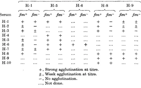
The cross-reactivity of fim+ a n d ^ m " suspensions of selected type strains were compared by agglutination with the H sera that had exhibited cross-reactions. The results (Table 3) confirmed that fimbrial antibody was responsible for the hetero-logous agglutination reactions seen with sera H-l, H-2 and H-3 with strain H-8 and sera H-5, H-6 and H-8 with strain H-l. Serum H-2 failed to agglutinate the
fimr variant of strain H-l and the agglutination titre of this variant with serum H-3
274 T. L. P I T T , Y. J . ERDMAN AND C. BITCHER Table 1. Agglutination of H-type strains of S. marcescens by
homologous and heterologous H-sera
Serum Titres of agglutination by the stated antisera of the type strains
against , • •—•—• • A • • — — v t y p e strains H I H 2 H 3 H 4 H 5 H6 H7 H 8 H9 H10 H l l H12 H13 H - l 3200 + . + + . . + + . H-2 + 3200 + + . . . . H-3 ± + 3200 . . . . + + + . . . H-4 . . . 3200 + H-5 ± • • + 6400 H-6 ± . . + + 3200 H-7 + . . . + . 1600 H-8 + + ± • • • • 6400 + . . ± . H-9 + + 6400 . . . . H-10 . . + . . . . + . 1600 H - l l 3200 H - l 2 3200 . H-13 3200
+ Strong agglutination at titre. + Weak agglutination at titre.
Table 2. Immobilization in agar of H-type strains by
homologous and heterologous H antisera
T i t r e of immobilisation of homologous s t r a i n b y t h e s t a t e d antisera a n d cross reactions w i t h t y p e strains
S e r u m H - l H - 2 H - 3 H - 4 H - 5 H - 6 H - 7 H - 8 H - 9 H - 1 0 H - l l H - 1 2 H - 1 3 H - l 1600 - - * - i - - i - - i -H - 2 - 1600 _ _ _ _ _ _ _ _ _ _ _ H - 3 - - 1600 _ - - - - _ _ _ - _ H - 4 - - 1600 _ _ _ _ _ _ _ _ _ H - 5 - - - - 3200 _ _ _ _ _ _ _ _ H - 6 + 1600 - _ - _ _ - -H - 7 _ _ _ _ _ _ 800 - - - -H - 8 _ _ _ _ _ _ _ 1600 + _ _ - _ H - 9 _ _ _ _ _ _ _ + 1600 -H - 1 0 _ - + _ _ _ _ _ _ 8 0 0 - - -H - l l _ _ _ i _ i _ _ j _ 800 i -H - 1 2 _ _ _ _ _ _ _ _ _ _ _ 1600 -H - 1 3 - - - i - i - - t - - t 8 0 0 + , Immobilized a t t i t r e . —, N o t immobilized. i, P a r t i a l inhibition of g r o w t h b y serum.
was four times lower than the titre with the fim+ culture. However, the fim~ variant of strain H-5 exhibited the same agglutination reactions as the fim+ form and this result suggested that flagellar or fimbrial antigens were not responsible for the cross-reactions of the strain.
Bacteriophage typing
The lytic pattern of the ten phages was determined by typing each propagating strain with all the phages diluted to a concentration of 10 x RTD. Most of the
Table 3. Cross-agglutination offimbriate and non-fimbriate variants
of selected H type strains by homologous and heterologous H antisera Agglutination by stated antiserum of suspensions of type strains
H-l H-5 H-6 H-8 H-9
Serum fim+ fim- fim+ fim~ fim+ fim~ ftm+ fim~ fim+ flm~
H-l + + + + + - + ± H-2 ± - + - ± ± H-3 + ± + - + H-4 + + H-5 ± - + + H-6 ± - + + + + H-7 + + + + H-8 + - + + + + H-9 + + + + H-10 + +
+ , Strong agglutination at titre. + , Weak agglutination at titre. —, No agglutination.
..., Not done.
phages produced defined clear plaques on the strains and lytic reactions were easy to read. The lytic pattern of each phage was distinct and one phage lysed all ten host strains. None of the phages were specific for their propagating strain alone. Intra-laboratory variation was assessed by typing 100 pairs of cultures from different sources twice at an interval of a week. Lytic reactions were scored as strong or weak. The percentages of pairs of tests that showed no, one, two or more strong reaction differences was calculated and the results are shown in Fig. 1. Sixty-nine per cent of strains gave identical patterns of susceptibility when typed twice; 26 % showed a difference of one, and 5 % showed differences of two or more strong reactions. Analysis of the results showed that phages 2, 5 and 9 were responsible for the majority of the discrepancies. Phage 2 was weakly active on many strains, although lysing its host strain strongly, so it was used at 100 x RTD for typing routine isolates.
Typing of cultures from outbreaks of infection
Two hundred and seventy-three cultures of S. marcescens were typed by bacterio-cine and bacteriophage sensitivity and by O and H serological methods. Only 1 % of the strains were not grouped by 0 serology, but 16% were non-typable by H-serology (H: NT); 96 % of strains were sensitive to bacteriocines and the same proportion were typable by bacteriophage. A strain was considered to be distinct by bacteriocine typing if its pattern of sensitivity to bacteriocines from 12 standard strains differed by more than three reactions from the modal pattern amongst strains from the same incident. Similarly, a rule of two or more major differences in sensitivity pattern was applied for phage typing. The 0-types of cultures were determined with specific sera in agglutination tests, and H-typing was performed
276 T. L. PITT, Y. J. ERDMAN AND C. BTTCHEB 80 8 3 60 S 40 8
I
20 _n „_ 0 1 2 3 Number of reactionsFig. 1. Reproducibility of phage typing; percentage of 100 cultures giving the indicated number of strong reaction differences.
in two stages; first, a strain was tested by agglutination with unabsorbed H sera and second, immobilization tests to confirm the H type were performed with those sera that had agglutinated the strain. A summary of the typing results is shown in Table 4.
Incident I
For 12 months there had been outbreaks of urinary tract infection with S.
marcescens involving 65 patients in a geriatric ward and the out-patient department
of a 600-bed general hospital in the UK. Thirty-nine cultures were received for typing and 36 of these were of serotype 0 3 :HNT. Within this serogroup, the bacteriocine sensitivity patterns of the cultures were similar and 31 were lysed by a single phage. The remaining 5 cultures were not typable. Two ONT :HNT strains were indistinguishable by bacteriocine typing but had distinct phage typing patterns.
A review by the hospital microbiologist (Dr P. O'Neill - personal communi-cation) suggested that the reservoir of infection was a group of chronically ill patients. The return of one of these to the urological ward for further treatment or investigation provided a new source of infection within the ward. The hygienic facilities in the ward were poor, and the demonstration that most of the infections were caused by the same strain of 8. marcescens resulted in an improvement.
Incident 2
Sixty-four patients in three wards of a general hospital in Southern Germany were involved. S. marcescens was isolated from the blood of 12 of them. Eighty-two cultures were received for typing, but details of their epidemiology were not available. Serotype 014 was found in 77 cultures, and H-typing divided these into H2 and H4. Phage typing distinguished the same two groups of strains. However, five different patterns of bacteriocine susceptibility were found amongst the 77 cultures, but the majority fell into two well-defined groups. The other three patterns were similar to those of the two groups, but were distinct on the application of the
e 4 . Results of typing strains of S . marcescen s from six outbreaks of infection: strain definition by three methods of members of the same 0 serogroup Outbrea k 1 2 3 4 5 6 O-grou p 3 N T 1 4 2 4 14 4 14 N T 1 4 12/1 4 1 4 12 5 Numbe r of strain s 3 6 2 7 7 3 2 3 9 2 1 13 2 1 9 3 3 8 1 1 2 2 9 25 4 8 1 2 39 Numbe r o f strain s o f H grou p 8 1 0 1 1 1 2 1 3 N T 3 6 2 2 1 1 3 1 8 No . o f groups * distinguishe d b y Bacterio -cin e 1 1 5 1 2 2 1 1 1 3 1 6 2 1 Bacterio -phag e 1 2 2 2 1 3 1 1 1 2 1 3 2 1
I
f
I
* Define d b y th e compariso n o f differen t moda l pattern s withi n eac h outbreak . NT , No t typable .278 T. L. PITT, Y. J. EBDMAK AND C. BTJCHER
three reaction difference rule. As there was complete agreement between H-typing and phage typing the results of bacteriocine typing probably reflects the inherent lack of reproducibility of the method rather than an increase in the discrimination between strains.
Of the 5 strains not grouped as 014, 3 were found to be of group 02 and 2 strains were of group 04. The results of each of the typing methods indicated that one of the strains of group 02 was distinct from the others but both 04 strains were indistinguishable from each other.
Incident 3
Within six months, strains of S. marcescens with similar antibiotic-resistance patterns were isolated from specimens from two intensive care units (ICU) in different hospitals in Austria. One was an ICU for premature babies, the other for a department of open heart surgery. In the latter, patients from the adjacent accident department were also treated. In both units there were severe infections with S. marcescens (pneumonia, septicaemia) and as other methods failed to eradicate the organism, both were closed for a period.
Thirty-nine cultures from both hospitals were received for typing. All were of the serogroup 014 :H4 and 33 had the same bacteriocine and phage type. The remaining six were sensitive to the same bacteriocines, but there were two patterns of phage sensitivity differing from each other by two strong reactions.
Incident 4
This outbreak with a multi-resistant S. marcescens was described by Meers, Foster & Churcher (1978). The organism was found in sputum, tracheal aspirate, wounds and urine, and two patients with positive blood cultures died. Twenty-one strains isolated from patients and from hand-washings of a member of the nursing staff were typed and were all of the same serogroup, O4:H12; they could not be further divided by bacteriocine or phage typing.
A second strain (014: H7) represented by 13 cultures was isolated intermittently from sputa, wounds and urines over many weeks from the same hospital but no common-source was identified. In addition two ONT cultures were found to be distinct by bacteriocine and phage typing.
Incident 5
This outbreak occurred in a surgical ward and most isolations were made from urine specimens. Twenty-three cultures from different patients were received for typing and 18 of these were of serogroup 014 :H7; a single 014 strain was not typable by H-serology. The bacteriocine typing patterns of 17 of the 19 cultures were indistinguishable but the patterns of the remaining strains were unique, and one of these also differed in its susceptibility to the typing phages.
Three cultures exhibited an 0-antigen cross-reaction that was not observed amongst the type strains. The cultures were serotyped as 012/014 :H13 and were indistinguishable by further typing. One culture that was ONT differed in H-type, bacteriocine and phage pattern from the other strains.
Incident 6
Fifty-four cultures were received from a general hospital in Switzerland. The majority of the cultures were isolated from the urine and sputum of patients in a surgical intensive care unit and an associated surgical ward. Some cultures were recovered from patients in other wards and units of the main hospital and also from patients in two outlying cottage hospitals.
Six O-serogroups were found but 38 of the 54 cultures were of serogroup 014. Within this O-group, four distinct strains were distinguished by H typing and there was good agreement between this method and phage typing which identified three groups of strains. However, the results of bacteriocine typing were variable; 6 strains were distinguished, but 29 of the 38 cultures were allocated to only two groups and three cultures were not typable. Eight of 11 cultures of serogroup 012 shared the same H antigen, but the results of bacteriocine and phage typing were equivocal, as 6 cultures were not typable by either method. The remaining cultures fell into four other O-serogroups and were therefore distinct from the outbreak strains.
Correlation of typing methods used to subdivide 0 serogroups
It is apparent from this and other studies (Wilfert et al. 1970; Negut, Davis & Washington, 1975; Anderhub et al. 1977) that epidemiologically valid results are obtained by the O-serogrouping of S. marcescens, and by this method we obtain broad but reproducible groups of strains. Therefore, for the purpose of our study of clinical isolates we have assumed that cultures belonging to different O-serogroups are different and have adopted O-serogrouping as our primary classification of strains.
To estimate the degree of correlation between H-serotyping, phage and bacterio-cine typing, strains of the same 0 group from the same incident of infection were classified into groups according to the following criteria. For H-serotyping, distinct groups of strains were defined as those of different H-types. Groups distinguished by phage typing were those whose typing pattern did not differ significantly from the modal pattern of their own group but were different by at least two strong reactions from other patterns seen within the same serogroup. Similarly, distinct groups by bacteriocine typing consisted of strains of the same modal pattern defined according to Anderhub et al. (1977) which differed from other patterns by at least three reactions.
An analysis of the typing results was performed by the use of a formula adapted from one used in numerical taxonomy to compare positive and negative results of biochemical tests on strains of different taxa (Stanier, Adelberg & Ingraham, 1977). A measure of the percentage similarity of two typing methods in the subdivision of strains of the same 0 serogroup was obtained by the formula
S/o =/ 0 a + b + c
_££_
where S is the percentage similarity between two typing methods, a is the percent-age of strains considered the same by both methods; b is the percentpercent-age of strains
280 T. L. PITT, Y. J. ERDMAN AND C. BUCHER
Table 5. Comparison of interpretation of results obtained by three methods of isolates
of S. marcescens of the same 0 serogroup in the same incident of infection
Percentage of strains considered
H serotyping H serotyping typing Phage typing typing V. V. V. phage typing bacteriocine bacteriocine
(a) the same by both, (6) different by both, and (c) the same only by
one of the («*) 69 56 71 stated typing A (6) 15 11 11 methods (c) \ 16 33 17 Percentage similarity, a + b + c 84 67 82
considered different by both methods; c is the percentage of strains considered the same by either one of the two methods but different by the other.
Clearly the degree of correlation observed is the ratio of these results which agree by both methods (a + b) to the total comparisons (a + b + c). Table 5 shows the percentage similarity calculated for H serotyping v. phage typing was 84 and reveals a high level of agreement in interpretation of results by these methods. However, H serotyping v. bacteriocine typing showed only 67% similarity, whereas phage typing v. bacteriocine showed 82 % similarity somewhat similar to H serotyping v. phage typing. The apparent discrepancy in comparison of H sero-typing v. the other two methods (84% and 67%) was due mainly to the number of non-typable strains found with this method (16 %).
DISCUSSION
Over the last decade S. marcescens has been isolated with increasing frequency in specimens from hospitalized patients in the USA and Europe (Ringrose et al. 1968; Wilfert et al. 1970; Daschner & Senska-Euringer, 1975; Schaberg et al. 1976). However, there have been few reports of outbreaks of infection in the UK (Black & Hodgson, 1971; Rogers & Gittens, 1974; Ball, McGhie & Geddes, 1977; Meers et al. 1978).
The factors which apparently contribute to the increased reporting of the isolation of 8. marcescens from clinical material include the natural antibiotic resistance of the bacterium and its ability to acquire new resistances (Traub, 1978; Schaberg etal. 1976), its presence in the hospital environment (MacArthur & Askerman, 1978) and the advent of commercially available identification schemes.
The purpose of epidemiological typing is for the identification of groups of infection coming from the same source, and to determine the source of infection either in groups of patients or in individuals. Typing may exclude strains that are different and from this one can infer that the source of infection is among the strains that are indistinguishable from the index strain. We have evaluated four methods for the type identification of 8. marcescnens first, by investigating the
biological factors which influence the efficiency of the methods, and second by typing strains from suspected outbreaks of infection in six hospitals.
O-serogrouping
It was relatively simple to prepare specific agglutinating sera of adequate titre but in practice it was found that one serogroup - 014 - was particularly prevalent and accounted for over two-thirds of the strains typed. This was expected, as other workers had reported the high incidence of this serogroup (Negut et al. 1975; Anderhub et al. 1977; Le Minor & Pigache, 1978) and therefore it may be worth searching for other antigens to subdivide this 0-group. Although most strains were typable by a conventional agglutination test and the results were repro-ducible when pairs of strains from the same patient were tested, the relative pre-dominance of a few serogroups suggests that O-typing alone is insufficiently dis-criminating for use in epidemiological studies.
H-serogrouping
H-antigen typing has been used fairly extensively by other workers to differenti-ate between strains of the sameO-group (Negut etal. 1975; Traub & Kleber, 1977; Le Minor & Pigache, 1978). Agglutination tests using sera raised against unheated cells of the 13 H-type strains revealed many cross-reacting antigens amongst them. Many of these could not be flagellar antigens as the results of agglutination tests could not always be corroborated by demonstrating immobilizing antibody to heterologous strains in the sera using a modification of the H-immobilization method of Le Minor (see Traub & Kleber, 1977). The main advantage of this test was that homologous titres were usually high while heterologous ones were low, though the method was laborious when used to type larger numbers of organisms because each serum agar tube had to be prepared individually. However, the immobilization test was useful in determining the ' true' H-type of a culture which had been agglutinated by more than one of the typing sera.
We investigated the role of fimbrial antigens in agglutination tests with un-absorbed H sera and found that they were responsible for some of the cross-reactions among type strains. This was expected as Nowotarska & Mulczyk (1977) had demonstrated that high titres of fimbrial antibody were present in sera raised against unheated suspensions of strains of S. marcescens. Moreover, the presence of cross-agglutination not attributable to these antigens, indicate that aggluti-nating antibody of other heat-labile constituents may be induced by whole cell unheated vaccine.
Eleven different H-antigen factors were detected amongst the clinical isolates examined and H4 was the most frequent type found. Although the percentage typability of H-typing was significantly lower than that obtained for 0 typing, it distinguished between strains more often than the latter method. Recently, seven new H-antigens have been described (Traub & Kleber, 1977; Le Minor & Pigache,
1978) and it may be expected that if antisera to these were included, the number of non-typable strains would be reduced.
282 T. L. PITT, Y. J. ERDMAN AND C. BUCHER Bacteriophage and bacteriocine typing
The typability of both methods was good but the phage typing patterns of strains were as a rule less variable than the bacteriocine patterns. The incon-sistency of bacteriocine pattern may have led to an interpretation of the typing results whereby strains were considered to be different when in fact they were not. The validity of a bacteriocine or phage type can be determined if the results by one of the methods are the same as those given by another. For example, in one outbreak due to the same O group the strains were sub-divided into 5 sub-groups by bacteriocine and 2 sub-groups by H typing and phage typing respectively. In this case we considered that the results of the latter methods were correct and that the apparent increase in discrimination by bacteriocine typing was due to the variability of the method.
Use of combined methods for typing
The four methods described here for the typing of S. marcescens suffer in varying degree from lack of discriminatory power; either because some types cannot be further divided or because less than complete reproducibility makes it necessary to introduce 'reaction-difference rules' to allow for the variability of the method (Anderhub et al. 1977). Therefore we propose that a combination of methods be used to type strains of S. marcescens in a hierarchical fashion as advocated by Meitert & Meitert (1966) who used O-serogrouping for the primary classification of P. aeruginosa strains and subdivided the serological groups by phage typing.
Our results suggest that either H-serotyping by immobilization tests or phage typing were the best alternatives as secondary methods to divide further strains of the same O-serogroup, and both of these methods are to be preferred to bacterio-cine typing for the routine type identification of strains of 8. marcescens.
REFERENCES
ADAMS, M. H. (1959). Bacteriophages. New York: Interscien.ee.
ANDERHTTB, B., PITT, T. L., EEDMAN, Y. J. & Wnxcox, W. R. (1977). A comparison of typing methods for Serratia marcescens. Journal of Hygiene 79, 89.
BALL, A. P., MCGHEE, D. & GEDDES, A. M. (1977). Serratia marcescens in a general hospital.
Quarterly Journal of Medicine 66, 63.
BLACK, W. A. & HODGSON, R. (1971). Search for Serratia. Journal of Clinical Pathology 24, 444.
DASCHSTER, F. & SENSKA-EURINGER, C. (1975). Kontaminierte Infusionen als Ursache nosokomialer Serratia-marcescens-sepsia bei Kindern. Deutsche Medizinische
Wochen-schrift 100, 2324.
DTJGTJID, J. P., ANDERSON, E. S. & CAMPBELL, I. (1966). Fimbriae and adhesive properties in Salmonellae. Journal of Pathology and Bacteriology 92, 107.
EDWARDS, P. R. & Ewnsro, W. H. (1962). In Identification of Enterobacteriaeceae, 2nd ed., p. 223. Minneapolis, Minnesota: Burgess.
FARMER, J. J. (1972). Epidemiological differentiation of Serratia marcescens — typing by bacterioein production. Applied Microbiology 23, 218.
FARMER, J. J. (1975). Lysotypie de Serratia marcescens. Archives Roumaines de Pathologie
Experimental et de Microbiologie 34, 189.
GRIMONT, P. A. D. & GRIMONT, F. (1978). Biotyping of Serratia marcescens and its use in epidemiological studies. Journal of Clinical Microbiology 8, 73.
LE MINOH, S. & PIGACHE, F. (1978). Etude antigenique de souches de Serratia marcescens isolees en France. II. Caracterisation des antigens O et individualisation de 5 nouveaux facteurs, frequence des serotypes et designation des nouveaux facteurs H. Annales
Micro-biologie (Paris) 129B, 407.
LIDWELL, O. M. (1959). Apparatus for phage-typing of Staphylococcus aureus. Monthly
Bulletin of the Ministry of Health and the Public Health Laboratory Service 18, 49.
MACABTHUB, B. S. & ACKEBMAN, N. B. (1978). The significance of Serratia as an infectious organism. Surgery, Gynecology and Obstetrics 146, 49.
MEEBS, P. D., FOSTER, C. S. & CHURCHER, G. M. (1978). Cross-infection with Serratia
•marcescens. British Medical Journal i, 238.
MEITERT, T. & MEITERT, E. (1966). Utilisation combinee du serotype et de la lysotypie des souches de Pseudomonas aeruginosa en vue d'approfondir les investigations epidemio-logiques. Archives Roumaines de Pathologic Experimental et de Microbiologie 25, 427. NEGTJT, M., DAVIS, B. R. & WASHINGTON, J. A. (1975). Biochemical and serological
charac-teristics of Serratia marcescens isolated from various clinical and environmental sources.
Archives Roumaines de Pathologie Experimental et de Microbiologie 34, 33.
NOWOTARSKA, M. & MTTLCZYK, M. (1977). Serologic relationship of nmbriae among Entero-bacteriaceae. Archivum Immunological et Therapie Experimentalis 25, 7.
PILLICH, J., HRADECNA, Z. & KOCTJB, M. (1964). An attempt at phage typing in the genus
Serratia. Journal of Applied Bacteriology 27, 65.
BUSTGBOSE, R. E., MCKOWN, B., FELTON, F. G., BARCLAY, B. O., MTTCHMOBE, H. G. & RHOADES, E. R. (1968). A hospital outbreak of Serratia marcescens associated with ultra-sonic nebulizers. Annals of Internal Medicine 69, 719.
ROGERS, K. B. & GITTENS, B. (1974). An epidemic due to Serratia marcescens in a neuro-surgical unit. Journal of Clinical Pathology 27, 930.
SCHABEBG, D. R., ALFOBD, R. H., ANDERSON, R., FARMER, J. J., MELLY, M. A. & SCHAFFNER, W. (197 6). An outbreak of nosocomial infection due to multiply resistant Serratia marcescens: Evidence of interhospital spread. Journal of Infectious Diseases 134, 181.
STANTER, R. Y., ADELBERG, E. A. & INGRAHAM, J. N. (1977). In General Microbiology, 4th ed., p. 507. Macmillan.
TRATJB, W. H. (1978). Antibiotic susceptibility of clinical isolates of Serratia marcescens compared with sensitivity to group A (phage tail) bacteriocins. Chemotherapy 24, 301. TBATJB, W. H. & KLEBER, I. (1977). Serotyping of Serratia marcescens: evaluation of Le
Minor's H immobilisation test and description of three new flagellar H antigens. Journal of
Clinical Microbiology 5, 115.
WHFEBT, J. N., BABBETT, F. F., EwnsrG, W. H., FINLAND, M. & KASS, E. H. (1970). Serratia
marcescens: biochemical, serological and epidemiological characteristics, and antibiotic