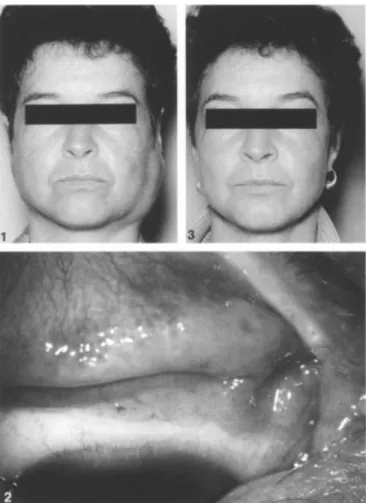
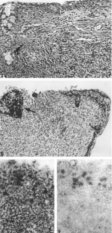
Diagnosis of MALT lymphoma by conjunctival biopsy: a case report
Texte intégral
Figure
Documents relatifs
A: large view of the cerebriform cystic lesion; B, C: higher magni- fication of two of the various lobes of the cyst including multiples scolices (black arrows); D, E: higher
We used an array including ALPL gene, genes of differential diagnosis COL1A1 and COL1A2 that represent 90% of OI cases, SOX9, responsible for CD, and 8 potentially modifier genes
Within the scope of a project aiming at automatic student’s modelling in algebra [9], we conducted tracks analyses with a variety of students from 8 and 9 grades, in order to
With regard to diagnosis and prognosis in cancer, they discussed the diagnostic value and the state-of-the-art of exo-miRNAs in cancer, by a survey of the available studies from 2013
emergence of a new W135 strain but clonal expansion within the electrophoretic type-37 complex. Burden of disease caused by Streptococcus pneumoniae in children younger than 5
The Bohr el Ghazal CDTI project coordinating officer, the Mvolo county supervisor, frontline health workers, CDDs ond community members f or giving the Mission
Diagnosability with disambiguation and an upper bounded energy budget (ULWUB diagnosability) is equivalent to a partial observation co-B¨uchi game [7] between a system and a
As the protocol follows the same steps and the cores numbered and analyzed separately, patients were grouped into 3 groups as follows: PCa diagnosed after 6
