ORIGINAL ARTICLE
Osteopathology induced by bisphosphonates and dental
implants: clinical observations
Christine Jacobsen&Philipp Metzler&Matthias Rössle& Joachim Obwegeser&Wolfgang Zemann&
Klaus-Wilhelm Grätz
Received: 14 August 2011 / Accepted: 27 February 2012 / Published online: 15 March 2012 # Springer-Verlag 2012
Abstract
Objectives Although there are many reports about risk factors for the development of BP-associated osteonecrosis of the jaws, the role of dental implants as a local risk factor is still discussed, especially in patients with oral BP treatment. Until now, a few case reports and surveys display a possible minor risk in patients with oral BP therapy, whereas the avoidance of implant placement is generally accepted in patients with intravenous BP therapy. Patient and methods In this study, the cases of 14 patients with osteonecrosis of the jaws in association with BP ther-apy and dental implant placement were analyzed carefully with a detailed literature review.
Results Of 14 patients, nine had underlying malignant disease and five patients had osteoporosis. In ten patients, implants were placed either in the posterior mandible or maxilla; the mean interval between implant insertion and disease onset was 20.9 months. Pain (n12) and signs of infection (n10) were the most common symptoms. Histologically, signs of infec-tion were found in nine of 11 analyzed patients with presence of Actinomyces in six patients. Two patients turned out to have infiltration of underlying malignant disease.
Conclusions Posteriorly placed implants seem to be of higher risk of development of osteonecrosis of the jaws.
Not only the implant placement but also the inserted implant itself seems to be a continuous risk factor.
Clinical relevance The herein elaborated risk factors help den-tists plan dental rehabilitation with implants in this high-risk group of patients and indicate careful and regular dental recall. Keywords Bisphosphonate-associated osteonecrosis of the jaws (BRONJ) . Dental implants . Risk factors . Posterior jaw . Infection
Introduction
About 100 years ago, phosphorus necrosis of the jaw was a common disease in match factory workers. In 2003, Marx et al. discovered an apparently similar type of osteopathology asso-ciated with bisphosphonate (BP) medication [1]. Since then, much has been published about “bisphosphonate-associated osteonecrosis of the jaw” (BRONJ) [2–4]. Because BRONJ is a major concern for patients and there is no remodeling tendency in the affected bone, BP medication became a new and important risk issue for dentists in general daily practice. Many efforts have been made to prevent this localized bone disease [5,6]. Thus, many guidelines and prevention protocols have been developed to guide the dental treatment of patients before and during BP medication [7,8]. The occurrence of BRONJ seems to be intimately connected with local risk factors; thus, elective invasive treatment (e.g., in dental implant placement) seems to be a high-risk procedure and is strongly advised against in various guidelines [9–11].
However, closer examination of the published data reveals a lack of information concerning dental implants and BRONJ. Several case reports and series have evaluated implant failures in patients, primarily those receiving intra-venous BP therapy. Other cohort studies showed minor risks
C. Jacobsen (*)
:
P. Metzler:
J. Obwegeser:
W. Zemann:
K.-W. GrätzDepartment of Craniomaxillofacial Surgery, University Hospital of Zurich,
Frauenklinikstrasse 24, 8091, Zurich, Switzerland e-mail: christine.jacobsen@usz.ch M. Rössle
Institute of Pathology, University Hospital of Zurich, Frauenklinikstrasse 24, 8091, Zurich, Switzerland
of osteopathology development in patients receiving oral BP therapy and dental implant insertion. On the other hand, the local and short-term use of BP has been shown to have a positive influence on osseointegration of all implants, dental or otherwise [12,13].
To date, guidelines concerning implant placement in patients receiving BP therapy are ambiguous. Intravenous BP therapy for underlying malignant disease seems to be a clear contraindication for the placement of dental implants, whereas implants can be placed in patients receiving oral or intravenous BP for the treatment of osteoporosis. Nev-ertheless, dentists are still uncertain whether, where, when, how and in which patients it is possible to place dental implants without a significant risk for the development of BRONJ.
In this study, 14 patients with osteopathology of the jaw associated with BP therapy and dental implant insertion were analyzed carefully. A detailed literature review was also conducted. We clarified the risk factors for implant placement in patients receiving BP therapy to help dentists distinguish high- and low-risk situations.
Patients and methods
The study sample included patients with BRONJ who were evaluated, treated and followed up in a special outpatient clinic at the Department of Craniomaxillofacial Surgery, University Hospital of Zurich between 2005 and 2010. During this period, 110 patients with osteopathology of the jaw were referred to the clinic. For all patients, a detailed medical history including underlying diseases, general risk factors, medications, other therapeutic measures performed, dental history and possible local risk factors was collected. Additionally, a meticulous clinical examination was performed.
In total, 14 (11 women, three men) of the 110 patients were referred due to osteopathology of the jaw associated with BP therapy and dental implants. Of these, nine patients were treated with intravenous BP for malignant diseases and five patients received BP therapy for the treatment of oste-oporosis. Of the nine patients with underlying malignant diseases, two had multiple myeloma, five had breast cancer, one had prostate cancer and one patient had lung cancer. The nine patients all received zoledronic acid (Zometa; Novartis, Basel, Switzerland) and one had previously received pamidronate (Aredia; Novartis).
The other five patients had no underlying malignant disease, but osteoporosis was the indication for BP treat-ment. Two of these patients received oral alendronate (Fosa-max; Merck, Sharp, & Dohme GmbH, Munich, Germany) therapy, one patient received intravenous ibandronate (Bon-viva; Roche Pharma AG, Basel, Switzerland) therapy, and
two received pamidronate (Aredia; Novartis) therapy in a 3-month regimen.
All implants in this study were conventional dental screwed implants without modification. Because all implants were inserted from different practitioners and patients were referred with apparent osteopathology, data about type of implant and manufacturer were not available in all cases. Therefore, this information was not collected and analyzed in this study.
To summarize the location of implant placement, all implants placed in or posterior to the second premolar region of the mandible or maxilla were defined as posterior implants and all implants placed in or anterior to the first mandibular or maxillary premolar were defined as anterior implants.
Cross-sectional imaging of both jaws was performed to confirm the diagnosis and to visualize the dimensions of the affected bone. A histological analysis was also performed to confirm the diagnosis and exclude other bone lesions, espe-cially the metastasis of underlying diseases.
During diagnosis and treatment, the infiltration of malig-nant underlying disease was found in two patients: one female patient with breast cancer (posterior mandible) and one male patient with multiple myeloma (anterior mandi-ble). Interestingly, the clinical and radiological presentations of these patients resembled classical BRONJ. The other 12 patients had BRONJ in association with previous dental implant placement.
To treat osteopathology, when general health status and patient approval allowed, BP therapy was stopped 3– 6 months before surgical treatment to encourage bone remodeling and necrotic bone sequestration. Surgical inter-vention was then performed to remove the infected necrotic bone and to allow soft-tissue healing. Additionally, systemic anti-infectious treatment with antibiotics (mean: 4 weeks) was performed.
All specimens were primarily obtained for medical pur-poses, with the informed consent of the patients. The study design fulfills the guidelines of the Declaration of Helsinki regarding ethical principles for medical research involving human subjects.
Literature review
The literature database PubMed, Embase and Ovid were searched for“osteonecrosis of the jaw” in combination with one of the following terms “bisphosphonate,” “dental implants,” “local risk factors” and “implant insertion” using the Boolean operator AND, in title, abstract and MeSH terms. The search included articles published between 1995 and 2011. In total, 50 articles were identified (refer-ences are available on request from the corresponding au-thor). Two of these publications were excluded because they were written in foreign languages not understandable for the
authors. During the assessment process, 17 articles were excluded because they described animal and clinical studies in which BP treatment was performed to improve osseointe-gration of dental implants without a link to BP-related osteonecrosis of the jaws, and three other articles gave a general overview about BRONJ without a focus on dental implants as a local factor. Of those, 21 articles were exclud-ed because they were either written in a language other than English, French or German or they did not refer to the topic of dental implant placement in patients with BP therapy. In total, 17 articles were excluded because they described cases or studies in which the application of bisphophonates was used to improve osseointegration of dental implant.
All publications identified in the literature searches were retrieved from online journals and selected on the basis of the inclusion criteria. The selected papers were assessed according to the following criteria for this study,
1. Study patients with BRONJ in association with dental implants.
2. Number of patients with BRONJ in association with dental implant placement.
3. Type of BP reported.
4. Underlying diseases of patients reported.
5. Location of BRONJ and thereof placed dental implants. 6. Symptoms and clinical picture.
Results
Fourteen patients had osteopathology of the jaw associated with BP therapy in an area of previous dental implant
placement; in two patients, we found infiltration of the underlying disease. The average duration of BP therapy before the onset of jaw problems was 38 months in the seven patients receiving intravenous BP therapy for under-lying malignant diseases and about 50 months in the five patients receiving BP treatment for osteoporosis. One fe-male patient with osteoporosis did not know the exact treatment duration but indicated that she had received BP treatment for more than 5 years.
The medical information and BP treatment of these patients is summarized in Table 1. In total, 23 dental implants were inserted in 12 BRONJ patients.
BRONJ localization
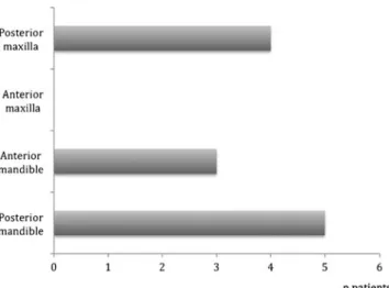
Osteopathology developed in the mandible in eight patients and in the maxilla in four patients. Nine patients received posterior implants: four patients received five implants in the posterior maxilla and five patients received six implants in the posterior mandible. In three patients, 12 implants were inserted in the anterior mandible. No patient with an osteo-pathology received an implant in the anterior maxilla (Fig.1).
Implant history
Nine patients were referred by dentists due to peri-implant problems or exposed bone around previously inserted implants. In one male patient, two dental implants had been explanted by the dentist after disturbed soft-tissue healing and two new implants were inserted in the same area; heal-ing disturbance persisted. In one female patient, massive
Table 1 Overview of patients with type of underlying disease, type of bisphosphonate medication, pathology report and presence of Actinomyces Number Diagnosis BP Localization Histology (additional finding to necrotic bone) Actinomyces 1 MC Z Mandible Purulent osteomyelitis 1
2 O A–P Maxilla No biopsy No biopsy
3 MM P–Z Mandible Acute purulent osteomyelitis 1 4 O P Mandible Acute and chronic osteomyelitis – 5 PC Z Mandible Destructing purulent osteomyelitis fibrinoleucocytic exudate – 6 MC Z Maxilla Acute and chronic inflammation, suspicion on infiltration of multiple myeloma 1
7 MM Z Mandible –
8 MC Z Mandible Purulent osteomyelitis 1
9 MC Z Mandible Acute inflammation 1
10 MC Z Mandible Subepithelial infiltration of carcinoma –
11 O I Mandible Chronic inflammation –
12 Lca Z Maxilla Active inflammation 1
13 O A Mandible No biopsy No biopsy
14 O A Mandible
MC mamma carcinoma, MM multiple myeloma, O osteoporosis, PC prostate cancer, Lca lung carcinoma, Z zoledronic acid, P pamidronate, A alendronate, I Ibandronate
infection resulted from a sinus lift and dental implant inser-tion; another female patient presented with massive con-comitant osteopathology of the jaws following the explantation of four interforaminal dental implants.
The mean interval between implant insertion and disease onset was 20.9 months. In patients with malignant underly-ing disease, the average interval was 17 months; in patients with osteoporosis, it was 25.6 months. Detailed information about the date of implant insertion was missing in one patient, and the two patients with malignant histology were excluded from this analysis.
Clinical presentation
Ten patients presented with pain. In nine patients, pus dis-charge could be observed; six patients presented with a submandibular or submental abscess. Four patients had par-esthesia of the mandibular or infraorbital nerve. Eight
patients presented with exposed bone and lack of healing. In four patients, a fistula was found; one extraoral fistula was present in the submental area. Two patients presented with involvement of the maxillary sinus. The male patient with multiple myeloma infiltration presented with a sub-mental abscess, and the female patient with breast cancer metastasis presented with hard, yellowish, exposed bone in the posterior mandible.
Diagnostic measures
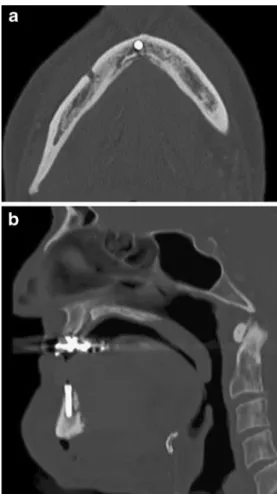
Cone beam or thin-layer computed tomography (CT) was performed to analyze volume expansion of the pathological bone structure. In five patients, magnetic resonance imaging (MRI) was also performed. All patients presented typical radiological signs of BP-associated osteopathology includ-ing thickeninclud-ing of the bone, massive sclerosis, poor cortico-medullary differentiation and periosteal new bone formation (Fig.2a–c). The two patients with infiltration of underlying malignant diseases showed the same radiological signs. No clear sign of infiltration or metastasis could be found in the radiological images (Fig.3a, b).
Histological analysis
Bone specimens were obtained from 12 patients for histo-logical analysis during surgical therapy or from a bone biopsy. Affected bone was available for histological analysis from all nine patients with underlying malignant disease. Necrotic bone was found in samples from all 12 patients. Metastasis or infiltration of the underlying disease with concomitant inflammation was also found in specimens from two patients. The bone specimens of the other ten patients showed typical histological findings of inflamma-tion in associainflamma-tion with BP-associated osteopathology. Acti-nomyces plaques were found in samples from seven patients (Table1and Fig.4). Two patients with osteoporosis refused
Fig. 1 Display of localization of bisphosphonate-associated osteonec-rosis of the jaw in 12 patients with dental implants as the local factor. The two patients with malignant histology were excluded from this figure
Fig. 2 a Coronal view of one female patient with previous implant insertion, sinus lift and oral bisphosphonate therapy. A sequester and sclerotiv changes of the lateral wall of the maxillary sinus are visible. b The same patient. Sagittal view of the cone beam tomography of the
right side shows sclerotic changes of the maxillary sinus walls with concomitant sinusitis. c Sagittal view of the left side of the maxilla shows no changes of the bone with well osseointegrated dental implants
to provide samples for histological analysis of the affected bone.
Treatment
Surgical treatment involving the removal of necrotic bone was performed in ten of the 12 patients (Fig.5). Two female patients refused surgical treatment. Complete remission was accomplished in nine of ten surgically treated patients (Fig. 6). One surgically treated female patient died during follow-up due to underlying diseases. All patients received additional anti-infectious treatment with antibiotics. In all patients, pain and hypesthesia symptoms improved during anti-infectious treatment.
The male patient with multiple myeloma infiltration was surgically treated with implant explantation, decortication and revision of the abscessed area. He also received chemo-therapy for multiple myeloma progression. Complete remis-sion of the infected jaw bone was accomplished. The female patient with breast cancer infiltration was treated conserva-tively due to her poor general condition. She died during follow-up.
Literature analysis
In total, 28 articles were included in the literature review, most of them were case reports or series (n15). The other articles were literature reviews, and only two articles were surveys or studies. Table 2 presents the numbers and per-centages of the papers meeting the different criteria.
Of the 28 included articles, only five presented patients with apparent diagnosis of BRONJ, thereof four case reports and one case series. In total, four original articles and
Fig. 3 a Axial view of a cone beam tomography of a male patient with submental abscess. The lingual aspect of the frontal mandibular bone shows signs of a sequestrum with surrounding sclerotic areas. b Sag-ittal view of the same patient shows periosteal reaction in the lingual area and massive submental swelling. However, histologically, the patient showed infiltration of multiple myeloma
Fig. 4 Histological depiction of removed bone. Central Actinomyces plaque (“Druse” surrounded by neutrophilic granulocytes, adjacent to necrotic bone (asterisk). Insert magnification of fusiform Actinomyces bacteria (modified Gram− stain). H&E stain of specimen of the same patient as Figs.5and6
Fig. 5 Intraoperative view of an area of osteonecrosis of the jaws in association with bisphosphonate therapy and dental implant insertion as a local factor. The implant surrounding necrotic bone area shows signs of infection
surveys reported about implant failure in patients with oral BP therapy, but a verified diagnosis of BRONJ has not been performed. In total, nine articles were about patients with either BRONJ in association with dental implants as a local factor or implant failure in patients with BP medication [14–22].
Thereof, in total, 31 patients with a total of 35 implants showed osteonecrosis due to BP treatment and dental im-plant placement. An analysis about the location of failed implants showed that in patients with clear diagnosis of BRONJ, eight implants have been placed in the posterior maxilla, three in the anterior maxilla, 19 in the posterior mandible and five in the anterior mandible. Of the total 35 implants, 77 % (n27) have been placed in the posterior jaw, whereas only 23 % (n8) have been placed in the anterior jaw area. Two studies gave information about the location of
failed implants in patients with BP medication but without the clear diagnosis of BRONJ. However, location was not analyzed due to the lack of information about the presence of BRONJ. All patients with BRONJ and implant failure showed clinical symptoms and signs of infection (peri-implantitis) as the first sign.
The underlying disease was osteoporosis in 15 patients, multiple myeloma in seven patients, breast cancer in seven patients and prostate carcinoma in two patients. However, 15 patients were treated with oral BPs and 16 patients with high potent intravenous BPs.
All patients in the published articles about implant failure in association with BP therapy had osteoporosis as the underlying disease, except 16 patients in the case series of Lazarovici et al. [18] with underlying malignant disease.
Discussion
Seven years after the detection of the possible risk of BP treatment on the jaw, it remains unclear whether BP treat-ment should be an exclusion criterion for dental implant insertion. Specifically, it remains unclear whether the inser-tion of dental implants should be avoided only in patients receiving intravenous BP therapy or if patients with osteo-porosis receiving oral BP treatment are also at a higher risk for osteopathology of the jaw [14]. In this study, 14 patients with clinically apparent osteopathology of the jaws in asso-ciation with BP therapy and dental implant placement were analyzed. The two key findings of this study were first, the predominant location of affected implants in the posterior jaw area and second, that the clinical, and also the histolog-ical, picture showed severe signs of infection in all exam-ined cases. About a third of these patients had osteoporosis as the underlying disease.
A few retrospective case series or cohort studies have examined the incidence of osteopathology associated with BP therapy and implant insertion [15, 19, 28, 31,33]. In 2010, Koka et al. published a retrospective review that examined implant survival in 55 postmenopausal women who received oral BP therapy in comparison with a control group of 82 women who did not receive BP therapy [33]. Of the 121 implants, 120 survived in the BP user group, with no patient presenting with osteonecrosis of the jaw. Among those who did not use BP, 163 of 166 implants survived. These authors suggested that implant therapy in BP users was a“safe and predictable” procedure that did not require a drug holiday. However, their follow-up period was short, and their sample size was relatively small [33]. Other studies have also examined the development of ONJ after the in-sertion of dental implants, with the same results [15,27,28]. However, Goss et al. published another interesting find-ing in their south Australian case series in 2010. They
Fig. 6 Complete remission 6 weeks after surgical revision of the left posterior maxilla (the same patient as Fig.5)
Table 2 Display of number and type of previous published articles of bisphosphonate-associated osteonecrosis of the jaws due to implant therapy
Articles n 50
Excluded 22
Publication in Dutch or Italian 2 Osseointegration-studies with bisphosphonates 17 General overview about BRONJ 3
Included 28
Case report [13,15,19–25] 8 About patients with BRONJ 4 About failed implants in patients with BP 1 About successful implant insertion in patients with BP 3 Case series[14,17,26–30] 6 About patients with BRONJ 1 About successful implant insertion in patients with BP 5 Original article (case control study)[31,32] 2 Survey [16,18] 2 Literature review [16,18,33–42] 10
collected data using a mail-administered questionnaire and calculated the rate of implant failure on the basis of the assumption that 5 % of the population took BP [17]. They calculated a failure rate of 0.89 % and mentioned that there is a“certain amount of risk” for patients taking oral BP. In their study, seven implants failed in patients receiving oral BP treatment. Interestingly, implants had integrated success-fully in four of those patients before the initiation of BP therapy. This finding indicates that not only the surgical insertion of the implant, but also the implant itself, is a local risk factor for BP-associated osteopathology [17]. In this study, the average time between implant insertion and the diagnosis of osteopathology of the jaw was 20.9 months. Implant insertion thus appeared not to be the only factor contributing to the etiology of the osteopathology. Before the appearance of the problem, typically as infection or exposed bone and threads, the inserted implants seemed to clinically osseointegrate well.
Lazarovici et al. [18] published similar findings in a case series of 27 patients with osteopathology of the jaw in association with dental implant placement and BP. In their study, implant insertion was a local factor in 18.6 % (n027) of 145 osteopathology patients. Eleven (41 %) of these patients were on oral BP treatment and 17 received intrave-nous BP treatment. Most (77.8 %) patients in their study developed clinical evidence of osteopathology more than 6 months after implant placement. Osteopathology after intravenous BP treatment occurred earlier, relative to im-plant insertion, than ONJ after treatment with oral BP. In four of 27 patients with osteopathology of the jaw, implant insertion was performed an average of 80 months before the start of BP therapy; thus, in those patients, the implant itself, and not the surgical insertion, seemed to be the local factor affecting the development of osteonecrosis.
Similar findings can be seen in the 12 patients presented here. The average interval between implant placement and clinical evidence of BRONJ was 21 months. The interval was longer in patients receiving BP treatment for osteopo-rosis (26 months) than in patients receiving intravenous BP therapy for underlying malignant disease (17 months), sim-ilar to the findings of Lazarovici et al. Only one patient developed clinical evidence promptly (2 months) after im-plant insertion.
Some other aspects were conspicuous. In this study, nine of 12 patients showed implant failure in the posterior max-illa or mandible, whereas only three had implant failure in the anterior jaw. Nineteen of 27 implants in the Lazarovici et al. study were placed in the posterior maxilla or mandible. One important risk factor may thus be implant location. Dental implants placed in the posterior region of the man-dible or maxilla seem to be at a higher risk of development of osteopathology of the jaw than implants inserted in the anterior region. Martin et al. [19] published a cohort study of
8,572 patients. Of these, 16 patients had ONJ related to BP therapy and dental implant insertion. In total, 44 implants failed: 15 in the posterior maxilla, 17 in the posterior man-dible and six each in the anterior maxilla and manman-dible. Other studies have reported findings consistent with these results; three-fourths of implants of the to-date published cases of BRONJ in association with implant insertion were placed in the posterior jaw [14,16, 20]. The influence of implant location may as well explain the low rates of osteo-pathology found in some other studies such as that of Shabestari et al. [31]. They analyzed 21 female patients with osteoporosis who received oral BP therapy after the place-ment of 46 dental implants and found no case of BRONJ. In addition to the relatively small sample sizes, 32 of the 46 implants were placed in the anterior mandible or maxilla; only 14 were placed in the posterior mandible or maxilla.
The etiopathological process of BRONJ remains unclear. However, a strong connection with infection seemed evident in these patients [43,44]. Acute and chronic inflammation with verification of Actinomyces, in addition to nonvital bone, was found in all patients who were analyzed histolog-ically in this study. After starting systemic anti-infectious treatment, discomfort and other symptoms such as hypoes-thesia resolved in all patients. The analysis of the literature showed the same result. All patients with BRONJ associated with dental implants had symptoms and signs of infection, for example, pus discharge [14,16,19]. For example, Favia et al. [16] reported a histological analysis of osteopathology of a 65-year-old multiple myeloma patient with implant failure in the posterior mandible. The apical area of the implant exhibited signs of osseointegration, but osteoblasts and signs of remodeling were absent. In other surface areas, dense connective tissue with inflammatory cells could be seen [16].
That the risk of osteopathology development in the pos-terior jaw is higher and that the development of osteopathol-ogy is not necessarily associated chronologically with implant insertion could be consistent with this conclusion. Because posterior dental implants are difficult for patients to clean, the development of peri-implant problems such as peri-implantitis followed by osteopathology of the jaw is more likely.
Another important side issue is the value of histological analysis of the affected bone. Both patients with malignant infiltration of affected bone in this study showed clinical and radiological signs characteristic of BRONJ, rather than signs of underlying disease infiltration. Only the histological anal-ysis showed the infiltration of malignant cells. Thus, histo-logical analysis of BRONJ-suspicious lesions in patients with underlying malignant disease remains of the utmost importance. The number of patients in this study is low; however, until now, evidence-based data about patients with BRONJ in association with BP treatment is still missing.
Conclusions
Several conclusions can be reached from this study. Implant- and BP-associated osteopathology of the jaw occurs in patients receiving oral BP therapy for osteoporosis and can be devastating. Implant insertion in the posterior jaw seems to increase the risk for the BP-associated devel-opment of osteopathology of the jaw; most reported cases of BRONJ and dental implants occur in the posterior jaw. This factor should be thoroughly considered before implant placement in patients receiving long-term oral BP treatment. Not only the surgical insertion of a dental implant, but also the inserted implant itself (particularly the peri-implant “danger zone”), appears to be a continuous risk factor for the development of osteopathology. Thus, in patients receiv-ing oral or intravenous BP therapy for osteoporosis, implant placement must be calculated and planned carefully, with special emphasis on the location of the implants, compliance and manual abilities of the patient. Furthermore, careful and frequent follow-up of patients with dental implants receiv-ing BP therapy is strongly recommended.
References
1. Marx RE (2003) Pamidronate (Aredia) and zoledronate (Zometa) induced avascular necrosis of the jaws: a growing epidemic. J Oral Maxillofac Surg 61:1115–1117
2. Ruggiero SL, Dodson TB, Assael LA, Landesberg R, Marx RE, Mehrotra B, Task Force on Bisphosphonate-Related Osteonecrosis of the Jaws AAoOaMS (2009) American Association of Oral and Maxillofacial Surgeons position paper on bisphosphonate-related osteonecrosis of the jaw—2009 update. Aust Endod J 35:119–130. doi:10.1111/j.1747-4477.2009.00213.x
3. Filleul O, Crompot E, Saussez S (2010) Bisphosphonate-induced osteonecrosis of the jaw: a review of 2,400 patient cases. J Cancer Res Clin Oncol 136:1117–1124. doi:10.1007/s00432-010-0907-7
4. Migliorati CA, Schubert MM, Peterson DE (2009) Bisphosphonate osteonecrosis (BON): unanswered questions and research possibil-ities. Rev Recent Clin Trials 4:99–109
5. Lodi G, Sardella A, Salis A, Demarosi F, Tarozzi M, Carrassi A (2010) Tooth extraction in patients taking intravenous bisphosph-onates: a preventive protocol and case series. J Oral Maxillofac Surg 68:107–110. doi:10.1016/j.joms.2009.07.068
6. Hellstein JW, Marek CL (2006) Bisphosphonate induced osteoche-monecrosis of the jaws: an ounce of prevention may be worth a pound of cure. Spec Care Dentist 26:8–12
7. Fehm T, Felsenberg D, Krimmel M, Solomayer E, Wallwiener D, Hadjii P (2009) Bisphosphonate-associated osteonecrosis of the jaw in breast cancer patients: recommendations for prevention and treatment. Breast 18:213–217. doi:10.1016/j.breast.2009.07.001
8. Advisory Task Force on Bisphosphonate-Related Ostenonecrosis of the Jaws AAoOaMS (2007) American Association of Oral and Maxillofacial Surgeons position paper on bisphosphonate-related osteonecrosis of the jaws. J Oral Maxillofac Surg 65:369–376. doi:10.1016/j.joms.2006.11.003
9. Dannemann C, Grätz KW, Riener MO, Zwahlen RA (2007) Jaw osteonecrosis related to bisphosphonate therapy: a severe second-ary disorder. Bone 40:828–834. doi:10.1016/j.bone.2006.11.023
10. Ruggiero SL, Dodson TB, Assael LA, Landesberg R, Marx RE, Mehrotra B, Surgeons AAoOaM (2009) American Association of Oral and Maxillofacial Surgeons position paper on bisphosphonate-related osteonecrosis of the jaws—2009 update. J Oral Maxillofac Surg 67:2–12. doi:10.1016/j.joms.2009.01.009
11. Fedele S, Kumar N, Davies R, Fiske J, Greening S, Porter S (2009) Dental management of patients at risk of osteochemonecrosis of the jaws: a critical review. Oral Dis 15:527–537. doi:10.1111/ j.1601-0825.2009.01581.x
12. Abtahi J, Tengvall P, Aspenberg P (2010) Bisphosphonate coating might improve fixation of dental implants in the maxilla: a pilot study. Int J Oral Maxillofac Surg 39:673–677. doi:10.1016/ j.ijom.2010.04.002
13. Zahid T, Wang B-Y, Cohen R (2010) Influence of bisphosphonates on alveolar bone loss around osseointegrated implants. J Oral Implantol. doi:10.1563/AAID-JOI-D-09-00114
14. Bedogni A, Bettini G, Totola A, Saia G, Nocini PF (2010) Oral bisphosphonate-associated osteonecrosis of the jaw after implant surgery: a case report and literature review. J Oral Maxillofac Surg 68:1662–1666. doi:10.1016/j.joms.2010.02.037
15. Bell BM, Bell RE (2008) Oral bisphosphonates and dental implants: a retrospective study. J Oral Maxillofac Surg 66:1022– 1024. doi:10.1016/j.joms.2007.12.040
16. Favia G, Piattelli A, Sportelli P, Capodiferro S, Iezzi G (2011) Osteonecrosis of the posterior mandible after implant insertion: a clinical and histological case report. Clin Implant Dent Relat Res 13:58–63. doi:10.1111/j.1708-8208.2009.00181.x
17. Goss A, Bartold M, Sambrook P, Hawker P (2010) The nature and frequency of bisphosphonate-associated osteonecrosis of the jaws in dental implant patients: a South Australian case series. J Oral Maxillofac Surg 68:337–343. doi:10.1016/j.joms.2009.09.037
18. Lazarovici TS, Yahalom R, Taicher S, Schwartz-Arad D, Peleg O, Yarom N (2010) Bisphosphonate-related osteonecrosis of the jaw associated with dental implants. J Oral Maxillofac Surg 68:790– 796. doi:10.1016/j.joms.2009.09.017
19. Martin DC, O'Ryan FS, Indresano AT, Bogdanos P, Wang B, Hui RL, Lo JC (2010) Characteristics of implant failures in patients with a history of oral bisphosphonate therapy. J Oral Maxillofac Surg 68:508–514. doi:10.1016/j.joms.2009.09.055
20. Shin E-Y, Kwon Y-H, Herr Y, Shin S-I, Chung J-H (2010) Implant failure associated with oral bisphosphonate-related osteonecrosis of the jaw. J Periodontal Implant Sci 40:90–95. doi:10.5051/ jpis.2010.40.2.90
21. Shirota T, Nakamura A, Matsui Y, Hatori M, Nakamura M, Shintani S (2009) Bisphosphonate-related osteonecrosis of the jaw around dental implants in the maxilla: report of a case. Clin Oral Implants Res 20:1402–1408. doi:10.1111/j.1600-0501.2009.01801.x
22. Starck WJ, Epker BN (1995) Failure of osseointegrated dental implants after diphosphonate therapy for osteoporosis: a case re-port. Int J Oral Maxillofac Implants 10:74–78
23. Ferlito S, Liardo C, Puzzo S (2011) Bisphosponates and dental implants: a case report and a brief review of literature. Minerva Stomatol 60:75–81
24. Pirih FQ, Zablotsky M, Cordell K, McCauley LK (2009) Case report of implant placement in a patient with Paget's disease on bisphosphonate therapy. J Mich Dent Assoc 91:38–43
25. Torres J, Tamimi F, García I, Cebrian JL, López-Cabarcos E, Lopez A (2008) Management of atrophic maxilla in severe osteo-porosis treated with bisphosphonates: a case report. Oral Surg Oral Med Oral Pathol Oral Radiol Endod 106:668–672. doi:10.1016/ j.tripleo.2008.03.008
26. Torres J, Tamimi F, Garcia I, Herrero A, Rivera B, Sobrino JA, Hernández G (2009) Dental implants in a patient with Paget disease under bisphosphonate treatment: a case report. Oral Surg Oral Med Oral Pathol Oral Rradiol Endod 107:387–392. doi:10.1016/j.tripleo.2008.11.024
27. Fugazzotto PA, Lightfoot WS, Jaffin R, Kumar A (2007) Implant placement with or without simultaneous tooth extraction in patients taking oral bisphosphonates: postoperative healing, early follow-up, and the incidence of complications in two private prac-tices. J Periodontol 78:1664–1669. doi:10.1902/jop. 2007.060514
28. Grant B-T, Amenedo C, Freeman K, Kraut RA (2008) Outcomes of placing dental implants in patients taking oral bisphosphonates: a review of 115 cases. J Oral Maxillofac Surg 66:223–230. doi:10.1016/j.joms.2007.09.019
29. Uebelhart B, Rizzoli R (2009) Osteoporosis. Rev Med Suisse 5:124–129
30. Leonida A, Vescovi P, Baldoni M, Rossi G, Lauritano D (2010) Immediate loading of dental implants in mandible full-arch: pilot study in patients with osteoporosis treated with bassoonists thera-py. J Oral Implantol. doi:10.1563/AAID-JOI-D-09-00132.1
31. Shabestari GO, Shayesteh YS, Khojasteh A, Alikhasi M, Moslemi N, Aminian A, Masaeli R, Eslami B, Treister NS (2010) Implant placement in patients with oral bisphosphonate therapy: a case series. Clin Implant Dent Relat Res 12:175–180. doi:10.1111/ j.1708-8208.2009.00150.x
32. Kasai T, Pogrel MA, Hossaini M (2009) The prognosis for dental implants placed in patients taking oral bisphosphonates. J Calif Dent Assoc 37:39–42
33. Koka S, Babu NMS, Norell A (2010) Survival of dental implants in post-menopausal bisphosphonate users. J Prosthodont Res 54:108–111. doi:10.1016/j.jpor.2010.04.002
34. Bornstein MM, Cionca N, Mombelli A (2009) Systemic conditions and treatments as risks for implant therapy. Int J Oral Maxillofac Implants 24:12–27
35. Edwards BJ, Hellstein JW, Jacobsen PL, Kaltman S, Mariotti A, Migliorati CA, Jaw ADACoSAEPoB-AOot (2008) Updated rec-ommendations for managing the care of patients receiving oral bisphosphonate therapy: an advisory statement from the American
Dental Association Council on Scientific Affairs. J Am Dent Assoc 139:1674–1677, 1939
36. Flichy-Fernández A-J, Balaguer-Martínez J, Peñarrocha-Diago M, Bagán JV (2009) Bisphosphonates and dental implants: current problems. Med Oral Patol Oral Cir Bucal 14:E355–360
37. Hwang D, Wang H-L (2006) Medical contraindications to implant therapy: part I: absolute contraindications. Implant Dent 15:353– 360. doi:10.1097/01.id.0000247855.75691.03
38. Javed F, Almas K (2010) Osseointegration of dental implants in patients undergoing bisphosphonate treatment: a literature review. J Periodontol 81:479–484. doi:10.1902/jop. 2009.090587
39. Madrid C, Sanz M (2009) What impact do systemically adminis-trated bisphosphonates have on oral implant therapy? A systematic review. Clin Oral Implants Res 20:87–95. doi: 10.1111/j.1600-0501.2009.01772.x
40. Mellado-Valero A, Ferrer-García JC, Calvo-Catalá J, Labaig-Rueda C (2010) Implant treatment in patients with osteoporosis. Med Oral Patol Oral Cir Bucal 15:e52–57
41. Montoya-Carralero J-M, Parra-Mino P, Ramírez-Fernández P, Morata-Murcia IM, MdC M-G, Calvo-Guirado J-L (2010) Dental implants in patients treated with oral bisphosphonates: a biblio-graphic review. Med Oral Patol Oral Cir Bucal 15:e65–69 42. Scully C, Madrid C, Bagan J (2006) Dental endosseous implants in
patients on bisphosphonate therapy. Implant Dent 15:212–218. doi:10.1097/01.id.0000236120.22719.02
43. Reid IR (2009) Osteonecrosis of the jaw: who gets it, and why? Bone 44:4–10. doi:10.1016/j.bone.2008.09.012
44. Kaplan I, Anavi K, Anavi Y, Calderon S, Schwartz-Arad D, Teicher S, Hirshberg A (2009) The clinical spectrum of Actinomy-ces-associated lesions of the oral mucosa and jawbones: correla-tions with histomorphometric analysis. Oral Surg Oral Med Oral Pathol Oral Radiol Endod 108:738–746. doi:10.1016/ j.tripleo.2009.06.019