Maternal transfer of antibodies induced by infection with
Eimeria maxima partially protects chickens against
challenge with Eimeria tenella
N. C. SMITH
1*, M.WALLACH
2!, M. PETRACCA
1, R.BRAUN
2W J . E C K E R T
11
Institut fur Parasitologie, Universitdt Zurich, Winterthurerstrasse 266a, CH-8057, Zurich
2 Institut fur allgemeine Mikrobiologie, Universitdt Bern, Baltzerstrasse 4, CH-3012, Bern, Switzerland
(Received 77 February 1994; revised 28 April 1994; accepted 28 April 1994)
SUMMARY
Infection of breeding hens with Eimeria maxima induces production of £jmm'a-specific IgG antibodies which are transferred to hatchlings via the egg yolk and confer a high degree of maternal immunity against homologous challenge and partial immunity to infection with another important species, Eimeria tenella. As an example, in an experiment using hatchlings from eggs collected between days 28 and 39 after infection of the hens with 20000 sporulated E. maxima oocysts, control chicks (challenged with 100 sporulated oocysts) excreted 6-8 + 1-2 million (mean + S.E., n = 10) or 5-8+1-2 million
(n = 8) oocysts of E. maxima or E. tenella, respectively, compared to 0-9+ 0-4 million (n = 5) E. maxima oocysts or 2-2 + 0-4
million (n = 9) E. tenella oocysts excreted by hatchlings of infected hens. This represents an 87 % reduction in oocyst excretion with regard to E. maxima and a 62 % reduction in oocyst excretion with regard to E. tenella in the progeny of the infected hens. In another experiment, eggs were collected from days 28 to 37 and again from days 114 to 123 after infection of the hens with E. maxima and hatchling oocyst excretion rates were 82 % and 62 %, respectively, reduced for
E. maxima and 43% and 4 1 % , respectively, reduced for E. tenella in the progeny of hens infected with E. maxima
compared to the progeny of uninfected hens. ELISA and Western blot analyses of maternally-derived IgG revealed a high degree of reactivity to antigens of E. maxima and E. tenella. Thus, maternally-derived, IgG-mediated cross-resistance to different species of Eimeria occurs in the chicken, most likely as a result of cross-recognition of conserved epitopes or proteins.
Key words: Eimeria maxima, Eimeria tenella, cross-protection, maternal immunity, IgG antibodies, Western blotting, ELISA.
INTRODUCTION
It is generally accepted that the immunity elicited by
Eimeria species which infect the domestic chicken is
highly specific as chickens completely immune to one species remain fully susceptible to infection with another (Wakelin & Rose, 1990). However, cross-species protection via maternally-derived immunity has not been investigated. Such an investigation would appear to be worthwhile for a combination of two reasons. First, it has been demonstrated that antibodies raised against one species of Eimeria cross-react with other species (Rose, 1987; Gilbert, Bhanushali & McDougald, 1988; Xie, Gilbert & McDougald, 1992) and, second, there is compelling evidence that maternal immunity to Eimeria is conferred by IgG antibodies. Thus, the egg yolk of a
* Reprint requests to Dr N. C. Smith, Queensland Institute of Medical Research, The Bancroft Centre, 300 Herston Road, Brisbane, QLD 4029, Australia,
t Present address: Dr M. Wallach, ABIC Ltd Pharma-ceutical and Chemical Industries, Industrial Zone, Kiryat Nordaw, POB 8077, Netanya, Israel.
normal chicken contains milligram quantities of IgG whereas there are only very small quantities of IgM or IgA in egg white (Losch et al. 1986). Parasite-specific IgG levels in egg yolk or the sera of young chicks from hens infected with Eimeria maxima correlate very well with immunity to Eimeria in chicks but parasite-specific IgM or IgA antibodies are not detectable (Smith et al. 1994). It has also been demonstrated that gamma-livetin globulin fractions of egg yolks from infected hens are able to be injected subcutaneously into naive chickens and thereby protect them against infection (Rose, 1972). Thus, in this study we have tested the ability of antibodies induced by infection with E. maxima to cross-react with E. tenella and protect chickens against heterologous challenge via maternal transfer of IgG. Breeding hens were infected with E. maxima, the eggs collected and the hatchlings challenged with either E. maxima or E. tenella and oocyst excretion monitored to assess resistance. Furthermore, the antibodies present in the egg yolks were tested, by enzyme-linked immunosorbent assay (ELISA) and Western blotting, for reactivity to the two species.
N. C. Smith and others
MATERIALS AND METHODS
Parasites and preparation of crude antigens
The Houghton strains of E. maxima and E. tenella were used throughout this study. Oocysts were passaged by oral inoculation (5000 sporulated oocysts) of 3 to 4-week-old female Lohmann Selec-ted Leghorn chickens which were purchased on the day of hatching from Animalco, Stetten, Switzerland. Faeces were collected from day 6 to day 9 post-infection, and unsporulated and sporulated oocysts were purified by centrifugation, salt flotation and treatment with sodium hypochlorite as previously described (Wagenbach, Challey & Burns, 1966). For antigen preparation, the oocysts were suspended in an equal volume of phosphate-buffered saline (PBS) and an almost saturating quantity of 1 mm glass beads was added (for 1 mg of protein, approximately 108 oocysts are required). The oocysts were then vortexed with the glass beads for 5 min. Rupture of the oocysts was confirmed microscopically. Residual, unruptured sporocysts and sporozoites were lysed by three cycles of freeze/thawing (liquid nitrogen/ 40 °C) followed by sonication in a Branson sonifier at 20 watts for 20 s at 4 °C. The homogenate was then centrifuged at 12 000 g for 5 min and the supernatant fraction retained.
For the preparation of merozoites, chickens were infected at 3—4 weeks of age with 200000 sporulated oocysts and at 96 h post-infection the chickens were killed under anaesthesia in accordance with Swiss animal protection regulations, their small intestines or caeca removed, slit open and washed in cold PBS. They were then cut into roughly 1 cm2 pieces which were incubated for a maximum of 30 min at 40 °C in Hank's medium (Gibco, pH 7-6) containing 0-025 % trypsin (Difco), 1 % taurocholic acid (Fluka) and 10 mM MgCl2. The large debris was then removed by filtering through gauze and, for E. maxima, smaller debris was removed by filtration through a 17/im polymon mesh (Swiss Silk Bolting Cloth Manufacturing Company, Zurich). The merozoites were centrifuged at 1000# for 10 min, the super-natant fraction discarded and the merozoites re-suspended in PBS. E. maxima merozoites were then filtered through a 10 /tm mesh and the centrifugation and washing steps repeated 3 times. E. tenella merozoites were purified using a DEAE-52 column. Merozoite extracts were prepared by suspending merozoites in PBS at l - 2 x l O8/ m l and freeze/ thawing and sonication was done as described for the oocysts. Approximately 108 merozoites were re-covered from each chicken.
For the preparation of gametocytes, chickens were infected with 10000 oocysts at 4 weeks of age. At 136-138 h (E. maxima) or 143-145 h (£. tenella) post-infection, the chickens were killed, the guts removed and washed with cold SAC (170 mM NaCl,
10 mM Tris-HCl, pH7, 10 mM glucose, 5 mM CaCl2, 1 mM phenylmethanesulfonyl fluoride, 1 mg/ml bovine serum albumin). For E. maxima, one end of the intestine was tied with string and the gut filled with 0-5 mg/ml hyaluronidase in SAC. The other end of the intestine was then tied and the filled gut was placed in 37 °C PBS in a shaking water bath for 20 min. At the end of this incubation the intestines were slit open and the contents discarded. For
E. tenella, the caeca were not tied but slit open and
incubated at 37 °C in a beaker with hyaluronidase in SAC (20 caeca in 200 ml). The intestines or caeca were placed on top of a 17 /tm mesh and the mucosa washed through with SAC (room temperature). The material left on the mesh was discarded and the flow through filtered through a 10 /tm mesh. The gameto-cytes accumulated on the filter and were washed off with SAC and centrifuged at 800 g for 5 min. The gametocyte extract was made by incubating the gametocytes at room temperature for 30 min in 0-5% deoxycholate in DEB (10 mM Tris-HCl, pH 7-4, 50 mM NaCl, 2 mM ethylenediamintetra acetic acid, 1 mM phenylmethanesulfonyl fluoride) -2 million gametocytes/ml. The ruptured parasites were then centrifuged at 1500^ for 20 min and the supernatant fraction collected and dialysed against 10 mM phosphate buffer (pH 8) plus 1 mM phenyl-methanesulfonyl fluoride at 4 °C overnight. The extract was then lyophilized and stored desiccated at - 2 0 °C.
The protein concentrations of all extracts were determined using a BioRad protein determination kit.
Preparation of egg yolks for analysis by enzyme-linked immunosorbent assay {ELISA)
Eggs were opened into a Petri dish, the yolk membrane pierced and yolk fluid withdrawn with a Pasteur pipette. The yolk was mixed with an equal volume of PBS containing 0-3% Tween 20, 0-02% sodium azide and 0-05 % bovine haemoglobin (hence-forth referred to as PBS-Tween). A small aliquot of this suspension was diluted to a final dilution of 1:500 with PBS-Tween and stored at - 2 0 °C until use. (Titration experiments had shown that a dilution of 1:500 was optimal for analyses of large numbers of yolk samples.)
Preparation of sera for analysis by ELISA
Sera were obtained from blood collected by cardiac puncture of young chickens under anaesthesia. The blood was allowed to clot for 1 h at room temperature and then at 4 °C overnight before centrifugation, separation of sera, dilution (1:100) in PBS-Tween and storage at — 20 °C. (Titration experiments had shown that a dilution of 1:1000 was optimal for analyses of large numbers of serum samples.)
Table 1. Protection against Eimeria tenella by maternally-derived antibodies from hens infected with
Eimeria maxima (20000 sporulated oocysts/hen)
(Results are means + S.E.)
Experiment no.*
1 2a 2b
Hatchling oocyst excretion ( x 10 6)
E. maxima Control Immunized 6-8 + 1-2 0-9 + 0-4 (n = 10) (« = S) 3-4 + 0-8 0-6 + 0-3 (n = 10) (n = 10) 5-7 + 0-7 1-8 + 0-4 (n = 10) (n = 24) E. tenella Control Immunized 5-8 + 1-2 2-2 + 0-4 (« = 8) (H = 9) 6-7 + 1-3 3-8 + 0-6 (w = 10) (n = 10) 3-7 + 0-6 2-2 + 0-5 (n = 10) (n = 12)
Yolk IgG ELISA absorbance 405 nmf Anti-unsporulated oocyst 0-58 + 007 (n = 10) 0-39 + 008 (n = 4) 0-26 + 003 (« = 16) Anti-sporulated oocyst 0-62 + 006 (n = 10) 0-93 + 007 (« = 4) 0-77 + 0-05 (n = 16) Anti-merozoite 0-81+0-12 (» = 10) 1-45+003 (» = 4) 0-79 + 0-06 (» = 16) Anti-gametocyte 0-29 + 006 (« = 10) 0-40 + 0-06 (n = 4) 0-38 + 005 (fi = 16)
* Experiment 1: eggs collected between days 28 and 39 after infection of the hens; Experiment 2 a: eggs collected between days 28 and 37 after infection of the hens; Experiment 2b: eggs collected between days 114 and 123 after infection of the hens.
t For comparison, the following mean+ S.E. ELISAabsorbance values were recorded for over 50 control yolks: 0-05 + 0-01 for unsporulated oocyst, 0-09±0-01 for sporulated oocyst, 0-22 + 002 for merozoite and 0-06 + 0-01 for gametocyte.
ELISA
Soluble antigens were diluted in 0-1 M carbonate/ bicarbonate buffer (pH 9-6) plus 0-02 % sodium azide. Then 100 /A aliquots of diluted antigens were allowed to absorb onto NUNC 'maxisorp' 96-well plates overnight at 4 °C. Unsporulated oocyst, sporulated oocyst and merozoite antigens were applied at 0-5 /<g/well whereas gametocyte antigen was applied at 1 /«g/well. The plates were then washed three times with 0-9% saline containing 0-3 % Tween 20, prior to addition of 100 fil aliquots of samples diluted in PBS plus 0-3 % Tween 20, 0-02 % sodium azide, and 0-05 % bovine haemo-globin. After incubation for 120 min at 41 °C, plates were again washed three times and 100 /A of diluted (1:2000) alkaline phosphatase-conjugated antibody to chicken IgG (Fc fragment specific, affinity purified, Bethyl Laboratories, Montgomery, Texas) was added to each well. After incubation at 41 °C for 120 min the plates were again washed three times and 100/^1 of a 1 mg/ml solution of £-nitrophenyl-phosphate (Sigma Chemical Ltd) in 0-05 M carbonate/bicarbonate (pH 9-8) was added to each well. After incubation at 37 °C for 30 min the reaction was stopped by addition of 50 /tl of 3 M NaOH to each well. Absorbance values were read at 405 nm in an automatic microelisa reader (MR610 autoreader, Dynatech Laboratories).
Western blotting
Antigens extracted from the various stages of development were separated on SDS-PAGE and Western transfer was performed as previously de-scribed (Wallach et al. 1989). Approximately 5-10 fig of protein from unsporulated oocysts, sporulated oocysts, or merozoites, or 50 fig gametocyte protein,
was electrophoresed in each lane (the use of a larger amount of gametocyte proteins was due to host contamination of this intracellular stage). These amounts were chosen according to the results of a titration experiment where the optimal amounts of the stage-specific proteins per lane were determined. Immune or normal chicken sera or yolks were used to detect antigens on the Western blot as previously described (Wallach et al. 1989). The immune sera and yolks were used at dilutions of 1:100 and incubated for 2 h at room temperature.
Experimental design
Vedette broiler breeder hens and roosters were purchased from SEG, Zell, Switzerland at 18 weeks of age, since our surveys had shown that
anti-Eimeria antibodies were much lower in
pre-pro-duction hens than in those which had already been moved into the production area. The hens were kept in a manner which, as far as possible, mimicked commercial conditions. Thus, the hens were kept on the floor, in accordance with Swiss animal protection regulations, with 16 h of light/day to maximize egg production and were fed a strictly limited amount (100 g/bird/day) of non-medicated food (Nafag AG, Gossau, Switzerland). However, very high levels of hygiene were maintained and litter was changed 1—2 times/week to minimize the possibility of infection. Furthermore, the birds were housed at a much lower stocking density (6 hens plus 1 male/3 m2 pen) than in commercial poultry houses and the litter was sampled (4 samples/pen of 7 animals) once/week to check for oocysts. Oocysts were only observed in the litter of groups experimentally infected with
E. maxima. Egg production commenced when the
N. C. Smith and others
reached by the time they were 30 weeks old. The hens were infected at this time.
In Experiment 1, a total of 13 hens was used. Six of these hens were infected with 20000 sporulated
E. maxima oocysts and the other 7 hens were left
uninfected. Eggs were collected between days 28 and 39 after infection of the hens and hatchlings were tested for maternally-derived immunity of E. maxima and E. tenella. A new group of hens was used in Experiment 2. Of these 6 were infected with 20000
E. maxima and 6 were left uninfected. Progeny of
these hens were used on two occasions to test for maternally-derived immunity to E. maxima and
E. tenella. Experiment 2 a used hatchlings from eggs
collected 28-37 days after infection of the hens and Experiment 2b used hatchlings collected 114—123 days after infection of the hens. Additional eggs were collected at various times after infection of the hens and sampled for antibody analysis. Hatchlings were challenged on day 3-4 post-hatching with 100 sporulated oocysts of E. maxima or E. tenella. This infective dose was chosen as a balance between the desire to use an inoculum which would reflect likely exposure levels encountered in the field and the need to use an infective dose which would ensure reproducible oocyst outputs. The chicks were housed in groups until the fifth day after infection, at which time they were individually caged. Faeces were collected from individually caged chicks daily from day 6 to day 9 post-infection and stored in 2 % po-tassium chromate. The 4 day collection of faeces from each chick was homogenized in water to a final volume of 500 ml using a Waring blender. Samples of these homogenates were diluted 1:10 with saturated saline and duplicate subsamples were counted using a modified McMaster chamber slide. The duplicates were counted by separate people, neither of whom was aware from which group the sample originated.
RESULTS
Protection against Eimeria tenella by maternally-derived antibodies from hens infected with Eimeria
maxima
The progeny of hens which had been infected with
E. maxima were resistant to challenge with either E. maxima or E. tenella (Table 1). In Experiment 1,
the mean oocyst excretion by hatchlings from infected hens challenged with E. maxima was reduced by, on average, 87 % compared to the progeny of untreated hens. Two out of five chicks from infected hens did not excrete oocysts. In the same experiment, the oocyst excretion in hatchlings of infected hens challenged with E. tenella was 62 % lower than that for control chicks. In Experiment 2a, E.
maxima-challenged progeny of infected hens excreted, on
average, 82 % fewer oocysts (6 birds out of 10 did not excrete oocysts) than control chicks. Excretion of
1.2 Anti'Unsporulated oocyst •
f
Anti-merozoiter:
- ,
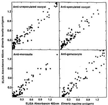
• • 1 • •1 * • * ' ^ * • Anti-sporuiated oocyst • • Anti-gametocyte • * 0.9 0.6 03 0.3 0.6 0.9 1.2 0.3 0.6 0.9 1.2 EUSA Absorbance 405nm Eimeria maxima antigensFig. 1. ELISA cross-reactivity of sera, from hatchlings of hens infected with Eimeria maxima, with antigen extracts of Eimeria tenella.
oocysts of E. tenella was also reduced: oocyst excretion by the progeny of infected hens was 43 % less than that of control chicks. Similar results were obtained in Experiment 2b. Thus, oocyst excretion by the progeny of infected hens was 68 % (2 birds out of 24 did not excrete oocysts) and 41 % reduced for challenge with E. maxima or E. tenella, re-spectively. In all the above cases, oocyst excretion was significantly lower in the progeny of infected hens
(P < 0-05, Wilcoxon test statistic).
Parasite-specific IgG antibodies in yolks of hens
infected with Eimeria maxima
Antibodies of the IgG isotype were readily detected, by ELISA, in the yolk of eggs from hens infected with E. maxima (Table 1 and Fig. 1). In all three of the protection trial experiments, the yolks of the eggs collected from the infected hens (Table 1) had significantly higher levels of £Ymma-specific anti-body, for all the developmental stages tested, than yolks of the untreated hens (P<0-05, one-way ANOVA, Student's t-test). Furthermore, yolk IgG reactivity to the E. maxima developmental stage extracts was very strongly correlated with reactivity to the corresponding E. tenella developmental stage extracts (Fig. 1; rs > 09, P < 00001), with lines of
best fit very close to linear (j> = 091x-|-0-10 for unsporulated oocysts; y = 0-82*+ 0-09 for sporu-lated oocysts; y = 0-92x — 0-01 for merozoites;
y = 0-96x + 0-03 for gametocytes).
Western blot analysis of protective sera
Sera were sampled from the chicks used in Ex-periment 2 on the day they were challenged with
A kDa — 205 — — 116 — — 97 — — 66 — — 45 — 8
Fig. 2. Western blot cross-reactivity of sera from hatchlings of hens infected with Eimeria maxima with antigen extracts of Eimeria tenella. (Sera from control chicks were non-reactive and the blots are not reproduced here.) (A) Sera pool from 3-day-old chicks from Experiment 2a; eggs collected 28-37 days after infection of the hens; maternally-derived protection versus E. maxima = 82%, protection versus E. tenella = 43%. (B) Sera pool from 3-day-old chicks from Experiment 2b; eggs collected 114—123 days after infection of the hens; maternally-derived protection versus E. maxima = 68 %, protection versus E. tenella = 41 %. Lane 1: E. tenella gametocyte extract; Lane 2: E. maxima gametocyte extract; Lane 3: E. tenella merozoite extract; Lane 4: E. maxima merozoite extract; Lane 5:
E. tenella sporulated oocyst extract; Lane 6: E. maxima sporulated oocyst extract; Lane 7; E. tenella unsporulated
oocyst extract; Lane 8: E. maxima unsporulated oocyst extract.
Table 2. Approximate molecular weights of prominent proteins identified by Western blotting of developmental stages of Eimeria maxima and Eimeria tenella
Unsporulated oocyst 190 160 150 110 105 77 75 40 Eimeria maxima Sporulated oocyst 230 175 _ _ Merozoite 230 175 _ _ Gametocyte 82 56 40 _ Eimeria tenella Unsporulated oocyst 210 140 85 70 Sporulated oocyst 230 95 80 _ Merozoite 230 95 80 65 Gametocyte 135 60
-E. maxima or -E. tenella (that is, at 3 days of age). T h e
sera from Experiment 2 a (hatchlings from eggs collected 28-37 days after infection of the hens) were pooled to provide a sample for Western blotting representing a group of chicks that was ultimately well protected against infection with E. maxima (82% reduction in oocyst excretion - Table 1). Similarly, the sera from Experiment 2 b (hatchlings from eggs collected 114-123 days after infection of the hens) were pooled to provide a sample for Western blotting representing a group of chicks that
was ultimately only partially protected against infection with E. maxima (68 % reduction in oocyst excretion - Table 1). Both pools recognized nu-merous proteins in all developmental stages of both parasites (Fig. 2). The pool from the well-protected hatchlings reacted somewhat more strongly than the pool from the partially protected chicks, but the same proteins were prominent. T h e approximate sizes of the more prominent proteins are sum-marized in Table 2. Sera from 3-day-old progeny of uninfected hens did not react on Western blots.
DISCUSSION
Infection of breeding hens with E. maxima causes the production of copious quantities of parasite-specific IgG antibodies which are transferred via the egg yolk to hatchlings (Smith et al. 1994). As a consequence, hatchlings are extremely well protected against infection with E. maxima, some totally, as assessed by oocyst excretion (Rose, 1972; Smith et
al. 1994). Progeny from hens infected with E. maxima are only partially protected against
challenge with E. tenella. However, the level of hatchling immunity to challenge with E. tenella (41—62 % ; Table 1), induced by infection of the hens with E. maxima, is comparable to the levels of resistance reported by Rose & Long (1971), where progeny were protected by between 43 and 73 % but only after the hens were infected on 4—5 occasions with E. tenella.
There are, in fact, a few reports of cross-resistance to Eimeria species in the chicken. For example, birds completely resistant to homologous challenge with high doses of E. maxima were also totally resistant to challenge with high doses of Eimeria brunetti as assessed by intestinal histology and faecal oocyst output (Rose, 1967 a). Likewise, chickens resistant to homologous challenge with E. brunetti were highly resistant to challenge with E. maxima. Similarly, chickens infected with Eimeria praecox and sub-sequently challenged with Eimeria acervulina were relatively resistant to the heterologous species (Gore & Long, 1982). The converse experiment, in which birds were first infected with E. acervulina before being challenged with E. praecox also demonstrated the existence of cross-species immunity. There is also evidence of cross-resistance between E. tenella and Eimeria necatrix (Rose, 19676). However, as pointed out by Rose (1987), these reports have not gained wide acceptance as being evidence for the existence of cross-species resistance because the results of the E. maxima/E. brunetti study were not reproducible (Hein, 1971) and the cross-protection observed in the other studies was relatively low, limited to certain developmental stages and ap-parently infective-site dependent. On the other hand, there are some examples where immunization with antigens derived from one species of Eimeria have induced a degree of cross-resistance (Danforth, Augustine & Jenkins, 1993).
The results of this study not only demonstrate the existence of cross-species resistance to E. tenella as a result of infection with E. maxima, but also provide an explanation for this phenomenon: ELISA and Western blot analyses reveal a large degree of cross-reaction of E. maxima and E. tenella antigens by yolk and hatchling sera resulting from infection of hens with E. maxima. Thus, ELISA absorbance values for all the developmental stages of the two parasites exhibit an almost perfect linear relationship. Western
blotting appears to confirm that E. maxima and
E. tenella share antigenic epitopes. There are several
proteins recognized in all stages of development in both the Eimeria species. However, the size of these proteins is generally different in the two. A notable exception to this is the 230 kDa protein band found in sporulated oocysts and merozoites of both species. This finding indicates that further investigation of this protein(s) as a potential candidate for inclusion in a maternally-based vaccine against coccidiosis, may be warranted.
In conclusion, the results presented here indicate that IgG antibody-mediated cross-resistance to different Eimeria species can occur. Furthermore, this resistance may be the result of cross-recognition of conserved epitopes or proteins in different species. The implications of these results for vaccination against coccidiosis are enormous as they suggest that a limited number of highly conserved antigens administered as a maternally-based vaccine may protect broiler chicks against a variety of Eimeria species.
The author would like to thank Roland Liechti for his care of the chickens and Marianne Kerkoc for technical assistance. This work was supported by the Bundesamt fur Bildung und Wissenschaft, Bern (Project: Coccidiosis, COST 89), the Roche Foundation and the Swiss National Science Foundation (Project Nos. 33757.92 and 31-35656.92).
R E F E R E N C E S
DANFORTH, H. D., AUGUSTINE, P. C. & JENKINS, M. C.
(1993). A review of progress in coccidial vaccine development. In Proceedings of the Vlth International
Coccidiosis Conference, (ed. Barta, J. R. & Fernando,
M. A.), pp. 49-60, Guelph, Canada: Moffitt Print.
GILBERT, J. M., BHANUSHALI, J. K. & McDOUGALD, L. R.
(1988). An enzyme-linked immunosorbent assay for coccidiosis in chickens: correlation of antibody levels with prior exposure to coccidia in the laboratory and in the field. Avian Diseases 32, 688-94.
GORE, T. c. & LONG, p. L. (1982). The biology and
pathogenicity of a recent field isolate of Eimeria
praecox Johnson, 1930. Journal of Protozoology 29,
82-5.
HEIN, H. (1971). Eimeria brunetti: cross infections in chickens immunized to E. maxima. Experimental
Parasitology 29, 367-74.
LOSCH, U., SCHRANNER, I., WANKE, R. & JURGENS, L.
(1986). The chicken egg, an antibody source. Journal
of Veterinary Medicine B33, 609-19.
ROSE, M. E. (1967a). Immunity to Eimeria brunetti and
Eimeria maxima infections in the fowl. Parasitology
57, 363-70.
ROSE, M. E. (19676). Immunity to Eimeria tenella and
Eimeria necatrix infections in the fowl. I. Influence of
the site of infection and the stage of the parasite. II. Cross-protection. Parasitology 57, 567-83.
ROSE, M. E. (1972). Immunity to coccidiosis: maternal transfer in Eimeria maxima infections. Parasitology 65, 273-82.
ROSE, M. E. (1987). Eimeria, Isospora and
Cryptosporidium. In Immunology, Immunopathology and Immunoprophylaxis of Parasitic Infections, vol. 3, Protozoa and Arthropods (ed. Soulsby, E. J. L.), pp.
275-312, Boca Raton, Florida, USA: CRC Press Inc. ROSE, M. E. & LONG, P. L. (1971). Immunity to
coccidiosis: protective effects of transferred serum and cells investigated in chick embryos infected with
Eimeria tenella. Parasitology 63, 299-313.
SMITH, N. C , WALLACH, M., MILLER, C. M. D., MORGENSTERN, R., BRAUN, R. & ECKERT, J. ( 1 9 9 4 ) .
Maternal transmission of immunity to Eimeria
maxima: ELISA analysis of protective antibodies
induced by infection. Infection and Immunity 62, 1348-57.
WAGENBACH, G. E., CHALLEY, J. R. & BURNS, W . C. (1966).
A method for purifying coccidian oocysts employing
chlorox and sulfuric acid-dichromate solution. Journal
of Parasitology 52, 1222.
WAKELIN, D. & ROSE, M. E. (1990). Immunity to
coccidiosis. In Coccidiosis of Man and Domestic
Animals, (ed. Long, P. L.), pp. 281-306, Boca Raton,
Florida, USA: CRC Press.
WALLACH, M., MENCHER, D., YARUS, S., HALABI, A. &
PUGATSCH, T. (1989). Eimeria maxima: identification of
gametocyte protein antigens and their possible role in protective immunity. Experimental Parasitology 68, 49-56.
XIE, M., GILBERT, J. M. & McDOUGALD, L. R. ( 1 9 9 2 ) .
Electrophoretic and immunological characterization of proteins of merozoites of Eimeria acervulina, E.
maxima, E. necatrix and E. tenella. Journal of Parasitology 78, 82-6.