EMERGENCY RADIOLOGY
Acute appendicitis: prospective evaluation of a diagnostic
algorithm integrating ultrasound and low-dose CT to reduce
the need of standard CT
Pierre-Alexandre Poletti&Alexandra Platon&Thomas De Perrot&Francois Sarasin& Elisabeth Andereggen&Olivier Rutschmann&Elise Dupuis-Lozeron&
Thomas Perneger&Pascal Gervaz&Christoph D. Becker
Received: 21 March 2011 / Revised: 25 May 2011 / Accepted: 13 June 2011 / Published online: 30 July 2011 # European Society of Radiology 2011
Abstract
Objectives To evaluate an algorithm integrating ultrasound and low-dose unenhanced CT with oral contrast medium (LDCT) in the assessment of acute appendicitis, to reduce the need of conventional CT.
Methods Ultrasound was performed upon admission in 183 consecutive adult patients (111 women, 72 men, mean age 32) with suspicion of acute appendicitis and a BMI between 18.5 and 30 (step 1). No further examination was recom-mended when ultrasound was positive for appendicitis, negative with low clinical suspicion, or demonstrated an alternative diagnosis. All other patients underwent LDCT (30 mAs) (step 2). Standard intravenously enhanced CT (180 mAs) was performed after indeterminate LDCT (step 3). Results No further imaging was recommended after ultrasound in 84 (46%) patients; LDCT was obtained in 99 (54%). LDCT was positive or negative for appendicitis in 81 (82%) of these 99 patients, indeterminate in 18 (18%) who underwent standard
CT. Eighty-six (47%) of the 183 patients had a surgically proven appendicitis. The sensitivity and specificity of the algorithm were 98.8% and 96.9%.
Conclusions The proposed algorithm achieved high sensitivity and specificity for detection of acute appendicitis, while reducing the need for standard CT and thus limiting exposition to radiation and to intravenous contrast media.
Keywords Appendicitis . Computed tomography . Sonography . Radiation dose . Emergency medicine
Introduction
Acute appendicitis is one of the most common acute abdominal disorders that require surgery. However, no standardized clinical and/or radiological guidelines have been validated for the identification of patients with appendicitis [1]. Computed tomography (CT) is recognized as the most accurate imaging method for the detection of acute appendicitis or alternative diagnoses in patients with right lower quadrant pain, and is often recommended as the primary imaging tool to be used in emergency settings [2– 6]. However, since appendicitis frequently affects young patients, systematic exposure to the large radiation dose delivered by a standard CT is controversial [7–9]. To obviate this problem, many authors advocate the use of ultrasound for the initial screening of right lower quadrant pain, especially in women in whom gynaecological conditions mimicking appendicitis may be detected by ultrasound [9–16]. However, the exact value of ultrasound in the assessment of patients with right lower quadrant pain is still debated: some authors reported ultrasound to be as specific as i.v. contrast enhanced CT in this setting, but limited in its sensitivity [6,10,12,15,17]. Others reported
P.-A. Poletti (*)
:
A. Platon:
T. De Perrot:
C. D. Becker Department of Radiology, University Hospital of Geneva, 4 rue Gabrielle Perret-Gentil,1211 Genève-14, Switzerland
e-mail: [email protected]
P.-A. Poletti
:
A. Platon:
F. Sarasin:
E. Andereggen:
O. RutschmannEmergency Center, University Hospital of Geneva, Geneva, Switzerland
E. Dupuis-Lozeron
:
T. Perneger Division of Clinical Epidemiology, University Hospital of Geneva, Geneva, SwitzerlandE. Andereggen
:
P. GervazDepartment of Surgery, University Hospital of Geneva, Geneva, Switzerland
that ultrasound achieves similar results to unenhanced CT, but is mainly restricted by a large number of indeterminate cases [18].
Recently, it has been suggested that a low-dose CT protocol (LDCT), delivering a dose of radiation close to that of an abdominal plain film, without intravenous (i.v.) contrast media, may be as accurate as standard CT for the diagnosis of appendicitis [19–21]. However, LDCT protocols have not been evaluated prospectively in routine practice.
The first objective of the current prospective study was to evaluate the diagnostic value of an algorithm integrating ultrasound and LDCT for the screening of patients admitted with right lower quadrant pain; the second objective was to determine to what extent this algorithm may reduce the need for standard CT and thus minimize the dose of radiation delivered to this population.
Materials and methods
A three step imaging diagnostic algorithm which integrated ultrasound (step 1), LDCT (step 2) and standard CT (step 3) was set-up for adult patients (>18 year-old) admitted with suspicion of appendicitis (Fig. 1). Pregnant women were excluded, as were patients with a BMI lower than 18.5 and higher than 30, because ultrasound and LDCT have limited performance in overweight patients [22,23] and in patients with a low BMI [20].
This study was approved by the research ethics committee of our institution (IRB 09-021R) which did not require informed consent, because the value of LDCT has already been evaluated for the diagnosis of appendicitis
[19–21] and also because the algorithm was adopted to reduce the mean radiation dose delivered to patients.
All consecutive patients with a suspicion of acute appendicitis, admitted during a 6 months period of time (December 2008 to May 2009) in our emergency center were included in the study. The clinician had first to indicate the patient’s BMI and rate the degree of clinical suspicion of appendicitis as high, moderate or low. Clinical suspicion was considered high in the presence of at least 4 of the 5 following criteria, usually recognized as common signs and symptoms of appendicitis [24]: 1) fever, 2) a suggestive history (migration of the pain from the periombilical region to the right lower fossa), 3) a typical abdominal examination (rebound, tenderness, guarding, Rosving’s sign, psoas sign) 4) a elevated leucocyte count and 5) a urinalysis not suggestive of urinary tract disease (less than 30 erythrocytes and 20 leukocyte cells per high-power field). The clinical suspicion was considered mild when two or three of these criteria were not typical for appendicitis, and low when four or five were not typical.
Diagnostic algorithm Step 1: Ultrasound
Ultrasound examinations were performed by the radiology resident on call with a prior training of at least 6 months in general ultrasound. Immediately after completion of the examination, the sonologist had to fill out a standardized form in the patient’s electronic file, indicating the presence or absence of ultrasound patterns, Sonography Standard CT Positive Appendicitis No further imaging* No appendicitis Indeterminate Negative Low clinical suspicion Alternative diagnosis Negative
High / moderate clinical suspicion
Indeterminate
LDCT
Surgery No further imaging*
Fig. 1 Imaging algorithm for the diagnosis of appendicitis in adult patients with BMI between 18.5 and 30. * For the diagnosis of appendicitis only
ered as direct or indirect signs of appendicitis (Table 1). An enlarged (≥7 mm) incompressible appendix was necessary to consider ultrasound positive for appendicitis. Ultrasound could only be considered negative in the absence of any direct or indirect sign of appendicitis. All other situation was considered indeterminate. No further imaging was recommended after a positive ultrasound for appendicitis (considered as a conclusive ultra-sound). Further imaging was also not recommen-ded in the following situations, when ultrasound was considered conclusive:
1) negative ultrasound with low clinical suspicion of appendicitis.
2) negative or indeterminate ultrasound for appendicitis, with depiction of an alternative diagnosis, which could explain the clinical presentation.
All other patients were referred to LDCT. Step 2: LDCT
LDCT was immediately interpreted, while the patient was still on the CT table, by one of the senior radiology residents of the emergency radiology unit, with a minimal background of at least 400 abdominal CTs and of 15 LDCTs under supervision. Based on the LDCT signs (Table1), the radiologist had to decide whether LDCT was positive (Fig.2), negative (Fig.3), or indeterminate for appendicitis. Radiologists had to make a diagnosis of appendicitis or absence of appendicitis, based on their own evaluation of the LDCT [20]. The following signs were considered highly suggestive for appendicitis: enlarged appendix (≥7 mm), periappendiceal fat stranding and arrowhead sign [25]. Appendicitis was excluded
when gas or contrast media was depicted in the appendiceal lumen and/or in the absence of any aforementioned signs of appendicitis.
A positive or negative LDCT for appendicitis was considered conclusive. LDCT was considered indeterminate when the appendix could not be identified.
Step 3: standard CT
When LDCT was reported indeterminate a standard CT was immediately obtained; otherwise, the patient was discharged from the CT facility.
As long as the current algorithm was under evaluation, it was accepted that a standard CT could be obtained after a conclusive ultrasound or LDCT examination. However, only an attending surgeon was authorized to modify the guideline. Additional standard CTs required in this setting were recorded separately, but not considered as part of the algorithm for appendicitis.
Technical imaging parameters
Ultrasound was performed transabdominally (no endovaginal examination) using a Philips IU 22 device (Philips Healthcare, Bothell, WA) with a sectorial 3.5–5 MHz probe for the assessment of intraperitoneal and retroperitoneal structures, and with a 12 mHz linear probe, for the assessment of the right lower quadrant,
LDCT and standard CT were performed using a 16-row Philips MX 8000 (Philips Medical Systems, Best, The Netherlands), from lung bases to pelvis. Four hundred mL of oral contrast material (Telebrix-Gastro, Guerbet, Zurich, Switzerland), were administrated to every patient 60 min before CT, without rectal contrast medium.
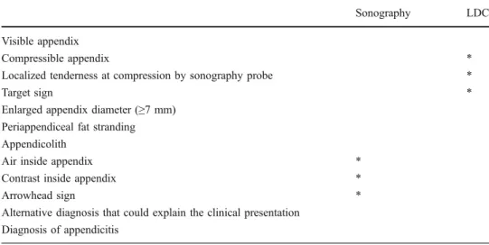
Sonography LDCT Visible appendix
Compressible appendix *
Localized tenderness at compression by sonography probe *
Target sign *
Enlarged appendix diameter (≥7 mm) Periappendiceal fat stranding Appendicolith
Air inside appendix *
Contrast inside appendix *
Arrowhead sign *
Alternative diagnosis that could explain the clinical presentation Diagnosis of appendicitis
Table 1 Sonography and LDCT signs used to assess patients with clinical suspicion of appendicitis
Evaluation form filled electroni-cally by the radiologist after com-pletion of sonography and LDCT examinations respectively. This form, including the overall appre-ciation for the presence or absence of appendicitis, was transmitted to the emergency physician. * Not applicable for this imaging method
LDCT was performed using 16×1.5 mm collimation, pitch 1.25, gantry rotation period 0.5 s, tube potential 120 kV, tube charge per gantry rotation 30 mAs, reconstruction slice thickness 3.0 mm.
Standard CT was performed using a power-injected bolus of 120 mL non-ionic intravenous contrast material (iohexol, AccupaqueR300, GE Healthcare AG, Opfikon, Switzerland),
16×1.5 mm collimation, pitch 1.35, gantry rotation period 0.5 s, tube potential 120 kV, tube charge per gantry rotation 180 mAs, reconstruction slice thickness 3.0 mm.
Effective dose calculation
The dose delivered by LDCT was estimated using the ImPACT CT patient dosimetry calculator [20, 26]. The effective dose of radiation (E) delivered by LDCT was:
E (women)=1.7±0.2 mSv and E (men)=1.2±0.1 mSv [26] The effective dose of radiation delivered by standard CT was: E (women)=10.2±1.2 mSv and E (men)=7.2±0.6 mSv
The mean dose of radiation delivered by the algorithm corresponds to the total dose delivered by LDCTs and standard CTs to women and men respectively divided by the total number of patients.
Definite diagnosis and follow-up
A definite diagnosis (reference standard) was obtained for every patient, based on the surgical findings and on the discharge summary. In patients who were managed non-operatively, a phone call to them or their family physician was performed 6 to 8 weeks after discharge.
Statistical analysis
The sensitivity and specificity were computed for each step of the diagnostic algorithm, using the surgical and the clinical follow-up as reference standard. The 95% percent confidence intervals were estimated with the Clopper-Pearson method. For the first two levels of the algorithm, indeterminate cases were considered as false negative test results for the computation of sensitivity and as false positive test results for the computation of specificity. This calculation will result in conservative estimates and will thus compromise both sensitivity and specificity. Positive predictive values (PPV) and negative predictive values (NPV) were also calculated; these analyses exclude naturally the indeterminate results. Finally we obtained positive and negative likelihood ratios (LR), well adapted for interpretation and use of non-dichotomous diagnostic test results [27]. LR ratios of 5 or more (or, equivalently, 1/5 or less) are considered to be clinically useful [27,28]. The mean dose of radiation actually delivered to our patients was compared (using Mann-Whitney test) to the mean dose that they would have received if standard CT were systematically performed; the significance level was fixed to 5%. All statistical calculations were performed by an independant center of clinical epidemiology, using R for Windows (Version 2.11.1).
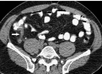
Fig. 3 31 year-old woman with extended colitis. She was admitted with right lower quadrant and periumbilical pain, slight fever and mild leukocytosis. Sonography (not shown) was considered indeterminate for appendicitis. LDCT axial image at the level of the pelvis shows an appendix of normal diameter, filled with oral contrast and without stranding of the adjacent fat (arrow). The diagnosis of pancolitis was assessed by the extended thickening of the large bowel (arrowheads). No further imaging was obtained. Patient was successfully treated by antibiotherapy after stool analysis revealed a bacterial origin of colitis Fig. 2 45 year-old man with acute appendicitis. Patient was admitted with right lower quadrant pain, no fever, vomiting, diarrhoea and leukocytosis. Sonography (not shown) was reported indeterminate for appendicitis and normal for the rest of the abdominal examination. Axial LDCT image at the level of the cecum (C) shows an enlarged appendix, without intraluminal contrast opacification (arrow) and a mild infiltration of the surrounding fat (asterisk), signs consistent with the diagnosis of appendicitis, which was confirmed at surgery
Results
Study population
A total of 213 consecutive patients with a right lower quadrant pain suggestive of appendicitis were admitted in our institution during the study period. Thirty (14%) met exclusion criteria: 4 (2%) women with a positive pregnancy test, 17 (8%) patients with a BMI>30, and 9 (4%) patients with BMI<18.5. The study population consisted of 183 patients, 111 (61%) women, 72 (39%) men, with a mean age of 32 years [median 30]. One hundred sixty-five (90%) patients were under 50 years of age. The clinical suspicion of appendicitis at outset was reported low in 30 (16%) patients, high/moderate in 153 (84%). A diagnosis of appendicitis was eventually confirmed at surgery in 86 (47%) patients (47 men, 39 women).
Algorithm diagnostic performance
Results obtained by each imaging method for the diagnostic work-up of right lower quadrant pain, with regard to the reference standard, are reported in Table2. According to the algorithm, 84 (46%) ultrasound examinations were considered conclusive (after exclusion from calculation of 8 negative examinations with a high/moderate clinical suspicion of appendicitis).
LDCT was performed in 99 patients in whom ultrasound was non conclusive (91 indeterminate plus 8 negative with high/moderate clinical suspicion). Eighty-one (82%) of them were conclusive (27 positive, 54 negative). Eigh-teen (18%) patients with indeterminate LDCT underwent standard CT.
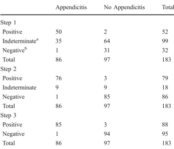
The distribution of the 183 patients, at each step of the algorithm, for the diagnosis of appendicitis with regard to the reference standards is reported in Table 3. Negative ultrasound examinations in patients with high/ moderate clinical suspicion of appendicitis (n=8) were considered indeterminate at Step 1, leading to a total number of 99 indeterminate examinations. In this section, negative cases for appendicitis (n=32) correspond to patients with a negative ultrasound examination, plus those with an alternative diagnosis. The distribution of the 183 patients after LDCT and CT were performed is reported at the“Step 2” and “Step 3” sections respectively.
The sensitivity, specificity, PPVand NPV, as well as the LR, were inferred from Table 3, and reported in Table 4. Ultrasound (step 1) achieved low sensitivity and specificity (58.1% and 31.9% respectively). These relatively low values
Appendicitis No Appendicitis Total Sonography
Positive 50 2 52
Indeterminate 32 59 91
Negative, high/ moderate clinical suspicion 3 5 8 Negative, low clinical suspicion 0 12 12
Alternative diagnosis 1 19 20
Total 86 97 183
LDCT among patients with indeterminate sonography or negative sonography with high suspicion
Positive 26 1 27
Indeterminate 9 9 18
Negative 0 54 54
Total 35 64 99
CT among patients with indeterminate LDCT
Positive 9 0 9
Negative 0 9 9
Total 9 9 18
Table 2 Results obtained by each imaging technique of the proposed algorithm for the diagnostic work-up of right-lower quadrant pain with regard to the reference standards (surgery and follow-up)
Numbers are related to a given diagnostic technique, independently to the others.
were due to the high proportion of indeterminate results which penalized the sensitivity and specificity. However, when a diagnosis was achieved by ultrasound, the results were excellent, with predictive values greater than 95%. Both LR reflected strong evidence, since they were more extreme than 25 or 1/25 (0.036 corresponds to 1/27.8). The combination of ultrasound and LDCT (step 2) led to an improvement of the sensibility and specificity (88.4% and 87.6% respectively). The proportion of indeterminate results fell to 9.8% (18/183), and the predictive values remained excellent, as did LR. Inclusion of standard CT (step 3) further improved sensitivity and specificity (98.8% and 96.9%), completely eliminated indeterminate results, kept predictive values at their very high level, and further strengthened LR.
Four (2.1%) of the 183 patients were misclassified for the diagnosis of appendicitis by the three steps algorithm:
1) A right Fallopian tube inflammation was reported at ultrasound; an appendicitis with secondary involvement of the Fallopian tube was found at surgery.
2) Appendicitis was reported at ultrasound; a complicated acute salpingo-oophoritis, with indirect periappendicitis, was found at surgery.
3) Appendicitis was reported at ultrasound; a necrotizing epiploic appendagitis was found at surgery.
4) LDCT was reported positive for appendicitis; patient was discharged home after complete resolution of her clinical symptoms without further imaging or surgery; her
follow-up was uneventful. In spite of the fact that this case might correspond to a spontaneously resolving appendicitis [29], it has been considered as a false positive LDCT. Ninety-seven (53%) of the 183 patients did not have appendicitis. Fifty-three were discharged home with a diagnosis of non-specific abdominal pain. The following alternative diagnoses were eventually considered in the 44 remaining patients: renal colic (n=9), colitis (n=9), pelvic inflammatory disease (n=5), ovarian cyst (n=5, Fig.4), primary mesenteric lymphadenitis (n=3), endometriosis (n=2), diverticulitis (n=2), small bowel occlusion (n=2), terminal ileitis (n=2), cholecystitis (n=2), primary dysmenorrhea (n=2), dermoid cyst (n=1).
Standard CTs performed outside protocol
Fourteen standard CTs were requested in 9 women and 5 men by an attending surgeon to confirm the presence or absence of appendicitis after a conclusive ultrasound or LDCT examination. These CTs were not recommended by the algorithm. None of these examinations modified the prior diagnosis or added clinically useful information.
Twelve standard CTs were requested by the clinicians to further investigate an alternative diagnosis reported at ultrasound and/or at LDCT, in 10 women and 2 men. No appendicitis was shown on any of these CTs.
Dose of radiation
If the algorithm were strictly followed, our patients would have received a mean dose of radiation of 1.73 mSv: 33 LDCTs (1.2 mSv) in men, 66 LDCTs (1.7 mSv) in women, 6 standard CTs (7.2 mSv) in men, 12 standard CTs (10.2 mSv) in women / 183. However, because of out-of-protocol standard CTs, they actually received a mean dose of 3.23 mSv. This mean dose was significantly lower (p<.0001) than the hypothetical mean dose that they would have received if a standard CT had been systematically performed at admission (9.02 mSv).
Table 3 Distribution of the 183 patients included in the series after completion of each step of the proposed algorithm for the diagnosis of appendicitis, with regard to the reference standards (surgery and follow-up)
Appendicitis No Appendicitis Total Step 1 Positive 50 2 52 Indeterminatea 35 64 99 Negativeb 1 31 32 Total 86 97 183 Step 2 Positive 76 3 79 Indeterminate 9 9 18 Negative 1 85 86 Total 86 97 183 Step 3 Positive 85 3 88 Negative 1 94 95 Total 86 97 183
Step 1 = distribution of the 183 patients after sonography. Step 2 = distribution after sonography and LDCT. Step 3 = distribution after sonography, LDCT and standard CT.
a
Patients with negative sonography and high/moderate clinical suspicion of appendicitis were considered as indeterminate
b
Patients with an alternative diagnosis were considered as negative for appendicitis.
Table 4 Evaluation of a three steps imaging algorithm for the detection of acute appendicitis in 183 patients admitted with right lower quadrant pain
Step 1 Step 2 Step 3
Sensitivity 58.1 (47.0–68.7) 88.4 (79.7–94.3) 98.8 (93.7–99.9) Specificity 31.9 (22.8–42.2) 87.6 (79.4–93.4) 96.9 (91.2–99.4) Positive predictive value 96.1 (86.8–99.5) 96.2 (89.3–99.2) 96.6 (90.4–99.3) Negative predictive value 96.9 (83.8–99.9) 98.8 (93.7–99.9) 98.9 (94.3–99.9) Likelihood ratio + 28.2 (7.1–112.4) 28.5 (9.4–87.3) 32 (10.5–97.4) Likelihood ratio - 0.036 (0.005–0.26) 0.013 (0.002–0.09) 0.01 (0.002–0.08) Step 1 = sonography. Step 2 = combination of sonography and LDCT. Step 3 = combination of sonography, LDCT and standard CT. First number in each box corresponds to estimate. 95% confidence intervals are given in parentheses.
Discussion
In the current study, we evaluated a three step diagnostic algorithm for the assessment of acute appendicitis, integrating ultrasound, LDCT and standard CT as complementary imaging techniques, in patients with a BMI between 18.5 and 30. This algorithm was 98.8% sensitive and 96.9% specific for the diagnosis of appendicitis, which is in the high range of what has been recently reported for standard CT [2, 30–32]. Hence, for a similar result as obtained with a systematic use of standard CT at admission, a large majority of our patients were investigated with ultrasound and LDCT, or with ultrasound only, thus avoiding exposure to high radiation dose and to i.v. contrast media.
In the current series, ultrasound achieved a lower sensitiv-ity (58.1%) and specificsensitiv-ity (31.9%) than reported in prior series [14,17,18,33,34]. These values are explained by the high proportion of ultrasound examinations reported as indeterminate (or negative in patients with high/ moderate clinical suspicion of appendicitis). Thus, sensitivity and specificity were penalized by indeterminate results, not by false negative or false positive examinations. The high prevalence of indeterminate results is consistent with prior series [18]. Other studies, including a meta-analysis, reported that, in spite of a high specificity, ultrasound may also be limited by a low sensitivity for appendicitis [17,33].
Ultrasound achieved high PPV and NPV, as well as excellent LR, and therefore constitutes a valuable screening
test to reduce the number of patients addressed to CT facility [27]. In the current study, ultrasound was conclusive in 86 (46%) of the 183 patients.
LDCT was the second diagnostic step of the algorithm. The combination of LDCT with ultrasound achieved high PPV and NPV (96.2% and 98.8% respectively) for the diagnosis of appendicitis. Some prior series already suggested that unenhanced LDCT may be used in this setting with an accuracy close to that of unenhanced standard CT [19], or even close to that of i.v. enhanced CT [20, 21]. However, these analyses were based on retrospective review of LDCTs. To our knowledge, no prior series evaluated LDCT prospectively to assess the diagnosis of appendicitis in a routine clinical practice, without systematically performing additional standard CT. Besides, only one series considered the possibility to report LDCT as inconclusive [21], as in our algorithm. Under these conditions, Seo et al hypothesized that a standard CT may be required in 8.2% to 10.2% of patients. This is consistent with our findings, since standard CT was recommended by our algorithm in 9.8% (18/183) of patients, when both ultrasound and LDCT were non conclusive for appendicitis. However, Seo et al did not perform ultrasound as an initial imaging technique, and we did not systematically obtain LDCT in our patients.
Two factors may explain the high percentage (18%) of indeterminate LDCTs in our series. First, it is possible that the most typical cases of appendicitis were already identified by ultrasound, and that LDCT had therefore to deal with more subtle cases (i.e. appendicitis at very early stage). Second, the diagnosis at LDCT had to be done in a short time period, while patients were still on the CT table, so that standard CT could be performed immediately if required. These conditions constitute probably the main difference between prospective and retrospective LDCT analysis. Further prospective series will be necessary to evaluate strategies for reducing the number of indetermi-nate LDCTs, in real condition of work, without affecting the diagnostic value of this imaging technique.
In this study, 24% (44/183) of patients had an alternative diagnosis, while 29% (53/183) were considered to have non-specific abdominal pain. This is in the range of previous series, reporting 21% to 37% of alternative diagnoses [19, 21, 35]. Our data show that standard CTs were required in 7% (12/183) of our patients to further investigate an alternative diagnosis suggested at ultrasound or at LDCT. This observation reveals that, in real condition of use, a certain amount of standard CTs must still be obtained after appendicitis have been excluded, which cannot be inferred from retrospective series [7, 21]. This may be considered a limitation of the algorithm, when compared to immediately performing a standard CT at admission. However, many authors advocate the use of
Fig. 4 32 year-old woman with complicated right adnexal cyst. Patient was admitted with right lower quadrant pain, no fever, and slightly elevated white cell count. Oblique sagittal sonographic image obtained with a 5 MHz linear probe at the level of the right ovary, adjacent to the uterus (U), shows a round well delineated hypoechoic collection with a hyperechoic sediment (arrow), surrounded by free peritoneal fluid (arrowhead). Patient was referred to gynaecological ward with the diagnosis of complicated adnexal cyst, without need of further CT imaging. Follow-up was uneventful after symptomatic treatment
standard CT without i.v. contrast agents for the initial screening of patients with right lower quadrant pain [19, 36]. When compared to the latter protocol, our algorithm has probably the advantage to reduce the preliminary dose of radiation to those patients in whom further examination with i.v. contrast media will be required. In the current series, the mean dose of radiation delivered to each patient was 3.23 mSv, including standard CTs performed outside the algorithm. This corresponds to an almost 3 fold dose reduction when compared to the systematic use of a standard CT, for a similar diagnostic value. These observations substantiate the conclusions of a recent multicenter series by Lameris et al. [37]. In their survey on 1021 patients with acute abdominal pain, the authors suggested that performing ultrasound first and limiting the use of CT to these patients with a negative or inconclusive ultrasound, would constitute the best imaging strategy to reduce exposure to radiations while maintaining a high diagnostic sensitivity.
Some other limitations of our algorithm must be discussed. First, this algorithm is limited to patients with BMI between 18.5 and 30. In our series, only 12% of patients did not meet these conditions. However, this percentage may be more important in other populations. Second, our study did not evaluate to what extent the combination of ultrasound and CT imaging might prolong patients’ stay time in the emergency department and thus increase their risk of appendiceal perforation [38]. Third, the cost-effectiveness of the algorithm cannot be inferred from the current data. Finally, the rationale for using ultrasound and LDCT for the screening of appendicitis after 50 of age is debatable, because this population (9.8% in the current series) is less vulnerable to the deleterious effects of radiation [8,39].
In conclusion, this prospective study shows that ultrasound and LDCT can be integrated in the imaging management of adult patients with right lower quadrant pain, to reduce the number of standard CTs without compromising on the diagnostic quality. Further prospective series are still mandated to evaluate the actual impact of this algorithm on the patient’s work-flow in the emergency department, its value on elderly population, and its cost effectiveness.
Acknowledgement This paper was supported by the grant for Research and Development of the University Hospital of Geneva (PRD-03-II-15).
References
1. Ralls PW, Balfe DM, Bree RL et al (2000) Evaluation of acute right lower quadrant pain. American College of Radiology. ACR Appropriateness Criteria. Radiology 215(Suppl):159–166 2. Nathan RO, Blackmore CC, Jarvik JG (2008) Therapeutic impact
of CT of the appendix in a community hospital emergency department. AJR Am J Roentgenol 191:1102–1106
3. Novelline RA, Rhea JT, Rao PM, Stuk JL (1999) Helical CT in emergency radiology. Radiology 213:321–339
4. Kamel IR, Goldberg SN, Keogan MT, Rosen MP, Raptopoulos V (2000) Right lower quadrant pain and suspected appendicitis: nonfocused appendiceal CT–review of 100 cases. Radiology 217:159–163
5. Morse BC, Roettger RH, Kalbaugh CA, Blackhurst DW, Hines WB Jr (2007) Abdominal CT scanning in reproductive-age women with right lower quadrant abdominal pain: does its use reduce negative appendectomy rates and healthcare costs? Am Surg 73:580–584, discussion 584
6. Wise SW, Labuski MR, Kasales CJ et al (2001) Comparative assessment of CT and sonographic techniques for appendiceal imaging. AJR Am J Roentgenol 176:933–941
7. Paulson EK, Coursey CA (2009) CT protocols for acute appendicitis: time for change. AJR Am J Roentgenol 193:1268–1271
8. Brenner DJ, Hall EJ (2007) Computed tomography–an increasing source of radiation exposure. N Engl J Med 357:2277–2284 9. Brenner DJ (2010) Slowing the increase in the population dose
resulting from CT scans. Radiat Res 174:809–15
10. van Breda Vriesman AC, Kole BJ, Puylaert JB (2003) Effect of ultrasonography and optional computed tomography on the outcome of appendectomy. Eur Radiol 13:2278–2282
11. Poortman P, Oostvogel H, Lohle P, Cuesta M, de Lange-de KE, Hamming J (2009) Ultrasonography and clinical observation in women with suspected acute appendicitis: a prospective cohort study. Dig Surg 26:163–168
12. Hernanz-Schulman M (2010) CT and US in the diagnosis of appendicitis: an argument for CT. Radiology 255:3–7
13. Rao PM, Feltmate CM, Rhea JT, Schulick AH, Novelline RA (1999) Helical computed tomography in differentiating appendicitis and acute gynecologic conditions. Obstet Gynecol 93:417–421 14. Birnbaum BA, Jeffrey RB Jr (1998) CT and sonographic
evaluation of acute right lower quadrant abdominal pain. AJR Am J Roentgenol 170:361–371
15. Gaitini D, Beck-Razi N, Mor-Yosef D et al (2008) Diagnosing acute appendicitis in adults: accuracy of color Doppler sonography and MDCT compared with surgery and clinical follow-up. AJR Am J Roentgenol 190:1300–1306
16. Toorenvliet BR, Wiersma F, Bakker RF, Merkus JW, Breslau PJ, Hamming JF (2010) Routine ultrasound and limited computed tomography for the diagnosis of acute appendicitis. World J Surg 34:2278–2285
17. Balthazar EJ, Birnbaum BA, Yee J, Megibow AJ, Roshkow J, Gray C (1994) Acute appendicitis: CT and US correlation in 100 patients. Radiology 190:31–35
18. Keyzer C, Zalcman M, De Maertelaer V et al (2005) Comparison of US and unenhanced multi-detector row CT in patients suspected of having acute appendicitis. Radiology 236:527–534 19. Keyzer C, Tack D, de Maertelaer V, Bohy P, Gevenois PA, Van
Gansbeke D (2004) Acute appendicitis: comparison of low-dose and standard-dose unenhanced multi-detector row CT. Radiology 232:164–172
20. Platon A, Jlassi H, Rutschmann OT et al (2009) Evaluation of a low-dose CT protocol with oral contrast for assessment of acute appendicitis. Eur Radiol 19:446–454
21. Seo H, Lee KH, Kim HJ et al (2009) Diagnosis of acute appendicitis with sliding slab ray-sum interpretation of low-dose unenhanced CT and standard-dose i.v. contrast-enhanced CT scans. AJR Am J Roentgenol 193:96–105
22. Josephson T, Styrud J, Eriksson S (2000) Ultrasonography in acute appendicitis. Body mass index as selection factor for US examination. Acta Radiol 41:486–488
23. Hormann M, Philipp MO, Eberl H et al (2004) The effect of varying low-dose protocols on perceived image quality in multidetector CT in a rabbit model of acute appendicitis. Eur Radiol 14:1465–1471
24. Paulson EK, Kalady MF, Pappas TN (2003) Clinical practice. Suspected appendicitis. N Engl J Med 348:236–242
25. Rao PM, Wittenberg J, McDowell RK, Rhea JT, Novelline RA (1997) Appendicitis: use of arrowhead sign for diagnosis at CT. Radiology 202:363–366
26. ImPACT (2006) Imaging performance assessment of CT scanners: a medical devices agency evaluation group. London UK www. impactscan.org/ctdosimetry.htm
27. Grimes DA, Schulz KF (2005) Refining clinical diagnosis with likelihood ratios. Lancet 365:1500–1505
28. Altman DG MD, Bryant TN, Gardner MJ (2000) Statistics with confidence. BMJ Books:105–115
29. Cobben LP, de Van Otterloo AM, Puylaert JB (2000) Spontaneously resolving appendicitis: frequency and natural history in 60 patients. Radiology 215:349–352
30. Rhea JT, Halpern EF, Ptak T, Lawrason JN, Sacknoff R, Novelline RA (2005) The status of appendiceal CT in an urban medical center 5 years after its introduction: experience with 753 patients. AJR Am J Roentgenol 184:1802–1808
31. van Randen A, Bipat S, Zwinderman AH, Ubbink DT, Stoker J, Boermeester MA (2008) Acute appendicitis: meta-analysis of diagnostic performance of CT and graded compression US related to prevalence of disease. Radiology 249:97–106
32. van Randen A, Lameris W, van Es HW et al (2010) Profiles of US and CT imaging features with a high probability of appendicitis. Eur Radiol 20:1657–1666
33. Doria AS, Moineddin R, Kellenberger CJ et al (2006) US or CT for diagnosis of appendicitis in children and adults? A meta-analysis. Radiology 241:83–94
34. Gracey D, McClure MJ (2007) The impact of ultrasound in suspected acute appendicitis. Clin Radiol 62:573–578
35. Lane MJ, Liu DM, Huynh MD, Jeffrey RB Jr, Mindelzun RE, Katz DS (1999) Suspected acute appendicitis: nonenhanced helical CT in 300 consecutive patients. Radiology 213:341–346 36. Rao PM, Rhea JT, Novelline RA, Mostafavi AA, Lawrason JN,
McCabe CJ (1997) Helical CT combined with contrast material administered only through the colon for imaging of suspected appendicitis. AJR Am J Roentgenol 169:1275–1280
37. Lameris W, van Randen A, van Es HW et al (2009) Imaging strategies for detection of urgent conditions in patients with acute abdominal pain: diagnostic accuracy study. BMJ 338:b2431 38. Pritchett CV, Levinsky NC, Ha YP, Dembe AE, Steinberg SM (2010)
Management of acute appendicitis: the impact of CT scanning on the bottom line. J Am Coll Surg 210(699–705):705–697
39. Shuryak I, Sachs RK, Brenner DJ (2010) Cancer risks after radiation exposure in middle age. J Natl Cancer Inst 102:1628–1636