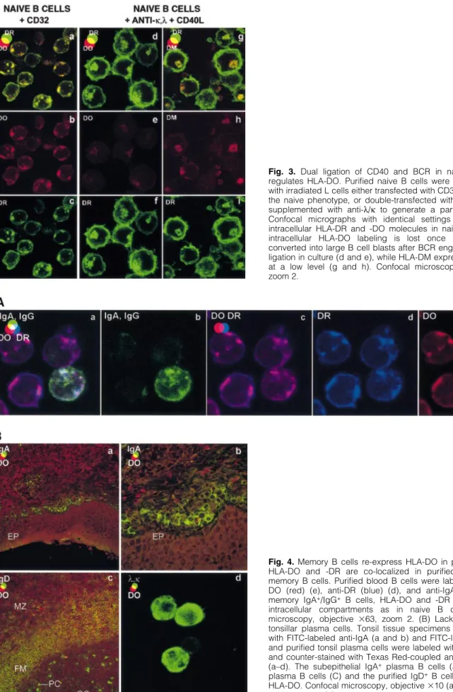
Human germinal center B cells differ from naive and memory B cells by their aggregated MHC class II‐rich compartments lacking HLA‐DO
10
0
0
Texte intégral
Figure
Documents relatifs