HAL Id: hal-02411397
https://hal.archives-ouvertes.fr/hal-02411397
Submitted on 24 Nov 2020
HAL is a multi-disciplinary open access
archive for the deposit and dissemination of
sci-entific research documents, whether they are
pub-lished or not. The documents may come from
teaching and research institutions in France or
abroad, or from public or private research centers.
L’archive ouverte pluridisciplinaire HAL, est
destinée au dépôt et à la diffusion de documents
scientifiques de niveau recherche, publiés ou non,
émanant des établissements d’enseignement et de
recherche français ou étrangers, des laboratoires
publics ou privés.
Underlying Oral Regeneration in the Sea Anemone,
Nematostella vectensis
Aldine Amiel, Hereroa Johnston, Karine Nedoncelle, Jacob Warner, Solène
Ferreira, Eric Röttinger
To cite this version:
Aldine Amiel, Hereroa Johnston, Karine Nedoncelle, Jacob Warner, Solène Ferreira, et al..
Charac-terization of Morphological and Cellular Events Underlying Oral Regeneration in the Sea Anemone,
Nematostella vectensis. International Journal of Molecular Sciences, MDPI, 2015, 16 (12),
pp.28449-28471. �10.3390/ijms161226100�. �hal-02411397�
Article
Characterization of Morphological and Cellular
Events Underlying Oral Regeneration in the Sea
Anemone, Nematostella vectensis
Aldine R. Amiel, Hereroa T. Johnston, Karine Nedoncelle, Jacob F. Warner, Solène Ferreira and Eric Röttinger *
Received: 8 September 2015; Accepted: 6 November 2015; Published: 1 December 2015 Academic Editor: Francesc Cebrià
Institute for Research on Cancer and Aging, Université de Nice Sophia-Antipolis UMR 7284, INSERM U1081, CNRS UMR 7284, Nice 06107 Cedex 02, France; aldine.amiel@unice.fr (A.R.A.); j.hereroa@hotmail.fr (H.T.J.); Karine.Nedoncelle@unice.fr (K.N.); warner.jacob@gmail.com (J.F.W.); solene.ferreira.ca@gmail.com (S.F.) * Correspondence: eric.rottinger@unice.fr; Tel.: +33-493-377-791
Abstract: Cnidarians, the extant sister group to bilateria, are well known for their impressive regenerative capacity. The sea anemone Nematostella vectensis is a well-established system for the study of development and evolution that is receiving increased attention for its regenerative capacity. Nematostella is able to regrow missing body parts within five to six days after its bisection, yet studies describing the morphological, cellular, and molecular events underlying this process are sparse and very heterogeneous in their experimental approaches. In this study, we lay down the basic framework to study oral regeneration in Nematostella vectensis. Using various imaging and staining techniques we characterize in detail the morphological, cellular, and global molecular events that define specific landmarks of this process. Furthermore, we describe in vivo assays to evaluate wound healing success and the initiation of pharynx reformation. Using our described landmarks for regeneration and in vivo assays, we analyze the effects of perturbing either transcription or cellular proliferation on the regenerative process. Interestingly, neither one of these experimental perturbations has major effects on wound closure, although they slightly delay or partially block it. We further show that while the inhibition of transcription blocks regeneration in a very early step, inhibiting cellular proliferation only affects later events such as pharynx reformation and tentacle elongation.
Keywords: regeneration; wound healing; tissue tracking; marine invertebrate; sea anemone; Nematostella vectensis
1. Introduction
Regeneration is the biological process that enables organisms to regrow missing body parts after amputation. This fascinating phenomenon has intrigued naturalists and scientists for more than 300 years. Among the first animals in which regeneration has been described was the freshwater polyp Hydra, a cnidarian that belongs to the group of hydrozoans. While regenerative potential has since then been described in other groups of cnidarians (Anthozoa [1,2], Cubozoa [3], and Scyphozoa [4]), the majority and most detailed cellular and molecular regeneration studies in this phylum have been carried out using Hydrozoa (reviewed in [5,6]). These studies highlight variations in the cellular mechanisms (e.g., cellular proliferation) underlying regeneration in different groups of cnidarians (the hydrozoan Hydra vs. the anthozoan Nematostella [7]). They also demonstrate variations within Hydrozoa (Hydra vs. Hydractinia [8]) and even within the same species; in Hydra, for example, the regenerative mechanism varies depending on the amputation site [9]. Therefore,
additional systems are required to determine whether common mechanisms govern regeneration throughout Cnidaria and to understand the evolution of this process within the animal kingdom.
The majority of cnidarian species belong to the Anthozoa class (sea anemones and corals) which diverges basally to their other cnidarian sister groups [10–12]. This places anthozoans at a key phylogenetic position to understand the evolution of the regeneration process among Cnidaria. Several anthozoans have been shown to display high regenerative capacities [1,2,13]. However, very little is known about the cellular/molecular mechanisms that underlie this process in these animals. The anthozoan sea anemone Nematostella vectensis (Nv) is a well-established system for the study of embryonic and larval development [13–18]. Interestingly, it is also able to regrow half of its oral or aboral body within five to seven days after bisection (Figure1) [12,13].
Int. J. Mol. Sci. 2015, 16, page–page
2
Therefore, additional systems are required to determine whether common mechanisms govern regeneration throughout Cnidaria and to understand the evolution of this process within the animal kingdom.
The majority of cnidarian species belong to the Anthozoa class (sea anemones and corals) which diverges basally to their other cnidarian sister groups [10–12]. This places anthozoans at a key phylogenetic position to understand the evolution of the regeneration process among Cnidaria. Several anthozoans have been shown to display high regenerative capacities [1,2,13]. However, very little is known about the cellular/molecular mechanisms that underlie this process in these animals. The anthozoan sea anemone Nematostella vectensis (Nv) is a well-established system for the study of embryonic and larval development [13–18]. Interestingly, it is also able to regrow half of its oral or aboral body within five to seven days after bisection (Figure 1) [12,13].
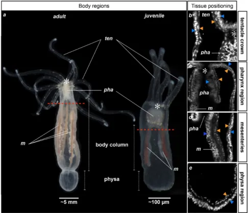
Figure 1. General anatomy of adult and juvenile Nematostella vectensis. Photographs illustrating the adult (left) and juvenile (right) morphology of Nematostella; (a) Polyps are oriented toward the oral region to the top and aboral region to the bottom. Red dotted lines indicate the future amputation site for the following experiments of this study. (b–e) Close-up confocal stack images of the tentacle (b); pharyngeal (c); mesenterial (along the body column) (d); or the physal (e) regions. Juvenile polyps were stained with DAPI to label the nuclei (white staining). An asterisk (*) indicates the position of the mouth. Orange and blue arrowheads indicate the gastrodermal and ectodermal epithelia, respectively. ten, tentacles; pha, pharynx; m, mesenteries.
Nematostella is a diploblastic animal comprised of an outer ectodermal epithelium and an inner
gastrodermis that are separated by muscle fibers and the mesoglea (Supplementary Figure S1A,B). The oral region consists of a pharynx and tentacles that surround the mouth opening. Freshly metamorphosed juveniles possess four tentacles that are used to catch food that floats by, while adult polyps have up to 16 [19]. The body column (or scapus) ends in its aboral-most region in a structure that is called the physa (Figure 1; [20–22]). Inside the body cavity, internal structures termed mesenteries stretch from the pharynx to the physa and correspond to the digestive and reproductive organs of the animal (Figure 1). Two primary mesenteries are distinctly visible in juveniles, while eight mesenteries that produce the germ cells are found in adult Nv (Figure 1) [23].
Figure 1. General anatomy of adult and juvenile Nematostella vectensis. Photographs illustrating the adult (left) and juvenile (right) morphology of Nematostella; (a) Polyps are oriented toward the oral region to the top and aboral region to the bottom. Red dotted lines indicate the future amputation site for the following experiments of this study. (b–e) Close-up confocal stack images of the tentacle (b); pharyngeal (c); mesenterial (along the body column) (d); or the physal (e) regions. Juvenile polyps were stained with DAPI to label the nuclei (white staining). An asterisk (*) indicates the position of the mouth. Orange and blue arrowheads indicate the gastrodermal and ectodermal epithelia, respectively. ten, tentacles; pha, pharynx; m, mesenteries.
Nematostella is a diploblastic animal comprised of an outer ectodermal epithelium and an inner gastrodermis that are separated by muscle fibers and the mesoglea (Supplementary Figure S1A,B). The oral region consists of a pharynx and tentacles that surround the mouth opening. Freshly metamorphosed juveniles possess four tentacles that are used to catch food that floats by, while adult polyps have up to 16 [19]. The body column (or scapus) ends in its aboral-most region in a structure that is called the physa (Figure1; [20–22]). Inside the body cavity, internal structures termed mesenteries stretch from the pharynx to the physa and correspond to the digestive and reproductive organs of the animal (Figure1). Two primary mesenteries are distinctly visible in juveniles, while eight mesenteries that produce the germ cells are found in adult Nv (Figure1) [23].
Int. J. Mol. Sci. 2015, 16, 28449–28471
Existing resources [24–27] as well as cellular and molecular tools developed by the Nematostella community (reviewed in [28], Layden et al., submitted) have over the past few years increased the interest to study regeneration in this cnidarian. However, studies describing the morphological, cellular, and molecular events underlying this process are still sparse and very heterogeneous in their experimental approaches [7,12,13,29–35]. In particular, different studies used (i) animals of different ages (juveniles vs. adults); (ii) different amputation sites (sub-pharyngeal, supra-physa); and (iii) isolated body parts were left to regenerate at different temperatures (between 15 and 27˝C). The potential to regenerate into a functional organism appears to be similar between the different body parts of an adult Nv [12]. The temperature aspect has recently been addressed by Bossert and colleagues, who showed in a study describing a Nematostella staging system for the regenerating adult isolated physa (NRSS) that the regeneration speed is temperature-dependent [30]. However, no particular reason was mentioned for the choices of the above-mentioned criteria by the authors; importantly, there is nothing currently known about the influence of age on the regenerative capacity and/or cellular mechanism deployed by Nematostella to reform the missing body parts.
The majority of the studies on Nematostella regeneration have focused on oral regeneration
following sub-pharyngeal amputation. In our present study, we show that there are no
apparent differences in the cellular mechanisms underlying oral regeneration after sub-pharyngeal regeneration in juveniles compared to adults. We further lay down the basic framework to study oral regeneration in juvenile Nematostella by taking advantage of the ease of animal imaging at this stage. We carefully analyze and describe morphological and cellular events that define specific landmarks of regeneration. Additionally, we propose assays to assess wound healing success and pharynx re-formation. Finally, we show the usefulness of these assays and landmarks for phenotype characterization from perturbation experiments.
2. Results
2.1. Head Regeneration Is Temperature—But Not Age-Dependent
While the age of adult Nematostella cannot yet be determined at a molecular or cellular level, one can distinguish juvenile and adult polyps from their anatomy (Figure 1). Previous studies describing regeneration in Nematostella have been carried out either in juveniles [7,34] or in adults [12,29–33,35]. However, it is not known if the regenerative capacities/mechanisms are conserved between the two. In order to determine if the timing of oral regeneration in Nematostella is age-dependent, we performed head amputation (bisection under the pharyngeal region; see red dotted line in Figure1a) experiments in Nematostella juveniles and adults while following the timing of regeneration. This sub-pharyngeal amputation site was used in all of the following amputation experiments performed in this study. We analyzed the morphology of the regenerating animals at 22˝C (temperature used in [7]) each day for one week. We observed that both juveniles (20 out of 20 cases) as well as adults (10 out of 10 cases) regenerate with a similar timing (Figure2).
We observed a specific sequence of events during the regeneration process (Figure 2). First, the mesenteries come into tight contact with the amputation site 24–48 h post-amputation (hpa, only observable in transparent juveniles). Next, at 72–96 hpa the tentacle bulbs become visible in juveniles and adults. Finally, there is a progressive elongation and formation of those structures up to 144 hpa (Figure2). Interestingly, we also observed that the number of new regenerating tentacles corresponds to the number of tentacles present in the polyp before amputation. The four-tentacle juveniles regenerate four new tentacles and the 12+ tentacle adults regenerate 12+ new tentacles (Figure2, 168 hpa).
In order to investigate if oral regeneration following sub-pharyngeal amputation is temperature-dependent as a function of age, we performed sub-pharyngeal bisections in Nematostella juveniles or adults and left them to regenerate at 16˝C (Supplementary Figure S2A,B). The timing of oral regeneration for both juveniles (20 out of 20 cases, Supplementary Figure S2A) and adults
(10 out of 10 cases, Figure S2B) is considerably slowed at 16˝C compared to 22˝C. In particular, the appearance of the tentacle bulbs is delayed by approximately 48–72 h in animals that regenerated at 16˝C. This observation is in line with a previous report that showed that the timing of regeneration for the isolated adult physa is temperature-dependent [30]. All following experiments were carried out at 22˝C for consistency and to be able to compare our findings to those reported by Passamaneck and Martindale and Bossert and colleagues [7,30].
Int. J. Mol. Sci. 2015, 16, page–page
4
regenerated at 16 °C. This observation is in line with a previous report that showed that the timing of regeneration for the isolated adult physa is temperature-dependent [30]. All following experiments were carried out at 22 °C for consistency and to be able to compare our findings to those reported by Passamaneck and Martindale and Bossert and colleagues [7,30].
Figure 2. Timing of oral regeneration is similar in juveniles and adults. Comparison of the duration
of oral regeneration between six-week-old juveniles (upper panel—four tentacles) and adults (bottom panel—12 tentacles). From left to right: regenerating polyps at 22 °C 24 h post-amputation (hpa), 48, 72, 96, 120, 144 and 168 hpa. At 72 hpa, in both juvenile and adult polyps, the tentacles bulbs are clearly visible (white arrows), and the pharynx starts to form in some individuals (only visible in transparent juveniles). Five days post-amputation (120 hpa), the juvenile and adult polyps are regenerated as indicated by the presence of the pharynx and elongated tentacles. The white asterisk at 168 hpa indicates the tentacles: four on the regenerating juvenile and 12 on the regenerating adult polyp. 2.2. Cell Proliferation Is Required during Head Regeneration in Adult Tissue
In Nematostella, cell proliferation is required for head regeneration in juveniles [7]. In order to determine the earliest time-point where this cellular proliferation is detectable, we performed EdU labeling at 1.30, 6, 12, 24, and 48 hpa. We found that while no staining is detectable at the two earliest time-points (data not shown), clear cellular proliferation is observed at 12 hpa at the amputation site (Figure 3Aa).
Cellular proliferation increases massively in this region between 24 hpa and 48 hpa, confirming a previous report (Figure 3Ab) [7]. Interestingly, and in contrast with previous observations, EdU-positive cells were not only detected in the ectodermal epithelium and the gastrodermis at the amputation site, but also in the oral-most regions of the mesenteries (Figure 3Ab).
Figure 3. Cont.
Figure 2.Timing of oral regeneration is similar in juveniles and adults. Comparison of the duration of oral regeneration between six-week-old juveniles (upper panel—four tentacles) and adults (bottom panel—12 tentacles). From left to right: regenerating polyps at 22˝C 24 h post-amputation (hpa), 48,
72, 96, 120, 144 and 168 hpa. At 72 hpa, in both juvenile and adult polyps, the tentacles bulbs are clearly visible (white arrows), and the pharynx starts to form in some individuals (only visible in transparent juveniles). Five days post-amputation (120 hpa), the juvenile and adult polyps are regenerated as indicated by the presence of the pharynx and elongated tentacles. The white asterisk at 168 hpa indicates the tentacles: four on the regenerating juvenile and 12 on the regenerating adult polyp.
2.2. Cell Proliferation Is Required during Head Regeneration in Adult Tissue
In Nematostella, cell proliferation is required for head regeneration in juveniles [7]. In order to determine the earliest time-point where this cellular proliferation is detectable, we performed EdU labeling at 1.30, 6, 12, 24, and 48 hpa. We found that while no staining is detectable at the two earliest time-points (data not shown), clear cellular proliferation is observed at 12 hpa at the amputation site (Figure3Aa).
Cellular proliferation increases massively in this region between 24 hpa and 48 hpa, confirming a previous report (Figure 3Ab) [7]. Interestingly, and in contrast with previous observations, EdU-positive cells were not only detected in the ectodermal epithelium and the gastrodermis at the amputation site, but also in the oral-most regions of the mesenteries (Figure3Ab).
Int. J. Mol. Sci. 2015, 16, page–page
4
regenerated at 16 °C. This observation is in line with a previous report that showed that the timing of regeneration for the isolated adult physa is temperature-dependent [30]. All following experiments were carried out at 22 °C for consistency and to be able to compare our findings to those reported by Passamaneck and Martindale and Bossert and colleagues [7,30].
Figure 2. Timing of oral regeneration is similar in juveniles and adults. Comparison of the duration of oral regeneration between six-week-old juveniles (upper panel—four tentacles) and adults (bottom panel—12 tentacles). From left to right: regenerating polyps at 22 °C 24 h post-amputation (hpa), 48, 72, 96, 120, 144 and 168 hpa. At 72 hpa, in both juvenile and adult polyps, the tentacles bulbs are clearly visible (white arrows), and the pharynx starts to form in some individuals (only visible in transparent juveniles). Five days post-amputation (120 hpa), the juvenile and adult polyps are regenerated as indicated by the presence of the pharynx and elongated tentacles. The white asterisk at 168 hpa indicates the tentacles: four on the regenerating juvenile and 12 on the regenerating adult polyp.
2.2. Cell Proliferation Is Required during Head Regeneration in Adult Tissue
In Nematostella, cell proliferation is required for head regeneration in juveniles [7]. In order to determine the earliest time-point where this cellular proliferation is detectable, we performed EdU labeling at 1.30, 6, 12, 24, and 48 hpa. We found that while no staining is detectable at the two earliest time-points (data not shown), clear cellular proliferation is observed at 12 hpa at the amputation site (Figure 3Aa).
Cellular proliferation increases massively in this region between 24 hpa and 48 hpa, confirming a previous report (Figure 3Ab) [7]. Interestingly, and in contrast with previous observations, EdU-positive cells were not only detected in the ectodermal epithelium and the gastrodermis at the amputation site, but also in the oral-most regions of the mesenteries (Figure 3Ab).
Figure 3. Cont. Figure 3. Cont.
Int. J. Mol. Sci. 2015, 16, 28449–28471 Int. J. Mol. Sci. 2015, 16, page–page
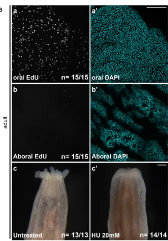
Figure 3. (A) Localized cellular proliferation in juveniles begins at 12 hpa at the amputation site. Overlapping confocal images in which the nuclei (DNA, cyan) were stained using DAPI and proliferating cells (red) were marked using an EdU labeling kit (Aa,Ab). Oral parts of the regenerating juveniles at 12 hpa (Aa) and 48 hpa (Ab). All animals are oriented with the amputation site to the top. The white arrow in (b) shows the presence of dividing cells in the most oral part of the mesentery tissues. Number of cases for the representative phenotype are in white at the bottom right of each image. m, mesentery; (B) Cell proliferation is necessary for adult tissue regeneration. Cell division is present during regeneration in adults after sub-pharyngeal amputation (Ba–Bb’). (Ba–Bb’) Confocal stack images in which the DNA (nucleus) is labeled with DAPI (cyan) and proliferating cells (white) were marked using an EdU labeling kit. (Bc,Bc’) Blocking cell proliferation using hydroxyurea (HU) blocks regeneration in the adult amputated polyps (c’) contrary to the regenerated untreated control (c). Number of cases for the representative phenotype are in white at the bottom right of each images (Ba,Bb,Bc,Bc’). Scale bar in (Ab) is 20 μm and applies to (Aa). Scale bar in (Ba’) is 50 μm and applies to (Ba,Bb,Bb’). Scale bar in (Bc’) is 1 mm and applies to (Bc).
In order to investigate if cell proliferation is also detectable during adult regeneration [7], we performed EdU staining on adult polyps 48 h after sub-pharyngeal amputation (Figure 3(Ba–Bb’)). While no EdU-positive cells are detected in aboral tissues (Figure 3(Bb,Bb’)), cellular proliferation is clearly visible at the amputation site in the adult tissue at 48 hpa (Figure 3(Ba,Ba’)). We further tested if cellular proliferation, similarly to juveniles [7], is required for adult regeneration. To do this we used the pharmaceutical cellular proliferation inhibitor hydroxyurea (HU). Continuous treatment of HU for six days post-amputation blocks oral regeneration in the adult polyp (Figure 3(Bc,Bc’)). Thus, cell division is also required for oral regeneration in adult Nematostella.
2.3. Wound Healing Occurs at 6 HPA
A recent report analyzed Nematostella wound healing after puncturing the body column with a glass needle [34]. The authors found that this process is visually completed after approximately 4 h (25 °C). In order to determine exactly when wound healing occurs in Nematostella juveniles, we
Figure 3. (A) Localized cellular proliferation in juveniles begins at 12 hpa at the amputation site. Overlapping confocal images in which the nuclei (DNA, cyan) were stained using DAPI and proliferating cells (red) were marked using an EdU labeling kit (Aa,Ab). Oral parts of the regenerating juveniles at 12 hpa (Aa) and 48 hpa (Ab). All animals are oriented with the amputation site to the top. The white arrow in (b) shows the presence of dividing cells in the most oral part of the mesentery tissues. Number of cases for the representative phenotype are in white at the bottom right of each image. m, mesentery; (B) Cell proliferation is necessary for adult tissue regeneration. Cell division is present during regeneration in adults after sub-pharyngeal amputation (Ba–Bb’). (Ba–Bb’) Confocal stack images in which the DNA (nucleus) is labeled with DAPI (cyan) and proliferating cells (white) were marked using an EdU labeling kit. (Bc,Bc’) Blocking cell proliferation using hydroxyurea (HU) blocks regeneration in the adult amputated polyps (c’) contrary to the regenerated untreated control (c). Number of cases for the representative phenotype are in white at the bottom right of each images (Ba,Bb,Bc,Bc’). Scale bar in (Ab) is 20 µm and applies to (Aa). Scale bar in (Ba’) is 50 µm and applies to (Ba,Bb,Bb’). Scale bar in (Bc’) is 1 mm and applies to (Bc).
In order to investigate if cell proliferation is also detectable during adult regeneration [7], we performed EdU staining on adult polyps 48 h after sub-pharyngeal amputation (Figure3(Ba–Bb’)). While no EdU-positive cells are detected in aboral tissues (Figure3(Bb,Bb’)), cellular proliferation is clearly visible at the amputation site in the adult tissue at 48 hpa (Figure3(Ba,Ba’)). We further tested if cellular proliferation, similarly to juveniles [7], is required for adult regeneration. To do this we used the pharmaceutical cellular proliferation inhibitor hydroxyurea (HU). Continuous treatment of HU for six days post-amputation blocks oral regeneration in the adult polyp (Figure3(Bc,Bc’)). Thus, cell division is also required for oral regeneration in adult Nematostella.
2.3. Wound Healing Occurs at 6 HPA
A recent report analyzed Nematostella wound healing after puncturing the body column with a glass needle [34]. The authors found that this process is visually completed after approximately 4 h (25 ˝C). In order to determine exactly when wound healing occurs in Nematostella juveniles, we performed DIC and confocal imaging on regenerating juveniles following sub-pharyngeal amputation at several time-points. While in some cases we were able to visualize a clear opening using DIC optics (Figure 4Aa), the three-dimensional folding and contraction of the tissues at the amputation site often made it hard to distinguish between a real wound or a depression that looked like an open wound (Figure4(Ab–Ae)). Thus, determining the wound closure simply by imaging proves to be difficult. In addition, using classical staining/imaging approaches we are unable to distinguish between a closed wound or contraction of the surrounding myo-epithelia.
Figure 4.(A) Wound closure. Oral opening during Nematostella regeneration. Example of DIC images of the oral part, oral view (ov), at the amputation site of an example of the opening at 2 hpa (white arrows in (Aa)) or the closed wound at 6 hpa (white arrows in (Ab)); (Ac–Ae) are confocal images of the oral-most part of the same polyp in lateral view (lv) or details of the oral view (Ad,Ae); (Ad) (yellow frame) or (Ae) (green frame) correspond to the double yellow or green arrow slice, respectively, in (c); Because of the folding that occurs at the amputation site during the first hours of regeneration, the dynamics of the wound healing are hard to assess in DIC optic or confocal images. Scale bars are 20 µm in (Aa–Ac) and 10 µm in (Ad,Ae); (B) Diagram of the compression assay during regeneration. The purple dots represent the nematosomes. The red dotted line represents the amputation site. The forceps are laterally compressing the regenerating polyp body; (C) Time series of the compression assay in an opened (Ca–Cc) or a wound-closed (Cd–Cf) polyp. The dotted double arrow in (Ca) indicates the axial orientation of the animals shown in (Ca–Cf). O, Oral; AbO, Aboral.
In order to have a more robust way to address wound healing after sub-pharyngeal amputation, we developed a compression assay to assess the state of the opening at the amputation site. This assay uses nematosomes (Supplementary Figure S3) as a marker to follow the fluid dynamics present in the gastric cavity of Nematostella (Figure4B,C).
Int. J. Mol. Sci. 2015, 16, 28449–28471
Nematosomes are cellular aggregates formed by cnidocytes that are freely circulating within the Nematostella body [36]. When compressing a juvenile with an open wound, the nematosomes will be expelled at the amputation site. On the contrary, when the wound is closed, the nematosomes will either remain in the gastric cavity or leak out of the body cavity through the aboral pore. This pore is an opening with an unknown function located at the aboral-most region of the body (Supplementary Figure S4) [37]. We assume that nematosomes will exit the body cavity through the wound or the aboral pore depending on the wound healing status. To use this wound healing assay, we compressed the body column of sub-pharyngeal amputated juveniles at 0, 1, 2, 4, 6, 12 hpa and followed the behavior of the nematosomes (FigureInt. J. Mol. Sci. 2015, 16, page–page 5).
Figure 5. Wound healing assay.
Interestingly, at 1 and 2 hpa in the majority of cases (90% n = 31 and 71% n = 24, respectively), the nematosomes are forced to exit the body cavity through the amputation site (Figure 5), showing that the wound is not healed yet. However, at 4 hpa, the majority of the juveniles (67%, 14 out of 21 cases) exhibited nematosomes leaking through the aboral pore. This is the case for 100% of juveniles (22 out of 22 cases) at 6 hpa and later time-points (Figure 5). Thus, in Nematostella, the wound is closed between 4 and 6 hpa following a sub-pharyngeal amputation.
2.4. Mesentery Behavior and Pharynx Formation as Specific Landmarks for Oral Regeneration
The size and transparency of juvenile Nematostella make them more amenable for detailed imaging experiments than adults. We thus analyzed in detail the tissue behavior during oral regeneration of juveniles. We performed DNA labeling on regenerating juveniles following sub-pharyngeal amputation and observed the sequential events every 12 h from 0 to 144 hpa using confocal imaging (Figure 6).
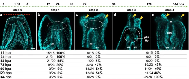
Focusing on the behavior of the mesenteries and the pharynx reformation, we distinguish four main characteristic features: (Step 0) 0–12 hpa, no contact between the remaining mesenteries and the surrounding oral epithelia (Figure 6a); (Step 1) 12–48 hpa, contact of the remaining mesenteries between each other at their most oral site and with the surrounding epithelia at the amputation site (Figure 6b); (Step 2) 60–96 hpa, emergence of a space between the mesenteries and the epithelia at the amputation site (Figure 6c). The epithelia of the amputation site seems dragged down towards the aboral region by the remaining mesenteries. This accentuates the protrusion of the developing tentacle bulbs; (Step 3) 72–120 hpa, the pharyngeal lip (basal part of the future pharynx) forms (Figure 6d). Interestingly, the pharyngeal lip appears to develop first from the oral-most part of the remaining mesenteries; (Step 4) 96–144 hpa, the pharynx is fully regenerated and the tentacles elongate (Figure 6e). Subsequently, the upper part of the pharynx forms (Step 4) progressively in the space that was previously created (Figure 6e) and corresponds to a highly proliferative region (Figure 3Ab).
Figure 5.Wound healing assay.
Interestingly, at 1 and 2 hpa in the majority of cases (90% n = 31 and 71% n = 24, respectively), the nematosomes are forced to exit the body cavity through the amputation site (Figure5), showing that the wound is not healed yet. However, at 4 hpa, the majority of the juveniles (67%, 14 out of 21 cases) exhibited nematosomes leaking through the aboral pore. This is the case for 100% of juveniles (22 out of 22 cases) at 6 hpa and later time-points (Figure 5). Thus, in Nematostella, the wound is closed between 4 and 6 hpa following a sub-pharyngeal amputation.
2.4. Mesentery Behavior and Pharynx Formation as Specific Landmarks for Oral Regeneration
The size and transparency of juvenile Nematostella make them more amenable for detailed imaging experiments than adults. We thus analyzed in detail the tissue behavior during oral
regeneration of juveniles. We performed DNA labeling on regenerating juveniles following
sub-pharyngeal amputation and observed the sequential events every 12 h from 0 to 144 hpa using confocal imaging (Figure6).
Focusing on the behavior of the mesenteries and the pharynx reformation, we distinguish four main characteristic features: (Step 0) 0–12 hpa, no contact between the remaining mesenteries and the surrounding oral epithelia (Figure6a); (Step 1) 12–48 hpa, contact of the remaining mesenteries between each other at their most oral site and with the surrounding epithelia at the amputation site (Figure6b); (Step 2) 60–96 hpa, emergence of a space between the mesenteries and the epithelia at the amputation site (Figure6c). The epithelia of the amputation site seems dragged down towards
the aboral region by the remaining mesenteries. This accentuates the protrusion of the developing tentacle bulbs; (Step 3) 72–120 hpa, the pharyngeal lip (basal part of the future pharynx) forms (Figure 6d). Interestingly, the pharyngeal lip appears to develop first from the oral-most part of the remaining mesenteries; (Step 4) 96–144 hpa, the pharynx is fully regenerated and the tentacles elongate (Figure 6e). Subsequently, the upper part of the pharynx forms (Step 4) progressively in the space that was previously created (Figure 6e) and corresponds to a highly proliferative region (Figure3Ab).
Int. J. Mol. Sci. 2015, 16, page–page
Figure 6. Dynamics of the oral tissue during regeneration. The dynamic behavior of the oral tissue
during regeneration was analyzed using confocal microscopy. (a–e) Confocal image stacks in which the DNA (nucleus) is labeled with DAPI (cyan). Five main phenotypes were observed between 0 and 144 hpa and are represented in this figure: (Step 0) the remaining parts of the mesenteries are separated together and from the epithelia at the amputation site (white dashed circle) (a); (Step 1) the remaining parts of the mesenteries are fused together and are in tight contact with the epithelia at the amputation site (white dashed circle) (b); (Step 2) an empty space forms between the remaining part of the mesenteries and the amputation site (white dashed circle) (c); (Step 3) the pharyngeal lip forms at the oral-most region of the remaining mesenteries, and the empty space becomes filled with nuclei (white dashed circle) (d); (Step 4) the pharynx is fully formed at the oral-most region of the remaining mesenteries (e). Numbers at the bottom of the image panel indicate the total number of analyzed specimens at 12, 24, 48, 72, 96, 120, 144 hpa and the number of cases representative of one of the described five steps in relation to the regeneration time in hours post-amputation (hpa). The amputation site is represented by a red dashed line in (a). Tentacle bulbs and elongated ones are shown by the yellow arrowhead (c–e). The white asterisk is the mouth opening (e). m, mesentery; pha lip, pharyngeal lip; pha, pharynx. Scale bar in (a) is 20 μm and applies to (b–e).
2.5. Fluorescence in the Pharyngeal Region as a Landmark for Pharynx Reformation
Nematostella, like other cnidarians, possesses endogenous fluorescence emitted by fluorescent
proteins and/or fluorescence of the tissues. The six-week-old Nematostella juveniles display a green
(excitation wave length at 488 nm) fluorescence (henceforth referred to as 488+) from a currently
unknown origin (Figure 7A).
Figure 7. Cont.
Figure 6. Dynamics of the oral tissue during regeneration. The dynamic behavior of the oral tissue during regeneration was analyzed using confocal microscopy. (a–e) Confocal image stacks in which the DNA (nucleus) is labeled with DAPI (cyan). Five main phenotypes were observed between 0 and 144 hpa and are represented in this figure: (Step 0) the remaining parts of the mesenteries are separated together and from the epithelia at the amputation site (white dashed circle) (a); (Step 1) the remaining parts of the mesenteries are fused together and are in tight contact with the epithelia at the amputation site (white dashed circle) (b); (Step 2) an empty space forms between the remaining part of the mesenteries and the amputation site (white dashed circle) (c); (Step 3) the pharyngeal lip forms at the oral-most region of the remaining mesenteries, and the empty space becomes filled with nuclei (white dashed circle) (d); (Step 4) the pharynx is fully formed at the oral-most region of the remaining mesenteries (e). Numbers at the bottom of the image panel indicate the total number of analyzed specimens at 12, 24, 48, 72, 96, 120, 144 hpa and the number of cases representative of one of the described five steps in relation to the regeneration time in hours post-amputation (hpa). The amputation site is represented by a red dashed line in (a). Tentacle bulbs and elongated ones are shown by the yellow arrowhead (c–e). The white asterisk is the mouth opening (e). m, mesentery; pha lip, pharyngeal lip; pha, pharynx. Scale bar in (a) is 20 µm and applies to (b–e).
2.5. Fluorescence in the Pharyngeal Region as a Landmark for Pharynx Reformation
Nematostella, like other cnidarians, possesses endogenous fluorescence emitted by fluorescent proteins and/or fluorescence of the tissues. The six-week-old Nematostella juveniles display a green (excitation wave length at 488 nm) fluorescence (henceforth referred to as 488+) from a currently unknown origin (Figure7A).
Int. J. Mol. Sci. 2015, 16, 28449–28471
Int. J. Mol. Sci. 2015, 16, page–page
8
Figure 6. Dynamics of the oral tissue during regeneration. The dynamic behavior of the oral tissue
during regeneration was analyzed using confocal microscopy. (a–e) Confocal image stacks in which the DNA (nucleus) is labeled with DAPI (cyan). Five main phenotypes were observed between 0 and 144 hpa and are represented in this figure: (Step 0) the remaining parts of the mesenteries are separated together and from the epithelia at the amputation site (white dashed circle) (a); (Step 1) the remaining parts of the mesenteries are fused together and are in tight contact with the epithelia at the amputation site (white dashed circle) (b); (Step 2) an empty space forms between the remaining part of the mesenteries and the amputation site (white dashed circle) (c); (Step 3) the pharyngeal lip forms at the oral-most region of the remaining mesenteries, and the empty space becomes filled with nuclei (white dashed circle) (d); (Step 4) the pharynx is fully formed at the oral-most region of the remaining mesenteries (e). Numbers at the bottom of the image panel indicate the total number of analyzed specimens at 12, 24, 48, 72, 96, 120, 144 hpa and the number of cases representative of one of the described five steps in relation to the regeneration time in hours post-amputation (hpa). The amputation site is represented by a red dashed line in (a). Tentacle bulbs and elongated ones are shown by the yellow arrowhead (c–e). The white asterisk is the mouth opening (e). m, mesentery; pha lip, pharyngeal lip; pha, pharynx. Scale bar in (a) is 20 μm and applies to (b–e).
2.5. Fluorescence in the Pharyngeal Region as a Landmark for Pharynx Reformation
Nematostella, like other cnidarians, possesses endogenous fluorescence emitted by fluorescent
proteins and/or fluorescence of the tissues. The six-week-old Nematostella juveniles display a green (excitation wave length at 488 nm) fluorescence (henceforth referred to as 488+) from a currently unknown origin (Figure 7A).
Figure 7. Cont.
Int. J. Mol. Sci. 2015, 16, page–page
Figure 7. (A) Image of 488+ detection in the pharynx. Fed (a,a’), starved (b–c’), regenerating 72 hpa
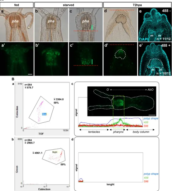
(d–e’) Nematostella polyp juveniles. DIC optic images (a–d). Epifluorescent images (a’–d’). The red dotted line labels the amputation site under the pharynx (c–d’). The green line in (c,c’) shows the 488+ fluorescence localized in the basal part of the pharynx. The area delimitated with the dotted line in d and d’ is the region where the 488+ re-emerged in the polyp at 72 hpa. (e,e’) Confocal images at 72 hpa labeled for DNA (nuclei in cyan) on the 488-negative (e) and 488-positive (e’) polyp juvenile. The area delimitated with the white dotted line in (e’) (488+ regenerating polyps) shows the pharyngeal lip/pharynx in formation that is absent from the 488-negative regenerating polyps in which only the contact between the two mesenteries is visible. pha, pharynx; m, mesentery. Scale bar in (Aa’) is 20 μm and applies to (Aa–Ae,Ab’–Ae’); (B) Biosorter. The dot plot in (a,b) contains the sorting results of the animal by density (Extension) vs. mass (Time Of Flight—TOF) or by 488 fluorescence intensity (Green) vs. density (Extension), respectively, using a Biosorter system. (c,d) Examples of the Biosorter profiles (signal vs. length) of the bright fluorescent group (Bright in b, which corresponds to the Alive group in (a)) or the non-fluorescent group (unlabeled group localized at the bottom left of the two dot plots (a,b)). O, Oral; AbO, Aboral. Scale bar is 100 μm in (Bc).
The 488+ is randomly distributed throughout the entire body in freshly fed animals (Figure 7(Aa,Aa’)). Interestingly, it became more and more localized to the pharynx when juvenile polyps were starved for one or two weeks (Figure 7(Ab,Ab’,Ac,Ac’)), suggesting a correlation of this staining with a metabolic state of the animals. The varied metabolic states within one batch of animals could explain the asynchrony of regeneration. It may also contribute to differences in the cell proliferation rate observed between animals (see table below Figure 6; Röttinger, unpublished data). After sub-pharyngeal amputation, we observed that the 488+ re-emerges in the regenerating polyp in nearly half of the cases around 72 hpa (Table 1).
Figure 7.(A) Image of 488+ detection in the pharynx. Fed (a,a’), starved (b–c’), regenerating 72 hpa (d–e’) Nematostella polyp juveniles. DIC optic images (a–d). Epifluorescent images (a’–d’). The red dotted line labels the amputation site under the pharynx (c–d’). The green line in (c,c’) shows the 488+ fluorescence localized in the basal part of the pharynx. The area delimitated with the dotted line in d and d’ is the region where the 488+ re-emerged in the polyp at 72 hpa. (e,e’) Confocal images at 72 hpa labeled for DNA (nuclei in cyan) on the 488-negative (e) and 488-positive (e’) polyp juvenile. The area delimitated with the white dotted line in (e’) (488+ regenerating polyps) shows the pharyngeal lip/pharynx in formation that is absent from the 488-negative regenerating polyps in which only the contact between the two mesenteries is visible. pha, pharynx; m, mesentery. Scale bar in (Aa’) is 20 µm and applies to (Aa–Ae,Ab’–Ae’); (B) Biosorter. The dot plot in (a,b) contains the sorting results of the animal by density (Extension) vs. mass (Time Of Flight—TOF) or by 488 fluorescence intensity (Green) vs. density (Extension), respectively, using a Biosorter system. (c,d) Examples of the Biosorter profiles (signal vs. length) of the bright fluorescent group (Bright in b, which corresponds to the Alive group in (a)) or the non-fluorescent group (unlabeled group localized at the bottom left of the two dot plots (a,b)). O, Oral; AbO, Aboral. Scale bar is 100 µm in (Bc).
The 488+ is randomly distributed throughout the entire body in freshly fed animals (Figure7(Aa,Aa’)). Interestingly, it became more and more localized to the pharynx when juvenile polyps were starved for one or two weeks (Figure 7(Ab,Ab’,Ac,Ac’)), suggesting a correlation of
this staining with a metabolic state of the animals. The varied metabolic states within one batch of animals could explain the asynchrony of regeneration. It may also contribute to differences in the cell proliferation rate observed between animals (see table below Figure6; Röttinger, unpublished data). After sub-pharyngeal amputation, we observed that the 488+ re-emerges in the regenerating polyp in nearly half of the cases around 72 hpa (Table1).
Table 1. The 488+ in hand-sorted regenerating polyps. Counting of the 488+ polyp juveniles at 24, 48, and 72 hpa. Two different experimenters performed blind counts. Hand-sorter 1 indicates the first experimenter and Hand-sorter 2 the second.
– 488 24 hpa 48 hpa 72 hpa
Hand-sorted + 0/41 0% 4/66 6% 44/77 57%
´ 41/41 100% 62/66 94% 33/77 43%
Bio-sorted + 3/41 7% 2/66 3% 31/77 40%
´ 38/41 93% 64/66 97% 46/77 60%
We thus used the localized green fluorescence in the pharynx of the uncut animal as a proxy to harmonize a batch of polyps before a series of cutting experiments. Sorting batches of uncut animals was performed using a large particle flow cytometer (Biosorter system, Union Biometrica) that enables the analysis of animals based on their length (time of flight, TOF), density (extinction), morphology (profiler), fluorescence, and the relative localization of the fluorescence along the animal body. In order to analyze the global amount of fluorescence intensity and its localization within the animals, we first defined debris (41% of the population; n = 264) based on morphology parameters
(TOF and extinction parameters of each polyp; dot plot Figure 7Ba). We then measured the
488+ fluorescence intensity (the mean of fluorescence intensity is mfi = 4961.1) within the same batch (59% of the population; n = 264; dot plot Figure7Bb). This 488+ can be localized within the profile of the animal using the Profiler software (Profile Figure7Bc). Strikingly, the highest amount of 488+ fluorescence is localized in the region of the polyp where the pharynx is supposed to be.
In order to sort them in an automatic manner with the Biosorter, we identified a profile of interest (Figure 7Bc). We then bio-sorted 84 juveniles with this selected profile, performed sub-pharyngeal amputation, and followed their regeneration. In parallel, we hand-sorted 31 animals that size-matched and appeared in good condition without the use of the green auto-fluorescence proxy. All animals were cut below the pharynx, removing the 488+ at 0 hpa. Animals were then placed back into culturing conditions and the re-emergence of the 488+ was assessed in the regenerating polyps from hand-sorted vs. bio-sorted batches (Table2).
Table 2. Emergence of the 488+ fluorescence during oral regeneration in the hand-sorted versus bio-sorted polyps at 72 and 120 hpa.
– 488 72 hpa 120 hpa
Hand-sorted + 14/33 42% 18/31 58%
´ 19/33 58% 13/31 42%
Bio-sorted + 38/84 45% 51/84 61%
´ 46/84 55% 33/84 39%
Interestingly, both batches, hand-sorted vs. bio-sorted, displayed a similar heterogeneity in their individual advancement through the regeneration steps as reflected by the numbers of 488+ vs. 488´ polyps in each batch of animals (Table2). While the 488+-based selection did not yield a better or more synchronous regeneration, this experiment shows that the Biosorter system can be used as a tool to physically sort animals with precise criteria and/or analyze them for a specific phenotype in large-scale experiments in an unbiased manner. These data also show that the viability of the animals and their regeneration rate are not affected by the Biosorter system.
Int. J. Mol. Sci. 2015, 16, 28449–28471
In order to determine if a correlation exists between the emergence of the 488+ and any previously described steps of regeneration, we sub-pharyngeally bisected juveniles and analyzed the phenotypes in detail of the 488+ and 488´ polyps at 72 hpa. We observed that the 488+ starts to emerge in the oral-most part of the remaining mesenteries around 72 hpa (Figure 7(Ad,Ad’)). Using confocal imaging on DAPI-stained (nucleus) animals we analyzed the detailed morphology of 488´ and 488+ regenerating juveniles at 72 hpa. We observed that 488´ are mainly at Step 2 (11 out of 12 cases) of the oral regeneration staging system described in Figures6and7Ae. No pharynx in formation is visible. Interestingly, the 488+ are mainly at Step 3 or 4 (10 out of 12 cases), with a clear pharyngeal lip or pharynx in formation (Figure7Ae’). These observations show a strong correlation between the initiation of pharynx formation (Steps 3 and 4) and the presence of 488+ in the regenerating juvenile after sub-pharyngeal amputation.
Int. J. Mol. Sci. 2015, 16, page–page
11
in formation is visible. Interestingly, the 488+ are mainly at Step 3 or 4 (10 out of 12 cases), with a clear pharyngeal lip or pharynx in formation (Figure 7Ae’). These observations show a strong correlation between the initiation of pharynx formation (Steps 3 and 4) and the presence of 488+ in the regenerating juvenile after sub-pharyngeal amputation.
2.6. The Regenerating Pharynx Forms from the Oral Part of the Remaining Mesenteries
Taken together, our observations of the sequential events (Figure 6) and the 488+ emergence (Figure 7A) during regeneration led us to hypothesize that the regenerating pharynx after sub-pharyngeal amputation may come from the oral-most part of the remaining mesenteries. To test this hypothesis, we performed in vivo tissue tracking experiments in juveniles overexpressing Kaede mRNA. KAEDE is a fluorescent protein that can be permanently photoconverted from green to red by exposing the expressing cells of interest to UV light [38]. For our tissue tracking experiments, we photoconverted three distinct regions of interest at 24 hpa: (1) the epithelia central to the amputation site (Figure 8a); (2) the epithelia lateral to the amputation site (Figure 8b); or (3) the oral tip of the mesenteries (Figure 8c).
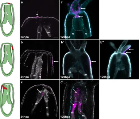
Figure 8. Tissue tracking experiment during regeneration. Spectral confocal images of Nematostella juveniles expressing Kaede photoconvertible fluorescent protein mRNA (a–c’). The Keade photoconverted region is represented in magenta and the non-photoconverted region in grey. Endogenous fluorescence is shown in turquoise to help visualize the morphology of the polyp (a’–c’); At 24 hpa, regions of interest (the epithelium central to the amputation site (a); the epithelium lateral to the amputation site (b), or the oral tip of the mesenteries (c)) were exposed to a UV laser resulting in permanent photo-conversion of the Kaede protein (magenta); The photoconversion at the central epithelium reveals integration of the converted patch (white arrows in (a’)) into the tentacles and most oral tip of the pharynx (white dotted line); Photoconversion of the lateral epithelium shows that this tissue remains in place during regeneration (white arrow in (b’)) or is incorporated into the adjacent tentacles (white arrows in (b’’)); The photoconversion of the oral tip of the mesentery results in integration of the converted patch (white arrows in (c’)) into the pharynx (white dotted line). Scale bar is 20 μm in (a).
Figure 8. Tissue tracking experiment during regeneration. Spectral confocal images of Nematostella juveniles expressing Kaede photoconvertible fluorescent protein mRNA (a–c’). The Keade photoconverted region is represented in magenta and the non-photoconverted region in grey. Endogenous fluorescence is shown in turquoise to help visualize the morphology of the polyp (a’–c’); At 24 hpa, regions of interest (the epithelium central to the amputation site (a); the epithelium lateral to the amputation site (b), or the oral tip of the mesenteries (c)) were exposed to a UV laser resulting in permanent photo-conversion of the Kaede protein (magenta); The photoconversion at the central epithelium reveals integration of the converted patch (white arrows in (a’)) into the tentacles and most oral tip of the pharynx (white dotted line); Photoconversion of the lateral epithelium shows that this tissue remains in place during regeneration (white arrow in (b’)) or is incorporated into the adjacent tentacles (white arrows in (b”)); The photoconversion of the oral tip of the mesentery results in integration of the converted patch (white arrows in (c’)) into the pharynx (white dotted line). Scale bar is 20 µm in (a).
2.6. The Regenerating Pharynx Forms from the Oral Part of the Remaining Mesenteries
Taken together, our observations of the sequential events (Figure6) and the 488+ emergence (Figure 7A) during regeneration led us to hypothesize that the regenerating pharynx after sub-pharyngeal amputation may come from the oral-most part of the remaining mesenteries. To test this hypothesis, we performed in vivo tissue tracking experiments in juveniles overexpressing Kaede
mRNA. KAEDE is a fluorescent protein that can be permanently photoconverted from green to red by exposing the expressing cells of interest to UV light [38]. For our tissue tracking experiments, we photoconverted three distinct regions of interest at 24 hpa: (1) the epithelia central to the amputation site (Figure8a); (2) the epithelia lateral to the amputation site (Figure8b); or (3) the oral tip of the mesenteries (Figure8c).
It is important to note that when we convert the oral tip of the mesenteries, the laser must pass through the lateral epithelia and this region is also converted. After conversion we analyzed the location of the converted red fluorescence at eight days post-amputation (dpa) using spectral confocal imaging (see materials and methods). Spectral imaging measures the complete fluorescent spectrum of each pixel and matches these to pre-calibrated profiles. In our experiments we calibrated the profiles to detect converted Kaede, unconverted Kaede, and endogenous fluorescence. We found that the central epithelia of the amputation site gave rise to the tentacles in 19 out of 19 cases (Figure8a,a’). Additionally, this region also gave rise to the mouth (oral-most part of the pharynx) in 15 out of 19 cases (four cases were undetermined) (Figure8a,a’). The lateral epithelia remained in the lateral tissues after regeneration in four out of 11 cases and in the tentacles in seven out of 11 cases (Figure 8b,b’,b”). In neither central epithelia nor lateral epithelia conversions did we observe converted cells contributing to the pharynx. In the case of photoconverted mesenteries we observed converted KAEDE-expressing tissues in the newly formed pharynx in seven out of seven cases (Figure8c,c’; Supplementary Figure S5). These animals also displayed converted tissues in their lateral epithelia and/or tentacles corresponding to the point of the laser entry during the conversion process. Since the lateral tissue remains in the lateral regions or ends up in the tentacles during regeneration but not the pharynx, we conclude that the converted tissue observed in the pharynx is indeed from the oral tip of the mesenteries, confirming our initial hypothesis (Figure8c,c’; Supplementary Figure S5).
2.7. De Novo Transcription Is Induced First in the Gastrodermis at the Amputation Site
In order to reform missing body parts, an injured organism requires rapid activation of rRNA and tRNA transcription for proper protein biosynthesis of existing or new transcribed mRNA, as well as for cellular proliferation [39,40]. To characterize the transcription in Nematostella, we used EU-Click-it chemistry (Life Technologies, Carlsbad, CA, USA) to detect de novo transcription in Nematostella after amputation (Figure9).
Int. J. Mol. Sci. 2015, 16, page–page
It is important to note that when we convert the oral tip of the mesenteries, the laser must pass through the lateral epithelia and this region is also converted. After conversion we analyzed the location of the converted red fluorescence at eight days post-amputation (dpa) using spectral confocal imaging (see materials and methods). Spectral imaging measures the complete fluorescent spectrum of each pixel and matches these to pre-calibrated profiles. In our experiments we calibrated the profiles to detect converted Kaede, unconverted Kaede, and endogenous fluorescence. We found that the central epithelia of the amputation site gave rise to the tentacles in 19 out of 19 cases (Figure 8a,a’). Additionally, this region also gave rise to the mouth (oral-most part of the pharynx) in 15 out of 19 cases (four cases were undetermined) (Figure 8a,a’). The lateral epithelia remained in the lateral tissues after regeneration in four out of 11 cases and in the tentacles in seven out of 11 cases (Figure 8b,b’,b’’). In neither central epithelia nor lateral epithelia conversions did we observe converted cells contributing to the pharynx. In the case of photoconverted mesenteries we observed converted KAEDE-expressing tissues in the newly formed pharynx in seven out of seven cases (Figure 8c,c’; Supplementary Figure S5). These animals also displayed converted tissues in their lateral epithelia and/or tentacles corresponding to the point of the laser entry during the conversion process. Since the lateral tissue remains in the lateral regions or ends up in the tentacles during regeneration but not the pharynx, we conclude that the converted tissue observed in the pharynx is indeed from the oral tip of the mesenteries, confirming our initial hypothesis (Figure 8c,c’; Supplementary Figure S5).
2.7. De Novo Transcription Is Induced First in the Gastrodermis at the Amputation Site
In order to reform missing body parts, an injured organism requires rapid activation of rRNA and tRNA transcription for proper protein biosynthesis of existing or new transcribed mRNA, as well as for cellular proliferation [39,40]. To characterize the transcription in Nematostella, we used EU-Click-it chemistry (Life Technologies, Carlsbad, CA, USA) to detect de novo transcription in
Nematostella after amputation (Figure 9).
Figure 9. De novo transcription at the amputation site of the regenerating polyp. Overlap of confocal images showing de novo transcription (EU) in red and nucleus (DNA) staining in cyan in the oral epitheliums (b–f) and gastric cavity (b’–f’) of the amputated juvenile polyp. The uncut control is in (a–a’). The white arrows in a,b,b’,c,f show the cells that are undergoing de novo transcription. The white dotted lines in (c’–f’) show the regions in the body gastric cavity that are undergoing massive
de novo transcription, the oral part of the mesenteries, and the epitheliums. All animals are oriented
with the amputation site to the top. Scale bar in (a) is 20 μm and applies to all Figure 9 and Supplementary Figure S6.
This technology allows the visualization of EU (Ethynyl Uridine), a modified Uridine analog, incorporated into nascent RNA [41]. In uncut controls, de novo transcription is barely detectable and only a few cells were EU-positive (EU+) throughout the body epithelia (Figure 9a,a’). After sub-pharyngeal amputation, between 1.30 and 24 hpa, the de novo transcription pattern is similar to controls in regard to the epithelia (Figure 9b,c; Supplementary Figure 6(Aa,Aa’)). However, in the Figure 9. De novotranscription at the amputation site of the regenerating polyp. Overlap of confocal images showing de novo transcription (EU) in red and nucleus (DNA) staining in cyan in the oral epitheliums (b–f) and gastric cavity (b’–f’) of the amputated juvenile polyp. The uncut control is in (a–a’). The white arrows in a,b,b’,c,f show the cells that are undergoing de novo transcription. The white dotted lines in (c’–f’) show the regions in the body gastric cavity that are undergoing massive de novo transcription, the oral part of the mesenteries, and the epitheliums. All animals are oriented with the amputation site to the top. Scale bar in (a) is 20 µm and applies to all Figure9and Supplementary Figure S6.
Int. J. Mol. Sci. 2015, 16, 28449–28471
This technology allows the visualization of EU (Ethynyl Uridine), a modified Uridine analog, incorporated into nascent RNA [41]. In uncut controls, de novo transcription is barely detectable and only a few cells were EU-positive (EU+) throughout the body epithelia (Figure9a,a’). After sub-pharyngeal amputation, between 1.30 and 24 hpa, the de novo transcription pattern is similar to controls in regard to the epithelia (Figure9b,c; Supplementary Figure S6(Aa,Aa’)). However, in the same time frame, some EU+ cells start to emerge in increasing numbers in the internal oral tissues such as the mesenteries and gastrodermis (Figure9b’,c’). At 48 hpa, a strong EU+ signal is detected in the oral epithelia, both the ectodermis and gastrodermis, and the oral-most part of the remaining mesenteries (Figure9d,d’; Supplementary Figure S6(Ac,Ac’)). At 96 hpa, EU+ cells are present in the elongating tentacles with the exception of the tentacle tips (Supplementary Figure S6Ba). Staining progressively decreases in the oral epidermis but remains dense in the oral gastrodermis and in the oral-most part of the mesenteries where the new pharynx is developing (Figure9e,e’). In the fully formed pharynx, at 144 hpa, only a few EU+ cells remain at the base of the tentacles and in the lower part of the pharynx (Figure9f,f’; Supplementary Figure S6(Ae,Ae’)).
2.8. Inhibition of Transcription or Proliferation has Different Effects on Regeneration
In order to determine the role of de novo transcription during regeneration in Nematostella, we treated amputated juveniles with the antibiotic Actinomycin D (AMD), an inhibitor of DNA-primed RNA synthesis [39]. In untreated controls, EU+ cells are massively detected at 48 hpa. As expected, at the same time-point no staining was observed in the regenerating juveniles that were treated with AMD from 36 to 48 hpa (Supplementary Figure S7a,b). Interestingly, we also observed that cell proliferation and regeneration were inhibited in AMD-treated animals (Supplementary Figure S7c,d). A recent study has used hydroxyurea (HU) to efficiently block proliferation and regeneration in Nematostella [7]. However, nothing is known about the precise phenotype caused by the inhibition of cellular proliferation during regeneration. We thus amputated juveniles below the pharynx, treated them with either AMD or HU from 0 to 144 hpa, and scored wound healing success in addition to the exact stage at which regeneration was blocked using the above-described assays and morphological landmarks at 12, 72, and 144 hpa (Figure10; Figure11).
Int. J. Mol. Sci. 2015, 16, page–page
same time frame, some EU+ cells start to emerge in increasing numbers in the internal oral tissues such as the mesenteries and gastrodermis (Figure 9b’,c’). At 48 hpa, a strong EU+ signal is detected in the oral epithelia, both the ectodermis and gastrodermis, and the oral-most part of the remaining mesenteries (Figure 9d,d’; Supplementary Figure 6(Ac,Ac’)). At 96 hpa, EU+ cells are present in the elongating tentacles with the exception of the tentacle tips (Supplementary Figure 6Ba). Staining progressively decreases in the oral epidermis but remains dense in the oral gastrodermis and in the oral-most part of the mesenteries where the new pharynx is developing (Figure 9e,e’). In the fully formed pharynx, at 144 hpa, only a few EU+ cells remain at the base of the tentacles and in the lower part of the pharynx (Figure 9f,f’; Supplementary Figure 6(Ae,Ae’)).
2.8. Inhibition of Transcription or Proliferation has Different Effects on Regeneration
In order to determine the role of de novo transcription during regeneration in Nematostella, we treated amputated juveniles with the antibiotic Actinomycin D (AMD), an inhibitor of DNA-primed RNA synthesis [39]. In untreated controls, EU+ cells are massively detected at 48 hpa. As expected, at the same time-point no staining was observed in the regenerating juveniles that were treated with AMD from 36 to 48 hpa (Supplementary Figure S7a,b). Interestingly, we also observed that cell proliferation and regeneration were inhibited in AMD-treated animals (Supplementary Figure S7c,d).
A recent study has used hydroxyurea (HU) to efficiently block proliferation and regeneration in Nematostella [7]. However, nothing is known about the precise phenotype caused by the inhibition of cellular proliferation during regeneration. We thus amputated juveniles below the pharynx, treated them with either AMD or HU from 0 to 144 hpa, and scored wound healing success in addition to the exact stage at which regeneration was blocked using the above-described assays and morphological landmarks at 12, 72, and 144 hpa (Figure 10; Figure 11).
Figure 10. Effect of the inhibition of cell proliferation or transcription on the wound healing process. Hydroxyurea (HU; orange) was used at 20 mM to block cell proliferation, and Actinomycin D (AMD; green) was used at 10 ug/mL to block transcription. Both drugs were used in a time window from 0 to 12 hpa. The compression assay was performed at 6, 12, or 24 hpa.
We used the compression assay we described above (Figure 4) to assess wound healing under these experimental conditions. At 6 hpa, wound healing is delayed in a fraction of the HU-treated juveniles (33%, n = 58). At 12 hpa, almost all of the animals treated with HU are completely healed (91%, n = 47), although the process was delayed by approximately 6 h (Figure 10). However, in AMD-treated juveniles, 29% (n = 71) are not healed at 6 hpa, and a similar fraction remains open at 12 hpa (24%, n = 56) as well as 24 hpa (28%, n = 45) (Figure 10). This observation suggests that wound healing is not blocked but delayed when cell proliferation only is inhibited. However, when both de
Figure 10.Effect of the inhibition of cell proliferation or transcription on the wound healing process. Hydroxyurea (HU; orange) was used at 20 mM to block cell proliferation, and Actinomycin D (AMD; green) was used at 10 ug/mL to block transcription. Both drugs were used in a time window from 0 to 12 hpa. The compression assay was performed at 6, 12, or 24 hpa.
We used the compression assay we described above (Figure4) to assess wound healing under these experimental conditions. At 6 hpa, wound healing is delayed in a fraction of the HU-treated juveniles (33%, n = 58). At 12 hpa, almost all of the animals treated with HU are completely healed (91%, n = 47), although the process was delayed by approximately 6 h (Figure 10). However, in AMD-treated juveniles, 29% (n = 71) are not healed at 6 hpa, and a similar fraction remains open at 12 hpa (24%, n = 56) as well as 24 hpa (28%, n = 45) (Figure10). This observation suggests that wound healing is not blocked but delayed when cell proliferation only is inhibited. However, when both de novo transcription and cell proliferation are blocked with the AMD treatment, wound healing does not occur in a subset of animals.
Int. J. Mol. Sci. 2015, 16, page–page
14
novo transcription and cell proliferation are blocked with the AMD treatment, wound healing does
not occur in a subset of animals.
Figure 11. Inhibition of de novo transcription blocks regeneration at an earlier step than inhibition of
cell proliferation. The experimental amputated polyps were treated with hydroxyurea (HU) to block cell proliferation or with Actinomycin D (AMD) to block de novo transcription from 0 to 144 hpa and analyzed at the indicated time-points. Confocal images showing the morphological phenotype using nuclear (DNA, cyan) staining in regenerating control (a,d,g) or experimental (b,c,e,f,h) polyps at 12 hpa (a–c); 72 hpa (d–f); or 144 hpa (g,h); The white arrows in (a,b,h) show the characteristic depression of the epithelium at the amputated site that correlates with the initiation of the contact between the remaining mesenteries and the surrounding epithelia (circle white dotted line in (a,b,h)); This depression and contact initiation are absent in the polyps treated with AMD (c). The green arrow in (d) indicates the forming pharyngeal lip. The white dotted lines in (d) to (f) indicate the mesenteries. The yellow arrowheads in (g) indicate the tentacles. m, mesenteries; pha, pharynx. All animals are oriented with the amputation site to the top. Number of cases for the representative phenotype are in white at the bottom right of each image. Scale bar in (a) is 20 μm and applies to all Figure 11.
Interestingly, we observed two strikingly different phenotypes at 12 hpa in the HU- versus AMD-treated juveniles. Similar to controls at 12 hpa, HU-treated polyps progress to Step 1 when the mesenteries are fused together and enter in contact with the surrounding epithelia at the amputation site (Figure 11a,b). In addition, a characteristic depression is present in the epithelia at the amputation site in the control as well as in HU-treated polyps (Figure 11a,b). However, the
Figure 11. Inhibition of de novo transcription blocks regeneration at an earlier step than inhibition of cell proliferation. The experimental amputated polyps were treated with hydroxyurea (HU) to block cell proliferation or with Actinomycin D (AMD) to block de novo transcription from 0 to 144 hpa and analyzed at the indicated time-points. Confocal images showing the morphological phenotype using nuclear (DNA, cyan) staining in regenerating control (a,d,g) or experimental (b,c,e,f,h) polyps at 12 hpa (a–c); 72 hpa (d–f); or 144 hpa (g,h); The white arrows in (a,b,h) show the characteristic depression of the epithelium at the amputated site that correlates with the initiation of the contact between the remaining mesenteries and the surrounding epithelia (circle white dotted line in (a,b,h)); This depression and contact initiation are absent in the polyps treated with AMD (c). The green arrow in (d) indicates the forming pharyngeal lip. The white dotted lines in (d) to (f) indicate the mesenteries. The yellow arrowheads in (g) indicate the tentacles. m, mesenteries; pha, pharynx. All animals are oriented with the amputation site to the top. Number of cases for the representative phenotype are in white at the bottom right of each image. Scale bar in (a) is 20 µm and applies to all Figure11.
Interestingly, we observed two strikingly different phenotypes at 12 hpa in the HU- versus AMD-treated juveniles. Similar to controls at 12 hpa, HU-treated polyps progress to Step 1 when the mesenteries are fused together and enter in contact with the surrounding epithelia at the amputation
Int. J. Mol. Sci. 2015, 16, 28449–28471
site (Figure11a,b). In addition, a characteristic depression is present in the epithelia at the amputation site in the control as well as in HU-treated polyps (Figure 11a,b). However, the AMD-treated regenerating juveniles appear to have been blocked in Step 0, right after the amputation, when the mesenteries are neither in contact with one another, nor with the epithelia of the amputation site (Figure11c). The characteristic depression in the epithelia at the amputation site is absent as well.
To further characterize the phenotypes resulting from AMD or HU treatment, we assessed pharynx formation using the appearance of 488+ as a proxy at 72 hpa. In untreated regenerating juveniles, the 488+ fluorescence is observed in 62% (38 out of 61 cases). In AMD-treated juveniles, we never observed 488+ in 100% (52 out of 52 cases). We obtained similar results in HU-treated polyps in which 488+ never becomes detectable in 85% (53 out of 62 cases). In addition, no pharyngeal lip or tentacle bulbs were visible in either of the treatments (Figure11e,f) as the polyps resulting from AMD or HU treatment remain blocked at Step 0 or Step 1, respectively. These data show that for both the inhibition of de novo transcription or cell proliferation, the pharynx never starts to form, suggesting that cell proliferation is required for pharynx formation.
At 144 hpa, HU-treated polyps still remain blocked at Step 1 (Figure 11h) compared to the controls in which a fully formed pharynx is present (Figure11g). At 144 hpa, AMD-treated polyps were highly opaque and degraded (data not shown), suggesting a lethal effect of long-term inhibition of transcription. All together these results show that cell proliferation is required for both pharynx formation and tentacle elongation.
3. Discussion
3.1. Morphological and Cellular Landmarks for Oral Regeneration in Juveniles
In the present study, we describe in detail the morphological and cellular landmarks for regeneration (Figure 12). We also present in vivo tools that can be used to assess wound healing and pharynx formation in juveniles after sub-pharyngeal amputation.
Figure 12.Diagram summarizing the morphological and cellular events underlying oral regeneration in the sea anemone, Nematostella vectensis. The table below the illustration provides definitions for the various regeneration steps.