HAL Id: inserm-00151947
https://www.hal.inserm.fr/inserm-00151947
Submitted on 5 Jun 2007HAL is a multi-disciplinary open access archive for the deposit and dissemination of sci-entific research documents, whether they are pub-lished or not. The documents may come from teaching and research institutions in France or abroad, or from public or private research centers.
L’archive ouverte pluridisciplinaire HAL, est destinée au dépôt et à la diffusion de documents scientifiques de niveau recherche, publiés ou non, émanant des établissements d’enseignement et de recherche français ou étrangers, des laboratoires publics ou privés.
Progestins and progesterone in hormone replacement
therapy and the risk of breast cancer.
Carlo Campagnoli, Françoise Clavel-Chapelon, Rudolf Kaaks, Clementina
Peris, Franco Berrino
To cite this version:
Carlo Campagnoli, Françoise Clavel-Chapelon, Rudolf Kaaks, Clementina Peris, Franco Berrino. Progestins and progesterone in hormone replacement therapy and the risk of breast cancer.. Journal of Steroid Biochemistry and Molecular Biology, Elsevier, 2005, 96 (2), pp.95-108. �10.1016/j.jsbmb.2005.02.014�. �inserm-00151947�
Progestins and progesterone in hormone replacement therapy and the
risk of breast cancer
Carlo Campagnoli
a,*, Françoise Clavel-Chapelon
b, Rudolf Kaaks
c, Clementina Peris
a, Franco
Berrino
da Unit of Endocrinological Gynecology, “Sant‟Anna” Gynecological Hospital, Corso Spezia 60, 10126 Torino, Italy b Equipe E3N, Institut National de la Santé et de la Recherche Médicale (INSERM), 94805 Villejuif, France
c Unit of Nutritional Cancer, International Agency for Research on Cancer (IARC-WHO), 150 cours Albert Thomas, 69372
Lyon, France
d Department of Preventive and Predictive Medicine, Istituto Nazionale dei Tumori, via Venezian 8, 20133 Milano, Italy
Abstract
Controlled studies and most observational studies published over the last 5 years suggest that the addition of synthetic progestins to estrogen in hormone replacement therapy (HRT), particularly in continuous-combined regimen, increases the breast cancer (BC) risk compared to estrogen alone. By contrast, a recent study suggests that the addition of natural progesterone in cyclic regimens does not affect BC risk. This finding is consistent with in vivo data suggesting that progesterone does not have a detrimental effect on breast tissue. The increased BC risk found with the addition of synthetic progestins to estrogen could be due to the regimen and/or the kind of progestin used. Continuous-combined regimen inhibits the sloughing of mammary epithelium that occurs after progesterone withdrawal in a cyclic regimen. More importantly, the progestins used (medroxyprogesterone acetate and 19-Nortestosterone-derivatives) are endowed with some non-progesterone-like effects, which can potentiate the proliferative action of estrogens. Particularly relevant seem to be the metabolic and hepatocellular effects (decreased insulin sensitivity, increased levels and activity of insulin-like growth factor-I, and decreased levels of SHBG), which contrast the opposite effects induced by oral estrogen.
Keywords: Breast cancer; Progesterone; Progestins; Estrogen; Hormone replacement therapy; Insulin; Insulin-like growth factor-I; Sex hormone binding globulin
Introduction
That there is a relation between endogenous sex hormones and breast cancer (BC) is demonstrated by a large mass of epidemiological and clinical evidence: for example the drop in BC incidence after menopause [1], the reduced risk of BC in ovariectomized women and those with early natural menopause [2], and the efficacy of antiestrogen drugs in preventing and treating BC [3]. Furthermore, prospective studies published over the last 10 years have shown beyond reasonable doubt that in postmenopausal women, serum levels of endogenous sex hormones (estrogens and androgens) affect the risk of subsequent appearance of BC [4]. In particular, the incidence of BC is two-to-three times greater in women with serum levels of estradiol or testosterone in the higher quartiles or quintiles of the distribution, and this association remains significant after reciprocal adjustment. With regard to sex hormones and BC prior to menopause, it is difficult to investigate any association because of the marked fluctuations in hormone levels that occur during the menstrual cycle. No study has clearly demonstrated a relation between BC risk and estrogen levels in premenopausal women [5], possibly because they are constantly above the threshold required to stimulate the growth of BC. By contrast, the association between androgen levels and BC is clear also in premenopausal women [6,7]. Regarding progesterone, a prospective study that controlled for the phase of the menstrual cycle in which blood was sampled, showed an inverse relation between its serum levels in the luteal phase and the successive diagnosis of BC [6]. This finding contrasts with the widely held opinion, sustained by the „estrogen augmented by progesterone‟ hypothesis [8], that progesterone produced physiologically during the cycle contributes to the development of BC.
*
Corresponding author. Tel.: +39 011 3134605; fax: +39 011 3134798.
The possibility that the sex hormones administered as hormone replacement therapy (HRT) to menopausal women increase the risk of BC has been intensely debated and widely studied. Two recent randomized controlled studies [9,10] and most observational studies published over the last 5 years indicate that the administration of estrogens alone does not increase BC risk [11–17] or does so only modestly [18–20]. By contrast, randomised controlled [21,22] and most observational studies indicate that the addition of synthetic progestins to estrogen increases the BC risk much more than estrogen alone. This finding is considered to provide important support to the „estrogen augmented by progesterone‟ hypothesis [23–25]. However, a study on a French cohort [26] suggests that when natural progesterone is used instead of synthetic progestins in HRT, the risk of BC is not increased.
The aim of this paper is to review the available data on the influence of progestins and progesterone on the risk of BC, to discuss the discordant findings, and hence provide suggestions for the prescription of HRT in climacteric women.
Progesterone and progestins in hormone replacement therapy
Why progesterone or progestins are included in hormone replacement therapy
In HRT, the progestin is added to the estrogen to protect against the risk of hyperplasia and adenocarcinoma of the endometrium. More than 30 studies have shown that the administration of estrogens alone to non-hysterectomized women considerably increases the risk of endometrial hyperplasia and cancer [23,27,28]. Even low doses of unopposed estrogens are associated with increased endometrial cancer risk [29]. Addition of a progestin at adequate dosage reduces that risk [27,28,30,31].
Regimens of progestin addition to estrogens
Progestins and estrogens can be combined in various regimens (Table 1). Starting in the middle of the 1960s and continuing for nearly 20 years, particularly in the US, estrogens were the sole active ingredient even for non-hysterectomized women, in order to avoid menstrual-like bleedings [32]. From the second half of the 1980s in the US, and somewhat earlier in Europe, a progestin was added to estrogen in a sequential regimen that mimicked the rise and fall of the hormones in the menstrual cycle [32]. Subsequently the so-called continuous-combined regimen (Table 1) was increasingly adopted [25], first in Europe [33] then in the US [34]. This regimen induces endometrial atrophy, and the consequent amenorrhea [27] is appreciated by women, increasing compliance [35,36].
Table 1 Main types of estrogen plus progestin regimens (modified after Ref. [27])
Regimen Estrogen Progesterone/progestin
Sequential Cyclic
Continuous-cyclic
Days 1–25 Daily
Last 10–14 days of the cycle 10–14 days every month Combined Continuous-combined Cyclic-combined Daily Days 1–25 Daily Days 1–25
Progestins used in hormone replacement therapy
The progestins mainly employed in HRT are synthetic compounds endowed with progesterone-like action on the endometrium but somewhat different from natural progesterone. Table 2 lists the principal progestins in use, divided into those structurally related to progesterone and those structurally related to 19-Nortestosterone.
Progestin use in different countries
The progestins used for HRT vary markedly between countries; there is also variation in the type of estrogen used.
In the US, the most commonly used progestin by far is medroxyprogesterone acetate (MPA). Generally, MPA is combined with conjugated equine estrogens (CEE) in formulations for oral administration [19]. Initially MPA was mainly employed in sequential regimen at the dose of 10 mg/day; subsequently the dose was reduced to 5 mg/day [27]. In the 1990s continuous-combined formulations of CEE and MPA came onto the US market, in which MPA is present in each day‟s pill, so there is no break in progestin assumption. The dose was originally 5 mg/day but current formulations have reduced this to 2.5 mg/day [27], added to CEE 0.625 mg/day. A continuous-combined formulation with 2.5 mg/day MPA was employed in the two recent randomized controlled studies [21,22].
Table 2 Classification of progestins used in hormone replacement therapy (modified after Ref. [90])
Progestins Preparation
Progesterone) Retroprogesterone Progesterone derivative
17α-Hydroxyprogesterone derivatives (pregnanes) 19-Norprogesterone derivatives (norpregnanes) 17α-Hydroxynorprogesterone derivatives (norpregnanes)
19-Nortestosterone derivatives (estranes) 19-Nortestosterone derivatives (gonanes)
Natural progesterone (micronized Dydrogesterone
Medrogestone
Medroxyprogesterone acetate (MPA), megestrol acetate, chlormadinone acetate cyproterone acetate Demegestone, promegestone, trimegestone
Nomegestrol acetate
Norethisterone = norethindrone, norethisterone acetate, lynestrenol
Norgestrel, levonorgestrel, norgestimate
In the UK where estradiol, oral or transdermal, as well as CEE are used, the progestins are mainly 19-Nortestosteronederivatives (norethisterone acetate, norgestrel and levonorgestrel), with only about 20% of treated women using MPA [18].
In Northern Europe, 19-Nortestosterone-derivatives are mainly used (norethisterone acetate, 1–0.5 mg, or levonorgestrel, 0.25 mg) in general combined with oral estradiol, both in sequential and continuous-combined formulations, while MPA is used by less than 20% of treated women, in sequential formulations [37–39].
By contrast, in central and southern Europe 19-Nortestosterone-derivatives are less used, while a range of progesterone-derivatives are used, and these are added to various kind of estrogens. In France, MPA or cyproterone acetate are used mainly with oral estradiol, while dydrogesterone, nomegestrol acetate and promegestone are mainly employed with transdermal estradiol [26]. France is also peculiar for the widespread use of micronized natural progesterone (mainly oral), in association with oral or transdermal estradiol [26]; the progesterone dose is 200 mg/day in sequential regimen [40] and 100 mg/day in combined, usually cyclic (25 days/month), regimen [41].
Epidemiological data on hormone replacement therapy and risk of breast cancer
A few controlled trials (Table 3) and several observational studies (Table 4) published over the past 5 years permit a comparison between the effects of unopposed estrogens and estrogens plus progestins. One must take into account, however, that the study populations are not the same, because unopposed estrogens are usually prescribed to hysterectomized women only [9]. Actually, hysterectomized women are frequently also ovariectomized (up to 40% in the US study populations) and, therefore, at lower BC risk, but several studies did not adjust for ovariectomy in the analysis [13,15,18,20], which makes comparisons somewhat difficult.
Unopposed estrogens
Two randomized studies (Table 3) on the use of unopposed estrogens have been published. The first [10]was a small trial on postmenopausal women (mean age 71 years) with a history of ischemic stroke or TIA and mean follow-up of 2.8 years. The trial suggested that the use of oral estradiol at the dose of 1 mg/day did not increase the risk of BC. The second is the large randomized Women‟s Health Initiative (WHI) trial [9]. This trial found that the use of oral unopposed CEE, 0.625 mg/day for a mean of 6.8 years in hysterectomized, mostly
overweight, women of age 50–80 years was associated with a reduction by about a quarter in the risk of BC compared to women who did not receive CEE. The reduced risk, expressed as hazard ratio (HR) was almost significant (HR 0.77; 95% confidence interval (CI) 0.59–1.01) [9].
Table 3 HRT and breast cancer: consequences of the therapy with estrogen alone (ET) or with estrogen continuously combined with progestin (ccEPT) (randomized controlled trials, USA)
Author ET (HR (95% CI)) ccEPT (HR (95% CI))
Viscoli et al. [10] 664 women
Hulley et al. (HERS II study) [22] 2321 women Chlebowski et al. (WHI study) [21] 16608 women WHI Steering Committee [9] 10739 women
1.00 (0.30–3.50) – – 0.77 (0.59–1.01) – 1.27 (0.84–1.94) 1.24 (1.01–1.54) –
Table 4 HRT and breast cancer: consequences of the therapy with estrogen alone (ET) or with estrogen plus progestin (EPT) (observational studies)
Author
ET (RR (95% CI))
EPT (RR (95% CI))Schairer et al. (USA) [20] cohort, 46335 women Ross et al. (USA) [11] case-control, 1897 cases Moorman et al. (USA) [12] case-control, 397 cases Chen et al. (USA) [13] case-control, 705 cases Newcomb et al. (USA) [19] case-control, 5298 cases Porch et al. (USA) [14] cohort, 17835 women Weiss et al. (USA) [15] case-control, 1870 cases Kerlikowske et al. (USA) [43] cohort, 374465 women Li et al. (USA) [16] case-control, 975 cases
Million Women Study (UK) [18] cohort, 828923 women Magnusson et al. (Sweden) [38] case-control, 2563 cases Olsson et al. (Sweden) [17] cohort, 28378 women Stahlberg et al. (Denmark) [39] cohort, 10874 women Bakken et al. (Norway) [139] cohort, 31451 women
1.20 (1.00–1.40) 1.06 (0.97–1.15)a 0.80 (0.50–1.20) 1.17 (0.85–1.60) 1.23 (1.09–1.39) 0.96 (0.65–1.42) 0.84 (0.67–1.06) 0.92 (0.84–1.00)b 1.00 (0.70–1.30) 1.30 (1.21–1.40 1.94 (1.47–2.55) 2.70 (1.47-4.96)c 0.71 (0.40–1.26) 1.96 (1.16–3.35) 1.80 (1.10–1.90) 1.40 (1.10–1.80) 1.24 (1.07–1.45)a 0.70 (0.40–1.10) 1.49 (1.04–2.12) 1.43 (1.18–1.74) 1.37 (1.05–1.78) 1.22 (0.99–1.50) 1.49 (1.36–1.63)b 1.90 (1.40–2.60) 2.00 (1.88–2.12) 1.63 (1.37–1.94) 2.95 (1.84–4.72)c 1.22 (0.74–2.00)d 2.45 (1.61-3.71)e 2.70 (1.96–3.73) 2.50 (1.90–3.20) a Per 5 years. b >5 years of use. c >10 years of use. d Sequential therapy. e Continuous-combined therapy.
Most US observational studies [11–16,20] (Tables 4 and 5) also found little if any increase in the risk of BC in relation to unopposed estrogen use, at least for the first several years of treatment. For example, in the large cohort studied by Schairer et al. [20] the relative risk (RR) of BC for unopposed estrogens became significant only beyond 12 years of treatment. The mean annual increase in RR was 0.01 in all women (Table 5), 0.03 in thin women and absent (−0.01) in overweight women. In contrast, an increase in BC risk with assumption of unopposed estrogens has been reported by most studies conducted in Europe [18,38,39]. Such heterogeneity might be partly explained by the higher prevalence of obesity in the studies carried out in the US (between 20 and 45%) [9,13,16,19,21] than in Europe (under 10%) [26,39], because the risk due to hormonal treatment, is higher for lean than for overweight women [18,20]. Postmenopausal overweight women are at higher basal BC risk [42].
Table 5 Per year modification of relative risk RR or OR of breast cancer in women using estrogen alone (ET) or estrogen plus progestin (EPT) (observational studies)
Author ET EPT
Magnusson et al. (Sweden) [38] Willett et al. (USA) [44] Ross et al. (USA) [11] Schairer et al. (USA) [20] Newcomb et al. (USA) [19]
+0.03 +0.03 +0.01 +0.01 +0.02 +0.07 +0.09 +0.04 +0.08 +0.04
Estrogens plus progestins
The risks of estrogens plus progestins have been addressed in two randomized studies (Table 3). In the HERS trial, CEE 0.625 mg plus MPA 2.5 mg in continuous-combined regimen was investigated in postmenopausal women with coronary disease and average age 67 years, followed for about 6 years. A non-significant increase in risk was associated with HRT use compared to non-use (relative hazard 1.27; 95% CI 0.84–1.94) [22]. A similar, this time significant, increase in BC risk (HR 1.24; 95% CI 1.01–1.54), was found in the much larger WHI study [21] among women, age 50–80 years, who were given CEE 0.625 mg/day plus MPA 2.5 mg/day in continuous-combined regimen for a mean of 5.6 years. This arm of the WHI study, therefore, had BC risk findings that were directly opposite to those reported for the unopposed estrogens arm of the study [9].
In accord with trial results, almost all observational studies published over the last 5 years have also reported an increase in BC risk associated with progestin use in HRT [11,13–20,38,39,43,44,139]. The reported increase in risk was 2–4 times greater than that associated with the use of unopposed estrogen (Tables 4 and 5). Furthermore, although there were exceptions [11,16,18,19], most studies providing information on the two regimens of progestin addition [14,15,17,38,39,139] found that the risk was greater with continuous-combined than sequential regimen (Table 6).
Table 6 Estrogen plus progestin therapy and breast cancer: consequences of the sequential regimen (sEPT) and the continuous-combined regimen (ccEPT) (observational studies)
Author sEPT (RR (95% CI)) ccEPT (RR (95% CI))
Ross et al. (USA) [11] Newcomb et al. (USA) [19] Weiss et al. (USA) [15] Porch et al. (USA) [14] Li et al. (USA) [16]
Million Women Study (UK) [18] Magnusson et al. (Sweden) [38] Olsson et al. (Sweden) [17] Stahlberg et al. (Denmark) [39] Bakken et al. (Norway) [139]
1.38 (1.13–1.68)a 1.57 (0.95–2.60)b 0.91 (0.67–1.24) 1.04 (0.74–1.46) 2.00 (1.1–3.70) 1.77 (1.59–1.97)c 2.12 (1.95–2.30)b 1.48 (1.08–2.04)d 1.89 (0.88–4.09)b,d 1.22 (0.74–2.00) 1.94 (1.26–3.00)d 1.70 (1.00–2.80)c 2.20 (1.30–3.80)b 1.09 (0.88–1.35)a 1.54 (1.15–2.07)b 1.29 (1.02–1.64) 1.82 (1.34–2.48) 1.80 (1.30–2.50) 1.57 (1.37–1.79)c 2.40 (2.15–2.67)b 1.41 (1.09–1.83)d 2.89 (1.66-5.00)b,d 2.45 (1.61–3.71) 4.16 (2.56–6.75)d 2.60 (1.90–3.70)c 3.20 (2.20–4.60)b a Per 5 years. b >5 years of use. c <5 years of use. d
Only 19-Nortestosterone-derived progestins.
These findings, which are consistent with the „estrogen augmented by progesterone‟ hypothesis [23–25] prompted suggestions that alternative ways of protecting the endometrium – that have no or reduced effects on breast tissue – should be tried, e.g. the use of an intra-uterine device with a progestin or the intravaginal administration of progesterone [23]. The elimination of progestins from HRT preparations was even suggested, since the increased risk of endometrial cancer with unopposed estrogens would be more than counterbalanced by the reduced risk of BC [18].
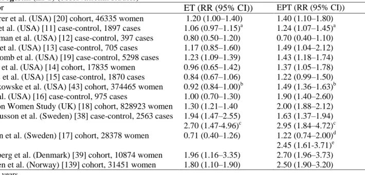
It is important to realize that recent findings relating to the use of natural progesterone, in sharp contrast with those referring to the use of progestins, are reassuring. These findings come from two cohort studies carried out in France, where oral micronized progesterone has been used by large numbers of menopausal women since over two decades. The first study was on a cohort of 3175 women followed for a mean of 8.9 years in a menopause clinic. Of these, 55% were classified as HRT users, the majority of whom received exclusively or mostly a combination of a transdermal estradiol plus either progesterone (60%) or progesterone-derived progestins other than MPA. No increase in the BC risk was found in women receiving these treatments [45]. The second, much larger study, based on the E3N-EPIC cohort, included 54,548 postmenopausal teachers who had not taken any HRT before enrolment and who were followed an average of 5.8 ± 2.4 [26]. At our knowledge this is the single prospective study in which women were followed up with periodic questionnaires since the beginning of exposure, thus avoiding the misclassification of treatment duration that may occur in the cohort studies with cross-sectional definition of exposure to HRT at the time of enrolment. Such a study design also avoids the bias of selectively enrolling only the women who have not developed BC after starting HRT, which systematically affects the studies based on the follow-up of women who have already started HRT before enrolment. In this study [26], oral micronized progesterone, contrarily to synthetic progestins, did not increase BC risk in women treated with transdermal estradiol. The RRs with respect to untreated women were, respectively: 1.2, 95% CI 0.8–1.8, for transdermal estradiol alone; 0.9, CI 0.7–1.2, for transdermal estradiol with micronized progesterone; 1.4, CI 1.2–1.7, for transdermal estradiol with synthetic progestins (Fig. 1).
Fig. 1. Relative risks associated with use of different hormones by women with incident HRT exposure, compared
with non-users: E3N-EPIC study [26]. TD-E2 = transdermal estradiol.
It should be noted that, at commonly employed doses, oral progesterone has peculiar pharmacokinetic properties [46] so that it could have different effects on target tissues than synthetic progestins, as suggested by its action at the endometrial level. Oral progesterone in sequential regimen efficiently protects the endometrium by counteracting the hyperplastic effect of oestrogen [30]. However, this is due more often to an antiproliferative effect [40], than to the induction of secretory changes like those generally induced by progestins [47], and also by vaginally administered progesterone [48] and high dose oral progesterone [40,47]. A similar peculiarity in the activity might be hypothesized also at the level of breast tissue.
Anyway, the E3N-EPIC study [26], provides evidence against the “estrogen augmented by progesterone hypothesis”, by suggesting that the increased BC risk found with the progestin addition is due to mechanisms other than the those exerted by natural progesterone.
Criticism of ‘estrogen augmented by progesterone’ hypothesis
According to this hypothesis, the increased risk of BC associated with estrogen is augmented substantially by progesterone [8]. The hypothesis is based on some findings from in vitro studies, on the results of in vivo studies on breast cell proliferation, on interpretations of the epidemiological relationship of BC with premenopausal menstrual irregularities and cycle length and, more recently, on the finding that BC incidence
and breast density are increased in women who use estrogen plus progestin HRT [23,24]. However, all these findings are open to different interpretations.
In vitro data bearing on the „estrogen augmented by progesterone‟ hypothesis
In vitro studies have established that estrogens markedly increase the mitotic rate of both normal and malignant breast epithelium cells; there is also evidence that estradiol and its metabolites are carcinogenetic to human breast epithelium [49,50]. Conversely, the picture is more complex for progesterone, which may affect mitotic activity of normal and malignant breast cells by various mechanisms [51–55] and may have proliferative or antiproliferative (antiestrogenic) effects depending on study parameters [24,56–58].
In vivo studies bearing on the „estrogen augmented by progesterone‟ hypothesis
The main evidence advanced in support of the hypothesis is the finding that the proliferation of breast epithelium increases in luteal phase, reaching a peak 9–10 days after ovulation. This increase in mitotic activity was first noted by morphological evaluation of breast tissue removed during mammoplasty [59] or autopsy [60] and was later found by thymidine index determination, Ki67 labeling, and PCNA labelling of breast tissue obtained during surgery [61–64] or by fine-needle-aspiration cytology [65]. The increase in proliferation occurs particularly in the terminal duct lobular unit (TDLU) [59,60,62] where most breast carcinomas arise [66]. However, it has not been established that the luteal phase cell proliferation peak is due to progesterone. An alternative hypothesis is that it is only estrogens that stimulate the proliferation of breast epithelium, but that there is a lag of 4–5 days between the estrogen peak and the proliferation peak [61,67]. Actually, breast epithelium does not appear as sensitive an estrogen target organ as the endometrium, probably because estrogens have an indirect effect on proliferation, which requires paracrine factors to mediate their signal [67]. It is noteworthy that studies on intact normal human breast tissue grafted subcutaneously to athymic nude mice found that estrogen, not progesterone, is the major epithelial cell mitogen [67,68]. Evidence that progesterone may in fact reduce estrogen-induced breast proliferation comes from a study in which gels containing estradiol or progesterone or a combination of both were applied daily to the breasts of postmenopausal women for 14 days prior to surgery not for malignancy [69].
Importantly, what does emerge from histological studies, is an increase in breast cell apoptosis during luteal phase and a sloughing of the breast epithelium in perimenstrual phase, which are related to the increase in post-ovulation progesterone levels and their decline in the immediate premenstrual phase. The number of apoptotic breast cells starts increasing a few days after ovulation (after the mitosis rate has already started increasing) and reaches a peak just before menstruation, to decline subsequently [59]. As described by Longacre and Bartow[60] the proliferative phase of the breast is characterized by small lobules with few terminal duct structures and uncommon mitoses; the secretory phase is characterized by increases in lobule size and number of terminal duct structures, and also by vacuolization and mitoses in duct basal epithelium; in the perimenstrual phase the breast undergoes lobular contraction with necrosis and sloughing of ductal epithelium. If greater or more prolonged epithelial cell proliferation is associated with greater risk of malignant transformation [23,24], then apoptosis and epithelial sloughing that follows are likely to be important protective factors [61].
Epidemiological data bearing on the „estrogen augmented by progesterone‟ hypothesis
The following epidemiological findings regarding premenopausal women have been cited in support of the hypothesis: reduced risk of BC in women with oligomenorrhea, in particular those who have had menstrual irregularities for prolonged periods after menarche, probably because of persistent lack of ovulation [70]; reduced risk of BC in obese premenopausal women, probably in relation with fewer ovulations [71]; and greater BC risk in women with short menstrual cycles, implying greater cumulative time in luteal phase, since cycle length varies mainly because follicular phase varies [6,64]. Note, however, that oligomenorrhea implies not only less progesterone but also fewer estradiol peaks and less cumulative estrogenic stimulation; while short cycles are either ovulatory implying greater cumulative exposure to estradiol, or are anovulatory implying reduced exposure to progesterone.
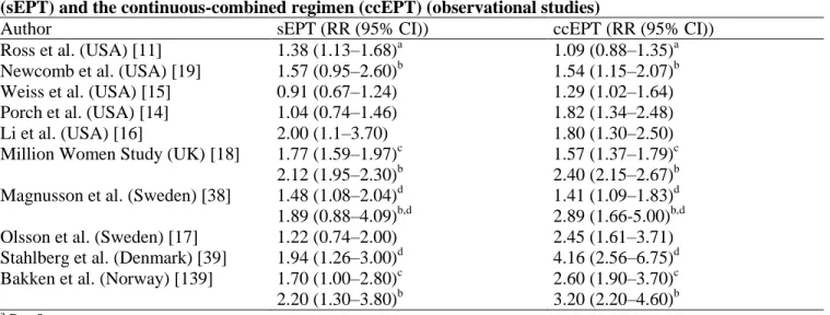
That normal or abundant progesterone production in premenopausal women may be even protective against BC was suggested by the results of a nested case-control analysis of a cohort of premenopausal women which sampled blood in luteal phase [6] (Fig. 2). Several previous case-control studies have suggested a similar conclusion [72–76]. Grattarola [77] was the first to propose that chronic anovulation – and hence low exposure to progesterone – was a risk factor, rather than a protective factor, for BC, based on the finding that a high
proportion of endometrial biopsies from young BC patients taken during the second half of the cycle, were still in proliferative phase.
Fig. 2. Relative risk of premenopausal breast cancer in women with regular menses according to mid-luteal
progesterone levels (40 cases and 108 matched control subjects from the ORDET cohort of 5963 premenopausal women) [6].
Breast density
The increase in mammographic breast density that occurs with the addition of progestins to estrogens in mainly continuous-combined but also sequential HRT regimens [78], and which is also observed when progesterone is administered orally [79], is cited as further evidence that progesterone augments the BC risk [24]. This is because high breast density is a strong independent risk factor for the development of BC [80]. However, the increase in density during hormone therapy is due not only to an increase in lobule volume but also to the intralobular stroma becoming loose and edematous, as also occurs during the luteal phase of the cycle [60]. Moreover, the density increase is quickly reversible at the hormone withdrawal [78]. It is doubtful, therefore, that the transient increase in breast density due to progesterone has the same biological significance as constitutional high density, which has a series of genetic [81], metabolic [82], and nutritional [83] as well as hormonal causes, most of which have been acting continuously since the breasts began to form [84].
Synopsis
We have seen on one hand, that the evidence adduced in favour of the „estrogen augmented by progesterone‟ hypothesis is open to different interpretations, and on the other that the physiological production of progesterone during the menstrual cycle may be associated with a lower risk of BC. The lack of increase in BC risk with HRT regimens cyclically containing natural progesterone, found by the E3N-EPIC study [26], is therefore biologically plausible. It is probable that the increase in BC risk found in other studies on HRT use is related to the continuous-combined regimen employed and/or to the fact that synthetic progestins rather than progesterone were used.
Differences between the various hormone replacement therapy regimens
As noted (see also Table 6), most observational studies that were able to provide results on both sequential and continuous-combined regimens found that the BC risk was greater with the latter, particularly when HRT use was longterm (Fig. 3). A comparison of the findings obtained in the different countries is rendered difficult because the estrogens employed also vary. Nevertheless, risk differences between sequential and continuous-combined regimens seemed more marked and consistent in studies conducted in Northern European countries than in those conducted in the US (Table 6, Fig. 3). This might partly be due to the fact that in the US, particularly in some states [85], the sequential regimen could have been privileged in women thought to be, and actually being, at high BC risk, because of widely advertised data suggesting a protective effect of the sequential
regimen [86]. More importantly, in Northern Europe the daily dose of 19-Nortestosterone-derived progestins (most often norethisterone acetate, 1 mg) was the same in both continuous-combined and sequential regimens, so that the monthly cumulative dose in the former was twice that of the latter, while in the US the daily MPA dose in combined regimen was much lower (2.5 mg) than that given in sequential regimen (5–10 mg), so that cumulative dose did not differ greatly between them.
Fig. 3. Relative risk (RR) or odd ratios (OR) of breast cancer in long term HRT users. (●) significant difference from
nonusers; () mainly medroxyprogesterone acetate (MPA); () 19-Nortestosterone-derived progestins and (20%) MPA; (*) only 19-Nortestosterone-derived progestins; () mainly 19-Nortestosterone-derived progestins.
A Swedish longitudinal study that used needle aspiration cytology to sample breast tissue [87] found a greater than four-fold increase in proliferation compared to baseline after 3 months of treatment with continuous-combined HRT, but no further increase at 6 months. A UK cross-sectional study, which assessed TDLU proliferation in surgical samples, found no differences between unopposed estrogen users, estrogen plus progestin users (some in sequential and some in combined regimen) and non-HRT users [88]. An increase in proliferation in women using combined-continuous HRT was however found by a cross-sectional study in the US on breast biopsies [62]. In this study, continuous-combined regimen was associated with increased TDLU proliferation compared to no HRT and unopposed estrogens, but this proliferation was modest in the early stages of treatment and increased progressively over the years, in contrast with the findings of the Swedish study [87]. The US study [62] also found that, for women on continuous-combined regimen, TDLU morphology was similar to that found in luteal phase premenopausal women [60].
It is possible that the increase in BC risk with continuous-combined regimens is in part due to the fact that no epithelial sloughing occurs, due to the lack of progestin withdrawal. Note that the absence of any increase in BC risk in association with natural progesterone HRT regimens reported by the French E3N-EPIC study [26] is consistent with this possibility. In France, in fact, natural progesterone is mainly used in cyclic (sequential and combined) regimens [41].
Non-progesterone activities of progestins
Suggestions that different progestins might be associated with differences in BC risk come from studies conducted in Europe. Again, however, comparisons are difficult because different progestins may be associated with different estrogens and various administration regimens. In two studies only [38,39] direct comparison of two progestins was possible since both were combined with oral estradiol in sequential regimens: no substantial differences in BC risk between MPA and progestins structurally related to 19-Nortestosterone were found. Furthermore, 19-Nortestosterone-derivatives and MPA in the Million Women Study [18], and 19- Nortestosterone- and progesterone-derivatives in the E3N-EPIC study [26] did not differ from each other with respect to BC risk. Nevertheless, as noted previously, the E3N-EPIC study did reveal some risk differences: in women treated with transdermal estrogen plus synthetic progestins the risk was significantly higher than when
natural progesterone was added to estrogen. Such heterogeneity was statistically significant in the first 4 years of treatment (the follow-up is too short to address the issue of longer term treatment) [26].
The progestins in predominant use in HRT preparations have activities that do not completely coincide with those of progesterone. In Northern European countries and in UK (Fig. 4) prevails the use of 19-Nortestosterone-derivatives (norethisterone acetate, norgestrel, levonorgestrel) which have androgenic activity [89,90], while in the US prevails the use of MPA which also is endowed with androgenic properties, even if to a lesser extent [90,91]. The increased BC risk found with the use of these progestins might be related to their „non-progesterone‟ activities. Before examining these activities it is useful first to examine the metabolic factors that increase BC risk and the effect of exogenous estrogens on these factors.
Fig.4. Progestins used in HRT in different countries. Metabolic factors increasing breast cancer risk
A key metabolic alteration that increases BC risk is the resistance to insulin action on carbohydrates (insulin resistance; reduced insulin sensitivity), due to genetic and nutritional factors, with consequent hyperinsulinemia [92,93].
Insulin resistance, hyperinsulinemia and high blood glucose are associated with increased risk of BC [94–98]. Elevated levels of insulin can directly stimulate the proliferation of cancer cells, an action probably mediated by the insulinlike growth factor-I (IGF-I) receptor. High insulin may also have indirect actions, by increasing the liver production of IGF-I, decreasing some IGF-binding proteins and sex hormone binding globulin (SHBG), and stimulating the ovarian production of androgens [99,100]. A randomized controlled study of dietary intervention in menopausal women showed that an insulin lowering diet can reduce the bioavailability of sex hormones and IGF-I [101,102].
Circulating IGF-I derives mainly from the liver [103]. Its production is stimulated by GH and facilitated by an affluent nutritional status, particularly by a high consumption of protein, and by insulin level. IGF-I bioavailability is regulated by IGF binding proteins (IGFBP) also produced in the liver. Levels of IGFBP-1 and IGFBP-2, which decrease IGFI bioavailability correlate inversely with blood insulin levels [104]. IGF-I has potent mitogenic and anti-apoptotic effects on BC cells. The mitogenic effect is synergistic with that of estrogens [105,106]. In particular estradiol increases the number of IGF-I receptors, and IGF-I is necessary for maximal activation of estrogenic receptors. Furthermore, both estradiol and IGF-I are capable of inducing the expression of the genes necessary for maximal cell proliferation [106]. As recently reviewed [107,108], most, but not all [109], prospective studies indicate that high IGF-I levels in premenopausal women (that is in women still producing estrogens) are a risk factor for later development of BC. Furthermore, one prospective study found a relation between IGF-I levels and BC risk in menopausal women on HRT [109], while another found a similar relation for overweight menopausal women [97].
SHBG is also produced by the liver, and its production is inhibited by insulin and IGF-I [92]. Low SHBG levels are a risk factor for BC in postmenopausal women [4,110] and possibly also in premenopausal
women [6]. SHBG specifically binds testosterone and – with lower affinity – estradiol. The principal consequence of low SHBG is that levels of free (bioavailable) testosterone are increased. BC cancer cells and surrounding stromal cells can aromatize androgens into estrogens. High levels of free testosterone have been identified as a risk factor for BC both before [6] and after [111] menopause. SHBG also decreases the bioavailability of the more active estrogens; moreover, through a specific receptor on the membrane of estrogen-sensitive BC cells, SHBG could have an antiestrogenic, antiproliferative effect [110,112].
Overall, these data indicate that metabolic factors play a crucial role in augmenting the effect of estrogen on breast tissue and on BC cells.
Effects of exogenous estrogens on metabolic risk factors for breast cancer
Estrogens, particularly orally administered estrogens, are able to counteract metabolic factors that increase the risk of BC. One way they do this is by increasing insulin sensitivity and hence lowering circulating insulin levels [113–119]. Oral estrogens, through their hepatocellular actions (accentuated by the first pass effects), also induce a significant reduction in circulating IGF-I and a sharp increase in circulating SHBG [110,113,120]. These effects are the opposite of those caused by obesity [121], a well established risk factor for postmenopausal BC [42,71]. Estrogens also increase circulating IGFBP-1 levels, again by a direct effect on liver cells, and this may further reduce the activity of circulating IGF-I as reviewed [122].
Most likely the above mentioned metabolic consequences of oral estrogens are more important in women with high metabolic risk, namely obese women. This would explain why BC risk decreased in the CEEs arm of the WHI study [9]; most women treated in this study, in fact, were overweight. Such protective effect may not be present in thin women. Actually a re-analysis of 51 epidemiological studies published in 1997 [123] showed a greater BC risk increase for thin than overweight women under HRT. Later cohort studies had a similar finding [18,20]. A further reason why oral CEEs are associated with lower BC risk could be that some components of CEE mixture, the 17 α-hydroxy-derivatives of equilin and equilenin, have a nonestrogenic or even an antiestrogenic effect on breast tissue, as suggested by some experimental findings [124].
Differences between progestins and progesterone
The progestins used in the countries where most epidemiological studies have been performed (Fig. 4) differ from progesterone because they could have direct effects on normal and malignant breast cells (described in Sections 6.3.1 and 6.3.2), and particularly because of indirect effects (metabolic and hepatocellular) which could stimulate BC cells in synergy with estrogens and increase estrogen bioavailability (described in Sections 6.3.3 and 6.3.4).
Estrogenic effects of progestins
In vitro studies have shown that progestins derived from 19-Nortestosterone exert an estrogen-like proliferative effect on BC cell lines [125,126]. The effect is probably mediated by reduced 5α metabolites, which interact with α and β estrogen receptors [127]. One might also conceive, however, that progestins with androgenic activity stimulate BC cell proliferation via interaction with androgen receptors.
Effects of progestins on cancer cell enzymes
Estrogen-sensitive cancer cells express the enzymes that enable them to produce estradiol from circulating androgens, estrone, and estrone sulfate [128,129]. Among the most important of these enzymes are aromatase, estrone sulfatase and 17 β-hydroxysteroid dehydrogenases (17 β-HSD) [130]. In vitro studies indicate that progestins can inhibit the activity of estrone sulfatase and influence the activities of 17β-HSD, so decreasing the formation of estradiol [130]. However, in some experimental conditions (but not in others) MPA seems to differ from progesterone and other progestins in being able to promote the reductive transformation of estrone into estradiol via 17β-HSD [56,130]. Such an effect might be important in women with high circulating levels of estrone, as occurs when oral estrogen-containing HRT is employed [56].
Metabolic effects and hepatocellular actions of progestins
Depending on their degree of androgenicity (Table 7), androgenic progestins reduce insulin sensitivity, opposing the action of estrogens [114–116,119,131–133].
The hepatocellular effects of androgenic progestins may also be described as opposite to those of estrogens. In general the strength of such effects is proportional to progestin androgenicity (Table 7) and is particularly marked when progestins are taken orally, thanks to the first pass effect through the liver. Some of these effects, e.g. the increase in circulating IGF-I activity and reduction in circulating SHBG, might increase the BC risk [120].
Table 7 Oral progestins in increasing order of metabolic and hepatocellular strength Natural micronized progesterone
Dydrogesterone; medrogestone; cyproterone acetate; 19-Norprogesterone derivatives
Medroxyprogesterone acetate (MPA)
19-Nortestosterone derivatives (norethisterone; norethisterone acetate; norgestrel; levonorgestrel)
(a) Increased IGF-I concentration and activity. When taken orally, androgenic progestins (e.g. norethisterone acetate and to a lesser extent MPA) provoke an increase in circulating IGF-I opposing the action of estrogens [134–136]. The increase is particularly marked when basal IGF-I levels are low [122]. These progestins also oppose the increase in IGFBP-1 caused by oral estrogens, and this effect probably contributes to the increase in IGF-I activity reviewed in [122]. By contrast, progestins with progesterone-like activity, like dydrogesterone, have essentially no hepatocellular effect and do not affect circulating IGF-I levels [122,134]. Androgenic progestins might therefore increase the risk of BC by increasing IGF-I levels [120,134,137].
(b) Reduced SHBG levels. Androgenic progestins, and to a much lesser extent MPA, also oppose the estrogeninduced increase in SHBG secretion by the liver [110,134,136,138]. Once again this effect is not exerted by the progesterone-like progestins [120,134,136].
SHBG binding
Thirty-five-to-forty percent of 19-Nortestosteronederived progestins circulate bound to SHBG [89]. This phenomenon, in combination with the progestin-induced reduction of SHBG production, results in increased levels of free androgens and estrogens. As the SHBG increase caused by oral estrogens is potentially protective [110], it is plausible that the reduction of SHBG levels and binding capacity caused by 19-Nortestosterone-derived progestins may increase the risk of BC [120,134].
Conclusion
The balance of the in vivo evidence is that progesterone does not have a cancer-promoting effect on breast tissue. This provides a biological rationale for the finding that oral micronized progesterone added to estrogens in sequential or cyclic-combined regimens does not increase the risk of BC [26]. The greater BC risk persistently related to the use of HRT preparations containing estrogen and synthetic progestins seems in all likelihood due to the regimen and/or to the kind of progestin used. The “non-physiological” continuous-combined regimen, could increase the risk because it does not allow sloughing of lobular duct epithelium (such as occurs when progesterone declines at the end of the normal menstrual cycle). More importantly, many of the progestins used have several non-progesterone like actions that potentiate the proliferative effect of estrogens on breast tissue and estrogensensitive cancer cells. We therefore suggest that when HRT is indicated, preparations containing progesterone and not a synthetic progestin should be used, according to a sequential or cyclic-combined regimen. In this way the risk of endometrial cancer is minimized without increasing the risk of BC.
References
[1] J. Clemmesen, Statistical studies in the aetiology of malignant neoplasms, 3, Acta Pathol. Microbiol. Scand. 209 (S1) (1969) S1–S58.
[2] D. Trichopoulos, B. MacMahon, P. Cole, Menopause and breast cancer risk, J. Natl. Cancer Inst. 48 (3) (1972) 605– 613.
[3] Early Breast Cancer Trialists‟ Collaborative Group, Tamoxifen for early breast cancer: an overview of the randomised trials, Lancet 351 (9114) (1998) 1451–1467.
[4] Endogenous sex hormones and breast cancer in postmenopausal women: reanalysis of nine prospective studies, J. Natl. Cancer Inst. 94 (8) (2002) 606–616.
[5] R.C. Travis, T.J. Key, Oestrogen exposure and breast cancer risk, Breast Cancer Res. 5 (5) (2003) 239–247.
[6] A. Micheli, P. Muti, G. Secreto, V. Krogh, E. Meneghini, E. Venturelli, S. Sieri, V. Pala, F. Berrino, Endogenous sex hormones and subsequent breast cancer in pre-menopausal women, Int. J. Cancer 112 (2) (2004) 312–318. [7] G. Secreto, B. Zumoff, Abnormal production of androgens in women with breast cancer, Anticancer Res. 14 (5B)
(1994) 2113–2117.
[8] T.J. Key, M.C. Pike, The role of oestrogens and progestagens in the epidemiology and prevention of breast cancer, Eur. J. Cancer Clin. Oncol. 24 (1) (1988) 29–43.
[9] G.L. Anderson, M. Limacher, A.R. Assaf, T. Bassford, S.A. Beresford, H. Black, D. Bonds, R. Brunner, R. Brzyski, B. Caan, R. Chlebowski, D. Curb, M. Gass, J. Hays, G. Heiss, S. Hendrix, B.V. Howard, J. Hsia, A. Hubbell, R. Jackson, K.C. Johnson, H. Judd, J.M. Kotchen, L. Kuller, A.Z. LaCroix, D. Lane, R.D. Langer, N. Lasser, C.E. Lewis, J. Manson, K. Margolis, J. Ockene, M.J. O‟Sullivan, L. Phillips, R.L. Prentice, C. Ritenbaugh, J. Robbins, J.E. Rossouw, G. Sarto, M.L. Stefanick, L. Van Horn, J. Wactawski-Wende, R. Wallace, S. Wassertheil-Smoller, Effects of conjugated equine estrogen in postmenopausal women with hysterectomy: the Women‟s Health Initiative randomized controlled trial, JAMA 291 (14) (2004) 1701–1712.
[10] C.M. Viscoli, L.M. Brass, W.N. Kernan, P.M. Sarrel, S. Suissa, R.I. Horwitz, A clinical trial of estrogen-replacement therapy after ischemic stroke, N. Engl. J. Med. 345 (17) (2001) 1243–1249.
[11] R.K. Ross, A. Paganini-Hill, P.C. Wan, M.C. Pike, Effect of hormone replacement therapy on breast cancer risk: estrogen versus estrogen plus progestin, J. Natl. Cancer Inst. 92 (4) (2000) 328–332.
[12] P.G. Moorman, H. Kuwabara, R.C. Millikan, B. Newman, Menopausal hormones and breast cancer in a biracial population, Am. J. Public Health 90 (6) (2000) 966–971.
[13] C.L. Chen, N.S. Weiss, P. Newcomb, W. Barlow, E. White, Hormone replacement therapy in relation to breast cancer, JAMA 287 (6) (2002) 734–741.
[14] J.V. Porch, I.M. Lee, N.R. Cook, K.M. Rexrode, J.E. Burin, Estrogen-progestin replacement therapy and breast cancer risk: the Women‟s Health Study (United States), Cancer Causes Contr. 13 (9) (2002) 847–854.
[15] L.K. Weiss, R.T. Burkman, K.L. Cushing-Haugen, L.F. Voigt, M.S. Simon, J.R. Daling, S.A. Norman, L. Bernstein, G. Ursin, P.A. Marchbanks, B.L. Strom, J.A. Berlin, A.L. Weber, D.R. Doody, P.A. Wingo, J.A. McDonald, K.E. Malone, S.G. Folger, R. Spirtas, Hormone replacement therapy regimens and breast cancer risk (1), Obstet. Gynecol. 100 (6) (2002) 1148–1158.
[16] C.I. Li, K.E. Malone, P.L. Porter, N.S. Weiss, M.T. Tang, K.L. Cushing-Haugen, J.R. Daling, Relationship between long durations and different regimens of hormone therapy and risk of breast cancer, JAMA 289 (24) (2003) 3254–3263.
[17] H.L. Olsson, C. Ingvar, A. Bladstrom, Hormone replacement therapy containing progestins and given continuously increases breast carcinoma risk in Sweden, Cancer 97 (6) (2003) 1387–1392.
[18] V. Beral, Breast cancer and hormone-replacement therapy in the Million Women Study, Lancet 362 (9382) (2003) 419–427.
[19] P.A. Newcomb, L. Titus-Ernstoff, K.M. Egan, A. Trentham-Dietz, J.A. Baron, B.E. Storer, W.C. Willett, M.J. Stampfer, Postmenopausal estrogen and progestin use in relation to breast cancer risk, Cancer Epidemiol. Biomarkers Prev. 11 (7) (2002) 593–600.
[20] C. Schairer, J. Lubin, R. Troisi, S. Sturgeon, L. Brinton, R. Hoover, Menopausal estrogen and estrogen-progestin replacement therapy and breast cancer risk, JAMA 283 (4) (2000) 485–491.
[21] R.T. Chlebowski, S.L. Hendrix, R.D. Langer, M.L. Stefanick, M. Gass, D. Lane, R.J. Rodabough, M.A. Gilligan, M.G. Cyr, C.A. Thomson, J. Khandekar, H. Petrovitch, A. McTiernan, Influence of estrogen plus progestin on breast cancer and mammography in healthy postmenopausal women: the Women‟s Health Initiative Randomized Trial, JAMA 289 (24) (2003) 3243–3253.
[22] S. Hulley, C. Furberg, E. Barrett-Connor, J. Cauley, D. Grady, W. Haskell, R. Knopp, M. Lowery, S. Satterfield, H. Schrott, E. Vittinghoff, D. Hunninghake, Noncardiovascular disease outcomes during 6.8 years of hormone therapy: Heart and Estrogen/progestin Replacement Study follow-up (HERS II), JAMA 288 (1) (2002) 58–66. [23] M.C. Pike, R.K. Ross, Progestins and menopause: epidemiological studies of risks of endometrial and breast
cancer, Steroids 65 (10–11) (2000) 659–664.
[24] R.J. Santen, J. Pinkerton, C. McCartney, G.R. Petroni, Risk of breast cancer with progestins in combination with estrogen as hormone replacement therapy, J. Clin. Endocrinol. Metab. 86 (1) (2001) 16–23.
[25] C. Stahlberg, A.T. Pederson, E. Lynge, B. Ottesen, Hormone replacement therapy and risk of breast cancer: the role of progestins, Acta Obstet. Gynecol. Scand. 82 (7) (2003) 335–344.
[26] A. Fournier, F. Berrino, E. Riboli, V. Avenel, F. Clavel-Chapelon, Breast cancer risk in relation to different types of hormone replacement therapy in the E3N-EPIC cohort, Int. J. Cancer 114 (2005) 448–454.
[27] Role of progestogen in hormone therapy for postmenopausal women: position statement of The North American Menopause Society, Menopause 10 (2) (2003) 113–132.
[28] D. Grady, T. Gebretsadik, K. Kerlikowske, V. Ernster, D. Petitti, Hormone replacement therapy and endometrial cancer risk: a metaanalysis, Obstet. Gynecol. 85 (2) (1995) 304–313.
[29] K.L. Cushing, N.S. Weiss, L.F. Voigt, B. McKnight, S.A. Beresford, Risk of endometrial cancer in relation to use of low-dose, unopposed estrogens, Obstet. Gynecol. 91 (1) (1998) 35–39.
[30] The Writing Group for the, PEPI, Trial, Effects of hormone replacement therapy on endometrial histology in postmenopausal, women. The Postmenopausal Estrogen/Progestin Interventions (PEPI), Trial, JAMA 275 (5) (1996) 370–375.
[31] J.E. Rossouw, G.L. Anderson, R.L. Prentice, A.Z. LaCroix, C. Kooperberg, M.L. Stefanick, R.D. Jackson, S.A. Beresford, B.V. Howard, K.C. Johnson, J.M. Kotchen, J. Ockene, Risks and benefits of estrogen plus progestin in healthy postmenopausal women: principal results from the Women‟s Health Initiative randomized controlled trial, JAMA 288 (3) (2002) 321–333.
[32] C. Campagnoli, L. Lesca, C. Cantamessa, C. Peris, Long-term hormone replacement treatment in menopause: new choices, old apprehensions: recent findings, Maturitas 18 (1) (1993) 21–46.
[33] L.A. Mattsson, G. Cullberg, G. Samsioe, Evaluation of a continuous oestrogen-progestogen regimen for climacteric complaints, Maturitas 4 (2) (1982) 95–102.
[34] U. Omodei, L. Speroff, Outlook on continuous oestrogen-progestin therapy, Contemp. Obstet. Gynecol. 31 (1988) S171–S173.
[35] M. Doren, G. Reuther, H.W. Minne, H.P. Schneider, Superior compliance and efficacy of continuous combined oral estrogenprogestogen replacement therapy in postmenopausal women, Am. J. Obstet. Gynecol. 173 (5) (1995) 1446–1451.
[36] R.G. Hahn, Compliance considerations with estrogen replacement: withdrawal bleeding and other factors, Am. J. Obstet. Gynecol. 161 (6) (1989) 1854–1858.
[37] H. Jernstrom, P.O. Bendahl, J. Lidfeldt, C. Nerbrand, C.D. Agardh, G. Samsioe, A prospective study of different types of hormone replacement therapy use and the risk of subsequent breast cancer: the women‟s health in the Lund area (WHILA) study (Sweden), Cancer Causes Contr. 14 (7) (2003) 673–680.
[38] C. Magnusson, J.A. Baron, N. Correia, R. Bergstrom, H.O. Adami, I. Persson, Breast-cancer risk following long-term oestrogen- and oestrogen-progestin-replacement therapy, Int. J. Cancer 81 (3) (1999) 339–344.
[39] C. Stahlberg, A.T. Pedersen, E. Lynge, Z.J. Andersen, N. Keiding, Y.A. Hundrup, E.B. Obel, B. Ottesen, Increased risk of breast cancer following different regimens of hormone replacement therapy frequently used in Europe, Int. J. Cancer 109 (5) (2004) 721–727.
[40] D.L. Moyer, B. de Lignieres, P. Driguez, J.P. Pez, Prevention of endometrial hyperplasia by progesterone during long-term estradiol replacement: influence of bleeding pattern and secretory changes, Fertil. Steril. 59 (5) (1993) 992–997.
[41] J.Y. Gillet, G. Andre, B. Faguer, R. Erny, M. Buvat-Herbaut, M.A. Domin, J.M. Kuhn, B. Hedon, E. Drapier-Faure, J. Barrat, Induction of amenorrhea during hormone replacement therapy: optimal micronized progesterone dose. A multicenter study, Maturitas 19 (2) (1994) 103–115.
[42] T.J. Key, P.N. Appleby, G.K. Reeves, A. Roddam, J.F. Dorgan, C. Longcope, F.Z. Stanczyk, H.E. Stephenson Jr., R.T. Falk, R. Miller, A. Schatzkin, D.S. Allen, I.S. Fentiman, T.J. Key, D.Y. Wang, M. Dowsett, H.V. Thomas, S.E. Hankinson, P. Toniolo, A. Akhmedkhanov, K. Koenig, R.E. Shore, A. Zeleniuch-Jacquotte, Berrino, P. Muti, A. Micheli, V. Krogh, S. Sieri, V. Pala, E. Venturelli, G. Secreto, E. Barrett-Connor, G.A. Laughlin, M. Kabuto, S. Akiba, R.G. Stevens, K. Neriishi, C.E. Land, J.A. Cauley, L.H. Kuller, S.R. Cummings, K.J. Helzlsouer, A.J. Alberg, T.L. Bush, G.W. Comstock, G.B. Gordon, S.R. Miller, C. Longcope, Body mass index, serum sex hormones, and breast cancer risk in postmenopausal women, J. Natl. Cancer Inst. 95 (2003) 1218–1226.
[43] K. Kerlikowske, D.L. Miglioretti, R. Ballard-Barbash, D.L. Weaver, D.S. Buist, W.E. Barlow, G. Cutter, B.M. Geller, B. Yankaskas, S.H. Taplin, P.A. Carney, Prognostic characteristics of breast cancer among postmenopausal hormone users in a screened population, J. Clin. Oncol. 21 (23) (2003) 4314–4321.
[44] W.C. Willett, G. Colditz, M. Stampfer, Postmenopausal estrogensopposed, unopposed, or none of the above, JAMA 283 (4) (2000) 534–535.
[45] B. de Lignières, F. de Vathaire, S. Fournier, R. Urbinelli, F. Allaert, M.G. Le, F. Kuttenn, Combined hormone replacement therapy and risk of breast cancer in a French cohort study of 3175 women, Climacteric 5 (4) (2002) 332–340.
[46] J.A. Simon, D.E. Robinson, M.C. Andrews, J.R. Hildebrand III, M.L. Rocci Jr., R.E. Blake, G.D. Hodgen, The absorption of oral micronized progesterone: the effect of food, dose proportionality, and comparison with intramuscular progesterone, Fertil. Steril. 60 (1) (1993) 26–33.
[47] M.I. Whitehead, D. Fraser, The effects of estrogens and progestogens on the endometrium. Modern approach to treatment, Obstet. Gynecol. Clin. North Am. 14 (1) (1987) 299–320.
[48] S.A. Pasquale, G.A. Bachmann, R.G. Foldesy, R.E. Blackwell, J.P. Levine, Peripheral progesterone (P) levels and endometrial response to various dosages of vaginally administered P in estrogen-primed women, Fertil. Steril. 68 (5) (1997) 810–815.
[49] J. Russo, M.H. Lareef, Q. Tahin, Y.F. Hu, C. Slater, X. Ao, I.H. Russo, 17Beta-estradiol is carcinogenic in human breast epithelial cells, J. Steroid Biochem. Mol. Biol. 80 (2) (2002) 149–162.
[50] J. Russo, I.H. Russo, Genotoxicity of steroidal estrogens, Trends Endocrinol. Metab. 15 (5) (2004) 211–214. [51] S.D. Groshong, G.I. Owen, B. Grimison, I.E. Schauer, M.C. Todd, T.A. Langan, R.A. Sclafani, C.A. Lange, K.B.
Horwitz, Biphasic regulation of breast cancer cell growth by progesterone: role of the cyclin-dependent kinase inhibitors, p21 and p27 (Kip1), Mol. Endocrinol. 11 (11) (1997) 1593–1607.
[52] C.A. Lange, J.K. Richer, K.B. Horwitz, Hypothesis: Progesterone primes breast cancer cells for cross-talk with proliferative or antiproliferative signals, Mol. Endocrinol. 13 (6) (1999) 829–836.
[53] E.A. Musgrove, C.S. Lee, R.L. Sutherland, Progestins both stimulate and inhibit breast cancer cell cycle progression while increasing expression of transforming growth factor alpha, epidermal growth factor receptor, c-fos, and c-myc genes, Mol. Cell. Biol. 11 (10) (1991) 5032–5043.
[54] R.L. Sutherland, C.S. Lee, R.S. Feldman, E.A. Musgrove, Regulation of breast cancer cell cycle progression by growth factors, steroids and steroid antagonists, J. Steroid Biochem. Mol. Biol. 41 (3–8) (1992) 315–321.
[55] R.L. Sutherland, O.W. Prall, C.K. Watts, E.A. Musgrove, Estrogen and progestin regulation of cell cycle progression, J. Mammary Gland. Biol. Neoplasia 3 (1) (1998) 63–72.
[56] B. de Lignières, Effects of progestogens on the postmenopausal breast, Climacteric 5 (3) (2002) 229–235. [57] J. Eden, Progestins and breast cancer, Am. J. Obstet. Gynecol. 188 (5) (2003) 1123–1131.
[58] J.R. Pasqualini, J. Paris, R. Sitruk-Ware, G. Chetrite, J. Botella, Progestins and breast cancer, J. Steroid Biochem. Mol. Biol. 65 (1–6) (1998) 225–235.
[59] D.J. Ferguson, T.J. Anderson, Morphological evaluation of cell turnover in relation to the menstrual cycle in the “resting” human breast, Br. J. Cancer 44 (2) (1981) 177–181.
[60] T.A. Longacre, S.A. Bartow, A correlative morphologic study of human breast and endometrium in the menstrual cycle, Am. J. Surg. Pathol. 10 (6) (1986) 382–393.
[61] L. Dahmoush, M.C. Pike, M.F. Press, Hormones and breast cell proliferation, in: R.A. Lobo (Ed.), Treatment of the postmenopausal women: basic and clinic aspects, Raven Press, New York, 1994, pp. 325–337.
[62] L.J. Hofseth, A.M. Raafat, J.R. Osuch, D.R. Pathak, C.A. Slomski, S.Z. Haslam, Hormone replacement therapy with estrogen or estrogen plus medroxyprogesterone acetate is associated with increased epithelial proliferation in the normal postmenopausal breast, J. Clin. Endocrinol. Metab. 84 (12) (1999) 4559–4565.
[63] C. Johansson, T.J. Anderson, R. Bergstrom, A. Lindgren, R. Persson, Epithelial proliferation in the normal human breast in relation to endogenous hormones and oral contraceptive use, Breast 7 (1998) 162–167.
[64] H. Olsson, H. Jernstrom, P. Alm, H. Kreipe, C. Ingvar, P.E. Jonsson, S. Ryden, Proliferation of the breast epithelium in relation to menstrual cycle phase, hormonal use, and reproductive factors, Breast Cancer Res. Treat. 40 (2) (1996) 187–196.
[65] G. Soderqvist, E. Isaksson, B. von Schoultz, K. Carlstrom, E. Tani, L. Skoog, Proliferation of breast epithelial cells in healthy women during the menstrual cycle, Am. J. Obstet. Gynecol. 176 (1) (1997) 123–128.
[66] J. Russo, B.A. Gusterson, A.E. Rogers, I.H. Russo, S.R. Wellings, M.J. van Zwieten, Comparative study of human and rat mammary tumorigenesis, Lab. Invest. 62 (3) (1990) 244–278.
[67] R.B. Clarke, Human breast cell proliferation and its relationship to steroid receptor expression, Climacteric 7 (2) (2004) 129–137.
[68] I.J. Laidlaw, R.B. Clarke, A. Howell, A.W. Owen, C.S. Potten, E. Anderson, The proliferation of normal human breast tissue implanted into athymic nude mice is stimulated by estrogen but not progesterone, Endocrinology 136 (1) (1995) 164–171.
[69] J.M. Foidart, C. Colin, X. Denoo, J. Desreux, A. Beliard, S. Fournier, B. de Lignieres, Estradiol and progesterone regulate the proliferation of human breast epithelial cells, Fertil. Steril. 69 (5) (1998) 963–969.
[70] B.E. Henderson, M.C. Pike, J.T. Casagrande, Breast cancer and the oestrogen window hypothesis, Lancet 2 (8242) (1981) 363–364.
[71] H. Vainio, F. Bianchini, Weight Control and Physical Activity, IARC Handbooks of Cancer Prevention, vol. 6, IARC Press, Lyon, 2002.
[72] L. Bernstein, J.M. Yuan, R.K. Ross, M.C. Pike, R. Hanisch, R. Lobo, F. Stanczyk, Y.T. Gao, B.E. Henderson, Serum hormone levels in pre-menopausal Chinese women in Shanghai and white women in Los Angeles: results from two breast cancer case-control studies, Cancer Causes Contr. 1 (1) (1990) 51–58.
[73] D. Drafta, A.E. Schindler, S.M. Milcu, E. Keller, E. Stroe, E. Horodniceanu, I. Balanescu, Plasma hormones in pre- and postmenopausal breast cancer, J. Steroid Biochem. 13 (7) (1980) 793–802.
[74] W.B. Malarkey, L.L. Schroeder, V.C. Stevens, A.G. James, R.R. Lanese, Twenty-four-hour preoperative endocrine profiles in women with benign and malignant breast disease, Cancer Res. 37 (12) (1977) 4655–4659. [75] F. Meyer, J.B. Brown, A.S. Morrison, B. MacMahon, Endogenous sex hormones, prolactin, and breast cancer in
premenopausal women, J. Natl. Cancer Inst. 77 (3) (1986) 613–616.
[76] G. Secreto, P. Toniolo, F. Berrino, C. Recchione, S. Di Pietro, G. Fariselli, A. Decarli, Increased androgenic activity and breast cancer risk in premenopausal women, Cancer Res. 44 (12) (1984) 5902–5905.
[77] R. Grattarola, The premenstrual endometrial pattern of women with breast cancer. A study of progestational activity, Cancer 17 (5) (1964) 1119–1122.
[78] L. Speroff, The meaning of mammographic breast density in users of postmenopausal hormone therapy, Maturitas 41 (3) (2002) 171–175.
[79] G.A. Greendale, B.A. Reboussin, S. Slone, C. Wasilauskas, M.C. Pike, G. Ursin, Postmenopausal hormone therapy and change in mammographic density, J. Natl. Cancer Inst. 95 (1) (2003) 30–37.
[80] A.M. Oza, N.F. Boyd, Mammographic parenchymal patterns: a marker of breast cancer risk, Epidemiol. Rev. 15 (1) (1993) 196–208.
[81] N.F. Boyd, G.S. Dite, J. Stone, A. Gunasekara, D.R. English, M.R. McCredie, G.G. Giles, D. Tritchler, A. Chiarelli, M.J. Yaffe, J.L. Hopper, Heritability of mammographic density, a risk factor for breast cancer, N. Engl. J. Med. 347 (12) (2002) 886–894.
[82] C. Byrne, G.A. Colditz, W.C. Willett, F.E. Speizer, M. Pollak, S.E. Hankinson, Plasma insulin-like growth factor (IGF) I, IGF-binding protein 3, and mammographic density, Cancer Res. 60 (14) (2000) 3744–3748.
[83] N.F. Boyd, C. Greenberg, G. Lockwood, L. Little, L. Martin, J. Byng, M. Yaffe, D. Tritchler, Effects at two years of a low-fat, highcarbohydrate diet on radiologic features of the breast: results from a randomized trial. Canadian Diet and Breast Cancer Prevention Study Group, J. Natl. Cancer Inst. 89 (7) (1997) 488–496.
[84] R.T. Chlebowski, A. McTiernan, Biological significance of interventions that change breast density, J. Natl. Cancer Inst. 95 (1) (2003) 4–5.
[85] C. Campagnoli, C. Abbà, S. Ambroggio, N. Biglia, R. Ponzone, Breast cancer and hormone replacement therapy: putting the risk into perspective, Gynecol. Endocrinol. 15 (S6) (2001) S53–S60.
[86] R.D. Gambrell, R.C. Maier, B.I. Sanders, Decreased incidence of breast cancer in postmenopausal estrogen-progestogen users, Obstet. Gynecol. 62 (3) (1983) 435–443.
[87] P. Conner, G. Soderqvist, L. Skoog, T. Graser, F. Walter, E. Tani, K. Carlstrom, B. von Schoultz, Breast cell proliferation in postmenopausal women during HRT evaluated through fine needle aspiration cytology, Breast Cancer Res. Treat. 78 (2) (2003) 159–165.
[88] D.F. Hargreaves, F. Knox, R. Swindell, C.S. Potten, N.J. Bundred, Epithelial proliferation and hormone receptor status in the normal post-menopausal breast and the effects of hormone replacement therapy, Br. J. Cancer 78 (7) (1998) 945–949.
[89] P.D. Darney, The androgenicity of progestins, Am. J. Med. 98 (S1A) (1995) S104–S110.
[90] A.E. Schindler, C. Campagnoli, R. Druckmann, J. Huber, J.R. Pasqualini, K.W. Schweppe, J.H.H. Thijssen, Classification and pharmacology of progestins, Maturitas 46 (S1) (2003) S7–S16.
[91] J.M. Bentel, S.N. Birrell, M.A. Pickering, D.J. Holds, D.J. Horsfall, W.D. Tilley, Androgen receptor agonist activity of the synthetic progestin, medroxyprogesterone acetate, in human breast cancer cells, Mol. Cell. Endocrinol. 154 (1–2) (1999) 11–20.
[92] R. Kaaks, Nutrition, hormones, and breast cancer: is insulin the missing link? Cancer Causes Contr. 7 (6) (1996) 605–625.
[93] R.R. Kazer, Insulin resistance, insulin-like growth factor I and breast cancer: a hypothesis, Int. J. Cancer 62 (4) (1995) 403–406.
[94] P.F. Bruning, J.M. Bonfrer, P.A. van Noord, A.A. Hart, M. Jong-Bakker, W.J. Nooijen, Insulin resistance and breast-cancer risk, Int. J. Cancer 52 (4) (1992) 511–516.
[95] D.A. Lawlor, G.D. Smith, S. Ebrahim, Hyperinsulinaemia and increased risk of breast cancer: findings from the British Women‟s Heart and Health Study, Cancer Causes Contr. 15 (3) (2004) 267–275.
[96] A. Malin, Q. Dai, H. Yu, X.O. Shu, F. Jin, Y.T. Gao, W. Zheng, Evaluation of the synergistic effect of insulin resistance and insulinlike growth factors on the risk of breast carcinoma, Cancer 100 (4) (2004) 694–700.
[97] P. Muti, T. Quattrin, B.J. Grant, V. Krogh, A. Micheli, H.J. Schunemann, M. Ram, J.L. Freudenheim, S. Sieri, M. Trevisan, F. Berrino, Fasting glucose is a risk factor for breast cancer: a prospective study, Cancer Epidemiol. Biomarkers Prev. 11 (11) (2002) 1361–1368.
[98] C. Schairer, D. Hill, S.R. Sturgeon, T. Fears, M. Pollak, C. Mies, R.G. Ziegler, R.N. Hoover, M.E. Sherman, Serum concentrations of IGF-I, IGFBP-3 and c-peptide and risk of hyperplasia and cancer of the breast in postmenopausal women, Int. J. Cancer 108 (5) (2004) 773–779.
[99] J.E. Nestler, D.J. Jakubowicz, Decreases in ovarian cytochrome P450c17 alpha activity and serum free testosterone after reduction of insulin secretion in polycystic ovary syndrome, N. Engl. J. Med. 335 (9) (1996) 617–623.
[100] L. Poretsky, M.F. Kalin, The gonadotropic function of insulin, Endocr. Rev. 8 (2) (1987) 132–141.
[101] F. Berrino, C. Bellati, S. Oldani, A. Mastroianni, G. Allegro, E. Berselli, E. Venturelli, A. Cavalleri, M. Cambie‟, V. Pala, P. Pasanisi, G. Secreto, DIANA trials on diet and endogenous hormones, in: E. Riboli, R. Lambert (Eds.), Nutrition and Lifestyle: Opportunities for Cancer Prevention, IARC Scientific Publications no. 156, Lyon, 2002, pp. 439–444.
![Table 1 Main types of estrogen plus progestin regimens (modified after Ref. [27])](https://thumb-eu.123doks.com/thumbv2/123doknet/13453892.411083/3.918.79.832.706.866/table-main-types-estrogen-plus-progestin-regimens-modified.webp)

![Fig. 1. Relative risks associated with use of different hormones by women with incident HRT exposure, compared with non-users: E3N-EPIC study [26]](https://thumb-eu.123doks.com/thumbv2/123doknet/13453892.411083/7.918.227.698.444.694/relative-risks-associated-different-hormones-incident-exposure-compared.webp)
![Fig. 2. Relative risk of premenopausal breast cancer in women with regular menses according to mid-luteal progesterone levels (40 cases and 108 matched control subjects from the ORDET cohort of 5963 premenopausal women) [6]](https://thumb-eu.123doks.com/thumbv2/123doknet/13453892.411083/9.918.210.712.146.342/relative-premenopausal-regular-according-progesterone-matched-subjects-premenopausal.webp)