HAL Id: hal-03263532
https://hal.uca.fr/hal-03263532
Submitted on 17 Jun 2021
HAL is a multi-disciplinary open access archive for the deposit and dissemination of sci-entific research documents, whether they are pub-lished or not. The documents may come from teaching and research institutions in France or abroad, or from public or private research centers.
L’archive ouverte pluridisciplinaire HAL, est destinée au dépôt et à la diffusion de documents scientifiques de niveau recherche, publiés ou non, émanant des établissements d’enseignement et de recherche français ou étrangers, des laboratoires publics ou privés.
Distributed under a Creative Commons Attribution| 4.0 International License
Overview of Studies Implementing Murine Models
Laura Joseph, Thomas Merciecca, Christiane Forestier, Damien Balestrino,
Sylvie Miquel
To cite this version:
Laura Joseph, Thomas Merciecca, Christiane Forestier, Damien Balestrino, Sylvie Miquel. From K. pneumoniae Colonization to Dissemination: An Overview of Studies Implementing Murine Models. Microorganisms, MDPI, 2021, 9 (6), pp.1282. �10.3390/microorganisms9061282�. �hal-03263532�
Microorganisms 2021, 9, 1282. https://doi.org/10.3390/microorganisms9061282 www.mdpi.com/journal/microorganisms
From
Klebsiella pneumoniae Colonization to Dissemination:
An Overview of Studies Implementing Murine Models
Laura Joseph †, Thomas Merciecca †, Christiane Forestier, Damien Balestrino ‡,* and Sylvie Miquel ‡
Laboratoire Microorganismes: Génome et Environnement, Université Clermont Auvergne, CNRS, F-63000 Clermont-Ferrand, France; laura.joseph@uca.fr (L.J.); thomas.merciecca@uca.fr (T.M.); christiane.forestier@uca.fr (C.F.); sylvie.miquel@uca.fr (S.M.)
* Correspondence: damien.balestrino@uca.fr † These authors contributed equally to this work. ‡ These authors contributed equally to this work.
Abstract: Klebsiella pneumoniae is a Gram-negative pathogen responsible for community-acquired
and nosocomial infections. The strains of this species belong to the opportunistic group, which is comprised of the multidrug-resistant strains, or the hypervirulent group, depending on their acces-sory genome, which determines bacterial pathogenicity and the host immune response. The aim of this survey is to present an overview of the murine models mimicking K. pneumoniae infectious pro-cesses (i.e., gastrointestinal colonization, urinary, pulmonary, and systemic infections), and the bac-terial functions deployed to colonize and disseminate into the host. These in vivo approaches are pivotal to develop new therapeutics to limit K. pneumoniae infections via a modulation of the im-mune responses and/or microbiota.
Keywords: Klebsiella pneumoniae; in vivo murine models; colonization and virulence factors; new
therapeutics
1. Introduction
Klebsiella pneumoniae is a Gram-negative non-motile and encapsulated bacterium
found in environmental conditions as diverse as soil, plant leaves, mammalian intestines, and waste waters [1,2]. It is an opportunistic pathogen that is able to colonize the mucosal epithelium of the gut and nasopharynx and to disseminate into the deep tissues and bloodstreams of susceptible patients, causing severe infections such as pneumonia, men-ingitis, endophthalmitis, pyogenic liver abscesses, and bacteremia [3–6]. The ability of this bacterium to form a biofilm on invasive medical devices leads to subsequent health care associated infections, particularly in the urinary and pulmonary tracts [4]. K. pneumoniae infections are difficult to treat, particularly because of the pathogen’s high endogenous antibiotic resistance. For example, K. pneumoniae is intrinsically resistant to ampicillin, ow-ing to the presence of β-lactamase (SHV-1) encodow-ing genes in its chromosomal genome [7]. In addition it was incriminated for the appearance of multidrug resistant (MDR) strains against third generation cephalosporins, fluoroquinolones, carbapenem and ami-noglycosides [8,9]. The correlation between its wide ecological range and its ability to carry multidrug resistance genes makes of K. pneumoniae a good candidate for dissemina-tion and horizontal gene transfer among the Gram-negative species. This pathogen con-tributes to a large diffusion of widespread antibiotic resistance genes especially in its di-verse niches [9,10]. However, the severity of the infections engendered by K. pneumoniae is rarely linked to the antibiotic resistance profile of the incriminated strains but depends rather on the accessory genome that conditions bacterial pathogenicity. The association between the clinical phenotype and the genomic variations is hard to elucidate because of
Citation: Joseph, L.; Merciecca, T.; Forestier, C.; Balestrino, D.; Miquel, S. From K. pneumoniae Colonization to Dissemination: An Overview of Studies Implementing Murine Models. Microorganisms 2021, 9, 1282. https://doi.org/10.3390/ microorganisms9061282
Academic Editors: Aida Duarte and Cátia Caneiras
Received: 28 May 2021 Accepted: 9 June 2021 Published: 12 June 2021
Publisher’s Note: MDPI stays neu-tral with regard to jurisdictional claims in published maps and institu-tional affiliations.
Copyright: © 2021 by the authors. Li-censee MDPI, Basel, Switzerland. This article is an open access article distributed under the terms and con-ditions of the Creative Commons At-tribution (CC BY) license (http://crea-tivecommons.org/licenses/by/4.0/).
an intensive gene-content turnover between the strains, both in the core and accessory genomes [10].
Clinical strains of K. pneumoniae can be divided into two main categories: the classical group (cKp) comprising of MDR strains, and the hypervirulent (hvKp) group of strains [6]. cKp strains are usually hosted in the gastrointestinal (GI) tract of patients with intes-tinal portage, the incidences of which are estimated to be between 6 and 19% [9,11]. The molecular mechanisms leading to the emergence of K. pneumoniae infections in immuno-compromised patients are unclear, but intestinal colonization is significantly associated with subsequent infections [12–14]. Indeed, whole genome sequencing of K. pneumoniae strains isolated from rectal swabs and clinical samples from the same patients showed that ~50% of K. pneumoniae infections result from the patients’ own microbiota [11]. The im-pairment of microbiota colonization resistance gives rise to a bloom of K. pneumoniae cells within the intestinal tract and is thus likely to represent an early step in the progression of these infections [15]. HvKp strains are emerging variants of cKp that, unlike cKp strains, cause organ and life-threatening infections even in healthy immunocompetent individu-als. hvKp strains are considered as strict pathogens that cause infections at multiple sites including pyogenic liver abscesses, pneumonia, endophthalmitis, meningitis, and ne-crotizing fasciitis followed by metastatic spreading [6,16]. The specific traits of hvKp were discussed in depth in a recent review published by Russo and Marr [17]. The understand-ing of the mechanisms of evolution from cKp to hvKp-specific virulence, however, is still incomplete. Hypervirulent strains mostly belong (around 70%) to K1 or K2 capsular sero-types [4,18–20]. HvKp strains always harbor one large plasmid, pLVPK (or highly similar pK2044), that is composed of many virulence genes such as rmpA, magA, and iro and iuc operons, which are responsible for aerobactin synthesis [21,22]. Although most hvKp iso-lates are susceptible to antibiotics, the acquisition of extensive or pan-antimicrobial re-sistance has the potential to create the ultimate superbug, notably by the acquisition of the genes encoding for extended-spectrum β-lactamases (ESBLs) and carbapenemases [7,10,23,24].
Several in vitro studies have characterized the pathogenicity of K. pneumoniae strains and described the underlying molecular mechanisms and their immune evasion strategies [6]. The development of innovative in vitro models is promising, but the complexity of the host physiology (immunity, microbiota, and the dynamics of physico-chemical condi-tions) is often difficult to reproduce in a single in vitro model. These data often need to be complemented by in vivo experiments to obtain a high reality scale and a better under-standing of host–pathogen interactions [25]. Although ethics statements applied to in vivo models impose strict controls and the limitation of their use, they are nevertheless indis-pensable to gain a fuller understanding of the infectious processes. Mammalian models are the most frequently used, owing to their reliability in mimicking human infections associated with K. pneumoniae [25]. The present review focuses on murine models (i.e., intestinal, pulmonary, urinary, and bacteremia) and their readouts to study the K.
pneu-moniae processes of colonization and infection, and the bacterial factors involved (Table
1). The potential therapeutic approaches to counteract the establishment or progression of the pathogen are also discussed.
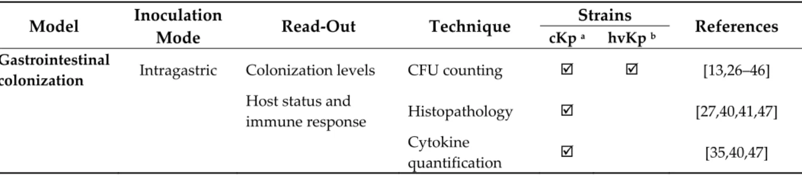
Table 1. In vivo investigated readouts in classical K. pneumoniae and hypervirulent K. pneumoniae.
Model Inoculation
Mode Read-Out Technique
Strains
References
cKp a hvKp b
Gastrointestinal
colonization Intragastric Colonization levels CFU counting [13,26–46]
Host status and
immune response Histopathology [27,40,41,47]
Cytokine
Inflammatory marker measurement [40–42,47,48] FISH [26,28,41,42,47] Microscopy [41] Microbiota modification 16S DNA sequencing [13,26,35,39,41] CFU counting [29,42] Systemic
dissemination Intragastric Colonization levels CFU counting [49–53]
Host status and
immune response Histopathology [49,51]
Cytokine
quantification [49]
Microscopy [54]
Lethality Survival [51,55]
Intravenous Colonization levels CFU counting [16,56–58] Host status and
immune response Histopathology [56,58]
Cytokine
quantification [56–58]
Microscopy [59]
Lethality Survival [56,58–60]
Intraperitoneal Colonization levels CFU counting [53,55,61–66] Host status and
immune response Histopathology [62,66–68] Cytokine quantification [63,64,66,69,70] Inflammatory marker measurement [65,66,70] Microscopy [61] Intraperitoneal Microbiota modification 16S Sequencing [66] Lethality Survival [16,55,61–66,68– 73] Subcutaneous Colonization levels CFU counting [74,75]
Host status and immune response
Liver abscess
measurement [74]
Lethality Survival [72,75–77]
Pulmonary
infection Intranasal Colonization levels CFU counting [78–97]
Host status and immune response Cytokine quantification [79,83,84,86,89– 92,95,98] Histopathology [78– 82,86,91,92,95– 97] Inflammatory marker measurement [79,82,84,86,88,92 ] Microscopy [92]
Lethality Survival [78,81,85,86,93,95 ,98] Intratracheal Colonization levels CFU counting [75,99–118]
Host status and immune response Cytokine quantification [100– 103,108,115,116] Histopathology [99–101,103– 105,110,113,116,1 19,120] Inflammatory marker measurement [100–103,109,114] Microscopy [105,113] Lethality Survival [75,77,100,101,11 1,112,115–120]
UTI c Intraurethral Colonization levels CFU counting [31,46,121–130]
Host status and
immune response Histology [127–129]
Microscopy [124,127,128]
CAUTI d Intraurethral Colonization levels CFU counting [131]
a cKp: Classical K. pneumoniae, b hvKp: Hypervirulent K. pneumoniae, c UTI: Urinary tract infection, d CAUTI: Catheter-associated urinary tract infection.
2. Intestinal Colonization
In humans, K. pneumoniae colonizes the intestinal microbiota and the establishment of this niche is considered as a starting point for dissemination and further infection [11,132]. Several murine models of intestinal colonization have thus been developed to study this pathophysiological process. Mice are an excellent tool to study K. pneumoniae gut colonization because they are easy to manipulate, the access to fecal content is easy, and they have a high reproductive rate. There is, however, no clear consensus concerning the genetic background of mice (OF1, BALB/c, CD1, CFW1, and C57BL/6), and no differ-ence in the dynamics of K. pneumoniae colonization between the various backgrounds.
Routinely, the murine model of intestinal colonization consists of an oral route of inoculation (gavage, and less frequently pipette feeding or the administration of the path-ogen in the drinking water) and the subsequent determination of the intestinal coloniza-tion by CFU counting in the intestinal or fecal contents, and an analysis of the immune host response (Table 1). Fecal shedding is currently used as a marker for gastrointestinal colonization by K. pneumoniae but an analysis of the different parts of the intestinal tract of mice that are orally contaminated showed an increasing gradient of colonization all along the intestine, with a preferential localization in the colon [26]. These approaches have highlighted the role of the local microbiota in K. pneumoniae implantation within the gut and characterized Klebsiella virulence and colonization factors.
2.1. Colonization Resistance of the Intestinal Microbiota to K. pneumoniae
Colonization resistance refers to the ability of the microbiota to prevent the expansion and persistence of exogenously acquired bacterial species [133]. Such a mechanism has been described in relation to the opportunistic pathogen K. pneumoniae (Figure 1A) [134]. Accordingly, the absence of intestinal microbiota in C57BL/6J germ-free (GF) mice re-sulted in higher levels of intestinal colonization by K. pneumoniae than those observed in conventional mice, and the introduction of a normal microbiota into K. pneumoniae-colo-nized mice resulted in the reduction and clearance of K. pneumoniae [26,27]. The induction
of microbiota dysbiosis, by antibiotics or chemical agents, is thus necessary to obtain effi-cient colonization by classical K. pneumoniae of the murine intestine, unlike the establish-ment of hvKp, which does not require microbiota disruption (Figure 1) [28,29,49].
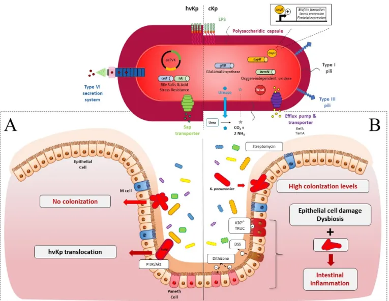
Figure 1. K. pneumoniae in the gastrointestinal (GI) tract in an in vivo murine model (A) In healthy immunocompetent
mice: (i) cKp (red oval, fine capsule) is not able to colonize the intestinal epithelium due to the presence of upright endog-enous microbiota (yellow, blue, green and purple ovals, full line); (ii) hVKp (red oval, hypermucous) deploys many viru-lence factors. Type VI Secretion System (T6SS): to counteract the microbiota barrier; transporters of antimicrobial peptides and amino-acids (Sap, Tdc) and enzymes with lysine decarboxylase activity (Cad). Hypercapsular phenotype due to the presence of pLVPK plasmid (black circle) protects the pathogen against the host immune reaction. HvKp use the PI3k/Akt pathway to cross the epithelial barrier. (B) In intestinal dysbiotic mice: Dysbiosis (yellow, blue, green and purple ovals, dotted line) chemically induced (streptomycin, Dextran Sodium Sulfate (DSS), dithizone) or endogenous to immune-de-ficient murine models (Il10-/-, T-bet−/− Rag2−/− ulcerative colitis (TRUC)) allows the implantation of cKp. Gut colonization by
K. pneumoniae associated with intestinal epithelium damage and dysbiosis enhanced the inflammatory state. Essential cKp
colonization factors are related to capsule synthesis (WcaJ, red circle), metabolism advantage (urease, oxidase, and gluta-mate synthase) (blue diamond, blue and green cylinder), GI stress resistance (EefA, TamA, OxyR,) (purple, yellow cylin-der) and surface cell adhesion (Type 1 and 3 pili).
Oral administration of streptomycin, an antibiotic that targets commensal facultative anaerobes, is the gold standard to establish cKp intestinal colonization, with bacterial in-oculums varying from 105 to 108 CFU [13,28,30–34,48,121,122]. Miller and Bonhoff ob-served that no Bacteroides were present in the feces of streptomycin-treated mice [135]. Accordingly, the ability of gnotobiotic mice to resist gut colonization by K. pneumoniae has
recently been reported to be due to the presence of the Bacteroidetes phylum [35]. Thus,
Bacteroides disruption by highly-concentrated streptomycin treatment led to a
“supershed-der phenotype” with high levels of fecal colonization (108 CFU/g feces) by the K.
pneu-moniae strain KPPR1S [67]. Constant streptomycin pressure is therefore necessary to
main-tain a stable and high colonization of cKp, from 108 to 1010 CFU/g feces. Lagrafeuille et al. showed that the colonization level of cKp decreased by 3 log units in one week if strepto-mycin was given at only one time point 48 h before inoculation [36]. However, although
K. pneumoniae becomes undetectable in the feces after the initial colonization, it seems to
persist at low levels in microniches in the colonic environment. Indeed, 15 days after CFU counts fell below the detection limit, the administration of ampicillin induced a rise above the detection limit of K. pneumoniae CFU in mice feces [37].
In 2003, Hoyen et al. analysed the effect of the administration of different antibiotics on the gut colonization abilities of ESBL-producing K. pneumoniae in a murine model [38]. The mice were intraperitoneally treated with either antibiotics targeting anaerobes such as clindamycin, piperacillin/tazobactam, ceftriaxone, and ceftazidime, or antibiotics with a low effect on anaerobes such as cefepime, levofloxacine, and aztreonam, and then orally inoculated with the SHV ESBL-producing P62 strain (103 CFU at day 0 and then 108 CFU at day 5).
Antibiotics with low effects on anaerobes did not influence K. pneumoniae intestinal colonization levels, whereas treatment with anaerobe-targeting antibiotics resulted in greater colonization levels (from 105 to 107 CFU/g feces) than treatment with a saline buffer. Only clindamycin promoted stable and rapid colonization of up to 109 CFU/g feces at 3 days post-inoculation [38]. In line with these results, Perez et al. showed that the in-hibition of anaerobes, especially the Bacteroides species, by daily subcutaneous treatment with clindamycin allows maximal K. pneumoniae gut colonization levels (up 1010 CFU/g feces) [29]. More recently, the correlation between microbiota composition and K.
pneu-moniae gut colonization was assessed by a deep sequencing approach. Treatment with
fi-daxomicin, a molecule with a narrow-antimicrobial spectrum, poorly modulated the mi-crobiota composition and achieved only low intestinal colonization (<104 CFU/g feces) by
K. pneumoniae. In contrast, the administration of vancomycin, which inhibits Firmicutes,
resulted in a greater level of colonization (106 to 1010 CFU/g of stool) [39].
Dysbiosis, which is required to impair colonization resistance, can also be induced by other chemical treatments (Figure 1B). For instance, the induction of intestinal dysbio-sis by the oral administration of 3% w/v Dextran Sodium Sulfate (DSS) results in a 3-log increase in the K. pneumoniae colonization level in feces of mice 3 days post-inoculation [27]. This high colonization level is however not linked to DSS-induced intestinal inflam-mation, since no increase in the colonization by K. pneumoniae was observed in a
Citrobac-ter rodentium model that generates inflammation but not dysbiosis [27].
The immune status of the host determines the pro-inflammatory levels of intestinal tissue in response to K. pneumoniae colonization. However, the precise role of each protag-onist (host immune response, endogenous microbiota, and K. pneumoniae) is hard to deci-pher owing to the complexity and diversity of the animal models used in the different studies (Figure 1B). Genetically modified mice models have been used to go further in the investigation of the host pathophysiology during intestinal colonization by K. pneumoniae. For instance, in Il10−/− mice with a low grade of intestinal inflammation, K. pneumoniae infant isolates potentiated inflammation and elicited colitis since fecal lipocalin-2 levels increased 1-month post-infection [40]. Similarly, oral administration of salivary K.
pneu-moniae isolates potently induced colonic TH1 cells in the same model [41]. The colitogenic properties of K. pneumoniae are also linked to the endogenous microbiota. The pro-inflam-matory effects of K. pneumoniae were higher in mono-associated or naturally colonized mice than in mice colonized with a full complement microbiota [40]. In addition, the K.
pneumoniae strains recovered from the feces of colitis-susceptible animals, T-bet−/− Rag2−/− ulcerative colitis (TRUC), were able to induce colitis in wild-type mice after oral
admin-istration, but only in the presence of endogenous microbiota [42]. These observations pro-vide epro-vidence that commensal microbiota work in concert with K. pneumoniae and other intestinal pathobionts to cause intestinal inflammation.
The role of K. pneumoniae in the exacerbation of intestinal inflammation has also been reported in mice with intestinal microbiota disruption due to chemically induced colitis. The administration of K. pneumoniae to new-born mice treated with dithizone, a molecule that induces Paneth cell ablation associated with microbiota dysbiosis, exacerbates intes-tinal inflammation, which leads to a necrotizing enterocolitis-like pathology [47,48]. The presence of colitogenic agents such as K. pneumoniae in gut microbial communities is thus necessary but not always sufficient to elicit intestinal inflammation in susceptible mice colitis models (either genetic or chemically induced). The intrinsic capacities of the strains also seem to be important, but the specific associated inflammatory factors remain to be identified.
2.2. K. pneumoniae Colonization Factors
The implantation of cKp strains in the gut and their persistent colonization involve competition with commensal bacteria and a variety of stressors for survival, including the limitation of resources. Bacterial fitness in this complex environment is mediated by a multitude of pathogen-associated factors, usually referred to as colonization factors, that enable the dense growth and persistence of the bacteria in the intestinal lumen.
Intestinal colonization models combined with the screening of K. pneumoniae trans-poson insertional mutant libraries (signature-tagged mutagenesis [STM] and multi-screening STM [MS-STM]), are powerful approaches that are able to identify the genetic factors involved in gut colonization. The mutants impaired in the intestinal colonization were mainly affected in metabolic pathways, membrane transporter synthesis, DNA-me-tabolism, transcriptional regulation, and unknown functions, and in lipopolysaccharides (LPS), phospholipids and fatty acid biosynthesis [43,122]. More recently, K. pneumoniae genes crucial for dense gut colonization were identified by the screening of a highly satu-rated transposon mutant library (<150,000 unique mutations) in antibiotic-treated mice orally inoculated with ~108 CFU of carbapenem-resistant K. pneumoniae (102 to 103 CFU of each transposon mutant per inoculum). The bacterial content of the fecal samples longi-tudinally collected for 4 weeks were sequenced by genome-wide transposon insertion se-quencing (INseq) [13]. This approach identified the genes that promote short- and long-term high-density colonization of the intestinal tract, including genes encoding for in-ner/outer membrane proteins, proteins involved in carbohydrate metabolism, DNA re-pair/metabolism, glutamate metabolism, and porphyrin metabolism. Some mutants with enhanced fitness were also identified with this methodological approach [13]. The crea-tion of isogenic mutants and their testing in individual or competitive assays have firmed the importance of the genes previously identified. For instance, Jung et al. con-firmed dramatic defects related to gut colonization for ΔtamA (transport system), ΔgltB (glutamate synthase) and ΔhemN (oxygen-independent oxidase) mutants in a 1:1 compe-tition assay with the wild-type K. pneumoniae strain, whereas the ΔfhlA (formate hy-drogenlyase transcriptional activator) mutant colonized the animal gut better than the wild-type [13]. The screening of mutant libraries in colonization models emphasizes the importance of the diversity of the bacterial functions that play prominent roles in bacterial establishment inside the gut. In particular, several metabolic functions are primordial for bacterial colonization since they confer advantages to outcompete resident microorgan-isms with a similar metabolism (Figure 1).
To validate the role of a bacterial function in intestinal colonization, the classical ap-proach is to test the isogenic mutant of the encoding-gene in a colonization model, either individually or in competition with the wild-type parental strain. The phenotype of the mutants will then strongly depend on the experimental model, i.e., individual or compe-tition testing. Mutants deficient in a factor required for compecompe-tition in an ecological niche could be deficient when tested in a competitive assay but could be unaffected when tested
individually. For instance, a cKp mutant defective in urease production (ΔureA) showed no colonization defect when tested individually but was impaired in its colonization ca-pacities when tested in competition with the wild-type strain [30]. However, authors rarely test the mutants in both models in their experimental methodology. Hsieh et al. showed that the cad and tdc operons, which confer adaptation of the hvKp NTUH-K2044 strain to bile salts and acidity, respectively, were involved in efficient intestinal coloniza-tion when the mutants were tested in competicoloniza-tion with the virulent wild-type strain. However, the phenotype of the mutants was not evaluated in an individual colonization assay [34]. Similarly, the tripartite efflux pump EefABC conferred a competitive ad-vantage on cKp for intestinal colonization, but its role was not analysed in an individual colonization assay. Growth experiments in the presence of different stresses showed that this efflux system confers acid tolerance on bacteria in response to an inorganic acid (e.g., chlorhydric acid), a condition encountered inside the intestinal tract [33].
Some other colonization factors contribute not only to competition with microbiota, but also to bacterial establishment and multiplication in the intestine (Figure 1). Thus, mutants deficient in these functions are deficient in both individual and competitive as-says. For instance, the role of capsular polysaccharides (CPS) in intestinal colonization was described in both individual and competitive assays by Young et al. [67]. The capsule-deficient mutants ∆manC and ∆wcaJ colonized poorly over the course of 15 days in indi-vidual colonization assays, and the ∆manC mutant presented a negative competitive index (∆manC/WT) in co-infection with the wild-type strain [67]. The role of the K. pneumoniae capsule in establishing robust gastrointestinal colonization was also observed by Favre-Bonté et al. in both individual and competitive assays with the wild-type strain [28]. In their study, a cKp capsule-deficient mutant colonized at lower levels (104 CFU/g feces) than the wild-type strain, which persisted in the intestinal tract at high levels (108 CFU/g feces), and in situ hybridization with fluorescence-labelled oligonucleotide probes on the colonic section in mice showed that the mutant formed clusters in the mucus layers, whereas the wild-type strain remained dispersed [28]. The role of certain fimbriae that are assembled by the chaperone-usher (CU) pathway has also been assessed in both individ-ual and competition models. An in silico predictive approach identified two novel CU systems encoded by kpj and kpg loci in cKp LM21. The deletion of the usher-encoding gene in these two operons significantly impaired intestinal tract colonization in a co-coloniza-tion assay with the wild-type strain. However, only the mutant deficient in the usher of the putative Kpj structure was tested in individual colonization assays, in which it also presented a colonization defect [32]. In a more recent study using INSeq, the deletion of
kpjC, which encodes the usher protein in the kpj operon, did not however significantly
alter the intestinal colonization of the K. pneumoniae carbapenem-resistant (CR-Kp) ST258 strain in a competitive assay [13]. The role of type 1 and type 3 pili have been mainly studied in murine intestinal competition assays by co-infecting a deficient mutant and the wild-type parental strain. Although the role of type 3 pili in efficient colonization is well established, studies are at a variance concerning the role of type 1 pili [13,31,32,44].
Bacteria can form multicellular biofilm-like communities in the gastrointestinal tract and therefore co-infection with a wild-type strain and a mutant deficient in individual colonization assays could form a mixed population (intraspecies) in the intestine, which would help compensate for the deficiency of the mutant strain. However, to our knowledge, no study has described this phenomenon. Several bacterial factors have been identified in individual colonization assays but their role in a competitive context has not been assessed. For instance, a K. pneumoniae oxyR mutant was drastically affected in an individual murine intestinal colonization assay after orogastric inoculation but its pheno-type was not determined in a competitive assay [45]. The transcriptional factor OxyR, a member of the LysR regulator family, is commonly associated with the detection of ele-vated levels of reactive oxygen species (ROS) such as hydrogen peroxide and the subse-quent regulation of the expression of antioxidant genes that protect the bacterial cells from
being killed. OxyR is also involved in the regulation of several phenotypes classically as-sociated with the process of gastrointestinal colonization, such as biofilm formation, fim-brial synthesis, motility, and resistance to intestinal stresses [136–138].
The use of animal models allows for a better understanding of the molecular mecha-nisms involved during gastrointestinal tract colonization by K. pneumoniae. The behavior of Klebsiella inside the intestine flora and its interactions with the other members of the microbiota are poorly documented however, and further elucidation would require the development of gnotobiotic models with controlled microbiota. Finally, although it is as-sumed that the intestinal colonization precedes the infection of secondary sites, the precise pathophysiology of Klebsiella infections is still unclear.
3. Systemic Infections
Extraintestinal infections involving bacteria from the intestinal microbiota result from both exogenous and endogenous contamination. Unlike cKp, hvKp strains are able to disseminate in immunocompetent patients, which increases the risk of community-ac-quired infections. Different murine models have been developed to assess the pathophys-iology of extra-intestinal infections and their associated mortality (Table 1). Oral inocula-tion of the pathogen, by mimicking the natural route of K. pneumoniae infecinocula-tion, makes it possible to study the phenomenon of bacterial translocation. However, extra-intestinal inoculations (i.e., subcutaneous, intraperitoneal, or intravenous) are usually performed to assess systemic bacterial fitness. While no clear consensus has been established regarding the genetic background of the mice used in experimental models (e.g., BALB/c, C57Bl/6, ICR, Swiss Webster), the BALB/c line is most often used [49,59,61,74]. Genetically im-paired models are also used to investigate the behavior of hvKp strains in immunocom-promised mice.
3.1. Spreading from the Intestinal Tract
Orogastric inoculation of the pathogen is a useful way to investigate the spatial and temporal kinetics of hvKp K. pneumoniae infection such as the ability of the bacteria to cross the epithelial barrier (Figure 1A) and to induce extraintestinal infections, particularly in the blood, spleen, and liver. Although intestinal translocation is not clearly established for cKp strains, oral gavages of BALB/c mice with 107 CFU of hvKp CG43 induced, as early as 24 h post-inoculation, colonization of the spleen with 3 to 4 log CFU/g, and subsequent dissemination to the liver, blood, and kidneys at 36 h, 48 h, and 72 h post-inoculation, respectively [49]. Four stages of hvKp strain infection have thus been determined: (i) in-testinal colonization rapidly followed by (ii) extrainin-testinal dissemination, (iii) hepatic replication and, (iv) sepsis [49]. Each of these infection steps is governed by specific bac-terial factors, especially those encoded by the virulence plasmid of hvKp strains since the hvKp CG43 pLVPK-cured strain is attenuated in the dissemination from the gastrointes-tinal tract to systemic circulation and to extraintesgastrointes-tinal organs compared to the parental wild-type strain [50].
After oral inoculation of the CG43 hvKp strain (107 CFU) in C57BL/6 mice, 28 STM mutants (impaired in cell metabolism, surface components and transporters, and regula-tion funcregula-tions) that attenuated liver and spleen disseminaregula-tion were identified. Of these, eight avirulent mutants specifically affected in the epithelial barrier translocation were identified; they did not cause mouse mortality, but their virulence was restored when in-oculated intraperitoneally [49]. The factors essential for the translocation process are as-sociated with type III fimbriae assembly, uracil permease synthesis, and quorum-sensing. The molecular mechanisms of intestinal barrier translocation remain incompletely de-fined but could involve the PI3K/Akt dependent pathway [54]. The same group identified the Sap complex as being involved in the dissemination process from the intestine after oral inoculation of hvKp Ca0437. The Sap transporter, an inner membrane complex con-ferring resistance on antimicrobial peptides in Salmonella, is associated with extraintestinal bacterial dissemination to the spleen and liver. A ∆sapA mutant had a three-fold reduced
bacterial load in the liver and spleen 72 h after oral inoculation compared to the Ca0437 wild-type strain. This attenuated phenotype was shown to be associated with a lower his-tological pathology score in the liver and colon of ∆sapA-inoculated mice than in those inoculated with the wild-type strain [51]. The type VI secretion system (T6SS), a molecular syringe with bactericidal activity harbored by certain pathogens, is also involved in hvKp virulence since it allows microbiota-encountered resistance during the colonization pro-cess to be bypassed [139]. Hsieh et al. identified a T6SS in the hypervirulent NTUH-K2044 strain that is critical for successful intestinal colonization and extraintestinal dissemina-tion. In mice inoculated with both the wild-type NTUH-K2044 strain and its T6SS deletion mutant (ratio 1:1), the mutant was detected at much lower levels than the wild-type strain in the intestine, liver, and spleen (competitive index of 0.123, 0.197, and 0.118, respec-tively) 7 days post-inoculation [52]. Intragastric inoculation can reproduce the first steps of the natural K. pneumoniae infectious route, but it does not allow the identification of the factors specifically involved in extraintestinal dissemination.
3.2. Spreading from Extraintestinal Sites
The systemic inoculation of hvKp in mice is performed intravenously, subcutane-ously, or intraperitoneally in order to bypass the gastrointestinal barrier. Such approaches have allowed the identification of specific hvKp-associated virulence factors in in vivo murine models.
As an essential component, the pLVPK plasmid, which carries inter alia, the rmpA and magA virulence genes, is largely responsible for the hypervirulent phenotype in hvKp. The presence of rmpA (a regulator of the mucoid phenotype) and magA (mucovis-cosity-associated gene A) genes on the pLVPK plasmid confers on hvKp a hypermucous phenotype [68,140]. The loss of RmpA and/or RmpA2 is associated with decreased capsule production, potentially making RmpA/RmpA2 the critical factors for the increased viru-lence of hvKp strains compared to cKp strains [17]. However, whereas prevaviru-lence studies showed an association of rmpA with infection severity in mice, its role in K. pneumoniae virulence remains unclear. The deletion of the plasmid-born rmpA gene in the hvKp NTUH-K2044 strain does not significantly reduce the virulence of K. pneumoniae in mice, either in mice survival assays or in in vivo competition assays after intraperitoneal inocu-lation [55]. MagA (renamed WzyKpK1) is a capsular polysaccharide polymerase specific to serotype K1 [68,71]. ∆magA mutants lose their mucoviscosity, become susceptible to neu-trophil phagocytosis, and are not lethal to mice [68,71]. Likewise, an uncapsulated ∆wcaJ mutant deficient in the WcaJ glycosyltransferase involved in the initial steps of capsule production is impaired in its ability to disseminate into the liver, lungs, and spleen of intraperitoneally infected mice in comparison to the parental strain [53]. After intraperi-toneal inoculation in mice, the serine protease hrtA mutant in a hvKp strain, abrogated in capsule production, had a 500-fold greater LD50 than the WT strain, thereby evidencing the primordial role of the capsule of K. pneumoniae in the infection process [141].
In vivo models have also been used to investigate the role of capsular polysaccharide composition in virulence. Pan et al. showed that intravenous inoculation of a KpL1 hvKp mutant deficient in gmd, a gene responsible for fucose synthesis, induces a mortality rate among mice of only 5% within 14 days compared to full mortality with the wild-type strain [60]. One explanation would be that the fucose-rich capsular serotype of virulent strains has less affinity for murine macrophages than the avirulent capsular-serotype strain [16]. Fucose-poor and mannose-rich capsules of classical KpU1 strains are highly adherent to mouse-isolated peritoneal macrophages in contrast to fucose-rich and man-nose-poor capsules of hvKp KpL1 strains, suggesting that a fucose-containing capsule has the advantage of bypassing the host defences [59]. Additionally, a KpL1 gmd mutant had a greater adherence to pre-recruited mouse peritoneal macrophages than its parental strain and was unable to disseminate via systemic circulation [60]. The large amount of mannose in cKp capsules would thus explain the tropism for phagocytes and the greater
clearance of cKp from the host compared to hvKp. In accordance, the strains of K.
pneu-moniae expressing Man-α2/3-Man or Rha-α2/3-Rha sequences in their capsule could be
effectively recognized by macrophage mannose receptors and ingested by macrophages, although hvKp K1 and K2 serotype strains that lack Man-α2/3-Man or Rha-α2/3-Rha in their capsule would thus not be recognized by the macrophage lectins [142,143]. In hvKp strains, capsular polysaccharides are thus essential virulence factors since they avoid, by their composition, recognition by macrophages. Thus, intraperitoneal injections in BALB/c mice showed that K1 and K2 capsular serotype strains were highly virulent, whereas the uncapsulated ∆K1 mutant (∆rfbP) had substantially attenuated virulence sim-ilar to that of the cKp K62 strain. Serotypes K1 and K2 had greater phagocytic resistance to neutrophils than the K62 and the ∆K1 mutants, and the determination of CFU in organs showed that mice lethality is linked to the inability to clear bacteria [62]. Time-dependent increases in cytokines and chemokines (TNF-α, IL-1ß, IL-6, IL-10, KC, and MIP-2) in the serum and liver were observed only in hvKp K1- and K2-infected mice in contrast to the inoculations of K62 and ∆K1, which did not result in similar strong immune responses [62].
Other surface structures, such as lipopolysaccharides, play a role in the expression of hvKp virulence in vivo. For instance, in a mouse intraperitoneal infection model, hvKp
wbbO mutants (NTUH-K2044 and NTUH-A4528 backgrounds) impaired in a
galactosyl-transferase that is required for serotype O1 were poor colonizers of mouse organs (i.e., liver, spleen, and blood) and induced a lower lethality rate than the wild-type strain [63]. Membrane lipoproteins Pal, LppA and YfgL are also involved in the virulence, dissemi-nation, and induction of inflammation: mice intraperitoneally infected with deficient mu-tants had a better viability, as measured by the determination of the LD50 or 28-day sur-vival rate, had a lower bacterial dissemination and a lower IL-6 production in the liver and spleen than the mice infected with the parental wild-type strain [64,69].
The production of siderophores also confers on K. pneumoniae a selective advantage for virulence in in vivo systemic infection models. A quantitative analysis of siderophores has demonstrated that hvKP strains produce more siderophores than cKp strains and that this trait enhances hvKP virulence, since a high siderophore concentration was strongly predictive of a hvKp isolate and an increased lethality in a mouse systemic infection model [76]. HvKp strains have the ability to produce four different siderophores: enterobactin (Ent), yersiniabactin (Ybt), salmochelin (Sal), and aerobactin (Aer), but only Sal and Aer are specific to hvKp. The iro and iuc operons, responsible for Aer synthesis, are carried by the virulent pLVPK plasmid in the hvKp strains [22]. Aerobactin accounts for the over-whelming majority of increased siderophore production in hvKp, independent of the gene copy number. The role of each siderophore in virulence has been evaluated in pulmonary, subcutaneous, and intraperitoneal infection models by measuring the mortality of out-bred CD1 mice after subcutaneous challenge with hvKP1 mutants deficient for one or more siderophores. Of all the mutants tested, only the hvKP1 isogenic Aer (ΔiucA) mutant showed attenuated virulence in all models [72,77]. The role of aerobactin in virulence is however lower than that of the capsular polysaccharides. K1 and K2 strains were highly virulent in BALB/c mice after intraperitoneal injections, whereas the uncapsulated ∆K1 mutant (∆rfbP), while still harboring the Aer encoding gene, had a substantially attenu-ated virulence, similar to that of the cKp K62 strain [62]. Fulfilling Koch’s postulate, the combined effects of the overproduction of the virulence factors and a high degree of phag-ocytosis resistance seem to be very useful in the dissemination of hvKp. The interactions with host immunity are largely related to capsule polysaccharides and to the virulence of the strains (e g cKp or hvKp) which is dependant of the bacterial accessory genome [6,62].
By mimicking host pathology-engendered susceptibility, immunocompromised in vivo models greatly contribute to the understanding of the pathophysiology of K.
pneu-moniae systemic infections. hvKp strains cause skin and tissue infections in
immunocom-promised patients which can be reproduced in neutropenic mice treated with an anti-Ly6G antibody [19,74]. Interestingly, in this model some CR-Kp strains are as virulent as
a hvKP strain in their ability to form subcutaneous abscesses and to disseminate to the liver. Intravenous infection of immunocompromised diabetic mice with the low virulent cKp (KpU1 UTI strain) results in more severe lethality than in non-diabetic mice. Simi-larly, a hvKP strain isolated from the liver abscess had a greater virulence in diabetic than in nondiabetic mice [56]. In the same study, intravenous infection with hvKP significantly decreased the blood TNF-α level in diabetes mellitus mice, whereas the IL-1β level tended to increase in the blood of both infected nondiabetic and diabetic groups [56]. Another study using diabetic mice treated with streptozotocin, which ensures the death of pancre-atic β-cells, evidenced the ability of cKp and hvKp to translocate into the liver via Kuppffer cell activation. In this model, diabetes mellitus caused enteric dysbiosis, which leads to the production of nitric oxide and the subsequent stimulation of Kuppffer cells [57]. Inter-estingly, IFN-γ KO mice intravenously infected with the hvKp 43,816 strain were no more susceptible than their wild-type counterparts. The overproduction of the liver cytokine IL-10 in IFN-γ KO mice 2 days post-infection compared to the production in wild-type mice (35 ng/mL vs. below detection limit, respectively) could explain the underlying re-sponse [58]. However, other studies showed that the inhibition of IL-10 activity using the anti-IL-10 antibody tends to enhance the survival of mice after intraperitoneal or intratra-cheal inoculations of K. pneumoniae [144,145]. The role of the IL-10 cytokine can vary sig-nificantly depending on the site of infection [58].
Systemic infection models can be used to investigate the dissemination of the patho-gen and its colonization of extraintestinal organs while avoiding the epithelial barrier translocation process. Since the prevalence of cKp strains in severe disseminated infec-tions is low, most studies use K1 or K2 hvKp strains with high infectious doses. However, cKp strains can cause community-acquired infections in susceptible hosts with comorbid-ities who are immunocompromised or who have an existing barrier breakdown. Thus, the development of animal models mimicking susceptible hosts will be a significant contri-bution to understanding the pathophysiology of these infections.
4. Pulmonary Infections
K. pneumoniae has been historically recognized as a common respiratory pathogen
since its discovery in 1882 and is involved in both community-acquired pneumonia (CAP) and hospital-acquired pneumonia (HAP) [4,146]. CAPs caused by K. pneumoniae are rare in Europe and North America but account for 15% of the total cases of CAP in Asia and South Africa, mainly due to the increasing prevalence of hvKp strains [146–150]. Medical ventilators are a major risk factor in HAPs caused by K. pneumoniae because they provide the pathogen with a surface on which to colonize and form biofilms [149,151].
4.1. In Vivo Models
To assess host–pathogen interactions during K. pneumoniae pulmonary infections, several in vivo models have been developed including non-mammalian models [25] (Ta-ble 1). However, murine models are the most widely used because they are easy to handle and because there is a significant similarity in the infectious process of murine and human pneumonia [152]. The first model was developed in 1947 and consisted of an intratracheal inoculation of bacteria in rats to evaluate antibiotic treatments [119,120]. Since then, sev-eral models of pneumonia have been used that are mostly based on two methods of inoc-ulation.
The first method involves the direct instillation of bacteria into the lower respiratory tract. It requires general anesthesia, either by endotracheal injection or oropharyngeal in-oculation. For endotracheal injection, the trachea is exposed by surgery and a volume of 10 to 50 µL of bacterial suspension is injected before the closure of the incision [99–104]. This method can lead to the inflammation of the area, especially of the surgical wound, that potentially affects the global immune response measured at the endpoint of the ex-periment. For oropharyngeal inoculation, animals are placed in a semi-vertical position and a volume of 10 to 50 µL is injected with a blunt-tipped needle either at the tracheal
entrance or directly into the bronchi [77,105–109,119,120]. These techniques allow the ad-ministration of a precise bacterial inoculum into the lungs, which results in a good repro-ducibility of the infection [152,153]. Both methods are tricky and can cause the unilateral infection of the left lung and consequently a disparity in the effects of the infection on animals [99].
The second popular method used to induce pneumonia involves the inoculation of bacteria in the upper respiratory tract by intranasal instillation. The animals are held in a semi-vertical position, after brief anesthesia, and droplets of the bacterial inoculum are deposited onto the nares, allowing the contamination of the lungs by aspiration during breathing. The volume of the inoculum is generally around 20 µL, administered sequen-tially by aliquots of 5 to 10 µL [78–84]. Intranasal instillation is less invasive than instilla-tion in the lower respiratory tract but the dose of bacteria that reaches the lungs is difficult to control [152]. The inoculum can (i) be expulsed from the nares during expiration, (ii) be diverted to the alimentary tract or (iii) stay in the upper respiratory tract, leading to a high variability of infection.
There is no consensus on the bacterial inoculum load in the overall experiments per-formed with murine pulmonary models. However, it should be noted that inoculation in the lower respiratory tract requires a lower dose than intranasal inoculation. Bacterial in-oculum varies from 103 to 109 CFU, depending on the virulence of the strain (hvKp or cKp) and the susceptibility of the mice [81,85,102,108]. Although there is no consensus on the background of the mice used to mimic the pulmonary infection, C57BL/6 mice are the most commonly chosen. However, studies related to the host physiology are more likely to use BALB/c, 129, CBA, or C3H backgrounds. In addition, genetically modified mice whose production of immunological markers is impaired are often used to characterize the host responses to bacterial pulmonary infection.
The classical method to evaluate the role of bacterial factors in the establishment of pneumonia and the impact of host immunity or treatments on the disease outcome is to determine the bacterial burden in the lungs and other tissues by standard quantitative culture. The determination of the bacterial load is sometimes supplemented by other ap-proaches such as survival rate, histological studies of lung tissues, or host immune re-sponse analysis in the tissues and the bronchoalveolar lavage fluid (BALF) (Table 1). An-other alternative, which is highly sensitive and non-invasive, is the quantification of the bioluminescence emitted by a pathogen genetically modified to express the biolumines-cence-encoding genes (lux operon). This technique, infrequently used with K. pneumoniae lung infection models, is a useful tool to monitor the bacterial burden and to analyse the kinetics of spreading, and in parallel, to contribute to reducing animal numbers in accord-ance with ethical regulations.
4.2. K. pneumoniae Pulmonary Virulence Factors
Lung infection models in mice were used to characterise K. pneumoniae virulence fac-tors (Figure 2) and tropism without any rationale concerning the choice of the inoculation methods which seemed to depend mainly on routine laboratory practices. The polysac-charide capsule was one of the first and most described virulence factors in in vivo pneu-monia models and is essential to establish pulmonary infection despite local immune pressure [86,99,105,110]. The deletion of the capsule in K. pneumoniae impairs its growth and survival in the lungs and aborts its ability to cause severe pulmonary infection and animal death in both intranasal and intrapulmonary in vivo models [78,86,105,110]. In addition to protecting against phagocytosis, the capsule reduces the host inflammatory response by shielding the LPS O-antigen from innate immunity recognition, thus limiting immune cell recruitment in BALF and the production of pro-inflammatory cytokines (TNF-α, IFN-γ and IL-6) in the early and late stages of infection in the intranasal model [79,86,98]. Capsule production is transcriptionally regulated by a number of proteins that are associated with bacterial virulence in the lungs [4]. For instance, a mutant deficient in
FimK, a pilus regulator involved in capsule production, produces significatively less cap-sule than the wild-type strain and is attenuated in virulence in murine intrapulmonary models compared to the parental strain, suggesting that the capsule protects bacteria from the host immune response in the lungs [103]. More recently, Zhang et al. used an intrap-ulmonary in vivo model to identify AmpR, a regulator of the WcaJ enzyme that initiates capsule synthesis, as essential for the virulence of both hypermucoviscous and non-hy-permucoviscous K. pneumoniae strains [111]. In addition, ΔrcsB and ΔrmpA mutant strains had lung colonization levels that were about 4-log lower than that of the parental strain KPPR1S at 24 h post-infection in an intranasal model of pneumonia in mice, and a ΔrmpC strain was also attenuated by nearly 2 logs [87]. Unlike ∆rmpA and ∆rcsB, the ΔrmpC mu-tants retained the hypermucoviscous phenotype, supporting the notion that this pheno-type is not simply due to the overproduction of capsular polysaccharides and is not di-rectly associated with virulence [87]. The expression of rmpA modulates capsule produc-tion under the control of the RcsB, KvrA, and KvrB regulators. Their deleproduc-tion in hvKp strains decreased the bacterial virulence in an intranasal model of pneumonia in a mouse [82]. With the cKp strain, the deletion of kvrA also affected the virulence but had no effect on capsule gene expression or capsule-related phenotypes, suggesting that the effect of KvrA on virulence is not exclusively mediated by capsule production [82].
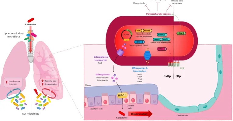
Figure 2. K. pneumoniae in the lungs in an in vivo murine model Local (respiratory) and distal (gut) microbiota (yellow,
blue, green, and purple ovals) can modulate the host immune response to K. pneumoniae (red oval) lung infection and K.
pneumoniae pathogenicity unlike dysbiotic gut microbiota (yellow, blue, green, and purple ovals, dotted line). Virulence
factors involved in lung colonization by classic (cKp, right side) or hypervirulent (hvKp, left side) K. pneumoniae strains:
Capsule, overproduced in hvKp and finely regulated by many pathways (FimK, RcsB, KvrA, KvrB, RmpA, RmpC); Lip-opolysaccharide (LPS), which depends on the wecA gene for the initiation of biosynthesis; Membrane proteins such as
transporters and efflux pumps (MdtJI, CopA, ProU and AcrAB); Siderophores enterobactin (Ent) and yersiniabactin (Ybt) and FepB siderophore transporters are present in cKp, but their role in virulence and dissemination through the lung epithelial barrier via hypoxia inducible factor-1α (HIF-1 α) stabilization was only characterized for the hvKp strains;
Crit-ical fitness genes involved in the synthesis of essential branched chain and aromatic amino acids (leuABC, ilvCD and aroE)
Many other bacterial factors involved in fitness during lung infections have been identified with global approaches in both in vivo models of pneumonia (intranasal and intrapulmonary infections). In 2007, Lau et al. used a PCR-based suppressive subtractive hybridization approach in an intrapulmonary mice model to compare two strains that differed significantly in their ability to cause disease: the classic IA565 and the hyperviru-lent KPPR1 strains [102]. In this model, the functions essential for causing pneumonia were related to the regulation of iron uptake, fimbrial-mediated adhesion, energy produc-tion and conversion, transcripproduc-tional regulaproduc-tion, signal transducproduc-tion, restricproduc-tion of endo-nuclease activity, and membrane transport [102]. In 2015, Bachman et al. performed a screening of a KPPR1 K. pneumoniae transposon library after intrapulmonary inoculation combined with high-throughput sequencing similar to that used by Jung et al. to identify the factors involved in intestinal colonization by the ST258 strain [13,107]. The determina-tion of the relative fitness of each mutant 24 h post-inoculadetermina-tion was based on the ratio of lung to inoculum read counts and the concordance in the location of transposon inser-tions. Over 300 mutants with at least a two-fold fitness defect in inducing lung infection and 69 with defects ranging from 10- to >2000-fold were identified. To validate the statis-tical approach using the CEDER p value as an efficient criterion to identify true-positive fitness genes in the lung, six isogenic mutants were constructed. Their subsequent indi-vidual analysis in an in vivo intrapulmonary model confirmed their fitness defect in the lung infection compared to the wild-type strain. Critical fitness genes included rfaH, which encodes a transcriptional factor involved in the transcription of several genes asso-ciated with virulence such as those involved in capsule and LPS production, those respon-sible for the synthesis of essential branched-chain and aromatic amino acids (aroE, ilvC and ilvD), and copA, which encodes a copper efflux pump required to prevent copper tox-icity [107]. Another screening in an in vivo intranasal murine model with a global STM approach identified 106 mutants of the hvKp KPPR1 strain that failed to either colonize the lungs or disseminate to the spleen. The first gene of the “enterobacterial common an-tigen” (ECA) synthesis locus, which encodes the WecA enzyme, appears to be detrimental as it is required for spleen dissemination upon lung infection. Although WecA is involved in both ECA and LPS production, the attenuation in virulence in the lungs of wecA mu-tants is the result of defects in LPS rather than ECA deficiency [78]. Willsey et al. identified other virulence factors involved in bacterial survival in the lungs by analyzing the in vitro transcriptional response of the K. pneumoniae strain MGH78578 to a purified pulmonary surfactant, a phospholipid-rich mixture coating the alveolar surfaces that has a role in the reduction in surface tension and host immunity modulation. The behaviour of isogenic KPPR1 deletion mutants in surfactant-induced genes was further investigated in an in-trapulmonary murine model of pneumonia. The study showed the importance of the MdtJI polyamine efflux pump and the ProU glycine betaine ABC transporter in pulmo-nary infection, and also the importance of the previously identified leuABC operon in-volved in branched-chain amino acid synthesis [109]. The involvement of other potential inner membrane transporters essential for hvKp in in vivo intrapulmonary models of in-fection such as the AcrAB efflux pump and PEG344 has also been reported [75,106]. More recently, Paczosa et al. characterized the K. pneumoniae virulence factors required to cause pneumonia in intranasally inoculated immunocompromised mice using a high-through-put screening of a transposon insertion mutant library [88]. They identified genes that promote the survival or growth of bacteria in the lungs in the presence of polymorphonu-clear cells (PMNs), including the genes responsible for mucoviscosity phenotypes (dsbC,
wzm–wzt, and ycgE), and in ROS- or reactive nitrogen species (RNS)-resistant (dedA, gntR, yaaA, and ycgE) [88].
In vivo intranasal and intrapulmonary murine models of pneumonia have also showed that certain siderophores (enterobactin, yersiniabactin, salmochelin, and aerobac-tin) are major virulence factors of hvKp, influencing pulmonary infections [77,85,108] (Fig-ure 2). Mutants attenuated in the import or export of siderophores also have a limited virulence. For example, a fepB mutant, unable to process enterobactin and enterocholin
through the periplasm, showed an attenuation of the bacterial burden in the lungs and was unable to disseminate to the spleen [80]. In addition to promoting bacterial survival and growth in the lung environment through their iron-chelating activities, siderophores secreted by K. pneumoniae in an intrapulmonary murine model contribute to activating the host stress responses by the induction of IL-6, CXCL1 and CXCL2 production and the stabilization of hypoxia inducible factor-1α (HIF-1α), the master transcription factor. Se-cretions within the lungs of pro-inflammatory cytokines (IL-6, CXCL1, and CXCL2) are necessary to protect the host from K. pneumoniae. In contrast, siderophore-dependent sta-bilization of HIF-1α has the opposite effect since it promotes bacterial dissemination from the lungs to the spleen. Although the mechanism involved in this systemic spread is un-known, HIF-1α is a global transcriptional factor that can control vascular permeability or induce the disruption of the epithelial barrier [108]. These data are consistent with those of a study published by Lawlor et al., who showed in a murine pulmonary model that K.
pneumoniae mutants deficient in Ent and Ybt production inoculated intranasally in mice
are significantly prevented from disseminating to the spleen after 96 h of infection [85]. Enterobactin, which is present in most K. pneumoniae isolates, is counteracted in the lungs by the host innate immunity protein lipocalin-2 (Lcn-2), which by a process of iron seques-tration limits the acquisition of iron by bacteria. The production of alternative sidero-phores (Ybt or glycosylated Ent [called salmochelin]) thus allows K. pneumoniae to avoid the action of Lcn-2 and bacterial growth in perivascular spaces, and the resulting systemic dissemination [112,113]. A subsequent study showed in an intrapulmonary murine model of pneumonia that Ybt is likely to confer on bacteria limited potential to disseminate and cause blood infection compared to Ent. Indeed, Ybt activity is limited by transferrin, an-other host iron-binding protein that is present in perivascular spaces [113]. In contrast to all these findings, Russo et al. showed in an intrapulmonary murine model that aerobac-tin, unlike other siderophores, plays a predominant role during pulmonary infection, con-trary to Ent, Ybt and Sal. These conflicting results could be due to differences in the overall production of siderophores among the hvKp strains [77].
Both intranasal and intrapulmonary inoculation methods contribute to obtaining an extended list of bacterial factors required for efficient infection of the lungs. The compar-ison of these technical approaches is still difficult as the use of both methods in a single study occurs rarely. However, the choice of the inoculation mode seems to be important in investigations into the host response and airway microbiota. Intranasal inoculation leads potentially to bacterial colonization of the upper respiratory tract. It is widely acknowledged that commensal flora has an impact on the behaviour of the pathogens but the role of the microbiota of the upper and lower respiratory tracts in the infection kinetics of K. pneumoniae is not fully understood.
4.3. K. pneumoniae in the Lungs: Host Immune and Microbiota Responses
The host immune response to lung infection can be elicited either by K. pneumoniae itself or by modulation of the local (airway) or distal (gut) microbiota (Figure 2). The com-plex interplay between the host immune response and the pathways developed by the bacteria to counteract and evade host defences has been well described by Bengoechea and Pessoa and Gonzalez Ferrer et al. [6,25]. Upper respiratory microbiota and oral mi-crobiota are still poorly documented but likely play a role in lung immunity states [154]. These microbiotas, along with the gut microbiota, can be activators of immune lung de-fences via the NLR ligands [89]. They could also provide a tool for the evaluation of the health status of the respiratory tract, since Morinaga et al. showed in an intrapulmonary model for mice that the alpha diversity of oropharyngeal microbiota is increased during pneumonia caused by K. pneumoniae [114].
The first insights into the “gut–lung axis” related to K. pneumoniae lung infections were reported by Fagundes et al., who showed that GF mice infected intranasally with K.
pneumoniae had an increased bacterial burden in the lungs, a greater dissemination of the
gut microbiota against K. pneumoniae pulmonary infection has been confirmed in other studies using intrapulmonary-infected GF or oral antibiotic-treated mice [89,90,115,116]. In both intranasal and intrapulmonary models, the lack of or the disruption of intestinal microbiota resulted in decreased proinflammatory cytokines, notably TNF-α, CXCL1, CXCL2 and IL-6, and increased levels of IL-10 anti-inflammatory cytokines in the lung tissue. A decrease in immune cell recruitment in the lungs, mainly of neutrophils and al-veolar macrophages, has also been observed in microbiota-defective mice [89,90,115]. As the ROS-mediated killing of alveolar macrophages primarily drives antibacterial activity in the lungs, the absence of commensal microbiota leads to a defect in bacterial clearance from the lungs. Pulmonary immune defences have shown to be restored by fecal trans-plant or oral transfer of the microbiota consortium, and by oral administration of NLR ligands (Nod1 and Nod2), which appear to be major receptors for the activation of lung defences by commensal flora [89,90].
The composition of microbial communities in the gut is thus likely to play an im-portant role in the host’s capacity to counteract a lung infection [155]. Members or phyla responsible for immune activation remain to be identified but there is evidence of the pro-tective immune effect of beneficial bacteria. For instance, the administration of
Bifidobac-terium logum protected mice from lung infection after intranasal administration of K. pneu-moniae, promoting bacterial clearance by the activation of the TLR-signaling pathway and
ROS production by alveolar macrophages [116]. Likewise, oral gavage with Lactobacillus
plantarum in mice subsequently infected intranasally by K. pneumoniae reduced the lung
inflammatory response due to the low recruitment of macrophages and neutrophils and cytokine production (KC, IL-6 and TNF-α) in BALF [84].
Intestinal dysbiosis is associated with an enhanced susceptibility to infection. How-ever, inter-individual variation in microbiota composition does not influence the patho-physiology of K. pneumoniae lung infection. Indeed, vendor-specific differences in the gut microbiota composition in genetically similar mice have no impact on the response to K.
pneumoniae lung infection [91].
5. Urinary Tract Infection
K. pneumoniae can infect the urinary tract of patients in either the absence or presence
of endogenous material such as catheters causing, respectively, urinary tract infections (UTIs) or catheter-associated urinary tract infections (CAUTIs). Classical K. pneumoniae strains also lead to severe disseminated infections that are difficult to treat owing to their intrinsic antibiotic resistance [156,157]. Even if the urinary tract is an environment without a dense microbiota, bacteria have to use different mechanisms to overcome adverse events such as osmotic stress, starvation, and the host immune response. To adapt to this com-petitive environment, K. pneumoniae possesses multiple factors that allow its survival and effective colonization of the urinary tract. Only a few in vivo studies have been performed using urinary models of infection in mice, probably because of the relative complexity of the experimental procedure, which prompts authors to use rats rather than mice.
To mimic UTIs, murine models (mice or rats) involve the emptying of the animal bladder by gentle pressure and then inoculating transurethrally a bacterial suspension (107 to 108 CFU in 50 µL) (Table 1) [46,123]. Fader et al. provided the first evidence of the attachment of K. pneumoniae to the urinary epithelium in 1980 using an in vivo model of infection of the rat bladder and showed the importance of fimbriae in adhesion to the urinary epithelium and in the severity of the UTI [124]. Further studies have since identi-fied the specific role of adhesion to the epithelial cells of type 1 pili that allows the initia-tion of the infecinitia-tion and bacterial survival within the bladder [31,46,125]. This adhesion phenotype requires the FimH adhesin, which recognizes mannosylated glycoprotein onto uroepithelium, as shown for the uropathogen E. coli (UPEC) [126,158]. The analysis of the phase-variable expression of fim genes in K. pneumoniae showed that the orientation of the
fim switch is on the “on” state during urinary colonization [31]. Nevertheless, K. pneu-moniae induces a urinary infection with lower bacterial loads than UPEC strains, especially