HAL Id: hal-02546644
https://hal.umontpellier.fr/hal-02546644
Submitted on 18 Apr 2020
HAL is a multi-disciplinary open access archive for the deposit and dissemination of sci-entific research documents, whether they are pub-lished or not. The documents may come from teaching and research institutions in France or abroad, or from public or private research centers.
L’archive ouverte pluridisciplinaire HAL, est destinée au dépôt et à la diffusion de documents scientifiques de niveau recherche, publiés ou non, émanant des établissements d’enseignement et de recherche français ou étrangers, des laboratoires publics ou privés.
Adaptive Support Ventilation Prevents
Ventilator-induced Diaphragmatic Dysfunction in
Piglet: : an in vivo and in vitro study.
Boris Jung, Jean-Michel Constantin, Nans Rossel, Charlotte Le Goff,
Mustapha Sebbane, Yannaël Coisel, Gerald Chanques, Emmanuel Futier,
Gerald Hugon, Xavier Capdevila, et al.
To cite this version:
Boris Jung, Jean-Michel Constantin, Nans Rossel, Charlotte Le Goff, Mustapha Sebbane, et al.. Adaptive Support Ventilation Prevents Ventilator-induced Diaphragmatic Dysfunction in Piglet: : an in vivo and in vitro study.. Anesthesiology, Lippincott, Williams & Wilkins, 2010, 112, pp.1435-1443. �10.1097/ALN.0b013e3181d7b036�. �hal-02546644�
Adaptive Support Ventilation Prevents Ventilator-induced
Diaphragmatic Dysfunction in Piglet
An In Vivo and In Vitro Study
Boris Jung, M.D.,* Jean-Michel Constantin, M.D., Ph.D.,† Nans Rossel, M.D.,‡ Charlotte Le Goff, M.D.,‡ Mustapha Sebbane, M.D.,* Yannael Coisel, M.D.,‡
Gerald Chanques, M.D.,* Emmanuel Futier, M.D.,§ Gerald Hugon,! Xavier Capdevila, M.D., Ph.D.,# Basil Petrof, M.D., Ph.D.,** Stefan Matecki, M.D., Ph.D.,†† Samir Jaber, M.D., Ph.D.‡‡
ABSTRACT
Background: Contrary to adaptive support ventilation
(ASV), prolonged totally controlled mechanical ventilation (CMV) results in the absence of diaphragm activity and causes ventilator-induced diaphragmatic dysfunction. Because main-taining respiratory muscles at rest is likely a major cause of ven-tilator-induced diaphragmatic dysfunction, ASV may prevent
its occurrence in comparison with CMV. The aim of our study was to compare the effects of ASV with those of CMV on both
in vivo and in vitro diaphragmatic properties.
Methods: Two groups of six anesthetized piglets were
venti-lated during a 72-h period. Piglets in the CMV group (n ! 6) were ventilated without spontaneous ventilation, and piglets in the ASV group (n ! 6) were ventilated with spontaneous breaths. Transdiaphragmatic pressure was measured after bilat-eral, supramaximal transjugular stimulation of the two phrenic nerves. A pressure–frequency curve was drawn after stimulation from 20 to 120 Hz of the phrenic nerves. Diaphragm fiber proportions and mean sectional area were evaluated.
Results: After 72 h of ventilation, transdiaphragmatic pressure
decreased by 30% of its baseline value in the CMV group, whereas it did not decrease in the ASV group. Although CMV was associated with an atrophy of the diaphragm (evaluated by mean cross-sectional area of both the slow and fast myosin chains), atrophy was not detected in the ASV group.
Conclusion: Maintaining diaphragmatic contractile activity
by using the ASV mode may protect the diaphragm against the deleterious effect of prolonged CMV, as demonstrated both in vitro and in vivo, in healthy piglets.
What We Already Know about This Topic
❖Controlled mechanical ventilation leads to diaphragmatic dys-function and atrophy
❖Adaptive support ventilation in which patient effort is included to trigger breathing hastens respiratory weaning in postoper-ative patients
What This Article Tells Us That Is New
❖In piglets, phrenic nerve-stimulated diaphragmatic strength was greater and histologic atrophy was less if they were ven-tilated for 72 h with adaptive support rather than controlled mechanical ventilation
* Assistant Professor of Anesthesiology and Critical Care, ‡ Research Fellow, ‡‡ Professor of Anesthesiology and Critical Care, Chairman, Department of Anesthesiology and Critical Care (DAR B), Intensive Care Unit, Anesthesia and Critical Care Department B, Saint Eloi Teach-ing Hospital, Equipe soutenue par la Re´gion et l’Institut National de la Sante´ et de la Recherche Me´dicale 25, Universite´ Montpellier 1, Centre Hospitalier Universitaire Montpellier, Montpellier, France. † Associate Professor of Anesthesiology and Critical Care, § Assistant Professor of Anesthesiology and Critical Care, Intensive Care Unit, Anesthesia and Critical Care Department, Hotel-Dieu Hospital, University Hospital of Clermont-Ferrand, Clermont-Ferrand, France. ! Research Technician, †† Professor in Physiology, Clinical Physiology Center, Arnaud de Ville-neuve Teaching Hospital, Equipe soutenue par la Re´gion et l’Institut National de la Sante´ et de la Recherche Me´dicale 25, Universite´ Mont-pellier 1, Centre Hospitalier Universitaire MontMont-pellier. # Professor of Anesthesiology and Critical Care, Chairman, Department of Anesthesi-ology and Critical Care (DAR A), Anesthesia and Critical Care Depart-ment A, Lapeyronie Teaching Hospital, Equipe soutenue par la Re´gion et l’Institut National de la Sante´ et de la Recherche Me´dicale 25, Universite´ Montpellier 1, Centre Hospitalier Universitaire Montpellier. ** Professor in Medicine, Meakins-Christie Laboratories, McGill Univer-sity, Montreal, Quebec, Canada.
Received from the Intensive Care Unit, Anesthesia and Critical Care Department B, Saint Eloi Teaching Hospital, Montpellier, France. Sub-mitted for publication September 10, 2009. Accepted for publication January 26, 2010. Supported, in part, by Hamilton Medical AG, Rha¨-zu¨ns, Switzerland who furnished the adaptive support ventilation tech-nology and the Gallileo®ventilator. Presented in part at the American
Society of Anesthesiologists congress, October 17–21, 2009, New Or-leans, Louisiana.
Address correspondence to Prof. Jaber: Intensive Care Unit, De-partment of Anesthesiology (DAR B), CHU de Montpellier, Hoˆpital Saint Eloi, 80 Avenue Augustin Fliche, 34295 Montpellier Cedex 5, France. s-jaber@chu-montpellier.fr. Information on purchasing re-prints may be found at www.anesthesiology.org or on the masthead page at the beginning of this issue. ANESTHESIOLOGY’s articles are made freely accessible to all readers, for personal use only, 6 months from the cover date of the issue.
P
ROLONGED totally controlled mechanical ventilation (CMV) results in the complete absence of neural activa-tion and mechanical activity of the diaphragm and has been shown to induce muscle atrophy, proteolysis, and reactive oxygen species liberation, leading to rapid losses in diaphrag-matic function, a syndrome known as ventilator-induced di-aphragmatic dysfunction (VIDD).1– 4Few countermeasuresto prevent VIDD have been evaluated. Continuous positive airway pressure,3intermittent spontaneous breathing,1or
as-sist-control mechanical ventilation4has been shown to
pro-tect the diaphragm against the deleterious effects of CMV in animals. Our group demonstrated that maintaining sponta-neous breathing activity with pressure support ventilation (PSV) reduced mechanical ventilation-induced proteolysis and inhibition of protein synthesis in comparison with CMV in an in vitro rat model.5 Adaptive support ventilation
(ASV), a complex mode recently approved by the Food and Drug Administration, is a new automatic ventilation mode, allowing assisted ventilation, in which minute ventilation is controlled by a combination of tidal volume (VT) and
respi-ratory rate (RR) based on respirespi-ratory mechanics. In sponta-neously breathing patients who are able to trigger a breath, the ventilator generates pressure support breaths, automati-cally adjusting inspiratory pressure to achieve the target VT,
and it delivers additional pressure-controlled breaths if the RR of the patient is less than the target RR.6 – 8It has recently
been shown that ASV may accelerate respiratory weaning after cardiac surgery in comparison with pressure-regulated, volume-controlled ventilation with an automode,9
synchro-nized intermittent ventilation,10and pressure-controlled or
pressure-support ventilation.11It has also been reported that
ASV is feasible in the more severely ill patients of an intensive
care unit12and reduces the work of breathing in comparison
with PSV when a dead space is added to the ventilator circuit (i.e., increase ventilatory demand) of critically ill patients.13
Therefore, we developed a model to examine whether ASV may protect the diaphragm against the detrimental ef-fects of CMV, both in vivo and in vitro, in the same animal. We hypothesized that ASV would induce, in a healthy piglet model, less diaphragmatic dysfunction than CMV after 72 h of mechanical ventilation.
Materials and Methods
This study, including care of the animals involved, was conducted according to the official edict presented by the French Ministry of Agriculture (Paris, France) and the recommendations of the Helsinki Declaration. Therefore, these experiments were conducted in an authorized labo-ratory and under the supervision of authorized researchers (S.J., X.C., and S.M.).
Animal Preparation
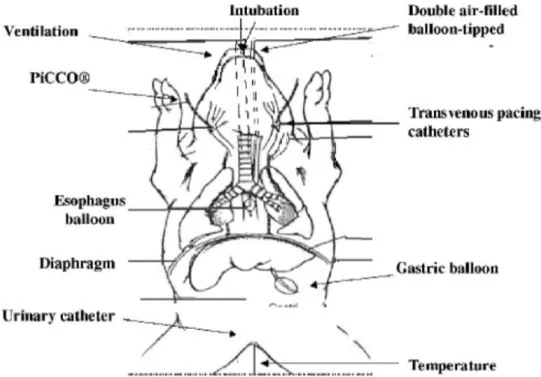
We used the same experimental design described in our pre-vious studies (fig. 1).14,15In brief, 14 piglets (15–20 kg) were
anesthetized with intravenous pentobarbital sodium (5– 6 mg/kg), intubated with a cuffed endotracheal tube, and me-chanically ventilated (Galileo®; Hamilton Medical AG,
Rha-zuns, Switzerland), with an inspired fraction of oxygen of 0.35, a VTbetween 10 and 12 ml/kg, an RR from 15 to 30
cycles/min to obtain normocapnia, and 5 cm H2O of
posi-tive end-expiratory pressure. Neuromuscular blocking agents were not used. The piglets were anesthetized with continu-ous intravencontinu-ous propofol (15–20 mg " kg"1"h"1),
midazo-Fig. 1. Diagram showing jugular, carotid catheter preparation and double air-filled balloon with transvenous jugular phrenic
pacing. PiCCO (Pulsion, Munich, Germany) ! pulse-induced contour cardiac output.
ASV Prevents Ventilatory-induced Diaphragm Dysfunction
lam (0.1– 0.3 mg/h), and ketamine (3– 4 mg " kg"1"h"1).
The level of sedation was monitored with bispectral index (BIS®; Aspect, Norwood, MA).16An oral gastric tube was
placed. A vesicostomy was performed, and a urine catheter was placed for urine collection. A rectal probe was used for frequent temperature measurements, and the animals were rested on soft cushions. Heating pads were used as needed to maintain a normal body temperature of 38.5°–39.5°C. A carotidal arterial catheter (PiCCO®; Pulsion, Munich,
Ger-many) was inserted for the monitoring of heart rate, arterial blood pressure, and cardiac output. Arterial pressure of car-bon dioxide levels was checked by using a capnograph (Del-tatrac®; Datex-Ohmeda, Helsinki, Finland) and then
veri-fied by arterial blood gases (iSTAT®; Abbott, Abbott Park,
IL). Parenteral nutrition was given from the first day (10% glucose solution and 20% amino acids solution, and Hypera-mine 20%®; Braun, Boulogne Billancourt, France)
provid-ing 30 –35 kcal " kg"1"day"1. All procedures were
per-formed aseptically. The animals received prophylactic intravenous antibiotics three times daily (amoxicillin– clavu-lanate, 100 mg " kg"1"day"1). At the end of the procedure,
costal region diaphragm samples were removed and then animals were killed by intravenous injection of pentobarbital sodium and potassium chloride. A physician provided round-the-clock supervision and animal care for the entire duration of the study. The two groups received the same care, except for the ventilatory mode.
Ventilatory Care
Seven piglets were ventilated for 72 consecutive hours in a totally controlled (CMV group) mode, and seven piglets were ventilated for 72 consecutive hours in a partially spontaneous mode (ASV group; fig. 2). In the CMV group, ventilatory settings were as follows: VTat 10 –12
ml/kg of the ideal body weight, RR from 15 to 30 min"1,
and positive end-expiratory pressure level at 5 cm H2O.
The absence of spontaneous breathing was verified on the ventilator trend graphs, and electromyographic activity of the diaphragm was measured in a few animals to ensure that no electrical activity was present in the diaphragm. In the ASV group, ventilatory settings were pediatric mode setting in phase with ideal body weight of the piglet, in-spiratory flow trigger at 0.3 l/min, percentage of mechan-ical ventilation between 100 and 150%, positive end-ex-piratory pressure level at 5 cm H2O, and expiratory trigger
at 25% of peak inspiratory flow. Active piglets were al-lowed to breathe spontaneously, and the ratio of pressure-assisted cycles was verified on the ventilator trend graphs. In both groups, oxygenation was maintained with FIO2
from 25 to 35% and minute ventilation to assess normo-capnia. The deepness of anesthesia was adjusted to blow out the respiratory drive in the CMV group and to allow spontaneous breathing without awakening in the ASV group.
Fig. 2. Diagram of mechanical ventilation parameters in the controlled mechanical ventilation (CMV) and adaptive support
ventilation (ASV) groups. From top to bottom airway flow (flow, l/s), airway pressure (Paw, cm H2O), esophageal pressure (Peso, cm H2O), transdiaphragmatic pressure (Pdi, cm H2O), and diaphragmatic electrical activity measured with an electromyogram
(EMG) are reported. Although the diaphragm was at rest without any spontaneous breathing activity in the CMV group, the diaphragm remains active, and spontaneous ventilation cycles occur in the ASV group.Arrows show spontaneous
Assessment of Diaphragm Muscle Activity during Mechanical Ventilation
First Part of the Study: In Vivo Assessment of Transdia-phragmatic Pressure. We measured transdiaTransdia-phragmatic
pressure (Pdi) for every 12 h to assess in vivo diaphragmatic contractile force in both groups as described in previous stud-ies.14,15In brief, double air-filled balloon-tipped catheters
were placed transorally into the distal third of the esophagus and in the stomach for measurement of Pdi. Bipolar trans-venous pacing catheters were introduced via each internal jugular vein and adjusted to achieve stimulation of the phrenic nerve and subsequent contraction of the diaphragm. Pdi was produced by supramaximal stimulation at fre-quencies of 20, 40, 60, 80, 100, and 120 Hz in a serial manner. Each train of impulses lasted for 2,000 ms, and each pulse had duration of 150 ms. A pressure–frequency curve was obtained for both groups at each 12-h period and then compared.
Second Part of the Study: In Vitro Assessment of Dia-phragm Histology. Biopsies (1 cm3) from the apposition
zone of the costal diaphragm from the entire midcostal mus-cle spanning were removed just before euthanasia. Each bi-opsy was partitioned, frozen in liquid nitrogen-cooled iso-pentane after 3–5 min for length equilibrium, and stored at "80°C. Immunochemistry was assessed on 10-!m unfixed serial transverse cryostat sections according to a well-de-scribed procedure.10 In brief, histologic analysis was
per-formed on six cross-sections from each muscle. Stained sec-tions were visualized under a Nikon optiphot-2 microscope (Nikon Instruments Europe, B.V. Amstelveen, The Nether-lands). All diaphragm images were obtained under identical conditions and with the same objective lens. The shape of each muscle fiber was accurately defined (on images #400), and a schematic drawing of each stained diaphragm section was recorded. Schematic representations of each muscle sec-tion were analyzed by Histolab program (version 5-13-1, Microvision Instrument, license number; 2497, Evry, France), and data were averaged per approximately 500 mus-cle fibers from the dissected diaphragms of piglets from each group.
Ten-micrometer unfixed cryostat sections of CMV and ASV group diaphragms were stained by hematoxylin and eosin. On each schematic drawing, the fiber cross-sectional areas were measured. To assess relative fiber percentages, im-munochemistry was assessed on 10-!m unfixed serial trans-verse cryostat sections. Two adjacent sections were incubated for 1 h in 1% bovine serum albumin with the respective primary antibodies for both the mouth anti–slow myosin heavy chain (M-8421; Sigma, Saint Louis, MO; dilution 1/8,000) and the mouth anti–fast myosin heavy chain (M-4276; Sigma; dilution 1/8,000). In addition, polyclonal an-tibodies against dystrophin (H4; dilution 1/5,000), pro-duced and characterized in our laboratory, were included in each of these stains to visualize fiber membranes. Immuno-reactions were detected with Cy3 and fluorescein isothiocya-nate-conjugated goat anti-piglet. After washing with
phos-phate-buffered saline solution, sections were incubated 1 h at room temperature with Cy3-goat anti-piglet IgG (Chemicon International, Molsheim, France; dilution 1/5,000) to detect myosin heavy chain, and with fluorescein isothiocyanate (Chemicon International, AP 132F; dilution 1/1,000) to de-tect dystrophin. Unbound antibodies were removed by washing the sections in phosphate-buffered saline. For neg-ative controls, only the second antibody was applied. Sec-tions were then visualized under adapted fluorescence. By using these stains, the myosin heavy chains that react with the chosen antibody on each section appeared orange, whereas the dystrophin at the membrane appeared green. We com-pared fiber type proportion measurements between the groups.
Statistical Analysis
Values are means and SD or medians and quartiles (25– 75th), as required. Normality of the distribution was assessed with the Kolmogorov–Smirnov test. A two-way analysis of variance with time (H0, H12, H24, . . . , H72) as one factor and modality (CMV vs. ASV) as the other factor was used. When appropriate, a Newman–Keuls test was used. Non-parametric paired Wilcoxon tests were used to compare data from days 1 and 3 for each animal in both the CMV and ASV groups. All P values were two tailed, and a P value less than 0.05 was considered significant (StatView®, version 5.0; SAS
Institute Inc., Berkeley, CA).
Results
Systemic and Biologic Response to Mechanical Ventilation
Among the 14 piglets, two died before the end of the study: one because of myocardial infarction at H10 (CMV group) and one because of septic shock (peritonitis after bladder catheterization resulting in small bowel perforation in the ASV group) after 28 h of ventilation. These two piglets were subsequently excluded from the analyses.
No significant differences were observed between the CMV group and the ASV group for all the studied baseline variables. Long-term mechanical ventilation, either in CMV or ASV, did not have consequences on body weight, intesti-nal transit, diuresis (data not shown), or hemodynamic vari-ables (table 1). BIS values were significantly different be-tween the CMV and ASV groups (72 $ 8 vs. 42 $ 12, P ! 0.02). Mean midazolam level administration remained at a higher level in the CMV group (3.8 $ 0.7 mg/h) than that in the ASV group (1.5 $ 0.7 mg/h) during the study (P % 0.05 between CMV vs. ASV after 12 h of ventilation). Doses of propofol did not significantly differ between the groups dur-ing the study (102 $ 8 and 90 $ 10 mg/h in the CMV and ASV groups, respectively).
Ventilatory Parameters and Spontaneous Breathing Data
In the CMV group, spontaneous activity of the diaphragm was present for no more than 5% of the delivered breaths, as
ASV Prevents Ventilatory-induced Diaphragm Dysfunction
determined by analysis of the trend graphs and the Pdi trac-ings. In the ASV group, active piglets triggered 80% of the delivered breaths, as assessed by the Galileo®trend graphs of
ventilator and, for a few animals, the surface electromyogram activity (fig. 2). Once normal gas exchanges were obtained, ventilatory settings were not modified throughout the study (table 1).
In Vivo Assessment of Diaphragmatic Force
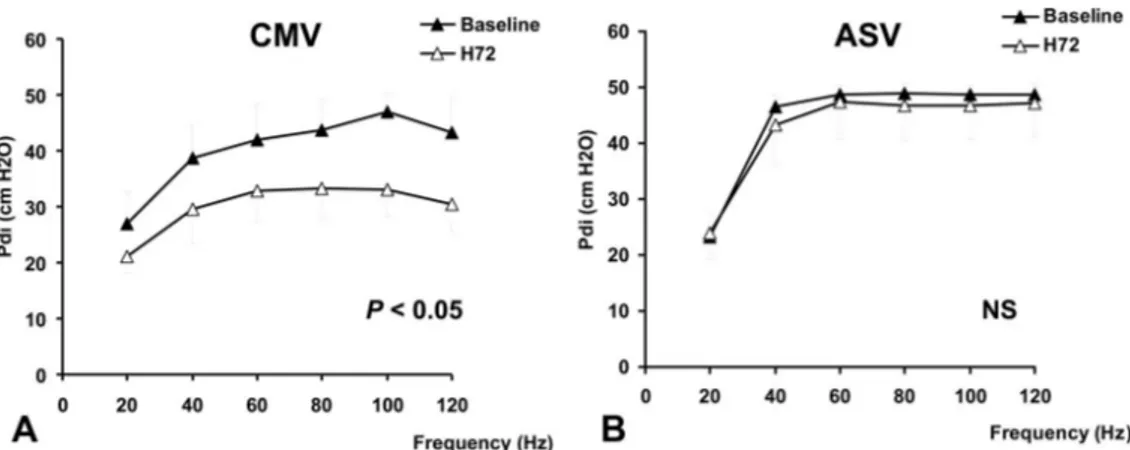
The baseline pressure–frequency curves of the two groups were not significantly different (Pdi ! 23.4 $ 2.6 vs. 24.6 $ 4.8 cm H2O at 20 Hz and 45.8 $ 3.0 vs. 46.8 $ 1.5 cm H2O
at 100 Hz for the CMV and ASV groups, respectively; fig. 3, A and B).
Although Pdi decreased significantly in the CMV group between baseline and H72 at all frequencies except 20 Hz
(P % 0.05; fig. 3A), it did not change significantly in the ASV group (fig. 3B).
Furthermore, the decrease in the Pdi between H0 and H72 in the CMV group ("26% ["17 to "32]) was signif-icantly higher than that in the ASV group ("2% ["8 to &3]; P % 0.05) at all frequencies of stimulation, except at 40 and 60 Hz at which the difference did not reach the statistical significance (fig. 3). Figure 4 shows the evolution of Pdi over time at a stimulation frequency of 100 Hz. In the CMV group, Pdi decreased significantly after 48 h of venti-lation (P % 0.05). In the ASV group, Pdi did not signifi-cantly differ during the whole study period.
In Vitro Assessment of Diaphragmatic Histology
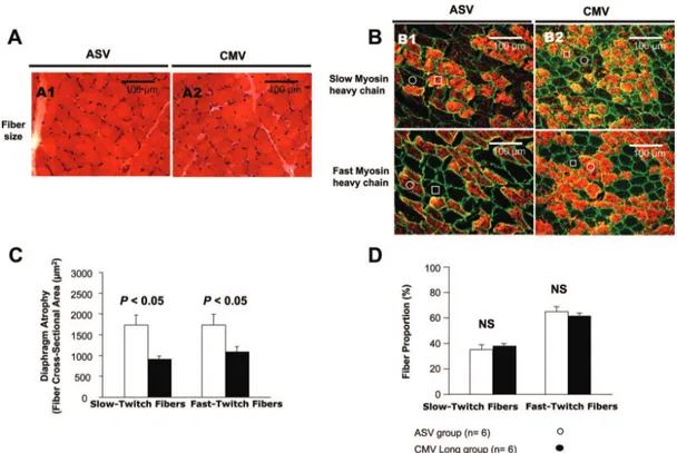
Figure 5A shows photomicrographs of typical diaphragm muscle cross-sections obtained in one animal of each group.
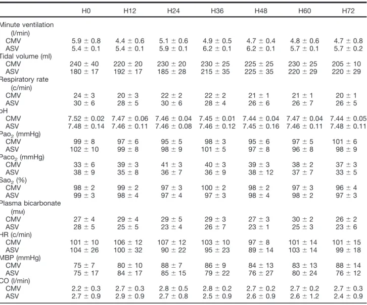
Table 1. Hemodynamic and Respiratory Variables for the Seven Steps of Measures between the CMV (n ! 6) and the ASV (n ! 6) Groups
H0 H12 H24 H36 H48 H60 H72 Minute ventilation (l/min) CMV 5.9 $ 0.8 4.4 $ 0.6 5.1 $ 0.6 4.9 $ 0.5 4.7 $ 0.4 4.8 $ 0.6 4.7 $ 0.8 ASV 5.4 $ 0.1 5.4 $ 0.1 5.9 $ 0.1 6.2 $ 0.1 6.2 $ 0.1 5.7 $ 0.1 5.7 $ 0.2 Tidal volume (ml) CMV 240 $ 40 220 $ 20 230 $ 20 230 $ 25 225 $ 25 230 $ 25 205 $ 10 ASV 180 $ 17 192 $ 17 185 $ 28 215 $ 35 225 $ 35 220 $ 29 220 $ 29 Respiratory rate (c/min) CMV 24 $ 3 20 $ 3 22 $ 2 22 $ 2 21 $ 1 21 $ 1 20 $ 1 ASV 30 $ 6 28 $ 5 30 $ 6 28 $ 4 26 $ 6 26 $ 7 26 $ 5 pH CMV 7.52 $ 0.02 7.47 $ 0.06 7.46 $ 0.04 7.45 $ 0.01 7.44 $ 0.04 7.47 $ 0.04 7.44 $ 0.05 ASV 7.48 $ 0.14 7.46 $ 0.11 7.46 $ 0.08 7.46 $ 0.12 7.45 $ 0.16 7.46 $ 0.11 7.48 $ 0.11 Pao2(mmHg) CMV 99 $ 8 97 $ 6 95 $ 5 98 $ 3 95 $ 6 97 $ 5 101 $ 6 ASV 102 $ 10 99 $ 8 98 $ 9 101 $ 5 97 $ 8 96 $ 8 98 $ 9 Paco2(mmHg) CMV 33 $ 6 39 $ 3 41 $ 3 40 $ 3 39 $ 3 38 $ 2 37 $ 3 ASV 38 $ 9 35 $ 8 36 $ 7 36 $ 9 38 $ 12 37 $ 7 33 $ 5 Sao2(%) CMV 98 $ 2 99 $ 2 97 $ 3 100 $ 2 98 $ 2 97 $ 3 96 $ 4 ASV 99 $ 3 98 $ 4 97 $ 4 97 $ 3 98 $ 4 98 $ 2 97 $ 3 Plasma bicarbonate (mM) CMV 27 $ 4 29 $ 4 29 $ 5 29 $ 3 27 $ 3 30 $ 2 26 $ 2 ASV 28 $ 5 25 $ 5 23 $ 4 26 $ 7 23 $ 1 25 $ 3 23 $ 6 HR (c/min) CMV 101 $ 10 106 $ 12 107 $ 12 103 $ 10 97 $ 8 101 $ 14 101 $ 15 ASV 104 $ 26 100 $ 32 90 $ 22 95 $ 23 89 $ 14 103 $ 14 99 $ 18 MBP (mmHg) CMV 75 $ 7 80 $ 10 88 $ 7 86 $ 9 84 $ 13 83 $ 13 88 $ 14 ASV 75 $ 17 84 $ 17 85 $ 15 79 $ 22 76 $ 27 80 $ 24 76 $ 12 CO (l/min) CMV 2.2 $ 0.3 2.7 $ 0.3 2.8 $ 0.5 2.8 $ 0.2 2.7 $ 0.2 2.7 $ 0.2 2.7 $ 0.3 ASV 2.7 $ 0.9 2.9 $ 0.9 2.7 $ 0.8 2.5 $ 0.9 2.6 $ 0.9 2.6 $ 1.2 2.4 $ 0.9
Data are presented as mean $ SD. There were no significant intergroup or intragroup differences for hemodynamic and for respiratory variables and arterial blood gases in both groups.
ASV ! adaptative support ventilation; CMV ! controlled mechanical ventilation; CO ! cardiac output; HR ! heart rate; MBP ! mean blood pressure; PaO2! arterial pressure of oxygen; PaCO2! arterial pressure of carbon dioxide; SaO2! oxygen saturation of arterial
Fibers that react with the antibody appear orange in figure 5B, whereas fibers not reacting with the antibody appear black. A representative slow-twitch fiber is indicated by an open square and a fast-twitch fiber by an open circle. In the CMV group, we observed a marked atrophy of both slow-and fast-twitch fibers after 72 h of totally controlled ventila-tion. Indeed, after 72 h of CMV, fiber cross-sectional area was decreased by 30 – 40% for both slow- and fast-twitch fibers (P % 0.05; fig. 5C). The atrophy reported in the CMV group contrasts with that reported in the ASV group, which was not associated with fiber cross-sectional area variation after 72 consecutive hours of ventilation (fig. 5C). Atrophy in the CMV group concerned both slow- and fast-twitch fibers, and fiber proportion at H72 was not different in com-parison with baseline in both groups (fig. 5D).
Discussion
This study demonstrates that maintaining spontaneous ven-tilation with ASV is efficient to protect the diaphragm against the occurrence of VIDD, both in vivo and in vitro, in the same piglet model. In addition, we report that in vivo, both diaphragmatic contractility and diaphragmatic atrophy occur when a totally controlled ventilation mode is applied but are prevented when spontaneous cycles with ASV are maintained.
By using in vitro animal models, several studies have re-ported that CMV-induced diaphragm inactivity decreases protein synthesis and increases degradation of key contractile proteins, resulting in diaphragmatic force loss.3,15,17–21
Pro-tein degradation is mediated via oxidative stress,15,22,23
apo-ptosis,24and proteasome proteolysis.5,25–27Few studies
eval-uating VIDD in vivo mainly with animals have been published3,15,21, although one was with humans.2 Some
other acute situations (i.e., myorelaxants, sepsis, acute hyper-capnic acidosis, and others) seem to induce VIDD in animal studies.14,28,29Sepsis may be associated with VIDD in the
intensive care unit, although mainly explored through exper-imental studies. Several studies described the mechanisms leading to the VIDD, showing inflammation, proteolysis, or nitric oxide pathways.28,30 –32Although one study
demon-strated that CMV decreases VIDD compared with sponta-neous breathing in septic rats, to the best of our knowledge,33
none compared assisted versus CMV.
In this study, we confirmed that prolonged CMV de-creased diaphragmatic contractile force in vivo in a healthy piglet model. In comparison with baseline, diaphragmatic force-generating capacity decreased by 25% after 72 h of ventilation in the CMV group (figs. 3A and 4).
Although it is widely accepted that VIDD occurs after several days of CMV, few countermeasures have been tested. Recently, it has been shown that administration of Trolox,34
an antioxidant, glutamine,35or leupeptin,25which decreases
Fig. 3. Diaphragmatic pressure–frequency curves obtained in the controlled mechanical ventilation (CMV) group (A) (n ! 6) and
in the adaptive support ventilation (ASV) group (B) (n ! 6). Transdiaphragmatic pressure (Pdi) after supramaximal phrenic nerve
stimulation at 20, 40, 60, 80, 100, and 120 Hz over 72 h of mechanical ventilation obtained in both groups. In the CMV group, Pdi significantly decreased between baseline and H72 at all frequencies (except at 20 Hz) (P % 0.05). Data are mean $ SD. P
values refer to between H72vs. baseline. NS ! not significant. No significant differences were observed for the values obtained
at all frequencies in the ASV group between H72 and baseline.
Fig. 4. Transdiaphragmatic pressure (Pdi) differences
eval-uated at a frequency of 100 Hz between the adaptive support ventilation (ASV) group (n ! 6) and controlled mechanical ventilation (CMV) (n ! 6) group during the whole study period. Pdi in the CMV group was lower than that in the ASV group after 48 h of ventilation. Data are mean $ SD. P values refer to between-group differences. *P % 0.05 vs. baseline values.
ASV Prevents Ventilatory-induced Diaphragm Dysfunction
calpain and cathepsin activity (proteases), prevents VIDD during mechanical ventilation in a rat model. Although med-ications to prevent or treat VIDD may be a point of interest in the future, ventilator strategies that allow diaphragmatic contractions seem to protect against VIDD.
Indeed, Sassoon et al.4reported the beneficial effect of
assisted controlled ventilation on in vitro contractile proper-ties of the diaphragm and the decrease in the muscle atrophy factor-box messenger RNA, a marker of gene atrophy in comparison with total CMV in rabbits. Gayan-Ramirez et
al.1also reported the beneficial effect of intermittent
spon-taneous breathing on in vitro contractility and myogenesis regulatory factors in the diaphragm. Recently, we reported that PSV was effective in reducing diaphragm proteolysis and inhibition of protein synthesis compared with continuous mechanical ventilation.5 This study is the first one
per-formed in an in vivo model of piglet, a large animal whose physiology is similar to human beings, to report that main-taining spontaneous activity of the diaphragm with ASV may protect diaphragm against VIDD evaluated both in vivo (contractility) and in vitro (atrophy and fibers repartition).
With the ASV mode, the ventilator software uses a modified version of the equation derived by Otis et al.7to minimize the
work of breathing.8,12,13,36,37For subjects who are unable to
trigger a breath, the ventilator generates a pressure-controlled cycle, automatically adjusting inspiratory pressure and time to achieve a target VTand a target RR. When the
spontane-ous activity of the diaphragm is sufficient to trigger a breath, the ventilator generates pressure-support breaths and, if nec-essary, delivers additional pressure-controlled breaths. In the ASV mode, similar to PSV, when an apnea occurs, the ven-tilator switches to the CMV mode but with an option that allows the patient to return to an assisted mode and triggers breaths.
In this 72 consecutive-hour study, the ASV mode al-lowed for a high proportion of pressure-supported cycles but ensured initiation of the apnea-induced CMV mode when necessary, with an optional return to PSV. In the ASV group, piglets were ventilated on pressure support more than 80% of the time, although they were ventilated in CMV mode the remainder of the time. On the contrary, in the CMV group, we could identify diaphragmatic activity
Fig. 5. Comparison of representative adaptive support ventilation (ASV) (A1, B1) and controlled mechanical ventilation (CMV)
(A2, B2) diaphragm-biopsy specimen with respect to fiber size or phenotype. Histologic analysis was performed on six
cross-sections from each muscle under a Nikon optiphot-2 microscope (Nikon Instruments Europe, B.V. Amstelveen, The Netherlands). (A) Hematoxylin and eosin coloration shows that neither inflammatory infiltrate nor necrosis is present in the CMV
and ASV group. (B) To assess for the slow- and fast-twitch fiber type cross-sectional area (CSA) and relative fiber percentages,
immunochemistry was assessed on 10-!m unfixed serial transverse cryostat sections. Two adjacent sections were incubated in 1% bovine serum albumin with the primary antibody 1 h for mouse anti–slow myosin heavy chain. The myosin heavy chains that react with the chosen antibody on each section appeared orange, whereas the dystrophin at the membrane appeared green. Schematic representation (on images #400) of each muscle section was analyzed by Histolab program (version 5-13-1; Microvision Instrument, Evry, France; license number: 2497), and data were averaged per approximately 500 muscle fibers from each dissected diaphragm of both the CMV and ASV groups. (C) Both the slow- and the fast-twitch fibers in the CMV group
are statistically smaller than those in the ASV group. (D) Fiber proportion (%) was not different between the CMV and ASV group.
during lesser than 5% of all breaths on the trend graphs of the Galileo®ventilator.
In this study, the piglets ventilated in ASV mode did not show any decrease in Pdi during the study period, contrary to those ventilated with CMV mode in whom Pdi decreased by 25% after 60 h of ventilation. We speculate that this protec-tive effect is related to the spontaneous activity of the dia-phragm because we did not compare ASV with pressure sup-port or any other spontaneous mode.
Some study limitations must be pointed out. First, we did not compare our results with a control group without anes-thesia, or mechanical ventilation, because large animals must be anesthetized for procedures such as phrenic stimulation and Pdi recording. Second, we did not compare ASV with PSV mode and therefore cannot conclude on the relative effects of spontaneous ventilation versus the specificity of the ASV mode. However, ASV may have beneficial effects com-pared with PSV because of the theoretical decrease in the work of breathing with the equation software derived by Otis
et al. and the possibility of an automated backup from the
apnea ventilation to ASV (i.e., spontaneous ventilation mode) and vice versa, which allowed piglets to breathe spon-taneously as much as possible. Third, although piglet respi-ratory muscles are close to those of humans, this study was limited because we compared the effect of CMV with that of ASV on healthy diaphragm muscles. Nevertheless, we can speculate that the reported alteration of Pdi would be even worse in pathologic situations, such as sepsis, that induce VIDD per se.28,38,39In the CMV group, sedation level was
more important than that in the ASV group to neutralize the centers of breathing. Although we cannot eliminate a direct effect of sedation on Pdi, a recent review on VIDD stated that the effect of sedation on diaphragm function was clearly lower than the direct effect of mechanical ventilation.40
Fur-thermore, in the clinical situation, sedation is frequently nec-essary to ensure patient–ventilator synchrony with the CMV mode and may lengthen the ventilation weaning process, thereby promoting VIDD.41,42Although this point
repre-sents clearly a methodological limit of our study, it reflects the clinical interaction among sedation, mechanical ventila-tion, and weaning.42,43
In conclusion, this study reports that maintaining dia-phragmatic contractile activity during 72 consecutive hours with the ASV mode may protect the diaphragm against the deleterious effect of total CMV in healthy piglets. ASV pre-vented the in vivo alteration of diaphragmatic contractility and in vitro diaphragmatic atrophy, contrary to total CMV, which was associated with both diaphragmatic atrophy and
in vivo alteration of the diaphragm contractility. Further
studies, including physiologic human studies, are required to more fully understand the effect of ASV on diaphragmatic function.
References
1. Gayan-Ramirez G, Testelmans D, Maes K, Racz GZ, Cadot P, Zador E, Wuytack F, Decramer M: Intermittent
sponta-neous breathing protects the rat diaphragm from mechan-ical ventilation effects. Crit Care Med 2005; 33:2804 –9 2. Levine S, Nguyen T, Taylor N, Friscia ME, Budak MT,
Rothenberg P, Zhu J, Sachdeva R, Sonnad S, Kaiser LR, Rubinstein NA, Powers SK, Shrager JB: Rapid disuse atro-phy of diaphragm fibers in mechanically ventilated hu-mans. N Engl J Med 2008; 358:1327–35
3. Sassoon CS, Caiozzo VJ, Manka A, Sieck GC: Altered dia-phragm contractile properties with controlled mechanical ventilation. J Appl Physiol 2002; 92:2585–95
4. Sassoon CS, Zhu E, Caiozzo VJ: Assist-control mechanical ventilation attenuates ventilator-induced diaphragmatic dysfunction. Am J Respir Crit Care Med 2004; 170:626 –32 5. Futier E, Constantin JM, Combaret L, Mosoni L, Roszyk L, Sapin V, Attaix D, Jung B, Jaber S, Bazin JE: Pressure support ventilation attenuates ventilator-induced protein modifications in the diaphragm. Crit Care 2008; 12:R116 6. Campbell RS, Branson RD, Johannigman JA: Adaptive
sup-port ventilation. Respir Care Clin N Am 2001; 7:425– 40, ix 7. Otis AB, Fenn WO, Rahn H: Mechanics of breathing in
man. J Appl Physiol 1950; 2:592– 607
8. Tehrani FT: The origin of adaptive support ventilation. Int J Artif Organs 2005; 28:1051–2
9. Petter AH, Chiolero RL, Cassina T, Chassot PG, Muller XM, Revelly JP: Automatic “respirator/weaning” with adaptive support ventilation: The effect on duration of endotra-cheal intubation and patient management. Anesth Analg 2003; 97:1743–50
10. Sulzer CF, Chiolero R, Chassot PG, Mueller XM, Revelly JP: Adaptive support ventilation for fast tracheal extubation after cardiac surgery: A randomized controlled study. ANESTHESIOLOGY
2001; 95:1339–45
11. Dongelmans DA, Veelo DP, Paulus F, de Mol BA, Korevaar JC, Kudoga A, Middelhoek P, Binnekade JM, Schultz MJ: Weaning automation with adaptive support ventilation: A randomized controlled trial in cardiothoracic surgery pa-tients. Anesth Analg 2009; 108:565–71
12. Arnal JM, Wysocki M, Nafati C, Donati S, Granier I, Corno G, Durand-Gasselin J: Automatic selection of breathing pattern using adaptive support ventilation. Intensive Care Med 2008; 34:75– 81
13. Jaber S, Sebbane M, Verzilli D, Matecki S, Wysocki M, Eledjam JJ, Brochard L: Adaptive support and pressure support ventilation behavior in response to increased ven-tilatory demand. ANESTHESIOLOGY2009; 110:620 –7 14. Jaber S, Jung B, Sebbane M, Ramonatxo M, Capdevila X,
Mercier J, Eledjam JJ, Matecki S: Alteration of the piglet diaphragm contractility in vivo and its recovery after acute hypercapnia. ANESTHESIOLOGY2008; 108:651– 8
15. Jaber S, Sebbane M, Koechlin C, Hayot M, Capdevila X, Eledjam JJ, Prefaut C, Ramonatxo M, Matecki S: Effects of short vs. prolonged mechanical ventilation on antioxidant systems in piglet diaphragm. Intensive Care Med 2005; 31:1427–33
16. Vivien B, Di Maria S, Ouattara A, Langeron O, Coriat P, Riou B: Overestimation of bispectral index in sedated intensive care unit patients revealed by administration of muscle relaxant. ANESTHESIOLOGY2003; 99:9 –17
17. Anzueto A, Peters JI, Tobin MJ, de los Santos R, Seidenfeld JJ, Moore G, Cox WJ, Coalson JJ: Effects of prolonged controlled mechanical ventilation on diaphragmatic func-tion in healthy adult baboons. Crit Care Med 1997; 25: 1187–90
18. Bernard N, Matecki S, Py G, Lopez S, Mercier J, Capdevila X: Effects of prolonged mechanical ventilation on respira-tory muscle ultrastructure and mitochondrial respiration in rabbits. Intensive Care Med 2003; 29:111– 8
19. Capdevila X, Lopez S, Bernard N, Rabischong E, Ramonatxo M, Martinazzo G, Prefaut C: Effects of controlled mechanical
ven-ASV Prevents Ventilatory-induced Diaphragm Dysfunction
tilation on respiratory muscle contractile properties in rabbits. Intensive Care Med 2003; 29:103–10
20. Le Bourdelles G, Viires N, Boczkowski J, Seta N, Pavlovic D, Aubier M: Effects of mechanical ventilation on diaphrag-matic contractile properties in rats. Am J Respir Crit Care Med 1994; 149:1539 – 44
21. Radell P, Edstrom L, Stibler H, Eriksson LI, Ansved T: Changes in diaphragm structure following prolonged me-chanical ventilation in piglets. Acta Anaesthesiol Scand 2004; 48:430 –7
22. Falk DJ, Deruisseau KC, Van Gammeren DL, Deering MA, Kavazis AN, Powers SK: Mechanical ventilation promotes redox status alterations in the diaphragm. J Appl Physiol 2006; 101:1017–24
23. Powers SK, Kavazis AN, DeRuisseau KC: Mechanisms of disuse muscle atrophy: Role of oxidative stress. Am J Physiol Regul Integr Comp Physiol 2005; 288:R337– 44 24. Powers SK, Kavazis AN, McClung JM: Oxidative stress and
disuse muscle atrophy. J Appl Physiol 2007; 102:2389 –97 25. Maes K, Testelmans D, Powers S, Decramer M, Gayan-Ramirez G: Leupeptin inhibits ventilator-induced dia-phragm dysfunction in rats. Am J Respir Crit Care Med 2007; 175:1134 – 8
26. McClung JM, Kavazis AN, DeRuisseau KC, Falk DJ, Deering MA, Lee Y, Sugiura T, Powers SK: Caspase-3 regulation of diaphragm myonuclear domain during mechanical ventila-tion-induced atrophy. Am J Respir Crit Care Med 2007; 175:150 –9
27. McClung JM, Whidden MA, Kavazis AN, Falk DJ, Deruis-seau KC, Powers SK: Redox regulation of diaphragm pro-teolysis during mechanical ventilation. Am J Physiol Regul Integr Comp Physiol 2008; 294:R1608 –17
28. Divangahi M, Demoule A, Danialou G, Yahiaoui L, Bao W, Xing Z, Petrof BJ: Impact of IL-10 on diaphragmatic cyto-kine expression and contractility during Pseudomonas in-fection. Am J Respir Cell Mol Biol 2007; 36:504 –12 29. Testelmans D, Maes K, Wouters P, Gosselin N, Deruisseau
K, Powers S, Sciot R, Decramer M, Gayan-Ramirez G: Rocuronium exacerbates mechanical ventilation-induced diaphragm dysfunction in rats. Crit Care Med 2006; 34: 3018 –23
30. Rabuel C, Samuel JL, Lortat-Jacob B, Marotte F, Lanone S, Keyser C, Lessana A, Payen D, Mebazaa A: Activation of the ubiquitin proteolytic pathway in human septic heart and diaphragm. Cardiovasc Pathol 2009; [Epub ahead of print] 31. Supinski GS, Vanags J, Callahan LA: Effect of proteasome inhibitors on endotoxin-induced diaphragm dysfunction. Am J Physiol Lung Cell Mol Physiol 2009; 296:L994 –1001 32. Supinski GS, Wang W, Callahan LA: Caspase and calpain activation both contribute to sepsis-induced diaphrag-matic weakness. J Appl Physiol 2009; 107:1389 –96 33. Ebihara S, Hussain SN, Danialou G, Cho WK, Gottfried SB,
Petrof BJ: Mechanical ventilation protects against dia-phragm injury in sepsis: Interaction of oxidative and me-chanical stresses. Am J Respir Crit Care Med 2002; 165: 221– 8
34. Betters JL, Criswell DS, Shanely RA, Van Gammeren D, Falk D, Deruisseau KC, Deering M, Yimlamai T, Powers SK: Trolox attenuates mechanical ventilation-induced dia-phragmatic dysfunction and proteolysis. Am J Respir Crit Care Med 2004; 170:1179 – 84
35. Oliveira GP, Oliveira MB, Santos RS, Lima LD, Dias CM, Ab’ Saber AM, Teodoro WR, Capelozzi VL, Gomes RN, Bozza PT, Pelosi P, Rocco PR: Intravenous glutamine decreases lung and distal organ injury in an experimental model of abdominal sepsis. Crit Care 2009; 13:R74
36. Burns KE, Lellouche F, Lessard MR: Automating the wean-ing process with advanced closed-loop systems. Intensive Care Med 2008; 34:1757– 65
37. Dongelmans DA, Veelo DP, Bindels A, Binnekade JM, Ko-ppenol K, Koopmans M, Korevaar JC, Kuiper MA, Schultz MJ: Determinants of tidal volumes with adaptive support ventilation: A multicenter observational study. Anesth Analg 2008; 107:932–7
38. Demoule A, Divangahi M, Yahiaoui L, Danialou G, Gvozdic D, Labbe K, Bao W, Petrof BJ: Endotoxin triggers nuclear factor-kappaB-dependent up-regulation of multiple proin-flammatory genes in the diaphragm. Am J Respir Crit Care Med 2006; 174:646 –53
39. Divangahi M, Matecki S, Dudley RW, Tuck SA, Bao W, Radzioch D, Comtois AS, Petrof BJ: Preferential diaphrag-matic weakness during sustained Pseudomonas
aerugi-nosa lung infection. Am J Respir Crit Care Med 2004;
169:679 – 86
40. Vassilakopoulos T: Ventilator-induced diaphragm dysfunc-tion: The clinical relevance of animal models. Intensive Care Med 2008; 34:7–16
41. Chanques G, Jaber S, Barbotte E, Violet S, Sebbane M, Perrigault PF, Mann C, Lefrant JY, Eledjam JJ: Impact of systematic evaluation of pain and agitation in an intensive care unit. Crit Care Med 2006; 34:1691–9
42. Girard TD, Kress JP, Fuchs BD, Thomason JW, Schweickert WD, Pun BT, Taichman DB, Dunn JG, Pohlman AS, Kinniry PA, Jackson JC, Canonico AE, Light RW, Shintani AK, Thompson JL, Gordon SM, Hall JB, Dittus RS, Bernard GR, Ely EW: Efficacy and safety of a paired sedation and ven-tilator weaning protocol for mechanically ventilated pa-tients in intensive care (Awakening and Breathing Con-trolled trial): A randomised conCon-trolled trial. Lancet 2008; 371:126 –34
43. Chanques G, Jaber S: Sedation assessment tool, sedation-algorithm, choice of sedation drugs: Intricate concepts of an emergent clinical practice. Intensive Care Med 2007; 33:554 –5