HAL Id: inserm-02477338
https://www.hal.inserm.fr/inserm-02477338
Submitted on 13 Feb 2020HAL is a multi-disciplinary open access archive for the deposit and dissemination of sci-entific research documents, whether they are pub-lished or not. The documents may come from teaching and research institutions in France or abroad, or from public or private research centers.
L’archive ouverte pluridisciplinaire HAL, est destinée au dépôt et à la diffusion de documents scientifiques de niveau recherche, publiés ou non, émanant des établissements d’enseignement et de recherche français ou étrangers, des laboratoires publics ou privés.
Heterogeneity of the early outward current in ventricular
cells isolated from normal and hypertrophied rat hearts
Jean-Pierre Benitah, Ana Maria Gómez, Patrick Bailly, Jean-Philippe da
Ponte, Guy Berson, Carmen Delgado, Paco Lorente
To cite this version:
Jean-Pierre Benitah, Ana Maria Gómez, Patrick Bailly, Jean-Philippe da Ponte, Guy Berson, et al.. Heterogeneity of the early outward current in ventricular cells isolated from normal and hypertrophied rat hearts. American Journal of Physiology, American Physiological Society, 1993, 469, pp.111 - 138. �10.1113/jphysiol.1993.sp019807�. �inserm-02477338�
Journalof Physiology (1993), 469, pp. 111-138 111
With9figures
Printed in Great Britain
HETEROGENEITY OF THE EARLY OUTWARD CURRENTIN
VENTRICULARCELLS ISOLATED FROM NORMAL AND HYPERTROPHIED RAT HEARTS
BY JEAN-PIERRE BENITAH*, ANA MARIA GOMEZt,
PATRICK BAILLY*, JEAN-PHILIPPE DA PONTE*, GUY BERSON*, CARMEN DELGADOt AND PACO LORENTE*
From *U195INSERMFaculte de Medecine, 63001 Clermont-Ferrand, France and the tInstituto de Farmacologia y
Toxicologia
(CSIC), Universidad Complutense,28040 Madrid, Spain (Received 23 June 1993)
SUMMARY
1. The nature, magnitude and kinetics of the 4-aminopyridine-sensitive early outward current (Ito) were analysed in isolated ventricular myocytes from the septum, the apexand the left ventricular free wall of rat ventricles using the whole-cell voltage clamp method. The modulatory effect of pressure overload-induced cardiac hypertrophy on the regional variations of Ito was assessed in each topographical class of cells.
2. Voltage clamp experiments were performed at room temperature (20-25°C) in the absence ofNa+ on both sides of the membrane and in the presence of 3mMCoCl2.
Ito
wasstudied from aholding potential
of -80mV anddeterminedby
subtractionof total outward currents elicited by the same protocols in the presence of 3mm
4-aminopyridine (4-AP) from those obtained in its absence.
3. In normal hearts, membrane passive properties were very similar in each topographical class of cells. Our results confirmed that the predominant early outward current in rat ventricular cells was 4-AP-sensitive, time and voltage dependent, and demonstrated that the magnitude of the current variedon aregional basis: current density of Ito in left ventricular free wall cells (30-1+9-2pA/pF at
+60 mV) was larger than in apex cells (20-2+1-7pA/pF) or in septum cells (11-9+3-3pA/pF). We noticedalargervariabilityindatafrom left ventricular free wall compared with other regions.
4. No shift insteady-state voltage dependence ofIto activation and inactivation wasfound. However, the maximal computed chord conductances were (in,uS/pF):
0 18 +0-07for leftventricularfreewallcells, 0-13+0-02forapexcells,and 0-08+0-02
for septum cells. Thesefindings might reflectadifferentialdistributioninfunctional
channel densities.
5. No difference in voltage-dependent
Ito
activation kinetics was present with respectto topography. However,inactivation time constants inseptumwerelonger than those of both other groups.6. Left ventricularhypertrophywasinducedbyabdominalaorticconstriction and MS 1562
J.-P. BENITAH AND OTHERS
its effects compared tothe findings from normal rats. Hypertrophied cells had similar resting potentials but higher capacitance values than normal cells. Although
It.
magnitude appeared not to be modified, the current density-voltage curves wereslightlyshifted to more positive potentials and significantly decreasedas compared
tonormal cells (inpA/pF,at + 60mV): 8-4 + 5-0 inthe left freewall group, 11-6+2-0 in the apex group, and 3-8+ 1-5 in the septum group. Steady-state activation and
inactivation parameters were not clearly modified, but kinetics were slowed down.
7. We conclude, therefore, that Ito is differentially distributed among different
regions of the normal rat ventricle and we propose that this regional heterogeneity
maybe related to differentdistributions of functional channeldensities, rather than
alterations in whole-cell kinetics or single-channel properties. Pressure overload-induced hypertrophy reducesIto currentavailability by decreasing currentdensities
without any significant change of whole-cell kinetics, while ahomogenizing tendency of the ionic profileis observed amongthe studiedregions. One possible explanation
for the hypertrophy-induced variations may be an absence of
Ito
channelneosynthesis, leading to a decrease of channel density per surface area unit.
INTRODUCTION
A transientoutward current was originally shown in sheep cardiacPurkinje fibres
clampedtomembrane potentials positiveto -20mV(Deck&Trautwein, 1964)and
for alongtime it wasconsidered to be a unique feature ofthistissuerelated torapid phase 1 repolarization (Fozzard& Hiraoka, 1973; Coraboeuf &Carmeliet, 1982). In recent years, with the improvement of patch clamp techniques, a wealth of experimental data has shown the presence of a transient outward current in other cardiac tissues from various species (Gintant, Cohen, Datyner & Kline, 1991), and has demonstrated the role of this current in initiating the repolarization ofaction
potentials(Tseng, Robinson & Hoffman, 1987). Magnitude andevenexistenceof the
current arestronglydependenton anumberof factors, suchasthe considered species (Josephson, Sanchez-Chapula & Brown, 1984), maturational changes during
development (Escande, Loisance, Planche & Coraboeuf, 1985; Kilborn & Fedida,
1990), regional differences in the distribution of the electrical properties (Giles &
Imaizumi, 1988; Furukawa, Myerburg, Furukawa, Bassett & Kimura, 1990;Fedida
& Giles, 1991), andpathology (Antzelevitch etal. 1991).
We have recently reported that in septal human ventricular myocytes isolated
frompatientswithpressureoverload-induced cardiachypertrophy, theslow inward
current is largely dominant compared to the outward currents. In contrast to the
observations in human atrial cells (Escande, Coulombe, Faivre, Deroubaix &
Coraboeuf, 1987), the transientoutwardcurrentinhypertrophichuman ventricular myocyteswasveryreduced or absent(Benitah, Bailly, D'Agrosa,DaPonte,Delgado &Lorente, 1992 a).Since thepresenceofatransientoutwardcurrenthasnotyetbeen
established with any certainty in the human ventricle, and due to the absence of
control data, it remains to be seen whether this pattern is related to regional differences in the properties of the cells or whether it is due to the pathological condition of the patients.
A differential distribution of transient outward currents in epicardial and endocardial layersoramongdifferentportionsofventricular tissue, has beeninitially
HETEROGENEITY OF TRANSIENT OUTWARD CURRENT 113 assumed on the basis of action potential data (Antzelevitch et al. 1991; Watanabe, Delbridge, Bustamente & McDonald, 1983). Recently reported voltage clamp data have lent support to this assumption by showingthat transient outward currents are significantly greater in epicardial than inendocardial cells dissociated from cat and rabbit left ventricles (Furukawa et al. 1990; Fedida & Giles, 1991).
In pressure overload-induced cardiac hypertrophy, the regulatory mechanisms of the action potential configuration are severely impaired. An increase in the duration of the action potential is the most commonly observed disturbance in the hypertrophied myocardium from rats with renal hypertension (Keung & Aronson, 1981), from cats submitted to pulmonary artery constriction (Tritthart, Leudcke, Bayer, Stierle & Kaufmann, 1975), and from guinea-pigs subjected to a progressive aortic banding (Nordin, Siri & Aronson, 1989). The question then arises as to which membranecurrents areinvolved inthese pathophysiological processes. This question is, however, currently a matter of considerable debate among electrophysiologists. Keung (1989) has previously stated that the action potential lengthening in hypertrophied rat myocardium is due to an increase in peak current density and a slower inactivation of ICa. But other investigators have presented experimental evidence in favour of the assumption that the action potential lengthening in myocardial hypertrophy is more probably due to a decrease in an early outward current rather than in an alteration in the magnitude or kinetics ofICa (Kleiman &
Houser, 1988; Scamps, Mayoux, Charlemagne & Vassort, 1990).
These considerations have prompted us to assess the role of the physiological heterogeneity of ventricular wall and the pressure overload-induced cardiac hypertrophy onthe magnitude and kinetics of the 4-aminopyridine-sensitive early outward currentinratventricle. The ratheart was regarded as an appropriate model of ventricular wallheterogeneity since it exhibits a4-aminopyridine-sensitive early outwardcurrentwhoseamplitude ismuchlargerthan inother species (Josephsonet
al. 1984). This large early outward current is composed of transient and maintained components (Dukes & Morad, 1991; Apkon & Nerbonne, 1991), and plays a
prominent role in the control of amplitude and duration of the action potential plateau (Josephson etal. 1984)particularlyduringthe initialphaseofrepolarization (Kilborn &Fedida, 1990). Moreover, thiscurrentseems toberesponsible forsomeof theheterogeneous electrical properties of ventricular tissue (Keung&Aronson, 1981; Watanabe etal. 1983; Antzelevitchetal. 1991). Accordingly, tosimulate thehuman cardiachypertrophy subsequent to aortic stenosis, weperformedourstudies on rat
hypertrophiedventricles after abdominal aortic constriction.
The purpose of this paper is to improve our understanding of the heterogeneity
within the ventricular wall of adult rat hearts. The following questions were
addressed. (i) Which typeof differentialdistribution forItoisphysiologically present inseptum, apex andleftventricular free wall as observedinisolatedratventricular
myocytes? (ii)How canthisphysiological pattern bealteredbycardiachypertrophy
induced by pressure overload?
Our results demonstrate the existence ofa differential distribution ofIto in the aforementioned regions of rat hearts, and a consistent modulation of the
physiological heterogeneity as a prominent feature of the cardiac hypertrophic process.
J. -P. BENITAH AND OTHERS
METHODS
Cell isolation
Data were obtained from thirteen adult male Sprague-Dawley rats (weight 380-495 g). Left ventricular myocytes were enzymatically dissociated through the use of a collagenase-based dispersion technique derived from theprocedure described previously by Powell, Terrar & Twist (1980). After intraperitoneal injection of 4 IU/g heparin, theanimal waskilled bydecapitation and
the heart was quicklyremoved, weighed and mounted on aLangendorff apparatus for retrograde perfusion of the coronary circulation. The heart was first washed out for 2 min with a Tyrode solution gassed with 100% 02 at 35 °C, and then perfused with a nominally calcium-free Tyrode solution until beating stopped (within 1-2min). The Tyrode solution contained(mM): NaCl, 140; KCl, 5-4; NaH2PO4, 0 4; NaHCO3, 5-8; MgCl2, 1; CaCl2, 1P8; glucose, 22; and Hepes, 5; pH was adjusted to 7-4 withNaOH. The preparation was next exposed for 16-5 min/(g of heart weight) to 60 IU/ml collagenase (Worthington, type II) dissolved in the calcium-free Tyrode solution. Thereafter collagenase was washed out for 3min with a 'Kraftbriihe' (KB) solution containing (mM): KCl, 30; KH2PO4, 10; MgCl2, 05; taurine, 15; glucose, 11; EGTA, 0 5;glutamate, 70; and Na2ATP, 5; with a pH adjusted to 7-4 withKOH.Theperfusion rate wasadjustedto10ml/min
with a peristaltic pump,except during the time exposure to therecirculating enzyme in which the flow rate was 8ml/min. After removing the atria, the ventricles were carefullydissected in three anatomical parts: the septum, the apex, and the left ventricular free wall, while the right ventricular free wall wasdiscarded; this partition allowed us to define three groups: the septum, the apex, and the left ventricular free wall cells. Each portion was separately cutoffandgently
stirred to obtain cells. Isolated myocytes were then filtered through a 250,um nylon mesh and centrifuged for 4min at 20g. The pellets were finally suspended and stored for 30 min before experiments in thestorage solution containing (mM): NaCl, 117;KCl,5-7;KH2PO4, 1P5;NaHCO3,
4-4; MgCl2, 1P7; CaCl2, 1; glucose, 11; Hepes, 21; creatine, 10; taurine, 20; and bovine serum
albumin, 0-1 mg/ml, with a pHadjusted to 7-4 withNaOH. This procedure yielded approximately 60% of Ca2+-tolerant cells, which were viable for about 10h.
Hypertrophic model
Left ventricular hypertrophy was induced in seven animals by the abdominal aortic stenosis technique previously described by Cutilleta, Dowell, Rudnick &Arcille (1975). Briefly, young male rats weighing 180-200 g were anaesthetized by an intramuscular injection of 0-25 ml/g Imalgene 1000 (RhoneMerieux, France). After opening the abdomen, the suprarenal abdominal aorta was freed fromconnective tissue and ahaemoclip (0-25 mm aperture) was placed around it using Weck forceps. Thisconstriction causes agradually progressive pressure overload on the left ventricle as theanimal grows. Careful postoperative attention was observedafter the operation and for the 3 days following surgery an antibiotic therapy (0-1 ml Borgal 7.5% from Distrivet, France) was
administered in order to prevent infection. Each operated rat waspaired with asham-operatedrat
ofthe sameweight and age. Thesham-operated rats underwent the same intervention exceptthat noclip wasplaced on the aorta. Previous work (Aronson &Nordin, 1984) showed that the sham-operated rats are not different from non-operated rats, and as a result they can be considered normal. Theanimals were fed ad lib. and watched over until killedfor experiments,6-7 weeks after surgery. Animals were killed and ventricular cells dissociated as described above. Only
hypertrophied hearts in which the heart weight/body weight ratio was over 30% greater than normal heart were selected. This selection criterion explains why our electrophysiological
experiments were conducted in only six hypertrophied hearts.
Electrophysiological recordings
Macroscopic current recordings were obtained through thewhole-cell voltage clamp method as
described by Hamill, Marty, Neher, Sakmann &Sigworth (1981), using apatch clamp amplifier
with a 100MCIfeedback resistance (Headstage CV-4 1/100, AxonInstruments, Foster City, CA,
USA). Therecording chamber was perfused at a flow rate of about 2ml/min with the standard superfusion medium. In order to record accurately pure early outward currents, other major overlapping currents had to be blocked. To do this, asodium-free solution wasusedas anexternal solution in order toeliminate the Na+ current contamination andexclude thepossible contribution of eitherNa+-activated potassium currents, or of transient currents generatedthroughtheNa+-K+
HETEROGENEITY OF TRANSIENT OUTWARD CURRENT 115
pump(Lefevre, Coulombe&Coraboeuf,1991); inaddition,CoCl2wasused to block the slow inward current. As a result, theexternal solution had the following composition(mM): choline chloride, 135;MgCl2, 1H1; CaCl2, 1P8; CoCl2, 2;mannitol, 0 4; glucose, 10; and Hepes, 10; pH was adjusted to 7-4 with KOH (total K+ was thus 4-5mM).The contamination by otherpotassium currentswas minimizedby the subtraction analysis of the 4-aminopyridine-sensitive earlyoutward current (I4)
beforeandafter applying3 mm 4-AP(Sigma,France). Because 4-AP is not atotallyspecific blocker for transient outward current (Kenyon & Gibbons, 1979), the magnitude of
I,
was measured as a function of membrane potential as the difference between the peak outward current and the current at the endof the pulse. All experiments were performed at roomtemperature (20-250C)inorder to slow down the current activation kinetics, and thereby improve voltage control. Patch pipettes,pulled from haematocritcapillaries (o.d.15-1P6mm), had tip resistances ranging between 1-5 and2-5MQl. Theinternal solutioncontained (mm): KCl, 150; MgCl2, 1; EGTA,5; Hepes, 5;
glucose, 10; disodium salt ofcreatinephosphate,5; and K2ATP, 5;pHwasadjustedto 7-2with
KOH (EGTAwasaddedtothepatch pipette solution to buffertheintracellularCa2+transientand thus minimize the contribution of the Ca2+-activated currents and the electrogenic Na+-Ca2+ exchange).Inwhole-cell configuration, theseries resistance determinedduring smallvoltage steps
(±10mV)fromaholding potentialof -70mVwascalculatedas
R.
=AVJ/I0, whereR.
is seriesresistance,
ATV.
is the amplitude ofvoltagestep andIois the maximum capacitive current value. As a result,R.
rangedfrom 3 to 6 MQ(mean±S.D. =4-84 + 0-85MQ,n=76),equal to 1-5 to2-foldthe pipette resistance. Neithercapacitance current nor series resistance with the cell membrane werecompensated. Asthepipette was in thesuperfusate,a5-15 mV junctionpotentialwasnoted. Therefore all voltage values subsequently obtained were corrected by the valueof the junction potential at the pipette tip. Voltageprotocol generation, data storage and analysis wereperformed
using a patch amplifier (Axopatch-1D, Axon Instruments) controlled by a microcomputer
(Compaq 386/85 Deskpro) equipped withanappropriatesoftware(pCLAMP,version5.5.1.,Axon
Instruments) and interfaced to theamplifier with a 125 kHz Labmaster board. Stati8twalmethOds
All data have been expressedas means +S.D. inthetext, and for sake ofclarityas means +S.E.M.
inthe graphs. Becausethe left ventricularfree wall cells showed a much higher variability than
septal andapexcells(confirmedby the analysis of thevarianceratios), nostatisticalanalysis of the 4, properties between the topographical groups (left ventricular free wall, septum, apex) were
performed. However, statistical significance levelswereevaluated forthehypertrophic indices, the morphological and the passive properties of the differentgroupsby using non-parametric tests
(Kruskal-Wallis and Mann-Whitney Utest), when appropriate. Differences with P < 0 05 were
consideredsignificant. Inspiteof the fact that the number of experimentswasnotequal for each
heartamong thetopographicalsubgroups, wecouldcheck that animal-to-animal variations did not affectcomparisons betweencells.
RESULTS
Characteristics of Ito in normal rats Passive
properties of single
ventricularcellsThe measurement of morphological and passive electrophysiological properties
wasperformedatthebeginning of the experimentin sevencontrolrats. Resultsare summarizedinTable 1. Onlyrod-shapedandquiescentsinglecells with well-defined regular cross-striations were chosen while moving the stage of the inverted microscope. Length and width were determined at the greatest span
parallel
and orthogonal tothe long axis of the cells at x400 magnification. Cellular width andlength were not significantly different between the septum, the apex, and the left ventricular free wall groups.However,opticallyestimatedsizesare anapproximation which underestimates the surface area by neglecting sarcolemmal
infoldings
thatconstitute more than 70% of the total membranearea (Severs,
Slade, Powel,
Twist & Warren, 1982). Hence, wepreferred
to refer to membranecapacitance
values.J.-P. BENITAH AND OTHERS
Membrane capacitance was measured in all cells by applying +10 mV amplitude
voltage steps from a -70 mV holding potential and calculated according to the equation:
Cm =
'cIo/Avm(I-
IOO/)where Cm is the membrane capacitance,
r,
is the time constant of the membrane capacitance,Io
is the maximum capacitive current value, AVm is the amplitude ofTABLE 1. Cell characteristics of normal and hypertrophied ventricles
Control Hypertrophied
Cell width Celllength Cm Cell width Cell length Cm
Cum) (#m) (pF)
Cum)
(#Im) (pF)Septum 17-9 + 5-8 813 + 12-3 177-4 + 26-1 269 + 6-6 79-4 + 94 2776 + 44-4
(n= 12) (n = 12) (n= 12) (n= 16) (n=16) (n=16) Apex 20-5+&85 78-3+8-7 159-8+52-1 27-5+ 10-6 76-5+11-6 242-7+57-9
(n=10) (n= 10) (n= 10) (n-10) (n=10) (n= 10) Left free wall 210O+40 790O+11P3 142-6+451 21P7+6-6 82-7+9-8 282-7 +46-5
(n=12) (n=12)
(n=12)
(n= 16) (n=16) (n= 16)Cm, membranecapacitance. Allvalues aremeans+ S.D.
voltagestepsand
I.
isthe amplitude ofsteady-state current. Whole-cell membranecapacitances were in the range 54-233 pF, similar values to those measured by Scamps etal. (1990) or Apkon & Nerbonne (1991). Statistical analysis showed that
the regional capacitance values were not significantly different. Accordingly, we
concluded thatanidenticalmorphometrycanbefoundinthewhole ventricular wall. Zero current potential of single myocytes bathed in normal external solution rangedbetween -75 to -90 mV. No significant differencewas noted between cells isolated from septum, apex and left ventricular free wall (in mV: -83-0 + 42 in
septal cells (n= 12); -79-4+641 in apex cells (n = 10); and -80-1+8-4 in left
ventricular free-wall cells (n = 12).
4-Aminopyridine sensitive early outward currents (Ito)
Basingourexperimentaldesignonthe presenceofarelatively large early outward
currentinadult rat cells (Josephsonetal. 1984)and thetopographicalheterogeneity
of action potentials inisolated rat ventricular myocytes (Watanabe etal. 1983), we decided to investigate the amplitude and kinetics ofthis current in ratventricular
cells from separate groups depending on the anatomical location as defined in the
Methods section.
To monitor macroscopic currents, ventricular cells were depolarized from a
holdingpotential (Vh) of -80 mV to voltage tests between -50 to + 60 mV in 10 mV
incrementsfor 300ms at aninterpulse interval of5 s(0-2Hz).The Vh of-80mVwas chosen since it is a membrane potential close to the resting level. Under control conditions (Fig. IA), depolarizingsteps tomembrane potentialsmorepositivethan
-30mVresultedinthe appearance oflargetime- andvoltage-dependent transient, orearly outwardcurrents which roserapidlyto peak followed byaslowerdecayto an apparent plateau as described by Josephson et al. (1984). To identify the ionic
HETEROGENEITY OF TRANSIENT OUTWARD CURRENT 117 nature of this earlyoutward current, we conducted our experiments in the presence of 4-AP, a well-known depressing agent of transient potassium currents. The subsequent exposure to 4-AP (Fig. 1B) completely abolished the transient outward current peak. However, in most cells, a very slowly decaying time-dependent
A 800pA 3 ms 'B' B = -~~~~~~~4 C
Fig. 1. Pharmacological dissection of the 4-AP-sensitiveearlyoutward current (Ito)in a
normalratventricularmyocyte.Outwardcurrents wereelicited fromaholding potential
of -80mV by 300ms depolarizing voltage-clamp steps (02Hz) delivered in 10mV
incrementsbetween -50and + 60mV (currentswerefilteredat10 kHzanddigitizedat
sample intervals of 600/is). A, total currents recordedin control conditions (Na+-free
solution and in presence of2 mm Co2+); B, plateau current traces after 3 min of 3mM
4-APsuperfusion;C, 4-AP-sensitive transientcurrentsobtainedbysubtraction of records
inBfrom records inA. Arrow indicateszero currentlevel.
J.-P. BENITAH AND OTHERS B 800 pA 30ms C Currentdensity (pA/pF) 8 i I I D -30 I -20 a
i
aI
gr'
aiI
;ai
I Currentdensity (PA/pF) a ---A A n L *; 30 20 10 a1 _ I,XX I T , I I, I I,TI , , I ., I , -, -60 -40 -20 0 20 40 60 80 -60 -40 -20 0 20 40 60 80 V(mV) V(mV)Fig.2.Current-voltage relationships for
It..
AandB, representativecurrenttracesofIt.
in normal(A) and in hypertrophied(B)myocytesisolated fromseptum(top traces), apex
(middle traces) and left ventricular free wall (bottom traces). Current traces were obtained by subtraction of records in the presence of 4-AP from those in control conditions induced by 300msdepolarizing pulses (0-2 Hz) from a holding potential of -80 mVto testvoltages ranging from -50to +60 mV(from lowesttohighest traces,in
10mV increments; currentswerefilteredat10kHz anddigitizedatsample intervals of 600/ss). C and D, mean current density-voltage relationships of I4: C, in normal
118
HETEROGENEITY OF TRANSIENrT OUTWARD CURRENT 119 outward current persisted after applying 4-AP. These results are similar to those measured by Apkon & Nerbonne (1991). The effect on the transient peak current which occurred at each membrane potential was achieved in 2-3min and was fully reversible in 3-4min. No inward current was elicited during this protocol. The slow inward current which would be expected to develop within this depolarizing step range was totally inhibited in the presence of 2mm CoC12. As a result, the elicited early outward current could not be activated by a rise of
[Ca2+]i
subsequent to the inflow of calcium current. Indeed, the second component that is known to depend on Ca2+ release from the sarcoplasmic reticulum in a number of species (Coraboeuf & Carmeliet, 1982; Escande et al. 1987; Giles & Imaizumi, 1988; Hiraoka & Kawano, 1989; Tseng & Hoffman, 1989) was never observed in our experiments. It may be assumed then that this component, if at all present, was inhibited by externalCoC12 and the internal buffer EGTA. Thus, the early outward current that was entirely suppressed by 3 mm 4-AP seemed to be a pure current, although acontribution froma second component cannot be excluded. The subtraction of the total outward currentsobtained with 4-AP (Fig. 1B) from those obtained without 4-AP (Fig. 1A) is represented in Fig. 1 C.
These findings were consistent with the observations of Josephson et al. (1984), Dukes & Morad (1991) and Apkon & Nerbonne (1991) arguing in favour of a single component for the transient outward current in rat ventricular myocytes: its
activation wasprobably independent of changes in
[Ca2+]i
and sensitive to the K+ channel blocker, 4-aminopyridine.Every analysis thereafter was made on the 4-aminopyridine-sensitive early outward current. Although transient outward currents have been studied in a
number of myocardial preparations, there has been no consensus concerning terminology for thiscurrent. Inthis reportwerefertothe4-aminopyridine-sensitive early outwardcurrent as
Ito
asthis termappropriately outlines the rapidly activating and inactivating characteristics of the measuredcurrentwaveformsas waspreviously proposed by Apkon & Nerbonne (1991).Topographical patterns for early outward currents weredetermined by studying the 4-AP-sensitive currents in the three cell groups. In Fig. 2A, we show representative 4-AP-sensitive membrane currents recorded in cells with similar membrane capacitances isolated from the septum, the apex and the left ventricular free wallof normal rats (from top tobottom respectively). Therewasclearevidence ofregional differencesintheItocurrent inthe ventricular wall.Fordepolarizingsteps positiveto -30mV, theamplitude of the 4-AP-sensitive outwardpeakcurrentwas
strikingly different as a function of cell location in spite of a similar voltage dependence. Currents increased monotonically as a function of the membrane voltage and atfirst glancethe
Ito
in septumappearedsmallerthaninthe apex and smaller inapexthaninthe left ventricular free wall. In addition topeak amplitude differences, visual inspection oftraces suggested that the time course ofdecay of septum currents was slowerthan the one of the apex andleft ventricular freewall.5-2
myocytes isolated from septum(@, n= 12), apex (0, n= 10) and left ventricular free wall (O:,n=12);D,inhypertrophiedmyocytes isolated from septum(@,n=16), apex
J. -P. BENITAH AND OTHERS
Ourattention wasalsodrawntoregionaldifferences inthedispersionofpeakcurrent
values. Thevariabilityof
It.
amplitudewasindeedveryhighinthe leftventricularfree wall group whose heterogeneous population seemed in fact composed of
subgroups: two cells with an Ito resemblingtheseptumcell current, two cells with a currentsimilar totheapex group current andeightcellsexhibitingamuchlargerIto.
Itisunlikely that the high variability observedinthefree wallgroup mayresult from
a rat-to-rat variability as it was not found in the other topographical groups (see
Methods section).
In order to compare the results obtained among the regional groups on a firmer
basis, the magnitude of 4-AP-sensitive outward currents was normalized to
membrane capacitance. There is clearevidencefrom thegraph displayedinFig. 2C to show that the Ito current density was quite different in septum, apex and left
ventricular free wall, whereas the activation threshold forIto current densities was not apparently affected by the topographical origin of cells.
Ito
began to activate between -45 to -25 mV (in mV: -31-7+665 in the septum group (n= 12);-34 0+5-7 inthe apex group (n= 10); and -350+7-4 inthe left ventricular free
wall group (n= 12)) and went on to increase as voltage steps were made more
positive. However, for voltage clamp steps positive to 0mV, the pattern of Ito densities clearly diverged accordingtothetopographical location withthe slopes of
the currentdensity-voltagerelationships assuming thefollowinghierarchical values: left free wall > apex > septum. The average Ito current density magnitudes for a + 60mV depolarizing voltage step reached (in pA/pF): 11-9+3-3 in the septum group (n= 12); 20-2 + 1-7 inthe apex group (n= 10); and
30'1
+9-2 in the left free wall group (n= 12), withthe largest variability obtainedintheleftventricularfree wall. Since the membrane capacitances did not differ significantly between thegroups asmentionedabove,it wassuggestedthat there was noobviouslink between cellularmorphology andIto densities.
Thesefindings indicated that the transient outward current prominent in rat cells is4-AP sensitive, time andvoltagedependent,aspreviouslydescribed byJosephson
et al. (1984), Dukes & Morad (1991), and Apkon & Nerbonne (1991). In addition,
there is aconspicuousinfluenceoftheregionaldistributionon somepropertiesof the
4-AP-sensitive early outwardcurrent. These observations ledus to comparefurther the time- and voltage-dependent regional properties of
Ito.
Voltage dependence of
It.
kineticsAssuggested bycurrentrecords visualizedinFigs 1 and2A,
Ito
seemedtoexhibitinactivation for depolarization steps more positive than -40 mV. In order to assess quantitatively this observation we determined the voltage dependence of
steady-state inactivation for
It0
by applying a 2 s conditioning pulse (at 0-2Hz) to variousmembranevoltages followed bya700 ms testpulseto + 40mV. Theprepulse potentialwasseparated from the testpulse bya3 ms return to Vhto allowabetterresolution ofcurrentfrom thevariable capacitive current. Themagnitude ofIto was maximumat a -60mVprepulse potential and gradually decreasedatlessnegative potentials so that inactivation was complete near membrane potential of +10mV
withoutunmasking the presence ofany inward current. Test currents (I) obtained from the different prepulse potentials were normalized to the maximal elicited
HETEROGENEITY OF TRANSIENT OUTWARD CURRENT
current ('max). Thus, the voltage dependence of Ito inactivation could be approximated by aBoltzmann distribution function of the form:
I/Imax
={1
+exp
[(Vo.5-Vc)/1k]}1,
where
Vc
is the voltage of the conditioning pulse, V0.5 the potential at which the conductance is half-maximally inactivated and k is theslope factor. Pooled data of steady-state inactivation kinetics from septal cells, apex cells and left ventricular free wall cells were plotted in Fig. 3A, B and Crespectively.The inactivation of Ito was a sigmoidal function of membrane potential. Inactivationwas almost completelyremovedat -50mVandcompleteinactivation
wasachievedatpotentialsbetween -10 and + 10 mV. Thethree inactivation curves
were nearly identical, which suggests lack of location-induced modulation in the kinetics of voltage-dependent steady-state inactivation in the three groups. No difference was observed for the mean values in either V0.5 (in mV: -35*7+47 for septum cells (n = 7); -36-1 +3'8 for apex cells (n= 7); and -36-0+5-8 for left ventricularfree wall cells (n = 6)), or k (in mV: -5-5+0-5 for septum cells (n=7); -
5.9
+0-6 for apex cells(n=
7); and -5-9+ I for left ventricular free wall cells(n=
6)).
Therefore, in contrast to the results of Josephson et al. (1984), Ito as described by Apkon & Nerbonne (1991) did not undergo steady-state inactivation at holding potentials negative to -50 mV;
Ito
inactivationbecame obvious at moredepolarized potentials. This apparent discrepancy could be due to plateau component contributions tosteady-state inactivation of the early outwardcurrentmeasuredby Josephson etal. (1984). Moreover, itmaylie inthefact that thevoltage dependence of steady-state activation and inactivation parameters is strongly modulated by both extracellularCa2" andNa+ (Dukes &Morad, 1991) andby the divalentcations(Agus, Dukes & Morad, 1991). Possibly in our experiments current properties were modifiedbythe use of a divalent cation (cobalt) to blockICa and the substitution of external Na+ for alarge impermeant cation (choline).
Assuming that the Ito current can be kinetically described as the product of inactivation and activation parameters, the voltage dependence ofIto steady-state activation was also studied. To do this, chord conductance was determined as a
function of membranepotential as previously reported by Lefevre et al. (1991): G=I/(Vm-Vrev),
where G is the chord conductance calculated at membrane potential Vm, I is the
currentmagnitude and
Vrv
is the apparentreversalpotential of thecurrentobtained by assuming thatIto
was apure potassium current, i.e. that it reversed at the K+equilibrium potential ( -94 mV in our experiments). As the maximum
experi-mental value of
Ito
did not reflect the maximum chord conductance, wedeterminedthe latterusingacomputer-calculated Boltzmann fit (FIT-Nonlinear CurveFitting Program for IBM PC) according to theequation:
G= Gmax/{1+exp[(V05-Vm)/k]},
where Gisthe chord conductance calculated at membrane potential Vm, Gmaxisthe maximum chord conductance, V0.5 the potential at which the conductance is
half-maximally activated and k is the slope factor describing the steepness of the
122 J. -P. BENITAH AND OTHERS A
"/max
1-0 0.8 0.6 0.4 0.2 i 0 B //"max 1.0 0.8 0.6 0-4 0-2 -0 C I/Imax 1.0 0.8 0.6 -0.4 -0.2 -0 -70 -60 -50 -40 -30 -20 -10 0 V(mV)Fig.3.Voltage dependence of steady-state inactivation of
I.
innormal septum(A), apex (B) and left ventricular free wall (C) myocytes.It4
steady-state inactivation was determined by using adouble pulse protocol. Conditioning pulses of2s applied from -80 mVtovariousvoltages from -60to +20mV(in10mVincrements)werefollowed (3ms delay) by a700 ms test pulse from -80 to + 40mV. Data werenormalized bydividingthetestcurrentby the maximal current elicited. Thesmoothcurves werefitted
tothemeandatapoints usingaBoltzmanndistributionasdescribed intext.Vertical bars represent +S.E.M.
-HETEROGENEITY OF TRANSIENT OUTWARD CURRENT 123 A
1.01
0.8 -0.6 B 0.8 0.6 C 0.8 0.6 -G/Gmax * V(mV)Fig. 4. Steady-state activation-voltage relationships of
It.
in normal rat ventricular myocytes. Normalized mean chord conductance in septum (A), apex (B) and left ventricularfree wall (C) myocytes wereplottedversusmembrane potential. Thecurves werefittedthroughthemeandatapointsaccordingtotheproceduredescribedinthetext.J. -P. BENITAH AND OTHERS
activation curve. Using the valueofGmax obtainedthrough this method, we plotted the normalized whole-cell conductance (G/Gmax) versusthe membrane potential.
Partly because
Vrev
is anextrapolated value, thesteady-state activation-voltagerelationships obtained with this method should be considered as approximations
only.Pooled dataof steady-stateactivationkineticsfromseptal cells, apex cellsand left ventricular free wall cells werepresented in Fig. 4A, B and C respectively. As
demonstrated by the fits to thedisplayedBoltzmann distribution, thesteady-state
activation data from the three groups follow nearly identical sigmoidal functions.
Hence location-induced modulation was again not observed in the kinetics of voltage-dependent steady-state activationand nosignificant differenceineither V0.5
(in mV: -2X4 + 86 for septum cells (n= 12); 3-5 + 4-6 for apex cells (n=9); and
4-1 +5-6forleft ventricularfreewallcells (n = 11)) or k (inmV: 12-3 + 3-2forseptum cells (n = 12); 12-0+2-6 for apex cells (n= 9); and 13-8 + 4-7 for left ventricularfree
wall cells (n= 11)).
Nevertheless, the
Gmax
values calculated asmentioned above were dependent on the considered location. The mean maximal chord conductances normalizedto themembrane capacitance were markedly different between groups (in ,sS/pF): 0-08+0-02forseptumcells (n= 12);0 13+0-02forapexcells (n= 9); and0 18+007 forleft ventricular free wall cells (n = 11).We noted again inthesefindingsalarger
variability in the maximum conductances from the left ventricular wall cells when
comparedtothe other groups.Assumingthat the steady-state activation curve may reflect the probability ofchannel opening for each membrane potential (McDonald, Cavalie, Trautwein & Pelzer, 1986), we can suggest that Gmax may represent an approximation of the maximal number of channels in the open state. We thus expected that the topographical differences in
Ito
magnitude might be themacroscopic expression of different functional channel densities in the ventricles since wedid not observe any statistical difference in membrane capacitances among the topographical groups.
After demonstratingthe lack of a differential distributioninsteady-statevoltage
dependence of both
Ito
activation and inactivation, we attempted to define the functional properties ofIto
by measuring the time courses of activation andinactivation.
Time course of current activation and inactivation
Current traces of Figs 1 and 2A show that Ito rises rapidly and then declines steadily during maintained depolarization, thus appearingtimedependent. Thetime courses of activation and inactivation were studied by measuring the time topeak
andanalysingthedecayof the4-AP-sensitiveearlyoutwardcurrent.Indeed, neither
capacitive current nor series resistance with the cell membrane were compensated. However, all current analyses were performed on the 4-AP-sensitive component determined by subtraction of the current recorded in the presence from the one recorded in the absence of 4-AP. Hence we can assume that the possible series resistanceerrorsdid notalterouranalysis (assumingthere are nochanges with time,
the subtraction procedure enabled elimination of most of the uncompensated capacitive transient, seeFig. 1). This is borne out by other experiments (data not shown) inwhich the protocols were carried out on the same cell with and without
HETEROGENEITY OF TRANSIENT OUTWARD CURRENT 125 A 4000-0. 4-c C-) 2000 0 0 30 60 Time(ms) 35 30 25- 20- 15-10 5-40 40 4 0 i 6 .J6
ilb
it lb -40 -20 0 20 40 60 V(mV)Fig. 5. Kinetics properties of activation ofIto in normal myocytes. A, representative example of the measurement procedure. 4-AP-sensitive currents were measured at
differentdepolarization voltages (-10to +60mV) fromaholding potential of -80 mV
andtimetopeak (T, inms)currentwasdetermined from theonsetof the currentriseto the peak current traces. B, pooled meandata points of timetopeak from septum (-), apex (0) and left ventricular free wall (K) were plotted versus membrane potential.
Vertical bars represent S.E.M.
B E -t a) 0 E I=
J. -P. BENITAH AND OTHERS
series resistancecompensation: under theseconditions, no appreciable difference in the analysis of 4-AP-sensitive early outward currents is seen.
The presenceofan artefact after subtractionin ourrecording system (Figs 1 and 2A) meant a detailed analysis of the activation time course was not possible.
However, some direction could be gleaned from the measurement of the
times-to-peakcurrent. As shown inFig. 5A, the timestopeakwere measured from thevery
beginning of theascent tothemaximum value ofthe4-AP-sensitive current traces.
Atavoltageclosetotheactivationthreshold(-30mV), the Ito time topeakwasless than 45 ms, and shortened monotonically at more positive voltages (Fig. 5B). No
appreciabledifference involtagedependence of this Ito activationkineticsindex was
observedbetween the three topographical groups. Duetomethodological problems related to the working temperature (20-25°C), an overestimation of the
physiological times to peak cannot be excluded. However, this voltagedependence ofthe time-to-peak amplitude has also been measured by Josephsonetal. (1984) and confirmed by Apkon&Nerbonne(1991)analysingthe activation rate constant of the
earlyoutwardcurrent, calculated from singleexponential fits to therisingphases of the currents. Moreover, Apkon & Nerbonne (1991) noted no distinguishable differencesinthe4-AP-sensitive componentactivationrates,from those determined from the total early outwardcurrents.
Achangeinthekinetics of
It.
decaymayresultfromanalteration of the K+ efflux. Inordertoevaluatethis possibility, we analysed thetime course ofItoinactivation. For depolarizing steps positive to -20mV, thedecaytime course was monophasicand usingtheCLAMPFIT program (Axon Instruments), the bestfit (r >099) was
obtained using amonoexponential model expressed asfollows:
Itot
=A7exp (-t/T) +Ac,
whereItotisthe total outward current, randAT are the time constant and the initial
amplitude ofthephaseofinactivationrespectively and
A,
theamplitude ofthetime-independent component. Figure 6A shows a representative example of fitting
currentdecaytimecoursesobtainedfordepolarizing pulses steppedto 0 to + 60mV
from a Vh of -80 mV. Pooled data of the time constant values obtained with the
fitting procedure from septal,apexand leftventricularfree wall cells wereplottedin
Fig. 6B. Individual values ranged from 81-1 to 15-7 ms. Within theexplored range
of membrane potentials (-20 to +60mV), there was a tendency for the time constants to show a nearly U-shaped relationship as a function of membrane potential. Although the voltage dependencepattern wassimilarfor the threegroups,
theinactivation time constantsforthe septum cells wereclearlylongerthanthose for the apex and left ventricular free wall cells.
Our findings on inactivation kinetics apparently disagree with some previously reported results (Clark, Giles & Imaizumi, 1988; Hiraoka & Kawano, 1989) on two aspects: (1) we could detect only one component for theIto current decay and (2) there was atendencytovoltagedependenceof the inactivation time constants. But
these reports dealt with experimental studies performed in rabbit atrial and ventricular cells. Indeed, all the authors working on isolated rat ventricular myocytes (Josephsonet al. 1984; Dukes & Morad, 1990; Apkon & Nerbonne, 1991) found that inactivation kinetics oftransientoutwardcurrentinthis specieswerebest
HETEROGENEITY OF TRANSIENT OUTWARD CURRENT 127 A T=48.9ms(+60) _ 45.9 (+50) 45.5(+40) 39.3(+30) 40.7(+0) 0 100 200 300 Time(ms) 20J -40
& tJI+si}s iI i
-20 0 20 40 60
V(mV)
Fig. 6. Time-dependent kinetics ofIt. inactivation in normal ventricular myocytes. A,
representative examples of kinetics properties of inactivation of Ito. 4-AP-sensitive
currentsweremeasuredat differentdepolarizing voltage clamp steps (0to +60mV,given
inparentheses)from aholding potentialof -80mV.Time constants wereobtainedby fittingcurrentdecayswithacomputercurve-fitting algorithm (as indicated,inms).The
bestfitwasobtained with asingle exponentialmodel.B,mean time constantin normal
myocytes isolatedfromseptum(-),apex (0)and left ventricular freewall(O) plotted versusmembranepotential.Vertical barsrepresent +S.E.M.
4000 -CL 40) 0
L-
3-2000 -0 B 80 70 60 50 4030-J. -P. BENITAH AND OTHERS
fitted by a single exponential function. Nevertheless,theydid notdetect any voltage dependence for the inactivation time constants. In the absence of a significant
criterion (see Methods section), we must stress that we do have some reservation
about the voltage dependence of inactivation kinetics suggested from our data. However, itshouldbenotedthat one singlecomponentfor theItoinactivation time course with a similar U-shaped voltage dependence has also been found (Tseng & Hoffman, 1989).
Characteristics ofIto in hypertrophy Morphometry and passive properties
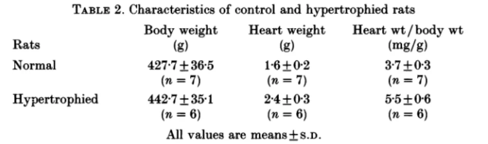
In the experiments conducted with six hypertrophied hearts, the body weight of
stenosed rats did not differ from that of normal rats. But as expected with left
ventricularhypertrophy, heart weight of stenosed rats was significantlyhigherthan
that of normal rats (Table 2). Thus, the heart weight/body weight ratio was
significantly greater inthe stenosedgroupthan in the normalgroup, whichwas our
selection criterion for left ventricular hypertrophy.
As regards cellular characteristics (Table 1), the hypertrophied myocytes were
wider but as long as the normal ones. However, no morphometric significant
difference was noted between the hypertrophic cellsisolated from septum, apexand
left ventricular free wall. Inkeepingwith the significantlyincreased cellwidth, the
C.
meanvalues weresignificantly higherinhypertrophiedmyocytesthan in normal ones, thus indicating an increased area of electrically chargeable membrane inhypertrophied cells which maybe simply attributed to the increaseof the apparent surface area. Whole-cell membrane capacitances were in the range of 143-373pF,
similar values to those measured by Scamps et al. (1990). The similar increases in
both heart weight/body weight ratio and myocyte surface area are in accordance with the expected rate of left ventricular hypertrophy as previously reported by Anversa, Loud, Giacomelli & Wiener (1978). Similarly in morphometric measure-ments, there were no significant topographical differences for Cm. The mean restingpotentialvalues for eachtopographicalgroup were notsignificantly different either (in mV: -81-3 + 4-9 for septum cells (n= 16); -81-6 +3*4 for apex cells
(n= 10); and -79-2+3-6 for left ventricular free wall cells (n= 16)).
Ito
inhypertrophied
myocytes
Inthe previoussection, wedemonstrated adifferentialdistribution ofItoinsingle
myocytesisolated from the septum, the apex and the left ventricularfree wall from normal rathearts. Notwithstandingitsunequal magnitude,
Ito
waspresent inall thestudied cells. Then in an attempt to explore more deeply the apparent lack of transient outward currents in human ventricular myocytes from hypertrophied
hearts (Benitah et al. 1992 a), we undertook a thorough study of the effects of myocardium hypertrophy on
Ito
in rat hearts.Inthese series of voltage clamp experiments, the sameprotocols and analyses as in normal myocytes were carried out. The averagemagnitude of
It.
appeared only slightly smallerinhypertrophiedcells (Fig. 2B)thaninnormal cells (Fig.2A).Inthe sameway as inthe normal group,therewas aconsiderablecell-to-cell variation inIto
magnitude fromthehypertrophied leftventricularfree wall.Infact,tencellsshowed
HETEROGENEITY OF TRANSIENT OUTWARD CURRENT 129
It.
characteristics closetothose of apex cells and the sixremaining
onesshowedIto
characteristics close to those of septal cells. As a result in hypertrophied cells, the topographical pattern of meanIto
magnitude valueswas:apex >left ventricular free wall > septum. Again, the large difference within-group variances precluded us from comparing the three topographical groups.TABLE2. Characteristics of control andhypertrophied rats Body weight Heartweight Heartwt/bodywt
Rats (g) (g) (mg/g)
Normal 427-7 +36-5
1P6±+02
3-7+0 3(n= 7) (n= 7) (n= 7)
Hypertrophied 442-7+3541 2-4+0 3 5-5+0-6
(n= 6) (n= 6) (n=6)
Allvalues are means+S.D.
The mean currentdensity-voltage relationships are shown in Fig. 2D, representing averaged data from sixteen septum, ten apex and sixteen left ventricular free wall hypertrophied myocytes. These findings clearly show that
It.
density was smallerinhypertrophied than in normal myocytes for the whole range of studied membrane voltages and for eachofthe considered groups. Theslopes of current density-voltage relationships were much less steep and Ito activation threshold was shifted to potentials more positive than in the normal groups. The differences in
It.
regional distribution between groups persisted, although they tendedtodecreaseascompared to the normal groups. The mean current threshold was (in mV): -26-9+6-5 inthe septum group (n = 16); -27-5 + 10-6 in the apex group (n= 10); and -21-7+6-5inthe left free wall group (n = 16); values did not differ greatly from eachother, but
shiftedto morepositivepotentialswhencomparedwithnormalgroups. The average
Ito
currentdensity
magnitudes
fora +60mVdepolarizing voltage
step
attained(in
pA/pF): 3-8+1-5inthe septum group (n = 16); 11-6+2-0 in the apex group (n= 10); and 8-4+5 0inthe left free wall group (n = 16) (with the largest variability recorded in the left ventricular free wall). Thus in the same way as with normal cells, we hypothesized that the smaller 4-AP-sensitive early outward currents found in
hypertrophied cells might reflecta reducedchannel density and otherdifferencesin
channel currentproperties: these possibilities are discussed below.
Thesteady-state inactivation curves were determinedinsevenseptum,eightapex and ten left ventricular free wall cells fromhypertrophied hearts (Fig. 7A,B and C respectively). Fitting the averagedataobtainedfromthosemyocytesto aBoltzmann
distribution functiongave the half-inactivation points at -33-7+3 5, -34*5+2-5,
and -31-2 + 6-5 mV (for the septum, apex and left ventricular free wall groups respectively) and aslopefactor of - 3+1-3, -5-3+ 10and -54 + 1 mV (forthe
septum, apex and left ventricular free wall groups respectively). As with normal groups, neither V0.5 nor k differed significantly between the different locations.
Furthermore, no significant difference was observed between normal and hyper-trophied myocytes (Figs 3 and 7). Thus therewas no hypertrophy-induced change
130 J. -P. BENITAH AND OTHERS
The Ito steady-state activation-voltage relationships in hypertrophied cells resembled those of normal cells with a similar sigmoidal dependence onmembrane potential. Theaveraged activation parameter data are plotted in Fig. 8A,B and C (for the septum, apex and left ventricular free wall groups respectively): the
A "//max B "//max C "//max 1.0 0.8 0.6 0*4 0.2 -1.0 -0.8 0.6 -0.4 0.2 -70 -60 -50 -40 -30 -20 -1o0 0 V(mV)
Fig. 7.Steady-stateinactivation-voltagerelationshipsof
It.
inhypertrophiedseptum(A), apex(B)andleft ventricular freewall(C)myocytes.The smoothcurves werefittedtothemean data points using a Boltzmann distribution as described in text. Vertical bars represent +S.E.M.
HETEROGENEITY
OF TRANSIENT OUTWARD CURRENT 131 1.0-0.8 0.6 a G/Gmax a C -80 -60 -40 -20 0 20 40 60 80 V(mV)Fig. 8. Steady-state activation-voltage relationships of
It.
in hypertrophied ratventricularmyocytes.Normalizedmeanchord conductancein septum (A),apex (B)and
left ventricular free wall (C) myocytes were plotted veraus membrane potential. The curves werefittedthrough themeandata pointsaccordingtotheproceduredescribed in thetext. Verticalbarsrepresent +S.E.M.
A
0.8
0-6
J.-P. BENITAH AND OTHERS
normalized data points for the threetopographical groups were fitted by Boltzmann distribution functions. With respect to the location, mean V0.5 was (in mV) -37 + 7-5 in septum cells (n = 15), 2-5 + 3-5 in apex cells (n= 9) and -1-6 + 7-2 in left
ventricular free wall cells (n= 15); the corresponding values for k were 8-7 + 2-6, 11-3 + 1-5 and 100+2-2 respectively. As with normal hearts, the activation parameters did not show any difference between the topographical groups (Figs 4 and 8). However, it could be observedthat k seemed smaller in the hypertrophied
groups than in the normal groups. This observation was consistentwith the current threshold shift to more positivepotentials,whilethe maximum Ito magnitude did not change.
As with normal groups, the mean maximalchord conductancesnormalized to the membrane capacitance weredifferentinhypertrophiedgroups(in,uS/pF): 0O02+001 for septum cells (n = 15); 007 +0.01 for apex cells (n= 9); and 0-05 +0'03 for left ventricularfreewall cells (n = 15). But inboth normal andhypertrophied cells, the
observed patterns might reflect differences in Cm, i.e. in membrane area. We therefore compared the mean maximal chord conductances before normalizing to membrane capacitance. Using this procedure, we no longer observednormal versus
hypertrophy differencesexceptforthe septal region: 6-7 + 2-9 versus 13-8 + 4-5,uS for
septum cells, 17-9 + 6-8 versus 18'6+7-8
#uS
for apex cells and 15-8 + 10-5 versus 26-0 + 130,uS for left ventricular free wall cells (in hypertrophy versus normalrespectively). Assuming Gmaxwascorrelated with thenumber of functional channels
asmentioned above, we consequently considered that there was a strong likelihood of differences in the functional channel densities in hypertrophied versus normal hearts for both the apex andleftventricular free wall regions, butnotinthenumber
offunctional channels. However, this hypothetical picture could not apply to the septal region inwhich wehaveto assume anabsolutehypertrophy-induced decrease
inthe number offunctional channels. A speculative explanation may be based onthe fact that hypertrophy would be unable to induce
Ito
channel neosynthesis (in the opposite way to the oneproposedbyScampsetal. (1991) regardingtheadaptationalchanges of
ICa,
L during the hypertrophic process in rat hearts). Furthermore, Ito channels in hypertrophied cells may be influenced by a modified environment thus altering their kineticbehaviour (asimilar situation may occurfor septalIto channelsin normal cells).
As shown in Fig. 2B, the rises of current traces from hypertrophied cells were
slowed down as compared to those recorded in a normal cell (Fig. 2A). This observation was borne out bythe plot ofmean times-to-peak data obtained within the range of explored membrane potentials in hypertrophied cells (Fig. 9A). No difference between location groups among hypertrophiedcells was noted, but there was a marked lengthening of activation in hypertrophied cells compared to the normal ones (Figs 5 and 9) for the whole range of explored voltages.
Figure 9B illustratesanothermajordifferencebetweenhypertrophied andnormal cellsfor the mean values of
Ito
inactivation time constants. Compare Figs6and9B;there is clear evidence that the time course of Ito inactivation was different in
hypertrophied myocytes than in the normal ones. If the general pattern of
inactivation timeconstants-voltagerelationshipswassimilar inbothgroups(absence of regional differences), the whole kinetics were markedly slowed down in
HETEROGENEITY OF TRANSIENT OUTWARD CURRENT 133 A 35 30 -25 20 (U I I 30 -15 0 E 0* B 80 70 60. E 50 40-30 20 -40 -20 0 20 40 60 V(mV)
Fig. 9. Comparison of kinetics properties of
40
in hypertrophied myocytes isolated from septum (@), apex (0) and left ventricular free wall (K). A, times topeak wereplottedversusmembrane potential.B, pooled mean data of inactivation timeconstants
obtained by curve-fitting algorithm were plotted versus membrane potential. Vertical
barsrepresent +S.E.M.
hypertrophied groups. As a result, the time constants of current decay seemed to undergo ahomogenizingtrend towards the level ofvaluesfoundinboth normal and
hypertrophiedseptumcells.Themeantimeconstantsmeasuredat
depolarizing
steps of +40mV were: 42-1 +6-8 versus 41-2+9-7ms for septum cells; 40-8+4-0 versus 32-5+8-3 ms for apexcells; and 42-1 + 6-8 versus 32-9 + 11-8 ms for the left ventricularJ.-P. BENITAH AND OTHERS
DISCUSSION
The majorfindingsof thispaper arethreefold.(1)Onthebasis ofourtopographical
model, there is clear evidenceof a differential distribution of Ito in septum, apex and
left ventricular free wall myocytes from normal rat hearts. (2) This physiological topographical heterogeneity ofItotendstodecreaseintheabdominalaortic
stenosis-induced left ventricular hypertrophy through a global reduction of the current
density. (3) The lowervariability ofcurrents in septum and apex groups compared
to the left ventricularfreewall group in both normal andhypertrophied cells lends
support to the assumption that a differentialdistribution of
Ito
may exist not only with reference to an epicardial-endocardial spectrum but also with respect to theanatomical considered regions.
Regional variation of Ito in normal rat hearts
Our findings demonstrated differences in the density of 4-AP-sensitive early
outward current intheseptum, theapex and the leftventricularfreewallof the rat heart. The mean current density inapex cells was clearly about twice as high as in septumcells andthemean currentdensity of left ventricular freewallcellswasabout three times higherthan in septum cells as shown in Fig. 2C. To ourknowledge, no
such differential distributionof Ito has beenreportedsofar. Yetthis distributionwas consistent, to a certain extent, with the previousfindings ofWatanabe etal. (1983)
who reported the fact that action potentials recorded from the apical region were shorter than those from the base of theleft ventricle.
Wefound that thevariabilityofIto ismuchhigherinthe left ventricular free wall groupthan inboth othergroupsand that such variabilityapparentlyresulted from
a rather heterogeneous composition of this group (see Results). Indeed, a large
variability for the transient outward current was previously reported in rabbit ventricle studies (Giles & Imaizumi, 1988), and as suggested bythese authors, we could not rule out the possibility that cell-to-cell differences in Ito size in left ventricular free wall were due to real differences in its distribution between
epicardium and endocardium in the rat heart. The assumption of a regional difference in action potential morphology between epicardium and endocardium,
probably related to a differential distribution of transient outward currents, was
supported by canine ventricular tissue studies (Litovsky & Antzelevitch, 1988). A 'spike and dome' configuration was recorded in epicardial preparations but was
absent in endocardium. Responses to different stimulation protocols and phar-macological interventions led to the proposal ofthe virtual absence ofa transient outward K+ current in endocardium as opposed to epicardium in this model. The
correlation of the presence of a notch in the epicardial action potential and a prominent transient outward current (Ito
i)
in isolated epicardial myocytes wasrecentlyascertainedby Furukawaetal. (1990) in cathearts;the opposite pattern was uncovered in subendocardial layers. In an attempt to describe the differential regional distribution of the 4-AP-sensitive outward currents (It) in rabbit left
ventricles, asimilar procedure analysing epicardial and endocardial layers separately
has recently been reported by Fedida & Giles (1991).
Differencesinthe resting potentialmightaltertheavailabilityand the recoveryof 134
HETEROGENEITY OF TRANSIENT OUTWARD CURRENT
the early outward current and thus induce apparent regional differences in the current characteristics. In fact, no differences for the zero current potential were recorded between the three groups. The differential distribution of Ito densities observedinnormal ratventricles could not be correlated to relative shifts of
steady-state activationand inactivation curves since therewere no significant differences in half-activation and inactivationvoltages, nor in slope factors between groups. There wasonlya slight non-significant tendency for the mean inactivation time constants of normal septum cells to assume higher values than those of both other groups within the + 20to + 60mV range (Fig. 6). Accordingly no consistent explanation for the regional variations in
It.
densities could be deduced from the steady-state activationand inactivation kinetics, nor were the different regional time courses of currentdecay of anysignificant value. Since no single channel studies were actually performed in this work, we could not exclude the existence of regional differences in the properties of the single transient outward current channels, though we shouldlike to consider the following assumptions: (1) the channel opening probability is related to thevoltage-dependent macroscopic current as suggested by McDonald et al. (1986) for theCaa2+ channel current and probably also for the transient outward current channels asproposed by Clark et al. (1988) on the basis of similar effects of stimulusfrequencyonthewhole-cell amplitudeand the meanopenprobability ofthe
channel; (2) the current-voltage relationship and slope conductances for single channels may be similar from one region to another. Provided that these assumptions
are correct, then the computed maximal conductance,
0max,
for septum, apex and left ventricular free wall cells may be viewed as an indirect estimate of the maximal numberof channelsinthe openstate. Continuing thus,it maybeexpected thatthedensity ofIto functional channels is reducedinseptal cells whencomparedtothe apex and left ventricular free wall cells in normal rat hearts. A similar conclusion was
recently drawn from single channel studies performed in rabbit left ventricles by Fedida &Giles (1991): single
It
channelsinrabbitendocardium andepicardium hadthe same amplitude and kinetics, whereas current density was higher in epicardial cellsthaninendocardial orpapillarymuscle cells.Moreover, the presentfindingson
hypertrophied rat heart seemed to agree with our assumption on a differential distribution of functional channel densities.
Distribution patternof Ito in hypertrophiedrat hearts
Regional differencesintheelectrophysiologicalproperties of the ventriculartissue
of the heartwerefirst outlinedby Keung&Aronson(1981).Workingonendocardial, epicardial and papillary muscle fibres obtained from hypertrophied and normal hearts, these authors showed that hypertrophy inducedby renalhypertension may
be anon-uniform processthat affectsthe ventricularwalllayersto adifferentdegree.
DirectevidenceforaglobaldecreaseinItocurrentdensityinthe
hypertrophied
rat ventricle results from our experiments. As mentioned above, it should be stressed thatthis decreasewasrelatedto a consistentreductioninmaximalIto conductances normalized to membrane capacitances. No hypertrophy-induced changes of the steady-state activation and inactivation parameters were involved in this process. Oneplausibleexplanation mightlie in the absenceofIt.
channelneosynthesisduringthe hypertrophic processleading to a decrease of channel density per surface area
J. -P. BENITAH AND OTHERS
unit, but notof the absolute number of channels per cell, at least in the apex and free wall regions. Such behaviour might be the reverseof the adaptationalprocessofCa2+ channels to pressure overload in which the total number of Ca2+ channels was increased whileCa2+channel density was maintained inhypertrophied rat ventricular cells (Mayoux, Callens, Swynghedauw & Charlemagne, 1988); conversely, it maybe comparabletothetime course pattern of other membrane proteins whose decreased density during cardiac overload suggests a non-regulation for the genes coding for these receptors (Mansier et al. 1990). Moreover, it should be mentioned that the
decrease ofIto currentdensity observedinhypertrophied cellswasofthe same order asthatobserved during theearly stagesofdevelopmentin ratventricular myocytes
(Kilborn & Fedida, 1990), eventhough furtherinvestigations would be mandatory
toelucidate and compare the molecular mechanisms for both these processes. As shown in normal hearts, the differentialdistributionamongthestudiedregions
persisted althoughittendedtodecrease. Regardingtimekinetics, wenotedaglobal lengthening of times to peak in all regions and a particular pattern change of
inactivation kinetics; time constants of current decay were not prolonged in
hypertrophied septal cells whereastheywere markedly lengthened in apexand left free wallregions,resultingin ahomogenization of valuesinallthe consideredregions. Thus the hypertrophy-induced inactivation kinetics changes associated with the
global reduction ofItodensitiesindifferent regions of the left ventricle may suggest a homogenizing process related to pressure overload-dependent structural and
functional alterations. Furthermore, assuming regional differences in the absolute
number of functional channels and an absence of channel neosynthesis during the
hypertrophic process, we may expect the appearance of environmental changes of channels within the membrane which should be able to modulate time-dependent
kinetics without altering both activation and inactivation parameters.
Wehave demonstrated inapreviousstudy (Benitahetal. 1992 a) that the calcium channel current is largely dominant as compared to transient outward currents in human septal cells from ventricular hypertrophy subsequent to chronic pressure overload. From our experimentaldata we expectthat twofactors canunderlie this
ionic pattern in the human heart; (1) the septum may be a region in which the transient outward current is not originally prominent, and (2) the hypertrophic
process isclearly apotent contributing factor toreducing this current.
We thank Dr M. Delmar for hishelpfulinformation and criticalreadingof themanuscript, and B.Calime for secretarial assistance. This workwassupported byagrant-in-aid from theGroupe
de Reflexion sur la Recherche Cardiovasculaire, the laboratoires Squibb (France), and Actions integreesFranco-Espagnol (92040).
REFERENCES
AGUS,Z.S., DUKES, I. A. &MORAD, M. (1991).Divalent cations modulate the transient outward
current in ratventricular myocytes. American Journal ofPhysiology261, C310-318.
ANTZELEVITCH, C., SICOURI, S., LITOVSKY, S. H., LUKAS,A., KRISHNAN, S. C., Di DIEGO, J. M.,
GINTANT, G.A. & Liu, D. W. (1991). Heterogeneity within the ventricular wall. Electro-physiology and pharmacology of epicardial, endocardial, and M cells. CirculationResearch69, 1427-1449.
HETEROGENEITY OF TRANSIENT OUTWARD CURRENT 137
ANVERSA, P., LOUD, A. V.,GIACOMELLI, F. & WIENER, J. (1978). Absolutemorphometricsuudyof myocardial hypertrophy in experimental hypertension. Laboratory Investigation 38, 597-605. APKON, M. & NERBONNE, J. M. (1991). Characterization of two distinct depolarization-activated
K+ currents in isolated adult rat ventricular myocytes. Journal of General Physiology 97, 973-1011.
ARONSON, R. S. & NORDIN, C. (1984). Electrophysiological propertiesof hypertrophied myocytes isolated from rats with renalhypertension. European Heart Journal 5, suppl. F, 339-345.
BEJNITAH, J.-P., BAILLY, P., D'AGROSA, M.-C., DA PONTE, J.-P., DELGADO, C. & LORENTE, P. (1992a). Slow inward current in single cells isolated from adult human ventricles. PflugersArchiv 421, 176-187.
BE1NITAH, J.-P., DELGADO, C., GOMEZ, A. M., BAILLY, P., DA PONTE, J.-P. & LORENTE, P. (1992 b).
Characteristics of the transient outward current within the ventricular wall in normal and hypertrophied rat ventricles. XIVth Congress of the EuropeanSociety of Cardiology, August 1992, Barcelona, Spain. European Heart Journal13, 397 (abstract 2313).
CLARK, R. B., GILES, W. R. & IMAIZUMI, Y. (1988). Propertiesof the transient outward current in rabbit atrial cells.Journal of Physiology405, 147-168.
CORABOEUF, E. & CARMELIET, E. (1982). Existenceof two transient outward currentsin sheep cardiacPurkinje fibers. Pflugers Archiv 392, 352-359.
CUTILLETA, A. F., DOWELL, T., RUDNICK, M. & ARCILLE, R. A. (1975). Regression of myocardial hypertrophy.I.Experimental model changes in heart weight nucleic acids and collagen. Journal of Molecular and Cellular Cardiology7, 767-781.
DECK, K. A. & TRAUTWEIN, W. (1964). Ionic currents in cardiac excitation.Pfliugers Archiv 280, 63-80.
DUKES, I. A. & MORAD, M. (1991). The transient K+ current in rat ventricular myocytes: evaluation ofitsCa2+and Na+ dependence. Journal of Physiology435, 395-420.
ESCANDE, D., COULOMBE, A., FAIVRE, J. F., DEROUBAIX, E. & CORABOEUF, E. (1987). Two types of transient outwardcurrentsinadult human atrial cells. AmericanJournal of Physiology252,
H142-148.
ESCANDE, D.,LOISANCE, D.,PLANCHE,C. & CORABOEUF, E. (1985). Age-related changes of action potential plateau shape in isolated human atrial fibers. American Journal ofPhysiology 249,
H843-850.
FEDIDA, D. & GILES, W. R. (1991). Regional variations in the action potentials and transient outward current in myocytes isolated from rabbit left ventricle. Journal ofPhysiology 442,
191-209.
FOZZARD, H. A. & HIRAOKA, M. (1973). The positive dynamic current and its inactivation
propertiesin cardiac Purkinje fibres. Journal of Physiology234,569-586.
FURUKAWA, T., MYERBURG, R.J., FURUKAWA, N., BASSETT, A. L. & KIMURA, S. (1990). Differences in transient outward currents of feline endocardial and epicardial myocytes. Circulation Research67, 1287-1291.
GILES, W. R. & IMAIZUMI, Y. (1988). Comparison of potassium currents in rabbit atrial and ventricular cells. JournalofPhysiology 405, 123-145.
GINTANT, G. A., COHEN, I. S., DATYNER, N. B. & KLINE, R. P. (1991). Time-dependent outward
currents in the heart. Transient outward current in cardiac fibers. In The Heart and Cardiovascular System, ed. FOZZARD, H.A., HABER, E., JENNINGS, R.G., KATZ, A. M. & MORGAN, H. E.,pp. 1138-1156. RavenPress, New York.
HAMILL, 0.P.,MARTY, A., NEHER, E., SAKMANN, B. &SIGWORTH, F.J. (1981). Improved patch
clamptechniqueforhigh-resolutioncurrentrecording from cells and cell-free membranepatches. PflugersArchiv391,85-100.
HIRAOKA, M. &KAWANO, S. (1989). Calcium-sensitive andinsensitive transientoutwardcurrent in
rabbit ventricular myocytes. Journalof Physiology410, 187-212.
JOSEPHSON, I.R., SANCHEZ-CHAPULA, J. & BROWN, A. M. (1984). Early outward currentin rat
single ventricular cells. Circulation Research54, 157-162.
KENYON, J. L.&GIBBONS,W. R.(1979).4-Aminopyridineand theearlyoutwardcurrentofsheep
cardiacPurkinjefibers. Journal ofGeneralPhysiology73, 139-157.
KEUNG, E. C. (1989). Calciumcurrentisincreasedinisolated adultmyocytesfromhypertrophied