HAL Id: hal-02047423
https://hal.archives-ouvertes.fr/hal-02047423
Submitted on 17 Dec 2020
HAL is a multi-disciplinary open access
archive for the deposit and dissemination of
sci-entific research documents, whether they are
pub-lished or not. The documents may come from
teaching and research institutions in France or
abroad, or from public or private research centers.
L’archive ouverte pluridisciplinaire HAL, est
destinée au dépôt et à la diffusion de documents
scientifiques de niveau recherche, publiés ou non,
émanant des établissements d’enseignement et de
recherche français ou étrangers, des laboratoires
publics ou privés.
Extracorporeal photochemotherapy induces arginase 1
in patients with graft versus host disease
E. Merlin, N. Goncalves-Mendes, Dalil Hannani, Anne de la Torre, M. C.
Farges, H. Laroye, F. Demeocq, J. Kanold, M. P. Vasson
To cite this version:
E. Merlin, N. Goncalves-Mendes, Dalil Hannani, Anne de la Torre, M. C. Farges, et al..
Extracor-poreal photochemotherapy induces arginase 1 in patients with graft versus host disease. Transplant
Immunology, Elsevier, 2011, 24 (2), pp.100-106. �10.1016/j.trim.2010.10.007�. �hal-02047423�
1
Extracorporeal photochemotherapy induces arginase 1 in patients with graft versus host disease.
E. Merlin1,2, N. Goncalves-Mendes3, D. Hannani4,5,6, A. de la Torre3, M. C. Farges3, H. Laroye7, F. Demeocq1,2, J. Kanold1,2, M. P. Vasson3.
Running title: arginine metabolism during ECP
1
CHU Clermont-Ferrand, Centre Régional de Cancérologie et Thérapie Cellulaire Pédiatrique, Hôtel-Dieu;
2 Inserm, CIC501; F-63001 Clermont-Ferrand, France. 3
Clermont Université, Univ Clermont 1, UFR Pharmacie, EA4233 Biochimie Biologie moléculaire et Nutrition, CRNH Auvergne, CLARA, F-63003 Clermont-Ferrand, France.
4
Inserm, U823, Immunobiologie et Immunothérapie des cancers, La Tronche, F-38706, France
5 Univ Joseph Fourier, F-38041 Grenoble, France. 6
EFS Rhône-Alpes, La Tronche, F38701 France
7 Centre Anticancéreux Jean Perrin, Unité de Nutrition, F-63000 Clermont-Ferrand, France.
Key words: Bone marrow transplantation – Cytokines – Nitric Oxide – Amino acids – Apoptosis – PBMC Arginine, ECP.
Corresponding author:
E. Merlin, Centre Régional de Cancérologie et Thérapie Cellulaire Pédiatrique, Hôpital Estaing
1, place Lucie Aubrac
63003 Clermont-Ferrand, France.
Phone: + 33 4 73 750 015 Fax: + 33 4 73 750 004 E-mail address: e_merlin@chu-clermontferrand.fr
2
Abstract
The benefits of extracorporeal photochemotherapy (ECP; psoralen and UVA exposure of blood mononuclear cells) in graft-versus-host-disease (GVHD) are well-recognized , but the mechanisms of action remain elusive. As the metabolism of L-arginine in immune cells is known to play a role in immune tolerance, we investigated the effect of ECP on arginine metabolism, and the influence of extracellular L-arginine concentration on the response to ECP in cells from patients on therapy by ECP for a GVHD and healthy donors cultured before and after ECP in the presence of different concentrations of arginine (0, 50, 100, 200 and 1000 µmol/l). At baseline arginine was not metabolized through the same pathway in patients and donors. When cells were exposed to ECP, the production of ornithine but not NO° was enhanced, while mRNA of arginase 1 was up-regulated but not INOS. In GVHD patients, increasing arginine concentration resulted in down-regulation of IFNγ and TNFα mRNA expression, whereas IL10 was up-regulated especially at physiological plasma levels (between 0 and 100 µM). Overall, our study shows that ECP orients the metabolism of arginine toward the arginase pathway together with shifting the cytokine profile toward IL-10, providing new insights into the enigmatic mechanism of action of ECP.
3
1. Introduction
Extracorporeal photochemotherapy (ECP) is a cellular therapy that has demonstrated efficacy in a number of T-cell-mediated diseases, such as T-cell cutaneous lymphoma, allograft rejection, and severe and/or steroid-resistant graft-versus-host-disease (GVHD) [1]. The process consists in an extracorporeal exposure of peripheral blood mononuclear cells to UVA-photoactivated 8-methoxypsoralen (8-MOP) and their subsequent reinfusion into the patient [2]. ECP induces immediate lymphocyte and delayed monocyte apoptosis [3], and it is widely suspected that its therapeutic effects are related to the infusion of apoptotic lymphocytes and tolerogenic dendritic cells [4]. The role of interleukin 10-induced synthesis (IL-10 is a potent anti-inflammatory and regulatory cytokine) by treated and untreated monocytes could be decisive [5-6], together with the generation of CD4+CD25+Foxp3+ regulatory T cells (Tregs) [4], although other signaling pathways are probably involved. In particular, ECP triggers significant metabolic changes, some involving fatty acids [7]. Thus, one possibility is that the effects of ECP are dependent on the patient’s metabolic status. GVHD is a multiorgan syndrome in which sustained inflammatory response, organ involvement (such as cholestasis, mouth ulcerations and colitis) together with recurrent infections can lead to impaired nutritional status. Moreover, some of our preliminary data suggest a significant decrease in free arginine rates in cases of gastro-intestinal involvement (unpublished data).
Two forms of GVHD are distinguished based on chronological, clinical and pathophysiological features. Classically, acute GVHD occurs prior to 100 days after hematopoietic cell transplantation and involves the skin, the gastrointestinal tact and/or the liver. Chronic GVHD is a pleiomorphic syndrome with onset generally occurring between 3 and 24 months after allogeneic hematopoietic cell transplantation. The highly variable clinical manifestations have features resembling autoimmune and other immunologic disorders [8]. They frequently involve the skin, liver, eyes, mouth, upper respiratory tract, esophagus, and less frequently involve serosal surfaces, lower gastrointestinal tract, female genitalia, and fascia. The biological mechanisms leading to chronic GVHD are not as well understood as those leading to acute GVHD. However, it is well establish that Donor T cells are critical for the development of both acute and chronic GVHD [9]. Schematically, target-tissue damage in acute GVHD is caused by cytopathic donor T cells that respond to the genetically disparate host polymorphic antigens, which are presented by host and/or donor antigen-presenting cells (APCs) [9]. The T cell response is amplified by several pro-inflammatory cytokines (such as IL-1, IL-6, IL-8 and tumor necrosis factor α) released in response to the conditioning regimen-induced epithelial injury [10]. .This T cell response can be counter-regulated by costimulatory inhibitors (CTLA-4 and ICOS) as well as regulatory cell populations (CD4_CD25hi Treg subset and NKT cells) [11]. Convserly, chronic GVHD is thought to result from an
“auto-4
reaction” against non-polymorphic antigens [9]. This reaction is caused by CD4_ T cells generated de novo from donor cells hematopoietic cells in a setting of thymic dysfunction caused by both conditioning regimen and previous acute GVHD. The mechanisms leading to organ injury involve both B cells and T cells, and are mediated at least by the secretion of TGF ß and agonistic antibodies against platelet-derived growth factor receptor (PDGFR). The role of regulatory T cells is much less clear than in acute GVHD [12].
L-arginine is an amino acid thought to become essential after injury. It plays a central role in several functions of the immune system [13]. In leukocytes, L-arginine can be metabolized by two pathways, under the control of two key enzymes, i.e. inducible nitric oxide synthase (iNOS) and arginase 1 leading to the modulation of available extracellular L-arginine and to the production of metabolites. Free L-arginine concentration in the environment was the first parameter shown to modulate T cell activation and function: arginine is known to enhance lymphocyte proliferation and IL2 utilization [14], whereas L-arginine starvation impairs T-lymphocyte function, at least partly by triggering the internalization of the CD3 zeta chain and impairing cell cycle progression by interfering with the cyclin D3 and cdk4 pathway [15]. Hence, L-arginine depletion under the action of arginase is known to contribute to tumor-induced immune suppression [16], as well as the counter-regulation of inflammatory purulent reactions in humans [17] as well as the maternal unresponsiveness toward the semi-allogenic fetus [18].
Aside of extracellular arginine concentration, the products of arginine metabolites are also involved in immune regulation. Inducible NO Synthase leads to the production of nitric oxide and citrulline, whereas the action of arginase results in the production of ornithine, proline and polyamines [19]. Depending on the predominant metabolic pathway, these products can mediate multiple biological responses. The increase of intracellular NO resulting from iNOS activity can mediate normal signaling or be cytostatic and cytotoxic, particularly at high concentration, in responses to cytokines [20]. When arginase and iNOS are concomitantly activated, the action of NOSynthase in the condition of low arginine level leads to the production of peroxynitrites (ONOO-) and triggers lymphocyte apoptosis [20]. Arginase activity not only leads to arginine depletion but also to the production of proline and ornithine which can be utilized to synthesize polyamines (putrescine, spermidine and spermine). Ornithine and polyamines are known to exert some immunomodulatory effects, in particular in the setting of allogeneic MLR [21]. Although the immunological role of spermine is not clear, a negative correlation has been found between blood spermine levels in patients with breast cancer and the impairment of IL12 synthesis in vivo. Moreover, it has been shown that in vitro exposure of dendritic cells (DCs) to spermine may impair their stimulatory function. [22] To our knownledge, nothing is known about the activity of arginase in
5
patients with GVHD. On the other hand, NO is suspected to be increased in patients with GVHD [23], but at present, the full range of effects of NO during GVHD is not yet fully described. The immunosuppressive effect of NO in acute GVHD is well established, but it is not clear whether this plays a deleterious or a protective role [24].
Whereas UV exposure is known to upregulate the activity of several enzymes such as ornithine decarboxylase (ODC, involved in the metabolism of ornithine and polyamines) in skin cells [25], to date, nothing is known about the combined effects of UVA and 8-MOP on arginine metabolism in human leukocytes during ECP. Moreover, given the impact of L-arginine availability on immune response, one possibility is that the response to ECP could be affected by L-arginine content in the cell environment. We therefore investigated the effects of ECP on arginine metabolism in the context of GVHD and in healthy donors, and determined whether the modulation of arginine availability can modify leukocyte response after ECP treatment.
Our work shows that ECP significantly impacts the metabolism of arginine toward the production of ornithine but not nitric oxide, and that under ECP condition arginine availability is an important factor for the modulation of TNFα in patients with GVHD.
2. Material and methods
2.1 Patients and healthy donors
The study enrolled 6 patients (4 males and 2 females aged 9 to 56, median 15) diagnosed with acute (n=3) or chronic (n=3) GVHD and 6 healthy donors (3 males and 3 females aged 24 to 35, median 28). Acute GVHD was graded according to Glücksberg criteria [26], and the severity of chronic GVHD was assessed as proposed by Shulman [27]. Complete response was defined as resolution of all organ manifestations. Partial response was defined as greater than 50 percent response of organ involvement. Absence of response was defined as stable or progressively worsening GVHD and/or the inability to taper other medication by at least 50 percent. The patients had undergone different immunosuppressive treatments but had not responded (table 1). Blood donations in this study conformed to institutional review board-approved protocols, and informed consent was obtained.
6
ECP was performed using the Vilber-Lourmat procedure, as previously described [28]. Briefly, mononuclear cells were isolated from patients by cytapheresis (Cobe Spectra, Gambro BCT, Bourg-la-Reine, France) then diluted in normal saline solution to obtain a final hematocrit <1%, and 8-methoxypsoralen was added at 200 ng/ml. After a 3-minute incubation, cells were transferred into a UV-A transparent bag (Macopharma) and exposed to UV-A light (340 nm) at 2 J/cm2 for 10 minutes (UVA-matic, Vilber-Lourmat, France). ECP sessions were performed three times a week (with a one-day interval between two ECP sessions) for three weeks, and then gradually reduced for those patients who were stabilized or who showed improvement. For each session, apheresis duration was 120 minutes. This allowed to collect between 1.6x10⁷ and 8.2 x10⁷ lymphocytes/kg(for patients with acute and chronic GVHD, respectively).
2.3 PBMC sampling and preparation
Two cell samples were collected from patients: one peripheral blood sample was collected by venous puncture in EDTA from patients immediately prior to apheresis (ie before ECP), and a second cell sample was collected from the buffy coat bag just prior to reinfusion (ie after ECP). Blood samples were also collected from healthy donors by venous puncture, but cytapheresis were not performed for healthy volunteers. PBMC were isolated by density gradient centrifugation over Ficoll-Histopaque (Sigma-Aldrich). The isolated cells were then washed twice in phosphate buffered saline (PBS, Gibco). Cells from the healthy volunteers were then separated into two fractions, and one was cultured without additional treatment (“non-treated”) while the other fraction was treated by PUVA exposure before being cultured (“ECP-treated”). The PMBC from the patients were cultured as control “non-treated” cells.
2.4 Media and cell cultures
”Non-treated” and “ECP-treated” mononuclear cells were cultured (37°C, 5% CO2) for 48 h in arginine-free and nitrate-free RPMI (1x10⁶ cells/ml) which was supplemented with L-arginine at 0, 50, 100, 200 or 1000 µM. 10% dialyzed fetal calf serum, 4mM L-glutamine and 100 UI/l penicillin-streptomycin were added. The cells were stimulated by phytohemagglutinin (5µg/ml). After 48 h culture, the supernatant was removed by centrifugation and stored at -80°C until NO and amino acid analysis. PBMC were washed in PBS then assessed for apoptosis and mRNA quantification of cytokines.
7
For arginase 1 and iNOS expression, 1x10⁶ cells/ml were cultured (37°C, 5% CO2) for 24 h in Minimal Essential Medium with 10% dialyzed fetal calf serum, 4mM L-glutamine and 100 UI/l penicillin-streptomycin. The cells were stimulated by LPS (1µg/ml).
2.5 Supernatant amino acid analysis
For amino-acid analysis, after deproteinization using sulfosalicylic acid, 30 µl of the supernatant was used for HPLC analysis with an ion-exchange column on a Dionex System. After post-column derivatization with ninhydrin, amino acids were detected at 570 nm. Data was collected and processed using Chromeleon® software, as previously described [29].
2.6 Supernatant NO analysis
Nitric oxide (NO) production was measured as nitrite plus nitrate using the Griess method after incubation with nitrate reductase (Cayman, France), as previously described [30].
2.7 Supernatant cytokine analysis
IL-10, TNFα and IFNγ assays were performed simultaneously using Cytometric Beads Array (CBA) kits on EPICS apparatus (Becton Dickinson Biosciences Pharmingen, San Diego, CA, USA) following the manufacturer's instructions.
2.8 Apoptosis
FITC-conjugated annexin-V coupled with 7-aminoactinomycin D (Beckman-Coulter) was used to detect apoptosis. Cells were washed twice in PBS then stained with FITC-libeled Annexin-V, 7-aminoactinomycin D (in a 100 µl of labeling solution) and incubated for 10 min in the dark according to the manufacturer’s protocol. Then, 400 µl of buffer was added, and stained cells (> 10,000 events) were analyzed by flow cytometry (Beckman-Coulter EPICS). This staining makes it possible to differentiate cells in early apoptosis (annexin + 7-AAD -), late apoptosis (annexin + 7-7-AAD +) and necrotic cells (annexin – 7-7-AAD+) from viable cells (annexin - 7-AAD -).
2.9 Real-Time RT-PCR quantification of the mRNA levels of cytokines and enzymes involved in arginine metabolism
8
PBMC total RNA was extracted using Trizol Reagent (Invitrogen) according to the manufacturer’s instructions. Reverse transcription was conducted from 1 µg of total RNA, giving rise to 20 µl of cDNA as previously described [31]. One µl was used for the PCR performed in real-time on a Lightcycler Instrument (Roche Diagnostics). Sample mRNA copy numbers were extrapolated from standard curves obtained with serially diluted purified PCR products. 18S RNA was used as internal standard, so the results were expressed as fold induction. IL-10, Interferon γ, Tumor Necrosis Factor-α, Arginase-1 and inducible NOSynthase mRNAs were detected using the primer probes reported in Table 2.
3. Results
3.1 Before ECP exposure, arginine is differentially metabolized in circulating leukocytes from ECP-treated GVHD patients and from healthy donors
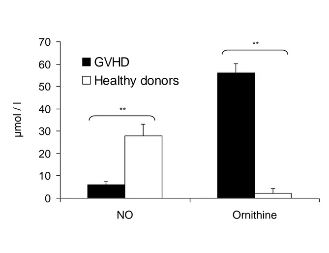
We investigated whether the metabolism of arginine was similar in cells harvested from ECP-treated patients as in cells from healthy donors. After a culture in nitrite-free medium, we measured the products of arginine catalysis that had been release into the supernatant. After a 48 hr-culture, NO production was lower in cells from patients with GVHD compared with cells from healthy donors (Figure 1, p<0.01). Moreover, the L-ornithine concentration in the supernatant was significantly higher in cells from patients treated by ECP for a GVHD than cells from healthy donors (p<0.01). This shows that in leukocytes from patients who were previously treated by ECP for a GVHD, arginine is preferentially metabolized toward the production of ornithine.
3.2 ECP induces the synthesis of L-ornithine but not nitric oxide
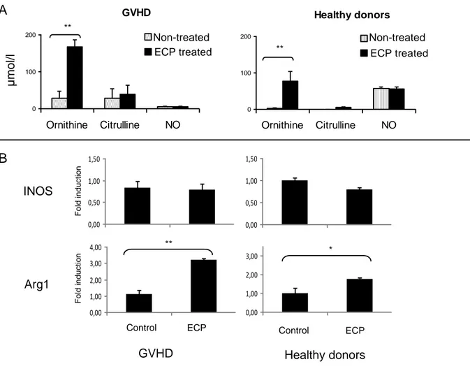
To assess whether the differences of arginine metabolism between patients and donors could be imputed to ECP, we measured the impact of ECP on the production of arginine metabolites. Cells were treated by ECP or not and then cultured during 48h. At the end of the culture, L-ornitine concentration in the supernatant has been evaluated. For both patients and healthy donors, L-ornithine concentration was significantly higher for ECP-treated cells (figure 2A) than for non-ECP-treated cells. This suggests that ECP activates the arginase pathway in treated-leukocytes. Through competition for arginine, the arginase can also modulate the production of nitric oxide (9). In leukocytes, the production of NO by iNOS is accompanied by the equimolar production of citrulline. Cells from both GVHD patients and donors showed similar NO and citrulline production with and without ECP treatment. Taken together, these results suggest that in ECP-treated leukocytes, arginine is metabolized through the arginase pathway, and does not serve as a precursor for the synthesis of N0.
9
3.3 ECP upregulates arginase expression.
In leukocytes, ornithine is produced from arginine by the enzyme arginase 1, whereas NO° is produced by inducible NOSynthase (iNOS). We found that arginase 1 but not iNOS was up-regulated in ECP-treated cells as compared with non-treated cells, in healthy volunteers and in patients with GVHD (figure 2B). These data are in line with those presented in figure 2A.
3.4 ECP affects cytokine synthesis
The effect of ECP on cytokine expression is still unclear. Some authors have reported an increase of Th1 cytokines [32] whereas other observed an increase of Th2 [33]. The discrepancies are attributed in part to the cells origin (healthy donors or patients). One well-established impact of ECP in the setting of GVHD is its upregulating effect on the synthesis of IL10 [5, 34], while TNFα is down-regulated. IL10 is a suppressive and regulatory cytokine which is involved in the triggering of Tregs [35], and which is able to down-regulate the expression of TNFα [36]. We evaluated ECP impact on cytokine synthesis by mRNA expression level in GVHD patients. After 48 hr culture with PHA, ECP induced a significant increase (up to 1.5-fold) in IL-10 mRNA expression and a decrease of TNFα and IFNγ mRNA expression (0.2 and 0.4-fold, respectively; figure 3).
3.5 After ECP, L-arginine availability impacts the production of ornithine but not NO
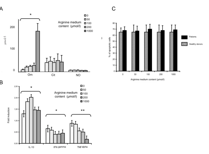
Arginine content of the culture medium is known to regulate the expression of some of the proteins of the metabolism of arginine, such as iNOS [37]. To assess the impact of extracellular L-arginine concentration on the metabolism of L-arginine in an ECP setting, cells from GVHD patients and healthy donors were treated by ECP then cultured for 48 hours in an arginine and nitrate-free RPMI 1640 supplemented with defined amounts of L-arginine (0, 50, 100, 200 and 1000 µmol/l) and then stimulated with PHA. In both GVHD patients and healthy donors, the final post-ECP supernatant concentration of arginine was about 50% lower than the initial concentration. Ornithine increase was strikingly dependent on arginine availability (figure 4A). Conversely, the concentration of L-arginine in the cell culture medium did not impact the final concentration of citrulline and NO in the supernatant. This indicates that under ECP conditions, the excess of arginine is only transformed in ornithine, and does not lead to an increase of NO synthesis, supporting data shown in figure 2.
10
3.6 After ECP, L-arginine availability impacts the expression of cytokine mRNA from GVHD patient mononuclear cells without impacting the triggering of apoptosis
We have shown that after ECP, increasing the availability of extracellular arginine leads to a dose-dependent increase of its derivative L-ornithine. Ornithine is known to exert some immunomodulatory effects; in particular it is able to inhibit the alloreactive proliferation in the setting of MLR [21]. To test whether the increase of free L-arginine impacts the immunological response of mononuclear cells to ECP via L-ornithine, we measured the synthesis of cytokine mRNAs after 48 hours in a culture media containing different amounts of L-arginine (0, 50, 100, 200 and 1000 µmol/l). Enriching the culture medium with L-arginine resulted in a significant dose-dependent decrease in TNFα mRNA transcription levels (figure 4B). IL10 and IFNγ mRNA transcription levels were significantly affected at concentrations ranging between 0 and 100 µM, but this effect plateaued out at above 100 µM. This difference in mRNA expression level could not be related to a variation of apoptosis, since the arginine content in the medium has no impact on the percentage of apoptotic cells after ECP (figure 4C).
11
4.
DiscussionThe metabolism of some free amino acids is known to play a central role in several immune system functions. We therefore investigated whether ECP induces significant changes in the extracellular concentration of amino acids after a 48-hr culture of PBMC. Our study shows that ECP significantly impacts the metabolism of arginine in leukocytes by upregulating the arginase pathway but not NO.
In leukocytes, arginine is metabolized either to ornithine by arginase or to citrulline and NO via iNOS, and the prevalent view is that these 2 pathways are antithetic [20]. Cells from ECP-treated GVHD patients produced less NO than those cells from healthy donors. This result is quite unexpected, since NO is known to be higher in plasma from patients with acute GVHD than in patients without GVHD [23]. Moreover, before ECP (but after that all but one patient had undergone several previous sessions) cells from GVHD patients produced more ornithine than cells from the healthy donors. Taken together, these data show that arginine is metabolized through different pathways in circulating leukocytes from ECP-treated GVHD patients than in healthy donors. Here we have shown that ECP induces ornithine synthesis via arginase 1. Hence, one possibility is that the difference in leukocyte arginine metabolism between patients and healthy donors results from the systemic effect of the previous sessions of ECP. In our study, GVHD patients naive from ECP are lacking as controls. It should be underlined that cells from patient 6 (harvested before the first ECP session) produced less ornithine than the other cells, both before (2 µmol/l) and after (16 µmol/l) ECP exposure. Thus, the effect of ECP on arginine metabolism could be cumulative. This needs to be investigated by serial assessments in GVHD patients starting prior to first exposure to ECP. Moreover, it is necessary to perform further serial analyses on a larger patient number. Thus, our results have to be considered preliminary, and additional studies are warranted.
There is growing evidence that ECP acts in part through the generation of CD4+CD25+Foxp3+ Tregs [38], but the exact mechanism of Tregs triggering is not known. Recent data have shown a pivotal role for NO in the suppression of Foxp3 and other Treg-specific molecules [39]. As arginase and iNOS are antagonistic pathways, our study suggests the hypothesis that ECP could promote Tregs by interfering in vivo with the release of NO through the activation of arginase pathway. To our knowledge, nothing is known about arginase activity in patients with GVHD. In humans, the modulation of T-cell response by arginase-induced arginine depletion is an important immunoregulatory pathway [40]. The expression and regulation of arginase in human leukocytes was thought to be limited to polymorphonuclear cells (PMN) [17] but there is increasing evidence that mononuclear cells are also involved. PMN contamination in our culture wells may have been an explanation for this
12
production of ornithine; however, after apheresis and density gradient centrifugation, PMN contamination was less than 1% of cells, as shown by standard-cytology white blood cell differential count. Moreover, arginase-producing myeloid-derived suppressor cells (MDSC) have recently been well characterized in the context of renal cell carcinoma. Although they look like polymorphonuclear cells, these myeloid CD11b+CD14- arginase-producing cells migrate within the monocyte gate on FACS [16]. Hence, given that we separated cells based on their density, the presence of MDSC in our experiment cannot be ruled out. Secondly, in patients with hepatocellular carcinoma, arginase-producing MDSC belong to the mononuclear cells, express CD14 and are capable of inducing IL-10 and regulatory CD4+CD25+Foxp3+ T cells [41] (two pathways involved in the mechanism of action of ECP). In mice, UV-induced immune tolerance to a specific hapten was shown to involve IL-10-secreting myeloid CD11b+/CMH II+ cells, which share phenotypic characteristics with these myeloid-derived suppressor cells [42]. Taken together, these data support the hypothesis of a role for arginase-producing suppressive cells in the mechanism of action of ECP.
In mice, the metabolism of arginine in leukocytes is strikingly dependent on the cytokines present in the micro-environment [20]. Induction of iNOS is controlled mainly by Th1 cytokines, such as interferon-γ (IFN-γ) and tumor necrosis factor-α (TNF-α), whereas Arg1 is induced by Th2 cytokines including IL-4, IL-13, transforming growth factor-ß (TGF-ß) and IL10 [43]. In humans, the regulation of arginine metabolism in leukocytes looks more complex. Whereas iNOS is expressed mainly by the presence of inflammatory mediators, the pathways regulating arginase-expressing cells remain less clear. It has been suggested that arginase expression is constitutive in and limited to the granulocytic population. However, other cell populations, such as alternatively-activated macrophages, also exhibit this enzymatic activity [44]. Moreover, increased arginase activity in human mononuclear cells has been demonstrated after severe trauma, and correlates with the plasma level of IL-10 [45]. Here we have shown that the link between arginase and IL10 also exists in leukocytes after ECP. ECP is known to decrease the number of TNFα and IFNγ-producing T-cells in patients with GVHD, while IL-10 secretion is at least preserved after ECP exposure [33]. IL-10 is known to downregulate TNFα [36] by promoting the degradation of its mRNA. Hence, the observed decrease in TNFα should reflect the induction of IL-10. IL-10 has recently emerged as an anti-inflammatory and anti-oxidant cytokine that inhibits the release of free oxygen radicals and suppresses the increase of nitrite concentration [36]. Hence, the combined effects of ECP on ornithine, NO and IL-10 production could be of interest in clinical situations where excessive NO synthesis by iNOS is deleterious (such as in sepsis) [46-47].
13
Another finding of our study was that increasing the extracellular concentration of arginine impacts the level of cytokine mRNA expression. The physiological plasma arginine concentration ranges between 80 and 120 µM. In patients with GVHD, especially in cases of gastro-intestinal involvement, this concentration can drop to 50 µM. Of note, we observed a significant and linear dose-dependent impact of arginine concentration variation between 0 to 100 µM. Above this physiological value, the observed effect only impacts TNFα mRNA. The observed effect of arginine on TNFα downregulation is intriguing, and requires further investigations. TNFα is known to upregulate the synthesis of NO through the induction of iNOS. However, little is known about the potential of L-arginine to regulate TNFα synthesis. In J774 cells, endogenous NO production has been shown to enhance
TNF-synthesis [48]. Moreover, TNFα synthesis is down-regulated by spermidine (one of the three polyamines, which are synthesized from ornithine downstream the arginase pathway) [49]. In our study ornithine was dramatically increased after ECP, whereas NO was not affected. Hence, the TNFα downregulation we observed could result from several mechanisms (increase of IL10 and ornithine derivatives) which reflect the global reorientation of leukocytes toward a “tolerogenic state” after ECP. TNFα is clearly recognized as deleterious in GVHD settings [50]. Hence, our data suggest that specific therapeutic intervention aimed at enhancing arginine availability for ECP-treated cells could prove useful. This intervention could be conceived at the global patient level, or limited to the irradiation bag.
In conclusion, we have shown that leukocyte exposure to psoralen and UVA during ECP significantly reorients the metabolism of arginine toward the production of ornithine via arginase 1 but not NO, concomitantly with an increase of IL10 and a decrease of TNFα. Moreover, the ECP-triggered downregulation of TNFα was significantly enhanced after arginine enrichment of the medium. This underlines the central role for arginine metabolism in immune tolerance induced by ECP in patients with GVHD.
14
5. References
[1] F. Heshmati, History of ECP. Extracorporeal Photochemotherapy and Transfusion Medicine, Paris : Help Medical, (2008).
[2] R.L. Edelson, Photopheresis: a new therapeutic concept, Yale J Biol Med, 62 (1989) 565-577.
[3] D. Hannani, F. Gabert, D. Laurin, M. Sall, J. Molens, O. Hequet, L. Chaperot, J. Plumas, Photochemotherapy induces the apoptosis of monocytes without impairing their function., Transplantation, 89 (2010 ) 492-499. [4] A. Maeda, A. Schwarz, K. Kernebeck, N. Gross, Y. Aragane, D. Peritt, T. Schwarz, Intravenous infusion of syngeneic apoptotic cells by photopheresis induces antigen-specific regulatory T cells, J Immunol, 174 (2005) 5968-5976.
[5] M. Di Renzo, P. Rubegni, A.L. Pasqui, G. Pompella, G. De Aloe, P. Sbano, A. Cuccia, C. Castagnini, A. Auteri, F. Laghi Pasini, M. Fimiani, Extracorporeal photopheresis affects interleukin (IL)-10 and IL-12 production by monocytes in patients with chronic graft-versus-host disease, Br J Dermatol, 153 (2005) 59-65. [6] A. Maeda, A. Schwarz, A. Bullinger, A. Morita, D. Peritt, T. Schwarz, Experimental extracorporeal photopheresis inhibits the sensitization and effector phases of contact hypersensitivity via two mechanisms: generation of IL-10 and induction of regulatory T cells, J Immunol, 181 (2008) 5956-5962.
[7] I. Wiswedel, J.U. Grundmann, D. Hirsch, H. Gollnick, Detection of enhanced monohydroxyeicosatetraenoic acid and F2-isoprostane levels in human plasma samples after extracorporeal photoimmunotherapy, Skin Pharmacol Appl Skin Physiol, 16 (2003) 372-378.
[8] A.H. Filipovich, D. Weisdorf, S. Pavletic, G. Socie, J.R. Wingard, S.J. Lee, P. Martin, J. Chien, D. Przepiorka, D. Couriel, E.W. Cowen, P. Dinndorf, A. Farrell, R. Hartzman, J. Henslee-Downey, D. Jacobsohn, G. McDonald, B. Mittleman, J.D. Rizzo, M. Robinson, M. Schubert, K. Schultz, H. Shulman, M. Turner, G. Vogelsang, M.E. Flowers, National Institutes of Health consensus development project on criteria for clinical trials in chronic graft-versus-host disease: I. Diagnosis and staging working group report, Biol Blood Marrow Transplant, 11 (2005) 945-956.
[9] T. Toubai, Y. Sun, P. Reddy, GVHD pathophysiology: is acute different from chronic?, Best Pract Res Clin Haematol, 21 (2008) 101-117.
[10] J.L. Ferrara, K.R. Cooke, T. Teshima, The pathophysiology of acute graft-versus-host disease, Int J Hematol, 78 (2003) 181-187.
15
[11] G. Socie, B.R. Blazar, Acute graft-versus-host disease: from the bench to the bedside, Blood, 114 (2009) 4327-4336.
[12] P.J. Martin, Biology of chronic graft-versus-host disease: implications for a future therapeutic approach, Keio J Med, 57 (2008) 177-183.
[13] C. Moinard, F. Caldefie, S. Walrand, C. Felgines, M.P. Vasson, L. Cynober, Involvement of glutamine, arginine, and polyamines in the action of ornithine alpha-ketoglutarate on macrophage functions in stressed rats, J Leukoc Biol, 67 (2000) 834-840.
[14] J.B. Ochoa, J. Strange, P. Kearney, G. Gellin, E. Endean, E. Fitzpatrick, Effects of L-arginine on the proliferation of T lymphocyte subpopulations, JPEN J Parenter Enteral Nutr, 25 (2001) 23-29.
[15] P.C. Rodriguez, D.G. Quiceno, A.C. Ochoa, L-arginine availability regulates T-lymphocyte cell-cycle progression, Blood, 109 (2007) 1568-1573.
[16] A.H. Zea, P.C. Rodriguez, M.B. Atkins, C. Hernandez, S. Signoretti, J. Zabaleta, D. McDermott, D. Quiceno, A. Youmans, A. O'Neill, J. Mier, A.C. Ochoa, Arginase-producing myeloid suppressor cells in renal cell carcinoma patients: a mechanism of tumor evasion, Cancer Res, 65 (2005) 3044-3048.
[17] M. Munder, H. Schneider, C. Luckner, T. Giese, C.D. Langhans, J.M. Fuentes, P. Kropf, I. Mueller, A. Kolb, M. Modolell, A.D. Ho, Suppression of T-cell functions by human granulocyte arginase, Blood, 108 (2006) 1627-1634.
[18] P. Kropf, D. Baud, S.E. Marshall, M. Munder, A. Mosley, J.M. Fuentes, C.R. Bangham, G.P. Taylor, S. Herath, B.S. Choi, G. Soler, T. Teoh, M. Modolell, I. Muller, Arginase activity mediates reversible T cell hyporesponsiveness in human pregnancy, Eur J Immunol, 37 (2007) 935-945.
[19] L. Cynober, J. Le Boucher, M.P. Vasson, Arginine metabolism in mammals. , J. Nutr. Biochem., 6: 402-413. (1995).
[20] V. Bronte, P. Serafini, A. Mazzoni, D.M. Segal, P. Zanovello, L-arginine metabolism in myeloid cells controls T-lymphocyte functions, Trends Immunol, 24 (2003) 302-306.
[21] B.M. Susskind, J. Chandrasekaran, Inhibition of cytolytic T lymphocyte maturation with ornithine, arginine, and putrescine, J Immunol, 139 (1987) 905-912.
[22] S. Della Bella, M. Gennaro, M. Vaccari, C. Ferraris, S. Nicola, A. Riva, M. Clerici, M. Greco, M.L. Villa, Altered maturation of peripheral blood dendritic cells in patients with breast cancer, Br J Cancer, 89 (2003) 1463-1472.
16
[23] I.C. Choi, P.C. Fung, A.Y. Leung, A.K. Lie, R. Liang, Plasma nitric oxide is associated with the occurrence of moderate to severe acute graft-versus-host disease in haemopoietic stem cell transplant recipients, Haematologica, 86 (2001) 972-976.
[24] P. Bobe, K. Benihoud, D. Grandjon, P. Opolon, L.L. Pritchard, R. Huchet, Nitric oxide mediation of active immunosuppression associated with graft-versus-host reaction, Blood, 94 (1999) 1028-1037.
[25] K.S. Lee, W.S. Lee, S.I. Suh, S.P. Kim, S.R. Lee, Y.W. Ryoo, B.C. Kim, Melatonin reduces ultraviolet-B induced cell damages and polyamine levels in human skin fibroblasts in culture, Exp Mol Med, 35 (2003) 263-268.
[26] H. Glucksberg, R. Storb, A. Fefer, C. Buckner, P. Neiman, R. Clift, K. Lerner, T. ED., Clinical manifestations of graft-versus-host disease in human recipients of marrow from HL-A-matched sibling donors., Transplantation, 18 (1974 ) 295-304.
[27] H. Shulman, K. Sullivan, P. Weiden, G. McDonald, G. Striker, G. Sale, R. Hackman, M. Tsoi, R. Storb, T. ED., Chronic graft-versus host syndrome in man: a long-term clinicopathologic study of 20 Seattle patients. , Am J Med., 69 (1980) 204-217.
[28] J. Kanold, E. Merlin, P. Halle, C. Paillard, A. Marabelle, C. Rapatel, B. Evrard, C. Berger, J.L. Stephan, C. Galambrun, C. Piguet, M. D'Incan, P. Bordigoni, F. Demeocq, Photopheresis in pediatric graft-versus-host disease after allogeneic marrow transplantation: clinical practice guidelines based on field experience and review of the literature, Transfusion, 47 (2007) 2276-2289.
[29] R. Minet-Quinard, I. Van Praagh, F. Kwiatkowski, G. Beaujon, V. Feillel, B. Beaufrere, P.J. Bargnoux, L. Cynober, M.P. Vasson, Pre- and postoperative aminoacidemia in breast cancer: a study vs. matched healthy subjects, Cancer Invest, 22 (2004) 203-210.
[30] M.P. Berard, J.F. Zazzo, P. Condat, M.P. Vasson, L. Cynober, Total parenteral nutrition enriched with arginine and glutamate generates glutamine and limits protein catabolism in surgical patients hospitalized in intensive care units, Crit Care Med, 28 (2000) 3637-3644.
[31] N. Goncalves-Mendes, L. Blanchon, A. Meiniel, B. Dastugue, V. Sapin, Placental expression of SCO-spondin during mouse and human development, Gene Expr Patterns, 4 (2004) 309-314.
[32] Y. Tokura, N. Seo, H. Yagi, H. Wakita, S. Moriwaki, F. Furukawa, M. Takigawa, Treatment of T lymphocytes with 8-methoxypsoralen plus ultraviolet A induces transient but biologically active Th1-skewing cytokine production, J Invest Dermatol, 113 (1999) 202-208.
17
[33] J. Bladon, P.C. Taylor, Early reduction in number of T cells producing proinflammatory cytokines, observed after extracorporeal photopheresis, is not linked to apoptosis induction, Transplant Proc, 35 (2003) 1328-1332.
[34] L.I. Craciun, P. Stordeur, L. Schandene, H. Duvillier, D. Bron, M. Lambermont, M. Goldman, E. Dupont, Increased production of interleukin-10 and interleukin-1 receptor antagonist after extracorporeal photochemotherapy in chronic graft-versus-host disease, Transplantation, 74 (2002) 995-1000.
[35] S.E. Allan, R. Broady, S. Gregori, M.E. Himmel, N. Locke, M.G. Roncarolo, R. Bacchetta, M.K. Levings, CD4+ T-regulatory cells: toward therapy for human diseases, Immunol Rev, 223 (2008) 391-421.
[36] J.J. Haddad, C.S. Fahlman, Redox- and oxidant-mediated regulation of interleukin-10: an anti-inflammatory, antioxidant cytokine?, Biochem Biophys Res Commun, 297 (2002) 163-176.
[37] S. El-Gayar, H. Thuring-Nahler, J. Pfeilschifter, M. Rollinghoff, C. Bogdan, Translational control of inducible nitric oxide synthase by IL-13 and arginine availability in inflammatory macrophages, J Immunol, 171 (2003) 4561-4568.
[38] E. Biagi, I. Di Biaso, V. Leoni, G. Gaipa, V. Rossi, C. Bugarin, G. Renoldi, M. Parma, A. Balduzzi, P. Perseghin, A. Biondi, Extracorporeal photochemotherapy is accompanied by increasing levels of circulating CD4+CD25+GITR+Foxp3+CD62L+ functional regulatory T-cells in patients with graft-versus-host disease, Transplantation, 84 (2007) 31-39.
[39] S. Brahmachari, K. Pahan, Suppression of regulatory T cells by IL-12p40 homodimer via nitric oxide, J Immunol, 183 (2009) 2045-2058.
[40] V. Bansal, J.B. Ochoa, Arginine availability, arginase, and the immune response, Curr Opin Clin Nutr Metab Care, 6 (2003) 223-228.
[41] B. Hoechst, L.A. Ormandy, M. Ballmaier, F. Lehner, C. Kruger, M.P. Manns, T.F. Greten, F. Korangy, A new population of myeloid-derived suppressor cells in hepatocellular carcinoma patients induces CD4(+)CD25(+)Foxp3(+) T cells, Gastroenterology, 135 (2008) 234-243.
[42] C. Hammerberg, N. Duraiswamy, K.D. Cooper, Active induction of unresponsiveness (tolerance) to DNFB by in vivo ultraviolet-exposed epidermal cells is dependent upon infiltrating class II MHC+ CD11bbright monocytic/macrophagic cells, J Immunol, 153 (1994) 4915-4924.
[43] E. Peranzoni, I. Marigo, L. Dolcetti, S. Ugel, N. Sonda, E. Taschin, B. Mantelli, V. Bronte, P. Zanovello, Role of arginine metabolism in immunity and immunopathology, Immunobiology, 212 (2007) 795-812.
18
[44] S. Babu, V. Kumaraswami, T.B. Nutman, Alternatively activated and immunoregulatory monocytes in human filarial infections, J Infect Dis, 199 (2009) 1827-1837.
[45] J.B. Ochoa, A.C. Bernard, W.E. O'Brien, M.M. Griffen, M.E. Maley, A.K. Rockich, B.J. Tsuei, B.R. Boulanger, P.A. Kearney, S.M. Morris Jr, Jr., Arginase I expression and activity in human mononuclear cells after injury, Ann Surg, 233 (2001) 393-399.
[46] C.C. McGown, Z.L. Brookes, Beneficial effects of statins on the microcirculation during sepsis: the role of nitric oxide, Br J Anaesth, 98 (2007) 163-175.
[47] S. Zhu, M. Ashok, J. Li, W. Li, H. Yang, P. Wang, K.J. Tracey, A.E. Sama, H. Wang, Spermine protects mice against lethal sepsis partly by attenuating surrogate inflammatory markers, Mol Med, 15 (2009) 275-282. [48] A. Deakin, A. Payne, B. Whittle, S. Moncada, The modulation of IL-6 and TNF-alpha release by nitric oxide following stimulation of J774 cells with LPS and IFN-gamma.
, Cytokine, 7 (1995) 408-416.
[49] J. Yu, S. Sauter, A. Parlesak, Suppression of TNF-alpha production by S-adenosylmethionine in human mononuclear leukocytes is not mediated by polyamines., Biol Chem. , 387 (2006) 1619-1627.
[50] J. Roy, B.R. Blazar, L. Ochs, D.J. Weisdorf, The tissue expression of cytokines in human acute cutaneous graft-versus-host disease, Transplantation, 60 (1995) 343-348.
Acknowledgments: This work was supported by Novartis nutrition (grant awarded under the control of the Société Francophone de Nutrition Clinique et Métabolisme [SFNEP])
19
Pt n° Age Sex Underlying disease Type of GVHD Grade of GVHD Organ involvement Immunosuppressive therapy before ECPInterval BMT-ECP (days) ECP sessions before enrolment Duration of ECP (days) Organ response Overall response to ECP
1 14 M ALL Acute 3 SL St – CyA – MMF 43 15 101 S Partial
2 9 M ALL Acute 2 S St - CyA 51 5 69 S Complete
3 56 M MDS Acute 3 SL St – CyA – MMF 67 3 27 - No
4 45 F AML Chronic E SL St - tacrolimus 212 8 250 S Partial
5 16 M ALL Chronic E SLG No 137 22 47 - No
6 12 F AML Chronic Lim S No 554 1 64 S Complete
Table 1. Patient characteristics. Abbreviations: ALL: acute lymphoblastic leukemia; AML: acute myeloblastic leukemia; E: extensive; G: gastro-intestinal tract; Lim: limited; L: liver; MDS: myelodysplastic syndrome; S: Skin; St: steroids; CyA: cyclosporin A; MMF: mycophenolate mofetil; S: skin. Acute GVHD was graded according to Glücksberg criteria (réf), and chronic GVHD according to Shulman criteria [Shulman 1980]
21
Table 2 Primers used to quantify mRNA levels by Real-Time RT-PCR.
Primers are listed 5’–3. F, Forward; R, reverse.
Primers Sequence (5’→3’) Arginase1 F: CTTGTTTCGGACTTGCTCGG R: CACTCTATGTATGGGGGCTTA iNOS F: CGGTGCTGTATTTCCTTACGAGGCGAAGAAGG R: GGTGCTGCTTGTTAGGAGGTCAAGTAAAGGGC IL10 F: AGATCTCCGAGATGCCTTCA R: ATTCTTCACCTGCTCCACGG IFNγ F: TTCAGCTCTGCATCGTTTTG R: TCAGCCATCACTTGGATGAG TNF F: AAGCCTGTAGCCCATGTTGT R: CAGATAGATGGGCTCATACC 18S F: GTCTGTGATGCCCTTAGA R: AGCTTATGACCCGCACTTAC
22
Figure 1. Arginine is differentially metabolized in circulating leukocytes from ECP-treated GVHD patients and from healthy donors. Mononuclear cells of 6 healthy donors and 6 patients on therapy by ECP for
a GVHD were cultivated for 48 hours on nitrate-free RPMI 1640 (arginine content: 1148 µmol/l) and stimulated with 5 µg/ml phytohemagglutinin. Ornithine and NO were measured in the supernatant by HPLC and the Griess reaction, respectively. Data are given as means ± SEM. Wilcoxon paired test. ** p<0.01
0
10
20
30
40
50
60
70
NO
Ornithine
µ
mol
/ l
GVHD
Healthy donors
**
**
23
Figure 2. Extracorporeal photochemotherapy impacts the metabolism of arginine toward the arginase pathway in patients and controls. Non-treated and ECP-treated mononuclear cells of 6 healthy donors and 6
patients on therapy by ECP for a GVHD were cultivated for 48 hours on nitrate-free RPMI 1640 (arginine content: 1148 µmol/l) and stimulated with 5 µg/ml phytohemagglutinin (A) or were cultivated for 24 hours on minimal essential medium and stimulated with 1 µg/ml LPS (B). Amino acids and NO were measured in the supernatant by HPLC and the Griess reaction, respectively. Expression of arginase 1 and iNOS were assessed by quantitative RT-PCR. Data are given as means ± SEM. Wilcoxon paired test. *p<0.05 ** p<0.01.
Healthy donors 0 100 200 Ornithine Citrulline NO Without ECP ECP treated GVHD 0 100 200 Ornithine Citrulline NO Without ECP ECP treated Non-treated Non-treated
INOS
Arg1
GVHD
Healthy donors
Control ECP Control ECP
0,00 0,50 1,00 1,50
TEMOIN PCE MELANGE
0,00 0,50 1,00 1,50
TEMOIN PCE MELANGE
0,00 1,00 2,00 3,00 4,00
TEMOIN PCE MELANGE
0,00 1,00 2,00 3,00
TEMOIN PCE MELANGE
** *
A
B
µ
mol
/l
Fold in d u ctio n Fold in d u ctio n ** **24
Figure 3. Extracorporeal photochemotherapy differentially impacts the expression of cytokines by PBMC. Mononuclear cells from 6 patients on therapy by ECP for a GVHD were cultivated after ECP on
nitrate-free RPMI 1640 and stimulated with 5 µg/ml phytohemagglutinin. After 48-hr culture, we measured mRNA expression by RT-PCR. Data are given as means ± SEM. Paired t-test *p<0.05.
0,00 0,50 1,00 1,50 2,00 2,50
IL-10 IFN gamma TNF alpha
Non-treated ECP-treated F o ld in d u cti o n
*
*
*
25
Figure 4. Arginine availability affects the production of ornithine synthesis and the transcription of TNF alpha without affecting apoptosis. Mononuclear cells from 6 patients on therapy by ECP for a GVHD were
cultivated after ECP on nitrate-free arginine-free RPMI 1640 supplemented with different concentrations of arginine (0, 50, 100, 200 and 1000 µmol/l). All were stimulated with 5 µg/ml phytohemagglutinin. After 48-hr culture, amino acids and NO were measured in the supernatant by HPLC and the Griess reaction, respectively (A); mRNA expression of IL10, IFN gamma and TNF alpha was measured by RT-PCR (B), and apoptosis by annexin staining (C). Data are given as means ± SEM. *p<0.05 **p<0.01.
0 10 20 30 40 50 60 70 80 0 50 100 200 1000 % o f a p o p to ti c c e lls
Arginine medium content (µmol/l)
Patients Healthy donors 0 100 200 300 Orn Cit NO 0,00 0,50 1,00 1,50 2,00 2,50 IL-10 IFN TNF 0 50 100 200 1000
Fo
ld
ind
uc
tio
n
Arginine medium content (µmol/l) 200 100 0 Orn Cit NO µm ol /l 0,00 0,50 1,00 1,50 2,00 2,50 IL-10 IFN TNF 0 50 100 200 1000 F o ld in d u cti o n Arginine medium content (µmol/l)