HAL Id: hal-01862530
https://hal-univ-rennes1.archives-ouvertes.fr/hal-01862530
Submitted on 5 Sep 2018HAL is a multi-disciplinary open access archive for the deposit and dissemination of sci-entific research documents, whether they are pub-lished or not. The documents may come from teaching and research institutions in France or abroad, or from public or private research centers.
L’archive ouverte pluridisciplinaire HAL, est destinée au dépôt et à la diffusion de documents scientifiques de niveau recherche, publiés ou non, émanant des établissements d’enseignement et de recherche français ou étrangers, des laboratoires publics ou privés.
Adrien Kaladji, Alexandre Villena, Remy Pascot, Florent Lalys, Anne
Daoudal, Elodie Clochard, Antoine Lucas, Alain Cardon
To cite this version:
Adrien Kaladji, Alexandre Villena, Remy Pascot, Florent Lalys, Anne Daoudal, et al.. Fusion imaging for EVAR with mobile c-arm. Annals of Vascular Surgery, Elsevier Masson, 2019, 55, pp.166-174. �10.1016/j.avsg.2018.06.006�. �hal-01862530�
M
AN
US
CR
IP
T
AC
CE
PT
ED
Original article 1Fusion imaging for EVAR with mobile c-arm
2
Adrien Kaladji1,2,3, Alexandre Villena1, Remy Pascot1, Florent Lalys4, Anne Daoudal1, Elodie
3
Clochard1, Antoine Lucas1,2,3, Alain Cardon1
4
1. Rennes University Hospital, Centre of Cardiothoracic and Vascular Surgery, F-35033
5
Rennes, France
6
2. INSERM, U1099, F-35000 Rennes, France
7
3. University Rennes 1, Signal and Image Processing Laboratory (LTSI), F-35000 Rennes,
8
France
9
4. Therenva, F-35000, Rennes, France
10
11
Corresponding author:
12
Adrien Kaladji, Centre of Cardiothoracic and Vascular Surgery, Rennes University Hospital,
13 F-35033 Rennes, France 14 [email protected] 15 16 Word count : 2575 17 18 19 20
M
AN
US
CR
IP
T
AC
CE
PT
ED
ABSTRACT 21 Introduction 22Fusion imaging is a technique that facilitates endovascular navigation but is only available in
23
hybrid rooms. The goal of this study was to evaluate the feasibility of fusion imaging with a
24
mobile C-arm in a conventional operating room through the use of an angio-navigation
25
station.
26
Methods
27
From May 2016 to June 2017, the study included all patients who underwent an aortic stent
28
graft procedure in a conventional operating room with a mobile flat-panel detector (Cios
29
Alpha, Siemens) connected to an angio-navigation station (EndoNaut, Therenva). The
30
intention was to perform preoperative 3D CT/perioperative 2D fluoroscopy fusion imaging
31
using an automatic registration process. Registration was considered successful when the
32
software was able to correctly overlay preoperative 3D vascular structures onto the
33
fluoroscopy image. For EVAR, contrast dose, operation and fluoroscopy time were compared
34
to those of a control group drawn from the department's database who underwent a procedure
35
with a C-arm image intensifier.
36
Results
37
The study included 54 patients and the procedures performed were: 49 EVAR, 2 TEVAR, 2
38
IBD, 1 FEVAR. Of the 178 registrations that were initialised, it was possible to use the fusion
39
imaging in 170 cases, i.e. a 95.5% success rate. In the EVAR comparison, there were no
40
difference with the control group (n=103) for fluoroscopy time (21.9 ± 12 vs. 19.5 ± 13 min,
41
p=0.27), but less contrast agent was used in the group undergoing a procedure with the
angio-42
navigation station (42.3 ± 22 ml vs. 81.2 ± 48 ml, p<0.001) and operation time was shorter
43
(114 ± 44 vs. 140.8 ± 38 min, p<0.0001).
44
Conclusion
M
AN
US
CR
IP
T
AC
CE
PT
ED
Fusion imaging is feasible with a mobile C-arm in a conventional operating room and thus
46
represents an alternative to hybrid rooms. Its clinical benefits should be evaluated in a
47
randomised series but our study already suggests that EVAR procedures might be facilitate
48
with an angionavigation system.
49
50
Key words: Fusion imaging; mobile c-arm; flat panel; EVAR; hybrid room; registration
51
M
AN
US
CR
IP
T
AC
CE
PT
ED
Introduction 53Fusion imaging is a technique used in vascular surgery in which a preoperative 3D CT scan is
54
projected onto 2D fluoroscopy. This makes it possible to navigate the vascular tree without
55
necessarily having to use iodinated contrast agent. An increasing number of studies are
56
reporting on the use of fusion imaging in aortic endovascular procedures and its benefits(1-5).
57
This imaging modality is however based on a certain number of prerequisites that limit the
58
number of centres using it. These prerequisites are currently only fulfilled by floor-mounted
59
fixed imaging systems that make automatic adjustments for movements of the operating table.
60
Fusion imaging involves alignment (registration) of the preoperative CT scan with the
61
fluoroscopy image. Once registration has been performed, whenever the table or C-arm
62
moves the system is capable of realigning the preoperative CT scan with the 2D fluoroscopy
63
image. In practice, only surgeons performing procedures in hybrid rooms have access to
64
fusion imaging and other modern navigation tools. The cost of hybrid rooms is a major barrier
65
to adoption of this technique.
66
The FUTUR study (whose French acronym stands for 'feasibility of computer-assisted aortic
67
and iliac endovascular procedures with mobile C-arm') set out to overcome the challenge of
68
performing fusion imaging with a mobile C-arm in a conventional operating room and
69
making it compatible with the clinical workflow.
70
71
Patients and Methods
72
The protocol and informed consent form were approved by the local institutional review
73
board, and all subjects gave informed consent.
74
75
Study design
76
The study was a single-centre, prospective, consecutive feasibility pilot study. The primary
M
AN
US
CR
IP
T
AC
CE
PT
ED
objective was to evaluate the feasibility of fusion imaging during aortic endovascular
78
procedures with a mobile C-arm through the use of the EndoNaut® angio-navigation station
79
(Therenva; Rennes, France). Secondary objectives were to evaluate the efficiency of the
80
system when deploying infrarenal aortic stent grafts to treat unruptured atheromatous
81
aneurysms (EVAR).
82
The primary endpoint was the feasibility rate of fusion defined as the convergence of bone
83
registration and the projection of the vascular 3D structure onto the fluoroscopy image
84
(number of initialised registrations/number of convergent registrations). If the accuracy and
85
robustness of the registration algorithm was already quantified in Duménil et al. (20), the
86
convergence of the algorithm was qualitatively performed by visual inspection of the main
87
angio-navigation system user solely. Registration failures were easily detectable even from a
88
non-expert eye as the algorithm gave an aberrant solution to the registration problem.
89
Secondary endpoints concerned radiation dose as measured by fluoroscopy time (FT in min),
90
dose-area product (DAP in Gy.cm2) and air kerma (AK in mGy).
91
92
Inclusion criteria
93
Patients eligible for endovascular treatment of aneurysm disease of the aorta.
94
Procedure performed in a conventional operating room equipped with a mobile flat-panel
95
detector (30×30 cm) (Cios-Alpha, Siemens®, Munich, Germany) and a floating table.
96
Patients who received written and verbal information about the protocol and did not object to
97
participating in the trial.
98
99
Non-inclusion criteria
100
-Patients who also required a conventional surgical revascularisation procedure or who
101
required an endovascular revascularisation procedure in another site.
M
AN
US
CR
IP
T
AC
CE
PT
ED
-Patients who underwent MR angiography during preoperative evaluation.
103
-Non-analysable CT angiogram (no or poor injection).
104
-Procedure performed in a hybrid room or in an operating room not equipped with a mobile
105
flat-panel detector.
106
107
Fusion imaging principle and operation of EndoNaut® station
108
The EndoNaut station is connected to the C-arm and retrieves the video signal generated by
109
the C-arm (Fig. 1). The station is positioned in front of the surgeon and becomes his or her
110
primary navigation interface. Sizing and planning data are derived from the preoperative
111
analysis carried out with EndoSize® software (Therenva; Rennes, France) and transmitted to
112
the station by importing a dedicated file from a USB storage device. The following data are
113
exported from the sizing software: aortoiliac mesh with key points used for sizing represented
114
as rings (below the lowest renal artery, iliac bifurcations – Fig. 1). All measurements and
115
aortic 3D screenshots (reporting c-arm angulation) are also exported. Human-computer
116
interaction is via a touch-sensitive tablet in one touch mode. Preoperative CT-scan is
117
searchable during all the procedure thanks to the tablet which controls every “image action”
118
(registration, navigation, measurements, numerical zoom...). To use fusion imaging
119
functionality, the registration must be initialised after setting C-arm/operating table angulation
120
as determined during preprocedural planning. The user then has to align, in an approximate
121
manner and in only one view, the bone 3D volume of the preoperative CT scan with the 2D
122
bony structures of the fluoroscopy image. Next, the software performs perfect, precise
123
alignment of bony structures using rigid 3D/2D registration. The duration of this geometric
124
transformation depends on the size of the image matrix and the C-arm used. For the C-arm
125
used in the present study, the average duration was 15 +/- 3s and never exceeded 22 seconds.
126
Each registration is valid until either the table or the C-arm position/angulation (i.e. C-arm
M
AN
US
CR
IP
T
AC
CE
PT
ED
pose) changes. A new registration becomes mandatory in that cases, and on the contrary to
128
fixed C-arm in hybrid room the fusion mask cannot be used to automatically position the table
129
or the gantry. After completion of registration, the vascular tree is projected with the planning
130
data defined using EndoSize®. Rings are visible in the planned deployment areas in the upper
131
and lower landing zones (Fig. 2A). For tortuous anatomy, in order to anticipate anatomical
132
deformations(6-8) caused by the extrastiff guidewire and the delivery system, a previously
133
simulated deformed 3D model is projected (Fig. 2B). Several studies have specifically
134
addressed simulation(9-11). These simulations are not carried out by the station but on a
135
workstation dedicated to sizing and simulation. For TEVAR (thoracic stentgrafts), FEVAR
136
(fenestrated stentgrafts) and IBD procedures (iliac branched device), rings are projected onto
137
the planned landing zones and the ostia of target vessels (Fig. 3 and 4). The precision of the
138
fusion in terms of location of target vessels (renal, internal iliac arteries) is always verified by
139
injection of a small volume of iodinated contrast agent. If there is a misalignment of the
140
projection of the 3D fusion mask and angiography due to the insertion of stiff guidewire,
141
adjustments are made.
142
143
Control group
144
For the EVAR procedures, contrast dose, fluoroscopy an operation time were compared to
145
those of a control group of patients who received an aorto-bi-iliac stent graft for non-ruptured
146
AAA using a mobile image intensifier system (OEC 9800, General Electric; GE, USA)
147
without an angionavigation station. These patients were drawn from the local EVAR database
148
and were operated upon between January 2013 and December 2014, the date on which the
149
mobile flat-panel detector in the study came into use.
150
151
Statistical analysis
M
AN
US
CR
IP
T
AC
CE
PT
ED
Quantitative data are expressed as mean ± standard deviation and qualitative data as a number
153
and corresponding percentage. Data were compared using Student's t-test. Significance was
154
set at p<0.05.
155
M
AN
US
CR
IP
T
AC
CE
PT
ED
Results 157Patients and procedures
158
From 1 May 2016 to 30 June 2017, 54 patients (49 men, 92.6%) of mean age 73.4 ± 9.3 years
159
were included in the study. Mean BMI was 28.3 ± 7.7 kg/m2. The procedures performed
160
were: 49 bifurcated stent grafts including 2 with unilateral internal iliac artery embolisation
161
and one with an anchoring system (Aptus, Medtronic); 2 branched iliac; 2 thoracic; and 1
162
fenestrated stent graft. The mean duration of the EVAR procedures was 103.6 ± 25.4 min. In
163
this cohort, proximal and distal seals were achieved with the successful introduction and
164
deployment of the device in the absence of surgical conversion or mortality, type I or III
165
endoleaks, or graft limb obstruction. Considering this definition, the technical success was
166
100%. Percutaneous access was performed for 50 (92.6%) patients (bilateral) without open
167
conversion by using 6 Fr Perclose Proglide device (Abbott Vascular, Redwood City, CA).
168
Every percutaneous access was ultrasound-guided. Patients were deemed unfit for
169
percutaneous access (n=4) when the majority of the anterior wall of the common femoral
170
arteries were calcified.
171
172
Primary endpoint
173
The mean number of registrations initialised per patient was 3.3 ± 1.1. Of the 178
174
registrations initialised across all patients, it was possible to project the vascular tree onto
175
fluoroscopy in 170 cases, i.e. a feasibility rate of 95.5%. The feasibility rate seems to be
176
similar for all procedure types (Table 1).
177
178
Secondary endpoints and comparison with control group
179
Table 1 shows the radiation and contrast dose for each procedure type. The control group
180
consisted of 103 patients who underwent EVAR (Fig. 4). Demographic data are compared
M
AN
US
CR
IP
T
AC
CE
PT
ED
between the EVAR group of the FUTUR study and the control group in the table 2. For
182
radiation data, no significant difference between the groups was found. On the basis of
183
comparable body mass indices, fluoroscopy time (21.9 ± 12 vs. 19.5 ± 13 min, p=0.27) and
184
dose-area product (70.6 ± 48 vs. 67.3 ± 74 Gy.cm2, p=0.77) did not vary between the two
185
groups. However, less contrast agent was used in the “FUTUR” group (42.3 ± 22 ml vs. 81.2
186 ± 48 ml, p<0.0001). 187 188 Discussion 189
Herein we report what is to our knowledge the first use of software capable of performing 3D
190
monomodal (CT)/2D fusion with a mobile C-arm for the placement of an aortic stent graft. In
191
all series reporting the use of fusion imaging a fixed system has been necessary(1, 3, 12-19).
192
The main reason is that the operating table has to be connected to the imaging system so that
193
the latter can realign the preoperative CT scan with the fluoroscopy image whenever the table
194
moves. In this regard, the system we report is not capable of realignment and this may
195
constitute in itself a limitation. However, in routine practice, fusion imaging is only useful at
196
very specific moments during placement of, for example, a bifurcated stent graft. During
197
deployment of the main body, the angle of the C-arm has already been determined in advance,
198
and the practitioner can focus on gradual deployment of the stents and needs to see the
199
position of the renal arteries to perfectly position the stent graft. At this stage, there are no
200
table or C-arm movements; on the contrary, good visualisation of the stent graft and aorta is
201
required. This is also true during deployment of the iliac limbs, in order to ensure precise
202
placement in relation to the internal iliac artery. During insertion of the guidewire, catheters
203
or stent graft, and even during catheterisation of the contralateral stump, table movements are
204
frequent, but projection of the vascular tree is not essential to the practitioner. So although the
M
AN
US
CR
IP
T
AC
CE
PT
ED
software reported here requires that registration be repeated after every table movement, in
206
our opinion this does not constitute a limitation in routine practice.
207
The technological challenge presented by fusion imaging with a mobile C-arm is to offer
208
registration that is as fast as possible and above all only requires one 2D fluoroscopy view so
209
that the system is compatible with the clinical workflow. The EndoNaut® station is the only
210
software that uses only one 2D view to perform registration with the 3D CT scan, as has been
211
previously reported(20). If the current system only uses information extracted from the
pre-212
operative 3D CT scan, other types of imaging modalities (MRI, non-enhanced CT,
213
ultrasound) could also be considered and is a subject of further improvement. The first
214
publications for fusion imaging described systems that could only perform 3D/3D
215
registration, which required CBCT at the start of the procedure(19, 21, 22). Given the
216
radiation emitted by CBCT, systems evolved towards 3D/2D registration(3) that restricted
217
CBCT to post-procedure assessment(23-26). Hence 2D fluoroscopy acquisition was necessary
218
but 2 views were needed to achieve sufficient precision and this is no longer the case in the
219
present study. With regard to the duration of the registration calculation, we did not report it
220
in a quantitative manner because short duration is a prerequisite for studies such as the present
221
one designed to demonstrate feasibility before envisaging studies to demonstrate clinical
222
benefits. The reported results demonstrate that fusion imaging is feasible in the vast majority
223
of EVAR cases. It was not feasible in cases with C-arm/operating table procedural
224
angulations that were extreme for a conventional operating room. Beyond 35-40° inclination
225
(regardless of orientation), the fluoroscopy image is contaminated by bony structures such as
226
the upper limbs which are not visible on the CT scan and which jeopardize alignment of the
227
two reference images. Nevertheless, this problem can be overcome by abducting and
228
externally rotating the arms during the procedure, for example.
M
AN
US
CR
IP
T
AC
CE
PT
ED
The majority of patients in our study underwent EVAR, as this is the predominant activity and
230
it is also the procedure with the highest reproducibility. The other stent graft cases we
231
reported are anecdotal and were mentioned in order to demonstrate that the station also works
232
with other procedures. Similarly, we did not report cases of iliac recanalization or renal and
233
mesenteric angioplasty, which are now systematically performed with the station.
234
Unsurprisingly, the radiation dose was not shown to be lower than in the control group. This
235
finding must be interpreted with the utmost caution because the comparison involves
236
completely different systems. The flat-panel detector does not use the same technology as an
237
image intensifier, nor does it have the same physical characteristics, both factors that affect
238
the delivered dose. Hence, drawing conclusions from this finding is fraught with bias.
239
Moreover, we are reporting the experience of one teaching hospital, one in which experienced
240
senior practitioners, senior practitioners who have become independent more recently and
241
junior practitioners under supervision participate in procedures. There is heterogeneity in the
242
use of X-rays beyond adherence to ALARA principles. For example, a junior practitioner will
243
attempt to track progress of the device through the aorta fluoroscopically, whereas a senior
244
practitioner will insert the device "blindly". The real comparison for radiation dose and X-ray
245
use would be a single-operator randomised series to overcome the anatomical differences
246
between patients that give rise to procedural difficulties. In comparison to image intensifiers,
247
flat-panel detectors offer higher image quality (more pixels per mm2), and the image matrix is
248
larger, so a higher radiation dose could have been expected, but this was not the case.
249
On the other hand, when we consider use of contrast agent, there is a clear difference. And it
250
is more logical to consider that the benefit is related to the station and not to the different
251
characteristics of the C-arms used. As a reminder, in the series of Hertault et al.(3), use of a
252
hybrid operating room did not lead to a significant reduction in the volume of contrast agent
253
used for bifurcated stent grafts (only for fenestrated/branched stent grafts). In this series, the
M
AN
US
CR
IP
T
AC
CE
PT
ED
X-ray dose reduction was very clear but it was comparing two different systems, hence it was
255
not the fusion imaging that led to this reduction.
256
Finally, the recent meta-analysis by De Ruiter et al.(4) concluded: "For equivalent
257
fluoroscopy times, the use of a fixed C-arm in noncomplex procedures leads to higher patient
258
radiation doses compared to a mobile C-arm". We therefore believe that the combination of a
259
mobile flat-panel detector with fusion imaging is a completely acceptable alternative to a
260
hybrid operating room.
261
262
Limitations
263
In the present study, we did not report any quantitative criteria for fusion precision. Several
264
publications(2, 6, 8, 12) have shown that, regardless of the registration method used, fusion
265
precision is found to be lacking when compared to subtraction angiography. Maurel et al.(6)
266
clearly showed, with the aid of perioperative CBCT, displacement of the ostia of renal and
267
visceral arteries in different planes due to deformations caused by insertion of a rigid material
268
(extrastiff guide, introducer) in the aorta. Several solutions have been considered to resolve
269
this phenomenon, such as "Image-based tracking fusion system" approaches. We decided not
270
to measure fusion precision here because currently no system is able to predict deformations
271
in a very precise manner. Even in cases with non-tortuous anatomy, there may be a mismatch
272
between the fused image and angiography, which in our opinion remains essential for
273
validating the fusion (7 ml to 30 ml/s suffice at the proximal landing zone). As long as
274
registration is based on bony structures (hence with rigid transformations), there will be a
275
mismatch with arterial structures which by nature are soft and deformable, which therefore
276
calls for approaches based on elastic registration(27), for example, or digital simulation
277
approaches that predict deformations using a biomechanical model. We have already
278
published several studies on this simulation approach(10, 11), which is integrated into the
M
AN
US
CR
IP
T
AC
CE
PT
ED
planning and fusion software. The objective of the present study was not to test the precision
280
of registration with a simulated and deformed model. A specific methodology, which was not
281
possible nor envisaged in the study design, needs to be developed for this purpose (using,
282
among others, perioperative CBCT). Moreover, we have already quantified the accuracy and
283
robustness of the registration algorithm in the dedicated methodological article presenting the
284
detailed principle of the 3d/2d registration (20). In this already published paper, a thorough
285
and precise validation scheme was proposed, and the mean registration error on the bony
286
landmarks was found to be < 0.5mm. Finally, the learning curve of the team and the fact that
287
we are a training center can lead to a bias in the reproducibility of the results and the
288
efficiency in the EVAR procedures. In this study, there was investigators with different
289
experiences (range from 10 to 300 EVAR procedures) but subgroups would not give enough
290
statistical power to reach statistical differences in radiation parameters.
291
292
Conclusion
293
Fusion of the 3D preoperative CT scan with 2D fluoroscopy is possible with a mobile C-arm
294
and compatible with the clinical workflow. In addition to the unquantifiable visual comfort
295
and the possibility of using all modern navigation tools, this technique also appears to reduce
296
the volume of contrast agent for EVAR procedures. The combination of a mobile flat-panel
297
detector with a computer-assisted surgery station is an acceptable alternative to hybrid
298
operating rooms for complex aortic procedures with high-performance imaging.
299
300
301
302
M
AN
US
CR
IP
T
AC
CE
PT
ED
Conflict of interest 304 no 305 306 Funding 307 None 308 309 Acknowledgements 310 311 References 312 313 3141. Dias NV, Billberg H, Sonesson B, Tornqvist P, Resch T, Kristmundsson T. The
315
effects of combining fusion imaging, frequency pulsed fluoroscopy, and
low-316
concentration contrast agent during endovascular aneurysm repair. J Vasc Surg
317
2016;63:1147-55.
318
2. Tacher V, Lin M, Desgranges P, Deux JF, Grunhagen T, Becquemin JP, et al. Image
319
guidance for endovascular repair of complex aortic aneurysms: comparison of
two-320
dimensional and three-dimensional angiography and image fusion. J Vasc Interv Radiol
321
2013;24:1698-706.
322
3. Hertault A, Maurel B, Sobocinski J, Martin Gonzalez T, Le Roux M, Azzaoui R, et al.
323
Impact of hybrid rooms with image fusion on radiation exposure during endovascular aortic
324
repair. Eur J Vasc Endovasc Surg 2014;48:382-90.
325
4. de Ruiter QM, Reitsma JB, Moll FL, van Herwaarden JA. Meta-analysis of
326
Cumulative Radiation Duration and Dose During EVAR Using Mobile, Fixed, or Fixed/3D
327
Fusion C-Arms. J Endovasc Ther 2016;23:944-56.
M
AN
US
CR
IP
T
AC
CE
PT
ED
5. Maurel B, Hertault A, Sobocinski J, Le Roux M, Gonzalez TM, Azzaoui R, et al.
329
Techniques to reduce radiation and contrast volume during EVAR. J Cardiovasc Surg
330
(Torino) 2014;55:123-31.
331
6. Maurel B, Hertault A, Gonzalez TM, Sobocinski J, Le Roux M, Delaplace J, et al.
332
Evaluation of visceral artery displacement by endograft delivery system insertion. J Endovasc
333
Ther 2014;21:339-47.
334
7. Kaladji A, Dumenil A, Castro M, Cardon A, Becquemin JP, Bou-Said B, et al.
335
Prediction of deformations during endovascular aortic aneurysm repair using finite element
336
simulation. Comput Med Imaging Graph 2013;37:142-9.
337
8. Kauffmann C, Douane F, Therasse E, Lessard S, Elkouri S, Gilbert P, et al. Source of
338
errors and accuracy of a two-dimensional/three-dimensional fusion road map for endovascular
339
aneurysm repair of abdominal aortic aneurysm. J Vasc Interv Radiol 2015;26:544-51.
340
9. Gindre J, Bel-Brunon A, Combescure A, Haigron P, Rochette M, Lucas A. Estimation
341
of clinically relevant indicators for EVAR using patient-specific finite element simulation.
342
Comput Methods Biomech Biomed Engin 2015;18 Suppl 1:1950-1.
343
10. Gindre J, Bel-Brunon A, Kaladji A, Dumenil A, Rochette M, Lucas A, et al. Finite
344
element simulation of the insertion of guidewires during an EVAR procedure: example of a
345
complex patient case, a first step toward patient-specific parameterized models. Int J Numer
346
Method Biomed Eng 2015;31:e02716.
347
11. Gindre J, Bel-Brunon A, Rochette M, Lucas A, Kaladji A, Haigron P, et al.
Patient-348
Specific Finite-Element Simulation of the Insertion of Guidewire During an EVAR
349
Procedure: Guidewire Position Prediction Validation on 28 Cases. IEEE Trans Biomed Eng
350
2017;64:1057-66.
M
AN
US
CR
IP
T
AC
CE
PT
ED
12. Sailer AM, de Haan MW, Peppelenbosch AG, Jacobs MJ, Wildberger JE, Schurink
352
GW. CTA with fluoroscopy image fusion guidance in endovascular complex aortic aneurysm
353
repair. Eur J Vasc Endovasc Surg 2014;47:349-56.
354
13. Kaladji A, Dumenil A, Castro M, Haigron P, Heautot JF, Haulon S. Endovascular
355
aortic repair of a postdissecting thoracoabdominal aneurysm using intraoperative fusion
356
imaging. J Vasc Surg 2013;57:1109-12.
357
14. Koutouzi G, Henrikson O, Roos H, Zachrisson K, Falkenberg M. EVAR Guided by
358
3D Image Fusion and CO2 DSA: A New Imaging Combination for Patients With Renal
359
Insufficiency. J Endovasc Ther 2015;22:912-7.
360
15. Tacher V, Desgranges P, You K, Ridouani F, Marzelle J, Kobeiter H. Feasibility of
361
Three-Dimensional MR Angiography Image Fusion Guidance for Endovascular Abdominal
362
Aortic Aneurysm Repair. J Vasc Interv Radiol 2016;27:188-93.
363
16. Alomran F, Desgranges P, Majewski M, You K, Kobeiter H. Image fusion for hybrid
364
repair of dislocated superior mesenteric branch of a branched endovascular aortic graft. J
365
Vasc Surg 2013;58:798-801.
366
17. Kaladji A, Daoudal A, Clochard E, Gindre J, Cardon A, Castro M, et al. Interest of
367
fusion imaging and modern navigation tools with hybrid rooms in endovascular aortic
368
procedures. J Cardiovasc Surg (Torino) 2017;58:458-66.
369
18. Koutouzi G, Sandstrom C, Roos H, Henrikson O, Leonhardt H, Falkenberg M.
370
Orthogonal Rings, Fiducial Markers, and Overlay Accuracy When Image Fusion is Used for
371
EVAR Guidance. Eur J Vasc Endovasc Surg 2016;52:604-11.
372
19. Kobeiter H, Nahum J, Becquemin JP. Zero-contrast thoracic endovascular aortic repair
373
using image fusion. Circulation 2011;124:e280-2.
M
AN
US
CR
IP
T
AC
CE
PT
ED
20. Dumenil A, Kaladji A, Castro M, Goksu C, Lucas A, Haigron P. A versatile
intensity-375
based 3D/2D rigid registration compatible with mobile C-arm for endovascular treatment of
376
abdominal aortic aneurysm. Int J Comput Assist Radiol Surg 2016;11:1713-29.
377
21. Dijkstra ML, Eagleton MJ, Greenberg RK, Mastracci T, Hernandez A. Intraoperative
378
C-arm cone-beam computed tomography in fenestrated/branched aortic endografting. J Vasc
379
Surg 2011;53:583-90.
380
22. Kaladji A, Dumenil A, Mahe G, Castro M, Cardon A, Lucas A, et al. Safety and
381
accuracy of endovascular aneurysm repair without pre-operative and intra-operative contrast
382
agent. Eur J Vasc Endovasc Surg 2015;49:255-61.
383
23. Biasi L, Ali T, Hinchliffe R, Morgan R, Loftus I, Thompson M. Intraoperative
384
DynaCT detection and immediate correction of a type Ia endoleak following endovascular
385
repair of abdominal aortic aneurysm. Cardiovasc Intervent Radiol 2009;32:535-8.
386
24. Biasi L, Ali T, Ratnam LA, Morgan R, Loftus I, Thompson M. Intra-operative
387
DynaCT improves technical success of endovascular repair of abdominal aortic aneurysms. J
388
Vasc Surg 2009;49:288-95.
389
25. Hertault A, Maurel B, Pontana F, Martin-Gonzalez T, Spear R, Sobocinski J, et al.
390
Benefits of Completion 3D Angiography Associated with Contrast Enhanced Ultrasound to
391
Assess Technical Success after EVAR. Eur J Vasc Endovasc Surg 2015;49:541-8.
392
26. Tornqvist P, Dias N, Sonesson B, Kristmundsson T, Resch T. Intra-operative cone
393
beam computed tomography can help avoid reinterventions and reduce CT follow up after
394
infrarenal EVAR. Eur J Vasc Endovasc Surg 2015;49:390-5.
395
27. Nasr B, Le Ven F, Savean J, Ben Salem D, Nonent M, Gouny P, et al.
396
Characterization of the Physiological Displacement of the Aortic Arch Using Non-Rigid
397
Registration and MR Imaging. Eur J Vasc Endovasc Surg 2017;53:282-9.
398
M
AN
US
CR
IP
T
AC
CE
PT
ED
Legends for figures and tables
401
402
Fig. 1: The angio-navigation station is placed in front of the surgeon (black arrow), who
403
interacts with it via a remote touch-sensitive tablet (white arrow) providing all image-related
404
actions (zoom, registration, CT scan interpretation, etc.). Screens of the c-arm are placed on
405
the right (yellow arrow).
406
407
Fig. 2: Fusion of preoperative CT scan for EVAR using a non-deformed model (A), which is
408
nullified by deformations caused by rigid tools. Adjustment of the fused image (B) by
409
projection of a model deformed by digital simulation.
410
411
Fig. 3: Regardless of the procedure, low-volume angiography is systematically performed to
412
verify the position of renal arteries in FEVAR procedures for example (A). The prosthesis is
413
deployed under fusion imaging guidance (B) and catheterisation is performed without the
414
roadmap (C).
415
416
Fig. 4: For TEVAR procedures in the descending thoracic artery, the prosthesis is deployed
417
frontally (A). A ring is projected onto the distal landing zone and the coeliac trunk (B-C).
418
This avoids a procedure with the C-arm sideways, which would increase the radiation dose to
419
the operator.
420
421
Fig. 5: Box plots comparing variables of patients operated on with the angio-navigation
422
station and the control group.
423
424 425
M
AN
US
CR
IP
T
AC
CE
PT
ED
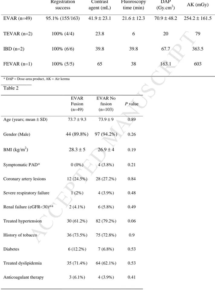
Table 1: Fusion feasibility rate and contrast and radiation dose by procedure type
Registration success Contrast agent (mL) Fluoroscopy time (min) DAP (Gy.cm2) AK (mGy) EVAR (n=49) 95.1% (155/163) 41.9 ± 23.1 21.6 ± 12.3 70.9 ± 48.2 254.2 ± 161.5 TEVAR (n=2) 100% (4/4) 23.8 6 20 79 IBD (n=2) 100% (6/6) 39.8 39.8 67.7 363.5 FEVAR (n=1) 100% (5/5) 65 38 163.1 603
* DAP = Dose-area product, AK = Air kerma
Table 2 EVAR Fusion (n=49) EVAR No fusion (n=103) P value
Age (years; mean ± SD) 73.7 ± 9.3 73.9 ± 9 0.89 Gender (Male) 44 (89.8%) 97 (94.2%) 0.26
BMI (kg/m2) 28.3 ± 5 26.9 ± 4 0.19 Symptomatic PAD* 0 (0%) 4 (3.8%) 0.21
Coronary artery lesions 12 (24.5%) 28 (27.2%) 0.84
Severe respiratory failure 1 (2%) 4 (3.9%) 0.48
Renal failure (eGFR<30)** 2 (4.1%) 6 (5.8%) 0.49
Treated hypertension 30 (61.2%) 82 (79.2%) 0.06
History of tobacco 36 (73.5%) 75 (72.8%) 0.9
Diabetes 6 (12.2%) 7 (6.8%) 0.53
Treated dyslipidemia 35 (71.4%) 64 (62.1%) 0.53
M
AN
US
CR
IP
T
AC
CE
PT
ED
* Peripheral Arterial Disease