HAL Id: hal-01097870
https://hal.archives-ouvertes.fr/hal-01097870
Submitted on 9 Jan 2015
HAL is a multi-disciplinary open access
archive for the deposit and dissemination of sci-entific research documents, whether they are pub-lished or not. The documents may come from teaching and research institutions in France or abroad, or from public or private research centers.
L’archive ouverte pluridisciplinaire HAL, est destinée au dépôt et à la diffusion de documents scientifiques de niveau recherche, publiés ou non, émanant des établissements d’enseignement et de recherche français ou étrangers, des laboratoires publics ou privés.
The Application of Variable Chlorophyll Fluorescence to
Microphytobenthic Biofilms
Rupert G. Perkins, Jacco C. Kromkamp, Joao Serôdio, Johann Lavaud, Bruno
Jesus, Jean-Luc Mouget, Sébastien Lefebvre, R. M. Forster
To cite this version:
Rupert G. Perkins, Jacco C. Kromkamp, Joao Serôdio, Johann Lavaud, Bruno Jesus, et al.. The Application of Variable Chlorophyll Fluorescence to Microphytobenthic Biofilms. Chlorophyll a Fluo-rescence in Aquatic Sciences: Methods and Applications, 2010, �10.1007/978-90-481-9268-7_12�. �hal-01097870�
The application of variable chlorophyll fluorescence to
microphytobenthic biofilms
Perkins, R.G.1*, Kromkamp. J.2, Serôdio, J.3, Lavaud, J.4, Jesus, B.5, Mouget, J-L.6, , Lefebvre, S.7, Forster, R.8
1. School of Earth and Ocean Sciences, Cardiff University, Main Building, Park Place, Cardiff, CF10 3YE, UK.
2. NIOO-KNAW Centre for Estuarine and Marine Ecology, P.O. Box 140, 4400 AC Yerseke, The Netherlands
3. Departamento de Biologia, Universidade de Aveiro, Campus de Santiago, 3810-193 Aveiro, Portugal
4. Université de La Rochelle, Technoforum, 23 avenue Albert Einstein, 17071 LA , OCHELLE cedex 9, France
5. Centro de Oceanografia, Faculdade de Ciências da Universidade de Lisboa, Campo grande, 1749-016 Lisboa, Portugal.
6. Laboratoire de Physiologie et de Biochimie végétales, MMS EA 2160, Université du Maine, Av. O. Messiaen, 72085 Le Mans Cedex 9, France 7. Laboratoire de Biologie et Biotechnologies Marine, Université de Caen,
Esplanade de la Paix, 14032 Caen cedex, France
8. Ecosystem Interactions, Centre for Environment, Fisheries and Aquaculture Science (CEFAS), Pakefield Road, Lowestoft, Suffolk NR33 0HT, UK
*corresponding author: Email: [email protected]; Tel.: 0044 (0)2920 875026. Fax.: 0044 (0)2920 875426
Abstract
The successful application of variable chlorophyll fluorescence methodology to higher plants and other phototrophs inspired workers in the 1990s to apply the methods to microalgal communities inhabiting benthic soft sediments, the microphytobenthos (MPB) of estuarine and other coastal habitats. It was quickly identified that particular aspects of the physiology (cellular vertical migration within the sediment matrix), photophysiology (high capacity for down regulation, e.g. NPQ, and chlororespiration in the dark) and the effects of the physical structure of the sediment/biofilm matrix (light attenuation by the matrix itself) confounded the interpretation of fluorescence information obtained. In this chapter, the authors attempt to explain these and other issues pertinent to MPB biofilms and to summarise how methods have been developed to alleviate the problems encountered. Although much work is still needed to fully understand fluorescence data for the MPB, studies to date have been highly illuminating with regard to rhythms of productivity, photoacclimatory mechanisms and the behavioural ecology and physiology of MPB at an integrated biofilm level and at a cellular level. This chapter therefore introduces benthic biofilms and relevant specific fluorescence methodological issues, expands on subsurface fluorescence signal and migration, discusses down regulatory non-photochemical quenching (NPQ) resulting from xanthophylls cycle induction, compares measurement of electron transport rate proxies, examines light curve methodology, and concludes by comparing fluorescence productivity measurements with those of other methodologies such as oxygen evolution and carbon uptake.
Contents
1. Introduction to benthic biofilms 2. The effects of subsurface signal
2.1 Microphytobenthic biofilms on soft sediments
2.2 Stromatolites - the effect of “layered” biofilms
2.3 Deconvolution of depth integrated signals
3. Down regulation through Non Photochemical Quenching
3.1 NPQ and the Xanthophyll cycle in diatoms
3.2 NPQ in the dark
4. The quantification of the microalgal biomass using fluorescence 5. Calculation of electron transport rate: ETR v rETR
5.1 Multiple and single turnover methods 5.2 The MT-method.
5.3 The ST-method
5.4 Assumptions and uncertainties.
5.5 Calculation of ETR in microphytobenthos studies
6. Light response curves
6.1 A brief overview of methodology
6.2 Steady sate light curves
6.3 Rapid Light Curves
6.4 Non-sequential Light Curves
6.5 Light Curves Summary
7. Comparison of fluorescence with other methodologies 8. General summary
1. Introduction to benthic biofilms
Community assemblages of diatoms, green algae and cyanobacteria comprise the microphytobenthos (MPB) inhabiting benthic sediment ecosystems (Admiraal 1984, Underwood and Kromkamp 1999, Consalvey et al. 2004). Particular attention has been paid top the analysis of intertidal soft sediment systems, e.g. cohesive mudflat and sandy substrata typical of estuarine habitats. Variable chlorophyll fluorescence has been applied to these systems since the 1990s, in an attempt to investigate the primary productivity and photophysiology of the integrated biofilms, when viewed as a “black box system”, and also the species level (Sections 5, 6 and 7). These transient biofilms are not confined to such soft sediment habitats however, and more recently application of fluorescence methodologies has been applied to biofilms inhabiting rocky shores and stromatolite systems (Kromkamp et al. 2007; Perkins et al. 2007). However the large majority of published work has centred upon benthic soft-sediment biofilms, due to their important ecosystem functions of carbon flow and sediment stability (Underwood and Kromkamp, 1999). In the former their high magnitude of productivity fuels carbon flow through invertebrate and bacterial food webs to support important trophic levels of anthropogenically exploited taxa, including coastal fish and shell fisheries and coastal avifauna. In the case of sediment stability, biogenic exopolymers, usually referred to as extracellular polymeric substances (EPS), produced by the MPB may contribute significantly to sediment stability, hence increasing the sediment resistance to hydrodynamic stresses and thus resistance to coastal erosion (e.g. Underwood and Kromkamp 1999 and citations there-in).
One attribute of MPB physiology has in particular contributed to confounding the application of fluorescence methodology to benthic biofilms. This is the behavioural adaptation of the MPB to migrate vertically in their sediment matrix habitat in response to environmental stimuli, as well as part of endogenous tidal and diel rhythms (Sections 2, 3, 4, 5 and 6). Consalvey et al. (2004) reviewed this migration in MPB biofilms and Consalvey et al. (2005) discussed the effect of this as part of a review of fluorescence methodology applied to MPB biofilms. Vertical migration appears to follow tidal and diel rhythms such that cells migrate to the surface of the sediment to coincide with daylight emersion periods, whilst migrating back in to the sediment for immersion periods (Consalvey et al. 2004 and citations there-in). In some instances this appears to be modulated by light environment such that low turbidity in the overlaying water column enables the MPB to remain at the sediment surface after the onset of immersion, and during bright moonlit emersion periods, MPB will migrate to the surface (authors pers. obs.).
Light is undoubtedly a major stimulus in MPB vertical migration. Several studies have shown that cells within MPB biofilms show negative or positive phototaxis. For example, cells will migrate down to avoid potentially harmful high light environments and migrate up to optimise the light environment when ambient light levels are low (Sections 2, 3 and 6). Cells also show “microcycling” such that their can be a constant turnover of taxa at the sediment surface, such that cells shade each other resulting in a reduction in photodose integrated over time (Section 2, 5,, 6 and 7). As well as affecting productivity, such “behavioural” down regulation of photosynthesis may act to make interpretation of fluorescence measurements difficult. For example, cells will migrate to different positions within the sediment matrix making the distance between the cells and the fluorometer probe variable and
unknown. This leads to a variable attenuation of applied actinic light, as well as the fluorescence yields used to study aspects of photoacclimation. Furthermore, cells may migrate in response to darkness applied for measurement of dark adapted fluorescence parameters, again altering fluorescence yields (Sections 2, 3 and 6). These effects, and others discussed in this chapter, lead to potential errors in the application of variable chlorophyll fluorescence to migratory MPB biofilms. As a result great care is needed in the interpretation of fluorescence data obtained. Benthic diatom taxa, which can comprise the majority of MPB biomass, also show differences in photophysiology from higher plants, making conventional interpretation of fluorescence data incorrect. For example, diatoms exhibit high levels of down regulation through diadinoxanthin/diatoxanthin xanthophyll cycling, non-photochemical quenching (NPQ), induced as a result of the trans-thylakoid proton gradient resulting from light induced electron transport (Section 3). Thus MPB cells can show behavioural and physiological down regulation. However chlororespiration during dark periods leads to retention of this proton gradient, retaining NPQ in the dark (Section 3). This can suppress maximum fluorescence yield in the dark (Fm) such that it is lower than operational maximum yield in low light (Fm‟), making conventional calculation of NPQ from the difference in these yields problematic (Sections 3 and 5).
In this review, the authors have summarised the main areas of fluorescence research as applied to benthic biofilms, principally concentrating on those of intertidal soft sediment ecosystems. The methodology is summarised, along with potential problems, as well as ways to minimise error and achieve correct interpretation of data obtained. The review covers subsurface signal as a result of vertical integration of fluorescence measurements (Section 2), non-photochemical quenching and the xanthophyll cycle (Section 3), measurement and calculation of fluorescence derived
electron transport rate (Section 5), methodology used to obtain light curve parameters of photophysiology, e.g. photoacclimation (Section 6) and concludes with a section comparing fluorescence to other methods including radio-labelled carbon uptake and oxymetry (Section 7).
2. The effects of subsurface signal
2.1 Microphytobenthic biofilms on soft sediments
Sediments colonised by microphytobenthos are optically dense, causing both downwelling light (including light generated by a fluorometer) as well as upwelling fluorescence to suffer from considerable attenuation within the microalgal biofilm/sediment matrix. As a result, the fluorescence levels Fs and Fm‟ measured near
the surface represent the integration of the fluorescence signals emitted at various depths, distorting the relationship between Fo and chl a as this relationship will thus
not only depend upon the total biomass, but also on the shape of the vertical biomass profile. Of more importance, this effect causes the measurement of the effective quantum yield, ΔF/Fm‟, at the surface to differ significantly from the true, inherent
value of the microalgae composing the biofilm, because the relationship between Fs
and Fm‟ varies with irradiance within the depth range over which the fluorometer
signal is integrated (Forster and Kromkamp 2004, Serôdio 2004).
The depth-integration effect was first predicted from observations of changes in ΔF/Fm‟ in undisturbed biofilms under constant irradiance (Underwood et al. 1999,
Perkins et al. 2001, 2002), and was studied in detail using numerical simulation models (Forster and Kromkamp 2004, Serôdio 2004). Studies showed that fluorescence measurements taken non-invasively at the surface of the sediment result in a substantial light-dependent overestimation of the inherent value of ΔF/Fm‟ and
relative electron transport rate, rETR, particularly important under supersaturating irradiances (Fig. 2.1). As a consequence, light curves derived from depth-integrated measurements are likely to appear to saturate at higher irradiances, or to be less photoinhibited when compared to the true physiological response of the biofilm-forming microalgae. The contribution of sub-surface fluorescence signals also affect the determination of the light response of non-photochemical coefficient NPQ, which is expected to be underestimated under high light when measured non-invasively in intact biofilms (Forster and Kromkamp 2004, Serôdio 2004).
While this effect may occur, although to a lesser extent, in other optically dense samples such as thick leaves or macroalgal thalli (Forster and Kromkamp 2004, Serôdio 2004, Susila et al. 2004), in the case of microphytobenthos the problem is further complicated by the occurrence of comparatively large scale vertical migration by motile diatoms. In this case, it becomes highly difficult to interpret fluorescence yields from biofilms where the cells move vertically within the sediment matrix, since the subsurface signal emanates not only from cells at unknown depth, but at a variable depth in the sediment. Additionally, signal strength is proportional to the distance between the fluorometer probe and the cells themselves, thus vertical migration may increase or decrease measured yields, so making interpretation of changes in yield or calculation of photophysiological parameters difficult. For example, a decrease in both Fm‟ and F can be due to induction of NPQ down regulation on exposure to
increasing PAR or to downward migration (negative phototaxis). It is often not possible to differentiate between the two processes using fluorescence methods. For this reason it can be prudent to use subsurface spectral reflectance for measurements such as biomass, see below, (Kromkamp et al. 2006, Morris et al. 2008) as the reflectance spectra are not influenced by NPQ.
It is hard to avoid artefacts of tidally induced vertical migration and positive or negative phototaxis but the effects can be minimized by not taking measurements during the first and last hour of the emersion period when vertical migration is maximal. In addition it is better to minimize the effects of phototaxis by keeping the duration of measurements as short as possible (e.g. minimise the duration of light steps during rapid light curves, RLCs, see later). PSII quantum efficiency often stabilizes before true steady state fluorescence is reached (authors pers. obs).
The issue of the subsurface signal from the cells within the subsurface sediment has been investigated in considerable depth (Kromkamp et al. 1998, Perkins et al. 2002, Forster and Kromkamp 2004, Serôdio 2004, Jesus et al. 2006). Kromkamp et al. (1998) discussed the issue of microcycling of cells such that the fluorescence yield was obtained from a varied surface community over time and that this could explain the persistent high ∆F/Fm‟ at high incident irradiance. Perkins et al. (2002)
reported over estimation of ETR in sediments due to subsurface signal from cells exposed to a lower light level than that applied at the surface and showed that the community composition could change during a light curve. In addition it appeared that the PSII signal was contaminated by PSI-fluorescence, shown by using a 680 nm bandpass filter compared to the more conventional 695 nm longpass filter. Underwood et al. (2005) used high resolution fluorescence imaging to report diel patterns in F/Fm‟ of several benthic diatom species which showed microcycling over
an artificially extended emersion period (Fig. 2.2). Serôdio (2004) and Forster and Kromkamp (2004) also demonstrated similar effects on measurements due to integration of the fluorescence signal over sediment depth. To make matters more complex, the degree of overestimation of ∆F/Fm‟, which manifests the greatest at light
biomass profile, and subsurface biomass maxima, resulting in the highest degree of overestimation of the true ∆F/Fm‟, and hence resulting in an overestimate of the
maximum electron transport rate, rETRmax, often by as much as of 60% (Forster and
Kromkamp 2004). The effect of this on estimates of depth integrated primary production will be discussed later when comparing fluorescence with other methods used to estimate primary production.
Vertical migration does not only influence the apparent value of ∆F/Fm‟, but it
also influences the measured minimal fluorescence yield, Fo (Forster and Kromkamp
2004, Perkins et al. 2001, Jesus et al. 2006), which is used as a proxy of biomass (Barranguet and Kromkamp 2000, Honeywill et al 2002). It is thus not surprising that Jesus et al. (2006) observed that tidally induced vertical migration resulted in either an over estimation or under estimation of biomass dependent upon the time the measurements were made within the emersion period.
Recent work using migration inhibitors (Cartaxana et al. 2008, Perkins et al. in prep.) and engineered non-migratory biofilms (Jesus et al. 2006a,b; Mouget et al. 2008) have investigated these issues further and confirm that vertical migration and the concomitant “deep layer fluorescence” lead to erroneous estimates of quenching coefficients and overestimation of ∆F/Fm‟.
2.2 Stromatolites - the effect of “layered” biofilms
Stromatolites are perhaps an extreme example of a layered biofilm where sub-surface cells “interfere” with the fluorescence signal from sub-surface cells and vice versa. The measurements become an integrated measurement of the sub surface cyanobacteria (Figure 2.3) and the surface cells, where diatom epiphytes can be present (Fig. 2.4, Perkins et al. 2007). Stromatolites consist of a microbial consortium
which trap ooids by extracellular polymers, mainly of cyanobacterial origin (Visscher et al. 1998, Reid et al. 2000). Reid et al. (2000) described 3 different developmental stages where the pioneering type 1 stromatolite has the lowest cyanobacterial diversity with Schizotrix gebeleinii as the dominant form. Type 2 is characterised by a micritic crust at the surface (often lacking diatom epiphytes, Kromkamp, pers. obs.) and in climax state type 3 the endolythic Solentia spp. bores into the ooids and fuses them together, giving the stromatolite its structure. Types 2 and 3 have the highest cyanobacterial diversity (Baumgartner et al. (2007). Figure 2.3 shows a typical example of a type 1 stromatolite (Kromkamp et al. 2007) with a clear subsurface layer, but the layer above also contain many cyanobacteria. This picture clearly highlights the difficulty when applying surface measurements using PAM fluorometry: the depth and thickness of the subsurface layer varies as well as the distance to the stromatolite surface. This means that quantitative analyses of both cyanobacterial biomass as well as effective quantum efficiency is very difficult as the apparent ∆F/Fm‟ and Fo is
integrated over an unknown depth interval with varying biomass distribution. This problem can be partly circumvented by measuring on a cross section by putting the PAM fiberoptics parallel to the mat surface (Perkins et al. 2007). However, this will disrupt chemical and light gradients, especially of oxygen and light, have greatly influence photosynthetic activity (Kromkamp et al. 2007).
The following example demonstrates the problems with working with cyanobacteria (Fig. 2.5). RLCs were made on two sections of the stromatolite where the depth of the subsurface layer was respectively at 4 and 1 mm below the surface. The layer above the surface contained cyanobacteria (cf. Schizotrix sp.) but the biomass was substantially lower than in the subsurface layer. After the RLCs were performed, the surface layer was removed until the subsurface layer was exposed and
new RLCs were made. In both cases the measured rETR was much higher when the surface layers were not removed. Several explanations are possible: the cyanobacteria in the surface layer have a higher photosynthetic activity then cyanobacteria in the subsurface biomass maximum. Deeper in the stromatolite the oxygen concentration might be low, causing rapid inactivation of the stromatolite (Kromkamp et al. 2007, Perkins et al. 2007). This might be the case for the layer 4 mm below the surface but is unlikely for the layer 1mm below the surface. Alternatively, fluorescence from the deeper layers, where the irradiance is lower and where ∆F/Fm‟ will thus be higher than
at the stromatolite surface, will cause an overestimation of true ∆F/Fm‟ of the cells at
the surface. The degree of overestimation depends on the depth (and thus the irradiance) of the subsurface layer (which is unknown without destructive sampling) and on the biomass in the subsurface maximum relative to the cyanobacterial biomass in the upper layers. Because the RLCs on the exposed subsurface layers are rather similar, the contribution of “deep layer fluorescence” seems the most likely explanation for the higher rates of rETR measured at the stromatolite surface. A comparison with measurements on cross sections (Mouget et al. in prep) seems to corroborate this conclusion.
2.3 Deconvolution of depth integrated signals
The inherent photophysiological status of the microalgae comprising a biofilm can be estimated from depth integrated fluorescence measurements made on undisturbed samples. A method has been proposed to estimate the inherent light response through the deconvolution of light-response curves based on depth-integrated measurements (Serôdio 2004). This approach is based on the relationship existing between the depth-integrated fluorescence levels measured at consecutive
light levels of a light curve and the depth attenuation of the fluorescence signal, which results in a set of recursive equations to be applied to depth-integrated light curves. This approach has assumptions and limitations (Serôdio 2004; Jesus et al. 2006): i) it assumes an homogeneous photophysiological light response for all the microalgae; ii) assumes an exponential vertical attenuation of incident actinic light and emitted fluorescence, which implies a vertically homogeneous photic zone; iii) it requires the attenuation coefficients for downwelling actinic light and upwelling fluorescence to be known; iv) it does not account for changes in surface biomass during the construction of the light curve on intact biofilms. Nevertheless, numerical simulations using published values for actinic light and fluorescence have shown a significant reduction in the differences between depth-independent light curves and those deconvoluted from depth-integrated curves (Serôdio 2004).
3. Down regulation through Non Photochemical Quenching
3.1 NPQ and the Xanthophyll cycle in diatoms
Diatoms of MPB biofilms are submitted to an extreme light environment which includes exposure to both high visible and UV irradiances, and fast and unpredictable light fluctuations. Additionally, other environmental pressures (nutrient limitation, extremes in temperature and salinity, etc.) can slow down the photosynthetic machinery and create a situation where the photosynthetically converted light energy cannot be entirely used for metabolic purposes (see Chapters 11 and 12). In order to maintain their photosynthetic productivity at an optimal level by preventing photoinhibition (i.e. decrease in quantum efficiencies Fv/Fm and
ΔF/Fm‟), diatoms need to acclimate to environmental changes through fast regulation
(Lavaud et al. 2002b), the non-photochemical quenching of chlorophyll fluorescence (NPQ) is believed to be one of the most important of these „photoprotective‟ (or „photoacclimative‟) mechanisms in diatoms (Lavaud 2007). Most of the diatoms of the MPB are able of vertical migration into the sediment as a behavioral photoprotective strategy in order to avoid excess light exposure at the surface (see above). However, vertical migration and NPQ appear to be complementary, NPQ induction may occur before the onset of the migratory response (Serôdio et al. in press, Underwood et al. 2005), although recent work using chemical inhibitors of migration and NPQ suggests that migration may be preferred to NPQ (Perkins et al. in prep.).
The NPQ mechanism is described in the Chapter 12 (paragraph 12.1). qE, the energy-dependent quenching, which is regulated by the build-up of a transthylakoid proton gradient (ΔpH) and the operation of the xanthophyll cycle (XC) (Lavaud and Kroth 2006), remains the best known component and plays a major role in the regulation of the diatom NPQ. The machinery triggering and controlling the NPQ amplitude and kinetics is now well known (Goss et al. 2006, Lavaud 2007). The major characteristic of NPQ in diatoms is its amplitude (Lavaud et al. 2002, Ruban et al. 2004) such that it can account for up to 90% of energy dissipation (Lavaud et al. 2002). Estuarine species of the MPB diatoms show a higher (up to 5 times higher than plankton species, Fig. 3.1) and faster (10 s induction) switch on/off of NPQ (Serôdio et al. in press; Serôdio et al. 2005, 2006, Herlory et al. 2007, Lavaud et al. 2007, Cruz and Serôdio 2008).
The mechanism of NPQ in diatoms shows other specificities regarding the pigments and proteins that are involved as well as their spatial organization (see Lavaud 2007). In particular, the xanthophylls cycle, which consists of the enzymatic de-epoxidation/epoxidation of the couple DD-DT, is employed (Fig. 3.2, Lavaud
2007). Accumulation of photoprotective diatoxanthin, DT, depends on the concomitant activity of two enzymes, a de-epoxidase and an epoxidase, the activity of which depends on the light intensity via the build-up of the transthylakoid ΔpH and the availability of co-factors (Fig. 3.2A).
The regulation of the XC in diatoms shows some striking peculiarities, the main one being the triggering of the diadinoxanthin, DD, de-epoxidase by a weak ΔpH (and thus a rather high lumen pH, Fig. 3.2B) (Jakob and Wilhelm 2001) so that the DD de-epoxidation already occurs at lower irradiances and shorter illumination times than in higher plants (Fig. 3.2B). Additionally, MPB diatoms can react to high light stress by accumulating large amounts of DD-DT (Rech et al. 2005, van de Poll et al. 2006, Schumann et al. 2007) in order to increase their photoprotection capacity via NPQ (Perkins et al. 2006, Schumann et al. 2007, Cruz and Serôdio 2008). All together, these differences in the regulatory components and mechanistic components of the NPQ process in diatoms have been suggested to ensure more flexibility and thus quicker response to the light environment (Lavaud 2007).
Field experiments have shown that the photoprotection ensured by both the XC and NPQ are essential for the MPB diatoms to maintain an optimal photosynthetic activity (Serôdio et al. 2005), even if not 100 % efficient (Serôdio et al. in press). Additionally, the differential ability of species to cope (including NPQ) with prolonged high light and UV (Waring et al. 2006) is believed to potentially control spatial distribution (Fig. 3.1) (Lavaud et al. 2007) as well as species succession within MPB biofilms (Tuji 2000, Serôdio et al. 2005, Underwood et al. 2005).
As described later in this chapter, RLCs have become a powerful approach to estimate the photosynthetic productivity and the photophysiology of MPB assemblages isolated from the field or directly in situ. Nevertheless, the assessment of
NPQ on MPB via RLCs is rendered problematic by the confounding effects on Fm and
Fm‟ levels of 1) downward vertical migration of the cells in the sediment, 2) the light
and fluorescence attenuation in the upper layers of the sediment, 3) the contribution of fluorescence originating in “deeper” layers with a concomitant lower irradiance, and 4) a sustained NPQ under prolonged darkness (see Serôdio et al. 2005, Jesus et al. 2006b). In order to solve this problem, Serôdio and co-workers proposed to assess NPQ following ΔαRLC, which is the variation of the initial slope of RLC under high
light exposure. αRLC indeed linearly correlates with NPQ irrespective of measurement
conditions, e.g. ex situ/in situ, summer/winter, low light/high light acclimation, etc. (Serôdio et al. 2005, 2006, in press, Cruz and Serôdio 2008) making it a good alternative to the classic calculation of NPQ as (Fm-Fm‟)/Fm‟ (or (F0-F0‟)/F0‟, see Fig.
3.3A) (discussed in Serôdio et al. 2006). Additionally, it has recently been reported that RLC construction itself can generate rapid endogenous changes of the photosynthetic activity which in turn modulates chl a fluorescence emission, and can potentially affect the ETR measurement and subsequent derivation of photophysiological parameters (Perkins et al. 2006, Cruz and Serôdio 2008). Together with the redox state of QA, NPQ has been shown to be one the main endogenous
mechanisms potentially disturbing chl a fluorescence emission (Perkins et al., 2006; Jesus et al., 2006b; Herlory et al., 2007).
There are a number of physiological/technical features which can strongly influence NPQ amplitude and kinetics, and subsequently the shape of RLCs in MPB communities dominated by diatoms: 1) the species composition (Perkins et al. 2002, Serôdio et al. 2005, Underwood et al. 2005), 2) the light/dark past history of the cells and hence the cells photoacclimation state (Perkins et al. 2006, Cruz and Serôdio 2008), 3) the accumulated light dose during the RLCs (Perkins et al. 2006, Herlory et
al. 2007 Cruz and Serôdio 2008), 4) the fluorometer used (Perkins et al. 2006, Cruz and Serôdio 2008). Figure 3.3 shows that, in the MPB diatom species Navicula
phyllepta, the length of each irradiance step (10, 30, 60 or 140 s) can influence partly
the level of NPQ induction (Fig. 3.3A), the profile of RLCs (Fig. 3.3B) and the subsequent determination of ETRmax as a function of the light history of the cells (Fig.
3.3C). However, it should be mentioned that this phenomenon is not always observed on intact MPB communities in situ as shown in Fig. 3.4.
3.2 NPQ in the dark
In diatoms, NPQ occurs not only during light exposure but also during prolonged darkness (several hours; Jakob et al. 1999, Consalvey et al. 2004, Serôdio et al. 2005). Dark NPQ is due to chlororespiration, the amplitude of which is especially high in diatoms (Dijkman and Kroon 2002, Lavaud et al. 2002b). This pathway allows electrons to flow from the NADPH, H+ to O2, both synthesized in
light, via the plastoquinone (PQ) pool: it is the respiratory chain of the plastids (Kuntz, 2004). In diatoms, the chlororespiratory pathway is switched on very rapidly after the onset of darkness (Dijkman and Kroon 2002). Its amplitude and duration directly relates with the irradiance and duration of the former illumination through accumulation of reducing equivalents (Lavaud et al., 2002b), in other words it depends on the past light history of the cells (Cruz and Serôdio 2008). By transferring electrons through PQ, the chlororespiratory pathway generates in darkness the build-up of a transthylakoid ΔpH (Ting and Owens 1993). This dark ΔpH has been shown to be sufficient to activate the DD de-epoxidase and to drive the synthesis of DT, and the development of a large NPQ (up to 2) (Jakob et al. 1999, Jakob and Wilhelm 2001; Serôdio et al., 2005, 2006). This is only possible because in diatoms, as specified above (Fig. 3.2B), the DD de-epoxidase needs only a weak acidification of the lumen
to be activated, even though the rate of chlororespiration and subsequent change in the ΔpH are low (Jakob and Wilhelm 2001). Such a process would allow the cells to prevent photoinhibition during a subsequent sudden exposure to high light by keeping activated the dissipative function of the LHC system; an obviously adaptive advantage for the diatom species growing in a fluctuating light environment. Dark NPQ is even more physiologically relevant for diatoms of the MPB which can spend more than 18 h per day in the dark in the sediment before migrating to the surface and experiencing high light exposure (Serôdio et al. 2005).
As well as the NPQ which develops in light, the dark NPQ generates a methodological problem due to our inability to instantaneously measure the „true‟ Fm
(and F0) level during dark-adaption of the cells in situ where their past light/dark
history is usually unknown. Indeed, to achieve correct measurement of fluorescence parameters, complete QA oxidation and NPQ relaxation are required, which is usually
reached after a short (15 min) dark-adaptation in controlled laboratory conditions but which might not be enough in situ (Perkins et al. 2001, Consalvey et al. 2004, Jesus et al. 2006a). Dark NPQ can easily quench Fm by at least 10-15 % depending on the
diatom species (Jakob and Wilhelm 2001). When the cells are further exposed to a low irradiance (below 150 µmol photons m-2 s-1, Mouget and Tremblin 2002) during the measurement following dark-adaptation (typically at the beginning of RLCs acquisition), the whole photosynthetic machinery is fully activated which 1) reoxidizes QA and the PS II, 2) dissipate the ΔpH and subsequently change the
equilibrium of the XC, hence relaxing the fluorescence quenching (Consalvey et al. 2004, Serôdio et al. 2005, 2006). Hence, it is rather common to observe Fm‟ level
transiently higher than the dark Fm level (Mouget and Tremblin 2002, Consalvey et al.
measurement and calculation of many fluorescence parameters. It can also significantly perturb the use of F0 and its changes as a proxy for the dynamics of MPB
diatom biomass at the surface and within the sediment (Consalvey et al. 2004, Jesus et al. 2006a). Solutions have been proposed to rule this problem out: 1) measurement of the „true‟ Fm level in the presence of DCMU which is only applicable in controlled
laboratory conditions (see Chapter 12, paragraph 12.4), 2) short exposure to low dose of far-red or low light instead of dark-adaptation in order to reoxidize PS II and dissipate the ΔpH and NPQ (Consalvey et al. 2004, Jesus et al. 2006a), 3) use Fm‟, m,
the maximum Fm‟ value measured as the „true‟ Fm, instead of the dark Fm level
(Serôdio et al. 2006, in press, Cruz and Serôdio 2008).
4. The quantification of the microalgal biomass using fluorescence
The quantification of the microalgal biomass of microphytobenthos biofilms is a methodological challenge, due to the thinness of the sediment photic zone, the large horizontal heterogeneity, and the rapid changes in microalgae near the surface due to vertical migratory movements. Serôdio et al. (1997) was the first study to investigate the possibility of using in vivo chlorophyll fluorescence to non-destructively quantify the microalgal biomass of microphytobenthos biofilms. They experimentally established a linear relationship between sediment chlorophyll a (chl a) content and minimum fluorescence yield Fo, showing that large Fo variations in the dark
represented changes in the amount of microalgae at the sediment surface, as the result of cell vertical migratory movements. The use of Fo as a biomass proxy was shown to
be preferable to other fluorescence parameters such as Fm or Fm‟ because it varied the
least with previous light history, temperature and microalgal group. Fo was shown to
sediment (defined as „photosynthetically active biomass‟; Guarini et al. 2000, Honeywill et al. 2002) but also of the microalgal biomass in the photic zone weighted by its contribution to depth-integrated photosynthesis (defines as „producitve biomass‟; Serôdio et al., 2001). This method allowed to overcome the operational difficulties with previously used methods, based on destructive and time-consuming procedures, and introduced considerable operational advantages, including the possibility to obtain repeated measurements in the same sample over time, without any physical disturbance of the sediment-air or –water interface, and allowing the concurrent measurement of other variables in the same sample (Serôdio et al. 1997).
However, the determination of Fo in MPB biofilms is not without problems
and has been the source of significant discussion (e.g. Consalvey et al. 2004, Jesus et al 2006b,c). These problems are related to MPB vertical movements during the dark adaptation (DA) period and to problems concerning the presence of NPQ in the dark (exhibited by diatom dominated biofilms). The determination of fluorescence parameters requiring dark-adaptation (Fo, as well other parameters and indices such as
Fm or Fv/Fm) requires that PSII reaction centres and QA be in their fully oxidised form
(Schreiber et al. 1986). To achieve this state it is conventional to place samples in full darkness until a dark adaptation steady state is reached. However, dark adapting microphytobenthic biofilms present a number of specific problems unique to this type of community.
Microphytobenthic biofilms are know to exhibit a behavioural photo-regulation mechanism where microalgae migrate vertically within the sediment matrix as a response to changes in ambient light in order to maintain an optimum light environment and avoid photo-inhibition (e.g. Perkins et al. 2002; Jesus et al. 2006b,c). This response to changes in ambient light can hinder the determination of Fo if
vertical migration occurs during the dark adaptation period necessary for QA and PSII
re-oxidation. This has been recognized as a potential problem since the first studies using fluorescence (e.g. Serôdio et al. 1997) and short adaptation periods have been suggested as alternatives. Several forms of determining the minimum fluorescence yield have been tested, depending on the length of dark-adaptation used (F', Fo', Fo2,
Fo5, Fo10, Fo15; the superscript number denoting the duration of the dark period in
minutes). By far, the most commonly used parameter is Fo15 (Kromkamp et al,1998;
Underwood et al. 1999; Barranguet and Kromkamp 2000; Perkins et al. 2001; Hagerthey et al. 2002; Honeywill et al. 2002; Underwood 2002; Consalvey et al. 2004; Defew et al. 2004; Jesus et al. 2006b,c ), followed by 5 min (Serôdio et al 1997, 2000, 2001, 2003; Jesus et al. 2006b,c) and 2 min (Serôdio et al. 2006, 2007 and 2008). However, the 15 min of DA can be excessive in some biofilms inducing significant downward migration during that period (e.g. Jesus et al. 2006a, b and c), suggesting that shorter time periods might be preferable. In fact, Jesus et al. (2006c) compared the relationship between chl a with Fo', Fo5 and Fo15 and found no significant
differences from Fo' to Fo15 in muddy sediment assemblages, suggesting that light
history had little or no effect in the epipelic assemblages and that no DA was necessary for a good relationship between chl a and fuorescence. However, in sandy sediments there was evidence of a light history effect and a 5 min DA period was necessary to remove this effect. Other authors (e.g. Serôdio et al. 2007) have found that shorter DA times (2 min) might be even better than 5 min for the determination of photosynthesis fluorescence indices that require the input of a minimum fluorescence yield parameter.
Intertidal biofilms exhibit the additional problem of not showing an homogeneous behavioural response to light stimulus throughout the tidal cycle, i.e.
biofilms close to the beginning of the emersion period will tend to increase Fo values
during the DA period as a result of cells migrating to the sediment surface, and measurements close to the end of the emersion period will be very sensitive to darkness and cells will migrate downwards quickly over the DA period. Although, this does not seem to be the case in all estuaries (e.g. Kromkamp et al,1998; Barranguet and Kromkamp 2000; Hagerthey et al. 2002; Honeywill et al. 2002;). To reduce this problem it was proposed that measurements are taken closer to the middle of the emersion period and that a low light or far-red treatment is used instead to the dark treatment (Jesus et al. 2006b). It is not clear why low light and far-red light work similarly but both treatments have to be applied at a reduced photon flux quantity to work properly. Thus, it is possible that this reduced photon flux promotes the dissipation of the chlororespiration trans-thylakoid proton gradient (pH) exhibited by diatoms in the dark (Ting and Owens, 1993, Dijkman & Kroon, 2002; Lavaud et al., 2002). The reduction in the pH promotes the exoxidation of diatoxanthin into diadinoxanthin. Therefore, decreasing the NPQ caused by diatoxanthin presence in the dark (Jakob et al. 1999, Jakob and Wilhem 2001). Another advantage of low light is that it seems to promote the presence of cells at the surface thus reducing the problem of migration downwards that occurs during the dark adaptation period (Jesus et al. 2006b).
5 Calculation of electron transport rate: ETR v rETR
5.1 Multiple and single turnover methods
The rate of electron transport by PSII depends on the amount of light absorbed by the antenna of PSII and the efficiency at which the absorbed light by PSII is used by the reaction centers (RCII) to drive charge separation. Basically two methods are used which are both based on the light doubling method originally proposed by
Bradbury and Baker (1981) which we will call the Multiple- turnover (MT) method and the single turnover (ST) method and which relate to the description of the pulse-amplitude modulation principle by Schreiber et al. (1986) and the pump and probe method (Falkowski et al. 1986b) respectively. The differences between these two approaches have recently been reviewed by Kromkamp and Forster (2003).
5.2 The MT-method.
The MT-method is usually used by scientists using the PAM family of fluorometers which uses a multiple turnover flash to measure the quantum efficiency of PSII. Because the flash duration used to measure the maximum fluorescence (Fm)
is relatively long (50ms - 1 sec) it allows for multiple charge separations during the flash and will thus lead to fully reduced QA, QB and PQ-pool. When the PSII effective
quantum efficiency (ΔF/Fm‟) is measured the absolute rate of electron transport per
unit area (ETRA) can be calculated as follows:
ETRA = E • AII • ФRC • F/Fm‟ (1)
AII is the fraction of the incident light (E) which is absorbed by PSII. ФRC is yield (in
electrons) of reduced QA per trapped photon, i.e. the maximum quantum yield of
photochemistry within PSII, and it is usually assumed to be equal to 1 (Kolber and Falkowski 1993). Note that ETRA is expressed in µmol electrons s-1 m-2 of surface area. When working with higher plants it is assumed that leaves absorb approx. 85 % of incident light, and about half of this is partitioned to PSII. Assuming that RC is close to 1, eq. (1) can be rewritten as:
ETRA 0.43 • E • F/Fm‟ (2)
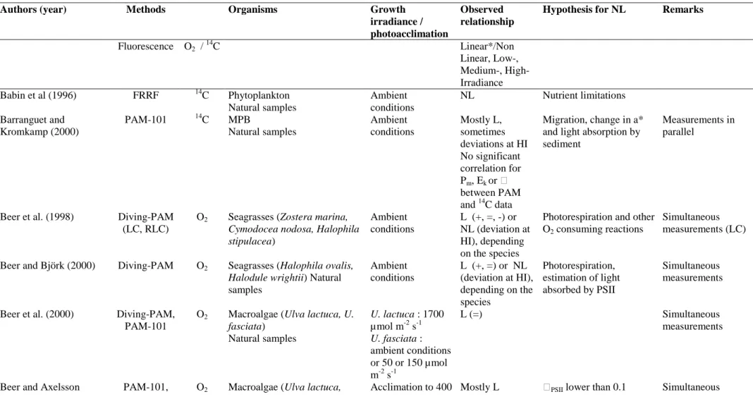
Often the fraction of absorbed light is determined by measuring the transmittance of light through a piece of macroalgal thallus using a light sensor (Beer et al. 1998, Longstaff et al. 2002). Implicit in this assumption is that the measured signal is equivalent to the integrated fluorescence yield over the entire path length, an assumption which might not be met, certainly when several cell layers are involved. The artefacts associated with this assumption were discussed in chapter 3 where the effects of “deep layer” fluorescence are described.
When working with optically thin suspensions of phytoplankton or unicellular algal cultures a slightly different approach is taken: in this case the ETR is usually calculated per mg chl a (µmol e- (mg chl a)-1s-1):
ETR = E • a‟*PSII • ΔF/Fm‟ • ФRC (3a),
Where a‟*PSII is the optical absorption cross section of PSII (here expressed in m2 (mg
chl a)-1, which is the product of the cross section of a single PSII unit and the number of PSII per mg chl (a‟*PSII = a*PSII • nPSII)). As it is rather difficult to measure the
absorption cross section of PSII it is normally assumed that a‟*PSII is half the optical
cross section of the cells (a* m2 (mg chl a)-1, i.e. it is assumed that 50% of the absorbed light is funneled to PSII and the other half to PSI). Below we will discuss this assumption. Thus, assuming ФRC equals 1we can rewrite eq. 3a as:
The optical cross section can be easily determined using a spectrophotometer equipped with an integrating sphere, i.e. by using the filterpad method for natural phytoplankton which requires concentration on a filter before the absorption measurements can be made (Tassan and Ferrari 1998, Simis et al. 2005).
5.3 The ST-method
Researchers using a saturating single turnover flash to measure Fm‟ usually
take the approach developed by Falkowski et al. (1986b) and Kolber and Falkowski (1993), based on the development of the pump and probe fluorometer, which was followed up by the Fast Repetition Rate Fluorometer (FRRF) (Kolber et al. 1998). Here the rate of photosynthetic electron transport is described as follows:
ETR = E • a*
PSII • nPSII • qP • Фtm • ФRC • f (4)
Here a*PSII is the optical cross section of a single PSII unit (m2/mol PSII) and nPSII is
the number of PSII units per mg chl a, usually to be assumed equal to 0.002 (thus assuming 500 chl a molecules per PSII (Falkowski 1981, Kolber and Falkowski 1993). The photochemical quenching coefficient qP (moles electrons transferred per mole
photons absorbed by PSII) reflects the proportion of oxidized PSII centers and is often used as a proxy of the number of open reaction centers. This is, however, only true when the PSII centers are not connected (Kramer et al. 2004). The trapping efficiency Φtm is the efficiency at which trapped photons in the pigment bed are transferred to an
open RCII and f is the fraction of functional PSII centers. The product of the optical PSII cross section and the trapping efficiency equals the effective PSII cross section (σPSII, units m-2 (mol PSII)-1):
σPSII = a*PSII • Фtm (5).
As the FRRF can measure the functional cross section σPSII from the rise of F to Fm
during the induction flashlet sequence, eq. 4 can be rewritten as
ETR = E • σPSII • nPSII • qP • Фtm • f (6).
Eq. 6 is the one that is proposed by Kolber and Falkowski (1993) and Kolber et al. (1998) for use with the pump and probe and FRR fluorometer. The fraction of inactive centers (f) was measured as Φsat/0.65, where Φsat is the maximum PSII
efficiency measured and 0.65 the assumed maximum PSII efficiency of healthy cells without non-functional PSII centers. Kromkamp and Forster (2003) argued that this factor should be omitted because non-functional PSII centers will affect the level of Fo‟, and this is already incorporated in the value of qP.
5.4 Assumptions and uncertainties.
Fraction of light absorbed by PSII. Both the MT as well as the ST method
usually use a number of a-priori assumptions. When using the MT method to calculate ETR it is necessary to know which fraction of the absorbed light is funneled to PSII. Using a combination of optical and biophysical techniques. Suggett et al. (2004) tested the hypothesis that about 50% of the light is absorbed by PSII. For a large range of species (diatoms, green alga, haptophytes and a cryptophyte) the fraction of light absorbed by PSII varied between 0.48 and 0.58, justifying this assumption. However, in the pelagophyte Aureococcus anophagefferens the PSII-antenna
absorbed about 36% of the light and in two cyanobacterial Synechoccocus species PSII absorbed 25-32% of the total light. Interestingly, the fraction of light absorbed by PSII was independent of the growth irradiance. A similar approach was taken by Johnsen and Sakshaug (2007), but they used a slightly different scaling procedure to match the fluorescence excitation spectra to the absorption spectra in order to arrive at the fraction of light absorbed by PSII: their results show that 48-88% of the light absorbed by the photosynthetic pigments was absorbed by PSII, which is generally higher than the estimates obtained by Suggett et al. (2004). Whether this difference is entirely due to methodological question or to growth conditions and different algal species remains an open question, but clearly this topic requires further research.
Estimates of the number of PSII. When using the ST protocol the absorption is
quantified by multiplying the measured functional cross section σPSII with the nPSII.
This latter factor is also rather difficult to measure, certainly using field material, and for this reason it is assumed that nPSII equals 0.002, i.e. a PSII contains 500 mol chl a
(mol PSII)-1 (Kolber and Falkowski, 1993), a value based on the determinations by the photosynthetic unit (PSU) size by Mauzerall and Greenbaum (1989). Assuming that a PSU contains 4 RCII it can be calculated from the data presented by the references in Table 1 that generally nPSII varies between 500-725 mol chl a per mol PSII, although
lower values have been reported for Isochrysis galbana.
Photoacclimation generally results in more nPSII when the cells are grown in
low irradiances (Table 1). Interestingly, Kromkamp and Limbeek (1993) observed that when the marine diatom Skeletonema costatum was grown in fluctuating light simulating vertical mixing, this resulted in smaller but more nPSII. This makes sense as
it allows the cells to both harvest the same amount as light as with large PSU, but it will result in a higher rate of maximal photosynthesis. Recently Suggett et al. (2004)
compared estimates of nPSII obtained using the ST turnover oxygen measurements
with a those obtained from a biooptical: nPSII = σPSII/a*PSII: they observed a linear 1:1
relationship between both methods and for most eukaryotic algae nPSII varied between
500-600 mol chl a per mol PSII. The pelagophyte A. anophagefferens had a larger nPSII and cyanobacteria seem to contain smaller nPSII (240-280 mol chl a per mol PSII).
Uncertainties in σPSII. Although the FRRF technique allows estimation of the measurement of the functional PSII cross section, it is necessary to stress that LED‟s (mainly blue) are used to induce the fluorescence induction curve from which σPSII is
estimated. Without spectral correction of the effective absorption of the FRRF in relation to the underwater light field, this may lead to an overestimation of σPSII
(Suggett et al., 2001).
5.5 Calculation of ETR in microphytobenthos studies
Most research measuring ETR on MPB or macroalgae have used a PAM-type fluorometer because a commercially available FRR-type fluorometer (such as Chelsea‟s FastTracka
or Satlantic‟s FIRe) that are able to measure σPSII , have not been
available (RandD versions of both instruments have however been used for coral reef research). As is clear from the above section on the MT-protocol, calculation of absolute rates of PSII electron transport requires knowledge of incident irradiance and the optical absorption cross section, and this is exceedingly difficult when working on benthic biofilms. For this reason often the relative rate of ETR (rETR) is calculated as ΔF/Fm‟ • E. Because the value of a* will change as a result of photoacclimation (time
scales of change are hours-days because they related to de-novo synthesis or break-down of pigments) it is not always possible to compare rates of rETR between publications.
Morris and Kromkamp (2003) cultured the benthic diatom Cylindrotheca
closterium at two different growth rates and compared the relationship between ETR
and oxygen evolution at a range of different temperatures during steady state growth. In general they observed that the relationship between ETR and oxygen evolution was not very sensitive to short-term changes in temperatures. However the relationship of rETR versus oxygen evolution was rather different between low and high growth rate, but when they examined the relationship on the basis of absolute ETR, the differences were minor. This was due to the a* values of the two cultures being different between the two growth rates. Nevertheless, often changes in rETR reflect changes in absolute ETR (Fig. 5.1): three out of the four different algal species showed a similar pattern in change in rETRmax and absolute ETRmax (expressed per cell), despite large changes
occurring in the photosynthetic physiology after transfer from replete to a P-free medium. The exception was Emiliania huxleyi which showed an unexpected increase per cell, because the optical absorption cross section per cell increased. If the absolute ETR was expressed per unit chl a all cells showed a good correlation between the changes in rETR and ETR (data not shown). This suggests that with some care it maybe possible to deduce changes in photophysiology from relative rates of ETR, but definitely more research is needed to confirm this.
A possible way to obtain MPB cells in order to measure a* spectrophotometrically would be to use the lens tissue method: however, this will only select for (a fraction) of the migrating species and the observed a* value might not be representative for the total MPB community. In order to avoid this problem Morris et al. (2008) reconstructed a* from HPLC pigment analyses using the procedure of Bidigare, (1990) and compared the absorption values to the ones measured using the filterpad method. From this it was concluded that the package
effect reduced the maximum absorption spectra obtained from the HPLC data by about 30%, in line with other studies on MPB (Mercado et al., 2004) and phytoplankton (Berner et al., 1989; Johnsen and Sakshaug, 2007) although Nelson et al. (1993) reported that less than 25% of the phytoplankton in Californian coastal waters showed a measurable package effect. Using the reconstructed and measured a* values Morris et al. (2008) demonstrated that the PAM derived quantum efficiencies for C-fixation (ΦC) on both intact diatom biofilms as on sediment slurries with
defined optical conditions matched the measured ΦC very well. Only when growth
rate of the diatom biofilm slowed down when the biomass reached an apparent steady state did the PAM derived ΦC of the biofilm overestimate the measured ΦC of the
sediment slurry, whereas ΦC of the slurry matched the measured ΦC. Most likely the
overestimate of the true ΦC was caused by fluorescence derived from deeper layers
(see above). This suggest that quantification of ETR on MPB biofilms is possible using reconstructed a* values from HPLC derived pigments, provided the diatoms in the biofilm are actively growing and the biofilm does not reach a high biomass.
6. Light response curves
6.1 A brief overview of methodology
Light response curves (the photosynthesis – irradiance curve following Falkowski and Raven (1997)) are used to determine a range of photophysiological and productivity parameters. Incremental increases or decreases in the actinic light environment (PAR, usually applied by the flourometer in use) are applied, with measurement of the quantum efficiency (F/Fm‟). The well known and accepted
equations for calculation of ETR are applied and the resulting values plotted as a function of PAR. The result is a saturating curvilinear response, encompassing a near
linear light limited phase, a light saturated phase and then and, in some instances, a third phase which is not always present and often attributed to down regulation/photoinhibition. Various methods of curve fitting have been applied to this curve, from which physiological parameters can be derived, including the method of Eilers and Peeters (1988) followed by curve fitting such as that following the Nelder-Mead model (Press et al., 2003). Parameters derived are usually:-
1. rETRmax – the maximum relative electron transport rate when light becomes saturating.
2. - the maximum light use coefficient
3. Ek - the light saturation coefficient, calculated as rETRmax /
4. - the coefficient of down regulation / photoinhibition
These parameters, except , along with the PAR at which saturation occurs (Es) can be calculated using the parameters derived from various models such Eilers and Peeters (1988).
The application of the methodology to benthic biofilms has been reviewed in Consalvey et al. (2005), but here it will be discussed principally to compare the three main types of light response curves: steady state light curves (SS), rapid light curves (RLC) and non-sequential light curves (NSLC). Light curve parameters (rETRmax,
etc.) are mostly interpreted in similar ways for all light curves, except for the coefficient . In SS light curves the decline in ETR indicated by the value of is attributed to photoinhibition, whereas in RLCs is a measurement of down regulation as the short duration of the light curve is not thought sufficient to induce photoinhibition (White and Critchley 1999). The other parameter worth mentioning here is rETRmax, where the subscript (r) preceding ETR denotes whether the
measurement of the light absorption coefficient a* (Sakshaug et al. 1997, Beer et al. 2001, Forster and Kromkamp 2004, Perkins et al. 2006). In most instances measurement of a functional value of a* is highly problematic for benthic biofilms, due to interference from the sediment matrix and the time required for measurement (see elsewhere in this chapter for more detail). As a result the majority of work has used rETR and hence determined rETRmax. Data are often considered to be
comparative as a result of this relative measurement, as opposed to absolute values for comparison to other measurements of productivity (e.g. 14C, O2 described later in this
chapter).
6.2 Steady sate light curves
Steady state (SS) light curves are those where the operational fluorescence yield (F or F‟) is allowed to reach a stable value after each incremental increase / decrease in PAR (Falkowski and Raven 1997). Thus the effects of QA
reduction/oxidation and NPQ induction/reversal are allowed to reach completion for the light environment applied. Once this has been achieved the saturating pulse is applied to determine the rise in fluorescence yield to Fm‟ and hence calculation of
F/Fm‟. SS light curves are often considered to be a measurement of the potential
photophysiology of the cells at the time of measurement, as opposed to their actual operational photophysiology at that time. Hence, they reflect the capacity for photosynthesis and for this reason allow comparison between SS light curves of different species and under different environmental conditions. SS light curves can thus be used for examination of phenotypic and genotypic adaptation. For example, the effects of light dose history prior to the light curve are greatly changed, or possibly negated, due to the photoacclimation during each step of the light curve. Obviously some aspects of prior photoacclimation will remain, such as changes in
pigment bed which take longer to be modified by the cells. In fact the time to reach steady state is a function of light history, e.g. the rate and magnitude of NPQ induction / reversal is a function of previous light dose history. This time period is therefore highly variable, and can be in the order of several minutes or longer. This has several drawbacks when applied to benthic biofilms:-
1. The long duration of the light curves (up to 40 minutes for an 8 step light curve1) often makes replication of measurements impossible.
2. Investigation of temporal or spatial variation is prohibited by the long time period spent obtaining a single set of measurements.
3. Changes in biofilm surface community may occur over this period of time due to microcycling (e.g. Kromkamp et al. 1998) and positive or negative phototaxis (e.g. Serôdio et al. 2006) induced by the applied PAR, as well as possible diel and tidal patterns in vertical migration (see elsewhere in this chapter), hence measurements at incremental light steps may be made on different cells.
As a result, SS light curves are usually only used with benthic biofilms to determine light parameters in theoretical investigations, rather than in determination of absolute values of light curve parameters. However even in these situations the issue mentioned in Point 4 above can make the light curve largely uninformative, or at least difficult to interpret due to the rapid changes in community structure between light steps. Perkins et al. (2002) demonstrated the complete change in surface community in a benthic biofilm using high resolution fluorescence imaging (HRFI) in an 8 minute light curve, changes being observed in surface diatom taxa and finally a complete
1
Walz fluorometers such as the Diving-PAM and Water-PAM are programmed to have 8 step light curves. However other manufacturer‟s fluorometers differ and can allow the number of steps to be dictated by the user.
change to a Euglena spp. dominated surface biofilm (Fig. 6.1). As a result SS light curves are often limited to work using cultures of cells or engineered biofilms; in both case migration is inhibited, or preferably prevented (e.g. Jesus et al. 2005; Perkins et al. 2006; Mouget et al. 2008).
6.3 Rapid Light Curves
The definition of a RLC is somewhat difficult: when does a light curve become “rapid”? For example Kromkamp et al. (1998) used light curves that were not SS curves, but had light steps of 2 minute duration. This followed on from the first “rapid” light curve work by Schreiber et al. (1994) and Schreiber et al. (1997), the former using 5 minute steps, the latter using truly rapid light curves of 10 seconds. It is the opinion of the authors that to be a true RLC, light steps should be of less than 60 s duration. Using this definition, RLCs were not applied to benthic biofilms until the work of Serôdio et al. (2005), with work on seagrass reviewing the use of RLCs by Ralph and Gademann (2005) in the same year. The methodology was further investigated using benthic diatom cultures and microphytobenthos suspensions (Perkins et al. 2006, Serôdio et al. 2006, Herlory et al. 2007, Cruz and Serôdio 2008) and is now an accepted method for field and laboratory measurements of operational photophysiological state.
In some cases RLCs can be considered to be a compromise between SS light curves and practical limitations of data acquisition and replication. This however is an error as RLCs measure an entirely different photophysiological state, namely the operational photophysiology at that time. The duration of light steps used is often a compromise between practical time limitations and obtaining the required measurements. The longer the duration of each light step, obviously the longer the
cells exposed to this new PAR will have to photoacclimate and the measurement becomes an intermediate between a RLC and a SS light curve. Indeed many workers now prefer RLCs between 10 and 30 s at each light step, with 60 s considered too long due to effects of photoacclimation during the light curve. Also an important consideration, as discussed above, is that the longer each light step, the longer the cells will have to migrate in response to the applied PAR or as part of diel and tidal patterns of vertical migration, hence altering the community structure being investigated during the light response curve itself.
Typically RLCs are of 10, 20 or 30 s at each light step and often have 8 incremental steps1 after an initial measurement in darkness (dark adaptation prior to the light curve is discussed below). Thus RLCs may be as short as 80 to 240 s. However migration and photoacclimation will still occur to some extent, with cells migrating vertically at speeds such as 0.17 – 0.28 m s-1 (Consalvey et al. 2004). Serôdio et al. (1997) estimated the effective measurement depth using fluorescence to be 270 m and Kromkamp et al. (1998) estimated detectable fluorescence measurements from only 150 m sediment depth. Both these estimates are well within the depths to which benthic diatoms migrate, demonstrating the ability of the surface community under investigation to change over even a RLC. Furthermore, Perkins et al. (2006) reported detectable photoacclimation by cultures of benthic diatoms in RLCs with light steps of 10, 30 and 60 s duration. In this case the level of photoacclimation increased as a function of the increase in light step duration, especially for light curves with incremental increases in, as opposed to decreases in, PAR (Fig. 6.2). They further concluded that RLCs with decreasing PAR light steps were preferable to minimise this effect and that light steps of 20 to 30 s duration were preferable. However if samples have been dark adapted prior to the light curve, then back
pressure on the electron transport chain due to the time required to reactivate dark reactions, e.g. RUBISCO activation, could result in under estimation of F/Fm‟. It
should also be noted that decreasing light steps are not appropriate for SS light curves in which reversal of NPQ is required.
Perkins et al. (2006) indicated that 10 s was not thought to be an acceptable duration due to limitations of measurements by the flourimeter used (Walz Diving-PAM control unit). When cells had been exposed to high light, their rapid induction of NPQ to quench the fluorescence yield after the incremental increase in PAR was such that there was a considerable change in operational fluorescence yield (F) between measurement of F and application of the saturating pulse and subsequent measurement of Fm‟. Where this happened, Fm‟ was under estimated relative to F
causing under estimation of F/Fm‟ and hence the calculated value of ETR.
While this question demands for further research, it is clear that the most appropriated light step to be used in each case will depend on the relative benefit of avoiding the mentioned artefact when applying 10 s, and of the confounding errors introduced by increasing the light step to 20s, namely short-term photoacclimation and migratory responses during the construction of the light curve. For example, the detection of diel rhythms in the photoacclimation status of microphytobenthos using RLCs has been shown to be possible when applying short light steps of 10 or 20 s, whilst becoming almost undetectable when using light steps of 40 s due to the rapid light-activation of dark-acclimated samples (Serôdio et al. 2005).
Despite the mentioned potential problems, incremental RLCs using 10 s light steps were shown to allow to characterise the steady-state photoacclimation status of samples acclimated to a wide range of ambient irradiances, through the estimation of the light-saturation parameter Ek from RLC parameters (Serôdio et al. 2006, Cruz and