HAL Id: hal-03181305
https://hal.sorbonne-universite.fr/hal-03181305
Submitted on 25 Mar 2021HAL is a multi-disciplinary open access archive for the deposit and dissemination of sci-entific research documents, whether they are pub-lished or not. The documents may come from teaching and research institutions in France or abroad, or from public or private research centers.
L’archive ouverte pluridisciplinaire HAL, est destinée au dépôt et à la diffusion de documents scientifiques de niveau recherche, publiés ou non, émanant des établissements d’enseignement et de recherche français ou étrangers, des laboratoires publics ou privés.
Whole blood mRNA levels of S1PR4 associated with
cerebral vasospasm after subarachnoid hemorrhage
Anne-Sophie Pulcrano-Nicolas, Alice Jacquens, Carole Proust, Frédéric
Clarençon, Claire Perret, Eimad Shotar, Louis Puybasset, Wilfried Le Goff,
Vincent Degos, David-Alexandre Trégouët, et al.
To cite this version:
Anne-Sophie Pulcrano-Nicolas, Alice Jacquens, Carole Proust, Frédéric Clarençon, Claire Perret, et al.. Whole blood mRNA levels of S1PR4 associated with cerebral vasospasm after subarachnoid hemorrhage. Journal of Neurosurgery, American Association of Neurological Surgeons, 2020, 133 (6), pp.1837-1841. �10.3171/2019.9.JNS191305�. �hal-03181305�
Whole blood mRNA levels of S1PR4 associated with cerebral vasospasm after subarachnoid
hemorrhage
Anne-Sophie Pulcrano-Nicolas, PhD1,2; Alice Jacquens, MD, MSc3,4; Carole Proust5;Frédéric
Clarençon, MD, PhD6,7; Claire Perret, MSc1,2; Eimad Shotar, MD7; Louis Puybasset MD,
PhD3,4; Wilfried Le Goff, PhD1,2; Vincent Degos MD, PhD3,4*;David-Alexandre Trégouët,
PhD5*; Sophie Garnier, PhD1,2
1 Sorbonne Université, UPMC, INSERM UMR-S 1166, F-75013, Paris, France
2 ICAN Institute for Cardiometabolism and Nutrition, Paris, France
3 Department of Anesthesia and Intensive Care, Pitié-Salpetrière Hospital, Assistance
Publique-Hopitaux de Paris, Paris, France
4 Université Paris 7, INSERM UMR 1141, Paris France
5 INSERM UMR_S 1219, Bordeaux Population Health Research Center, University of
Bordeaux, Bordeaux, France
6 Sorbonne Université, UPMC, Groupe de Recherche Clinique Biosfast, Paris, France
7 Department of Neuroradiology, Pitié-Salpêtrière Hospital, Paris France
* These authors equally contributed to the work
Corresponding Author:
David-Alexandre Trégouët, PhD
INSERM UMR_S 1219, Bordeaux Population Health Research Center,
146 rue Léo Saignat, 33076 Bordeaux, France.
Tel: 05 47 30 42 54
Key words:
Subarachnoid hemorrhage
Clinical trials Observational study (Cohort, Case control)
Risk factors in epidemiology
Gene expression study
Cerebral vasospasm
Running Title: Whole blood transcriptome in patients with cerebral vasospasm
Abstract word count: 144 words
Text word count: 2597 si on prend les descriptions des tables et les références) words
Number of references: 14
Number of table and figures: 3
Abstract
Objectives: To identify mRNA biomarkers of cerebral vasospasm in whole blood of patients
suffering from subarachnoid hemorrhage (aSAH).
Methods: A prospective transcriptomic study for vasospasm was conducted in whole blood
samples of 44 aSAH patients developing (VSP+, n = 22) or not (VSP-, n = 22) vasospasm. All
patients’ samples were profiled for 21,460 mRNA probes using the Illumina Human
HT12v4.0 array. Differential statistical analysis was performed using linear mixed model.
Results: This study revealed that patients who developed vasospasm after aSAH presented
with a significant (p = 8.03 x 10⁻⁶) increase of Sphingosine-1-phosphate receptor 4 (S1PR4)
mRNA level compared to patients who did not.
Conclusions: This result, consistent with previous experimental investigations conducted in
animal models supporting the role of S1PR4 and its ligand, S1P, in arterial-associated
vasoconstriction, suggests that S1PR4 could be used as a biomarker for cerebral vasospasm in
Introduction
Cerebral vasospasm is one of the main severe delayed complications after aneurysmal
subarachnoid hemorrhage (aSAH). It occurs in ~30% of aSAH patients and is associated with
increased mortality/morbidity.13 Vasospasm is characterized by a prolonged contraction of
arterial smooth muscle cells, arterial lumen narrowing, cerebral hypoperfusion and
subsequent potentially severe neurological deficit(s) secondary to delayed cerebral ischemia.
Diagnosing cerebral vasospasm before it becomes clinically symptomatic is crucial as, when
a patient becomes symptomatic and presents a neurological deficit, the vasospasm may have
already been responsible for definitive sequelae and may not be responsive anymore to
aggressive treatment. Many studies have suggested potential biomarkers (see 7) to diagnose or
predict the risk of cerebral vasospasm after aSAH, including S100B9 and MMP9 12. None of
these candidate biomarkers have yet been robustly validated in independent studies nor are
routinely used in daily practice. Today, clinicians still lack robust tools to identify which
patients will develop vasospasm secondary to aSAH. Additionally, the armamentarium to
treat this delayed complication may sometime be inefficient, or even harmful. Patients
admitted to Neuro-Intensive Care Units for aSAH are usually administered an invasive and
aggressive treatment with severe side effects, which have been reported as frequently as in
15%-20% of aSAH patients.10,5 A key element in the process of developing clinical prediction
tools lies in the identification of disease’s biomarkers that would hopefully be easily
measurable and/or drug-targetable.
Assuming that the use of whole blood could be a good model tissue to discover vasospasm’s
biomarkers, we set up the VASOGENE cohort composed of aSAH patients prospectively
followed-up for vasospasm and for which whole blood biobank was constituted.8 Using a
microRNA profiling strategy in VASOGENE samples, we recently identified
a genome-wide search for whole blood gene mRNA levels that could associate with
Methods
Standard Protocol Approvals, Registrations, and Patient Consents
Study participants were patients with aSAH selected from the prospective VASOGENE study
whose design has been previously reported.8 All patients were hospitalized at the
Neuro-Intensive care unit of the Pitié-Salpêtrière Hospital (Paris, France) within 48 hours of
aneurysm rupture and treated by embolization or surgery within the first 96 hours. The
VASOGENE study was carried out in accordance with the principles of the Helsinki
Declaration, was approved by its local ethic committees and was declared in ClinicalTrial
(https://www.clinicaltrials.gov/ct2/show/NCT01779713). All subjects or their legally
authorized representatives signed written informed consent.
Readers are referred to supplement materials of 8 for extensive descriptions of the technical
procedures and the study design of the VASOGENE study.
Study Design
The present work was conducted in a nested case-control sub-sample of the whole
VASOGENE study that is composed of 89 aSAH patients aged ≥18 years and of Caucasian origin, excluding Africans, Hispanics, and Asians. All patients were followed up for at least
12 days and at each day a transcranial Doppler (TCD) was performed. In case of significant
vasospasm depiction on TCD, a digital subtraction angiography (DSA) was performed to
confirm vasospasm. Once detected by TCD and confirmed by DSA, vasospasm was defined
as significant if patient had to be treated with intra-arterial pharmacological and/or
mechanical dedicated therapy. From this sample of 89 aSAH patients, we selected 23 patients
that developed significant vasospasm during the 12 days following aSAH (VSP+) and for
matched for age, sex and hemorrhage severity to 23 aSAH patients that did not develop
significant vasospasm (VSP-) during the 12 days period.
mRNA and miRNA preparation
Using an arterial catheter, blood samples were collected daily, on 2.5ml PAXgene blood
RNA tubes, for a 12 days follow up period starting from the admission day in the
neuro-intensive care unit. No heparin was present in the arterial line sample. After 4 hours at room
temperature, tubes were then stored at -80°C. For the 23 VSP+/VSP- pairs, we extracted
RNAs from biosamples collected at the admission day (D0) and 3 days (Dv3) before the day
VSP+ patients experienced vasospasm (or the corresponding day for their matched VSP
-patient). RNAs were extracted and purified using the PAXgene blood RNA kit (Qiagen) that
also collects miRNAs.
Gene expression profiling using mRNA microarray
Genome wide gene mRNA levels were profiled using the Illumina Human HT12v4.0 array
(that includes ~47,000 probes) and processed using the GenomeStudio software. We selected
for analysis only probes with detection p-values < 0.05 in at least 5% of the samples. Data
were then normalized using the variance stabilization transformation (VST) and quantile
normalization methodologies as implemented in the Lumi package.4 Principal components
analysis was performed to identify transcriptomic outliers. One VSP+/VSP- pair was thus
excluded leaving 22 pairs for statistical differential analysis.
Readers are invited to refer to Pulcrano-Nicolas et al.8, for a comprehensive description of the
experimental protocol and bioinformatics pipeline adopted for this miRNA sequencing
profiling.
Statistical Methods
Associations of mRNA levels with vasospasm were tested using a linear mixed model taking
into account the repeated measurements at D0 and Dv3 of a given aSAH patient as
implemented in the lme4 package available in R environment.2 To identify mRNA levels
whose changes over time may differ between VSP+ and VSP-, linear regression analyses were
conducted where the difference in mRNA levels between D0 and Dv3 was used as the
outcome. A Bonferroni threshold correcting for the number of tested mRNAs was used to
declare study-wide statistical significance.
Note that D0 and Dv3 time points were overlapping for some patients who developed
vasospasm earlier than 3 days after the hemorrhage, making the computation of the difference
in mRNA levels between D0 and Dv3 feasible for only 17 VSP-/VSP+ pairs. For identified
mRNA candidates, we looked for miRNAs correlates, available in 12 VSP-/VSP+ pairs, that
could also associate with vasospasm.8 All analyses were adjusted for age and sex.
Data Availability Statement
Normalized transcriptomic data are available in the European Genome-Phenome Archive
platform under the acronym access code VASOGENE. Data access will be granted after
request examination by Data Access Committee.
Results
After quality controls, 21,460 probes corresponding to 14,889 distinct genes were considered
to be highly expressed and kept for statistical association analyses.
Full results of the association scan for mRNA levels associated with vasospasm are given in
Supplementary Table 1. The search for mRNA levels that differ between VSP+ and VSP
-patients did not reveal any statistical association that reached the pre-specified threshold of
2.3 x 10⁻⁶ (~0.05 / 21,460). The strongest association reached p = 5.3x10⁻⁵ and was observed
for TP53INP1.
By contrast, the search for probes with mean levels difference between D0 and Dv3 differing
between VSP+ and VSP- patients revealed an interesting finding. Even though no association
satisfied the Bonferroni correction, the Quantile-Quantile plot (Figure 1) summarizing these
associations clearly shows that one probe demonstrated stronger statistical association than all
the others. This probe (ILMN_1784737) maps to Sphingosine-1-Phosphate Receptor 4
(S1PR4) with p = 8.03x10⁻⁶. Full association results are provided in Supplementary Table 2.
As shown in Figure 2, S1PR4 mRNA level slightly increased over time in VSP+ (+0.17 ±
0.25) while an opposite and more pronounced decrease (-0.29 ± 0.26) was observed in VSP-.
In terms of prediction, the model including age, sex and the difference of S1PR4 mRNA level
over time was associated with an area under the receiving operating characteristic curve
(AUC) of 0.927 [0.846-1] compared to 0.597 [0.399-0.794] for age and sex only. Of note, the
mRNA levels of 10 candidate biomarkers previously proposed for vasospasm 7 were available
in our mRNA study but none performed better than S1PR4 (Supplementary Table 3), the
strongest AUC being observed for endoglin (AUC = 0.75, p = 0.06).
We then sought for miRNAs, whose whole blood level changed between D0 and Dv3, and
correlated with S1PR4 mRNA levels, but did not find any (Supplementary Table 4).
Finally, in the subsample of VASOGENE patients with both mRNA and miRNA data (npairs=
0.896 [0.768 – 1]. This value increased, but not significantly (p = 0.12), to 0.931 [0.833 – 1]
when the model additionally incorporated whole blood miRNA levels of hsa-miR-3177-3p
we have previously shown to associate with vasospasm. 8
Discussion
This study is so far the largest investigation for global gene mRNA levels in whole blood
of aSAH patients followed for cerebral vasospasm. Having access to whole blood samples at
the admission day in neuro-intensive care unit and 3 days before vasospasm enabled us to
look for mRNAs differentially expressed between VSP+ and VSP- at both time points but also
for mRNAs whose change over time differed between the two patient groups. While the
former analysis did not reveal promising findings, the latter strongly suggested that patients
with decreased S1PR4 mRNA level after aSAH were at lower risk of vasospasm.
Several experimental arguments support S1PR4, that is mainly expressed in neurons at
later developmental stages1, as a good candidate biomarker for vasospasm. S1PR4 belongs to
the family of G-protein coupled receptors that are able to trigger the activation of specific
signaling pathways following their binding to Sphingosine-1-phosphate (S1P), a sphingolipid
mainly present in platelets. Activation of the S1P signaling was reported to play a role in
multiple biological processes including immunity, inflammation3 and could stimulate the
production of spasmogenic substances.11 Indeed, a study performed in a dog model for
vasospasm showed that S1P exerts a vasoconstrictor activity in cerebral artery via the
activation of the Rho-kinase pathway and an increase of Ca2+ in smooth muscle cells during
the synthetic phase of the cells.11 S1P has also been demonstrated to enhance myogenic tone
in a mouse model of SAH.14 In addition, another study performed in hypertensive rats
demonstrated that S1PR4 prompted vasoconstriction of pulmonary vascular smooth muscle
Given all these observations, we hypothesize that, following cerebral hemorrhage, platelets
present in blood release S1P which then induces vasoconstriction through a S1PR4 related
mechanism that needs to be deeply investigated but that could parallel that observed in the
above-mentioned animal models. As the sphingolipid S1P was not measurable by the
transcriptomic Illumina array, we could not assess its correlation with S1PR4 mRNA level in
our samples. Of note, as shown in Supplementary Table 5, none of the other S1PR genes
expressed in whole blood (S1PR1, S1PR3 and S1PR5) showed association with vasospasm.
Despite being the largest prospective cohort of aSAH patients followed up for vasospasm
and profiled for mRNA whole blood level, this study suffers from several limitations. Its low
sample size likely hampered our chance to detect study-wise statistical associations and
decreased the general power of our study. Consequently, we cannot rule out that we have
missed other key findings. We may also have missed some vasospasm-associated mRNAs
due to the use, when the mRNA study was launched, of a microarray technology to measure
gene expression while a next generation sequencing profiling would be nowadays more
efficient. The main limitation of this work relates to the lack of formal replication of the
association observed at S1PR4. Unfortunately, we are not aware of any similar prospective
study that could be used to replicate this association. Further clinical and/or experimental
works are definitively mandatory to definitively validate our finding. In particular, it would
be important to assess how much our S1PR4 biomarker compares to clinical scores such as
Fisher grade in predicting vasospasm in a standard clinical setting which was not possible to
address here since VSP+ and VSP- patients were matched by design according to the
hemorrhage severity. In addition, further clinical investigations would be needed to determine
whether the measurement of S1PR4 could be used to monitor the risk of vasospasm and
whether a pharmaceutical approach aimed at controlling S1PR4 regulation could help
Conclusions
In conclusion, our transcriptomic study identifies whole blood S1PR4 mRNA level as a
strong candidate biomarker for the risk of vasospasm in aSAH patients. The availability of
S1PRs antagonists or other pharmaceutical therapies targeting S1PRs1 open new avenues for
developing therapeutic agent protecting against vasospasm’s occurrence by modulating
S1PR4 regulation.
Disclosures
Pr. Frederic Clarençon has a consultant or advisory relationship to disclose (Balt, Medtronic
and Penumbra). Paid lectures. The other authors report no disclosures.
Acknowledgements
The VASOGENE study was financially supported by grants from "Comité d'orientation et de
suivi des essais cliniques" (COSSEC) of the Institut National pour la Santé Et la Recherche
Médicale (INSERM) and "Association pour la recherche clinique et expérimentale en
anesthésie réanimation" (ARCEaR) of La Pitié-Salpêtrière hospital.
MiRNA sequencing was funded by the European Society of Cardiology (ESC) Grant for
Medical Research Innovation and performed on the iGenSeq platform (ICM, Institut du
References
1. Arish M, Alaidarous M, Ali R, Akhter Y, Rub A. Implication of sphingosine-1-phosphate
signaling in diseases: molecular mechanism and therapeutic strategies. J. Recept. Signal
Transduct. Res. 2017;37(5):437–446.
2. Bates D, Mächler M, Bolker B, Walker S. Fitting Linear Mixed-Effects Models Using
lme4. J. Stat. Softw. 2015;67(1):1–48.
3. Brinkmann V. Sphingosine 1-phosphate receptors in health and disease: mechanistic
insights from gene deletion studies and reverse pharmacology. Pharmacol. Ther.
2007;115(1):84–105.
4. Du P, Kibbe WA, Lin SM. lumi: a pipeline for processing Illumina microarray.
Bioinforma. Oxf. Engl. 2008;24(13):1547–1548.
5. Lee KH, Lukovits T, Friedman JA. “Triple-H” therapy for cerebral vasospasm following subarachnoid hemorrhage. Neurocrit. Care 2006;4(1):68–76.
6. Ota H, Beutz MA, Ito M, Abe K, Oka M, McMurtry IF. S1P4 receptor mediates
S1P-induced vasoconstriction in normotensive and hypertensive rat lungs. Pulm. Circ.
2011;1(3):399–404.
7. Przybycien-Szymanska MM, Ashley WW Jr. Biomarker discovery in cerebral vasospasm
after aneurysmal subarachnoid hemorrhage. J Stroke Cerebrovasc Dis 2015: 24(7):
8. Pulcrano-Nicolas A-S, Proust C, Clarençon F, Jacquens A, Perret C, Roux M et al.:Whole
Blood miRNA Sequencing Profiling for Vasospasm in Patients With Aneurysmal
Subarachnoid Hemorrhage. Stroke. 2018;49:2220–2223.
9. Sanchez-Peña P, Pereira A-R, Sourour N-A, Biondi A, Lejean L, Colonne C et al.: S100B
as an additional prognostic marker in subarachnoid aneurysmal hemorrhage. Crit. Care Med.
2008;36(8):2267–2273.
10. Solenski NJ, Haley EC, Kassell NF, Kongable G, Germanson T, Truskowski L et al.:
Medical complications of aneurysmal subarachnoid hemorrhage: a report of the multicenter,
cooperative aneurysm study. Participants of the Multicenter Cooperative Aneurysm Study.
Crit. Care Med. 1995;23(6):1007–1017.
11. Tosaka M, Okajima F, Hashiba Y, Saito N, Nagano T, Watanabe T, et al.: Sphingosine
1-phosphate contracts canine basilar arteries in vitro and in vivo: possible role in pathogenesis
of cerebral vasospasm. Stroke 2001;32(12):2913–2919.
12. Triglia T, Mezzapesa A, Martin JC, Verdier M, Lagier D, Dufour H et al.: Early matrix
metalloproteinase-9 concentration in the first 48 h after aneurysmal subarachnoid
haemorrhage predicts delayed cerebral ischaemia: An observational study. Eur. J.
Anaesthesiol. 2016;33(9):662–669.
13. Vergouwen MDI, Ilodigwe D, Macdonald RL: Cerebral infarction after subarachnoid
hemorrhage contributes to poor outcome by vasospasm-dependent and -independent effects.
14. Yagi K, Lidington D, Wan H, Fares JC, Meissner A, Sumiyoshi M, Ai J, Foltz WD,
Nedospasov SA, Offermanns S, Nagahiro S, Macdonald RL, Bolz SS. Therapeutically
Targeting Tumor Necrosis Factor-α/Sphingosine-1-Phosphate Signaling Corrects Myogenic
Reactivity in Subarachnoid Hemorrhage. Stroke 2015;46(8):2260-70.
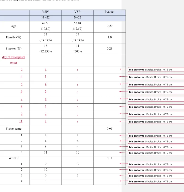
Table 1 Description of the clinical characteristics of the transcriptomic VASOGENE cohort
Figure 1 Quantile-Quantile plot summarizing the association of mRNA levels differences
(Dv3-D0) with the risk of vasospasm.
Figure 2 S1PR4 mean differences expression levels separately in aSAH patients with (VSP+)
Table 1 Description of the transcriptomic VASOGENE cohort VSP+ VSP- Pvalue1 N =22 N=22 Age 48.50 (10.80) 53.04 (12.52) 0.20 Female (%) 14 (63.63%) 14 (63.63%) 1.0 Smoker (%) 16 (72.73%) 11 (50%) 0.29 day of vasospasm onset 3 2 - 4 3 - 5 4 - 6 2 - 7 4 - 8 3 - 9 2 - 11 2 - Fisher score 0.91 1 2 2 2 4 6 3 5 4 4 11 10 WFNS2 0.11 1 9 12 2 10 4 3 0 3 4 3 3
Mis en forme : Droite, Droite : 0,76 cm Mis en forme : Droite, Droite : 0,76 cm Mis en forme : Droite, Droite : 0,76 cm Mis en forme : Droite, Droite : 0,76 cm Mis en forme : Droite, Droite : 0,76 cm Mis en forme : Droite, Droite : 0,76 cm Mis en forme : Droite, Droite : 0,76 cm Mis en forme : Droite, Droite : 0,76 cm
Mis en forme : Droite, Droite : 0,76 cm Mis en forme : Droite, Droite : 0,76 cm Mis en forme : Droite, Droite : 0,76 cm Mis en forme : Droite, Droite : 0,76 cm Mis en forme : Droite, Droite : 0,76 cm Mis en forme : Droite, Droite : 0,76 cm Mis en forme : Droite, Droite : 0,76 cm Mis en forme : Droite, Droite : 0,76 cm
5 0 0
GCS3>13 18 16 0.13
1Association test P-value derived from ANOVA and Chi-square test statistics for quantitative
and qualitative data, respectively.
Shown data: mean (SD) for quantitative variables and count (%) for qualitative variables.
2World Federation of Neurological Surgeons score 3Glasgow Coma Scale