HAL Id: inserm-00916134
https://www.hal.inserm.fr/inserm-00916134
Submitted on 9 Dec 2013HAL is a multi-disciplinary open access archive for the deposit and dissemination of sci-entific research documents, whether they are pub-lished or not. The documents may come from teaching and research institutions in France or abroad, or from public or private research centers.
L’archive ouverte pluridisciplinaire HAL, est destinée au dépôt et à la diffusion de documents scientifiques de niveau recherche, publiés ou non, émanant des établissements d’enseignement et de recherche français ou étrangers, des laboratoires publics ou privés.
Arrestins in host-pathogen interactions.
Stefano Marullo, Mathieu Coureuil
To cite this version:
Stefano Marullo, Mathieu Coureuil. Arrestins in host-pathogen interactions.. Handb Exp Pharmacol, Vsevolod Gurevich, 2014, 219, pp.361-74. �10.1007/978-3-642-41199-1_18�. �inserm-00916134�
Handbook of Experimental Pharmacology
Title: Arrestins -‐ Pharmacology and Therapeutic Potential Editor: Vsevolod Gurevich
Arrestins in host-‐pathogen interactions
Stefano Marullo1,2,3, Mathieu Coureuil3,41: Inserm, U1016, Institut Cochin, Paris, France 2: Cnrs, UMR8104, Paris, France
3: Université Paris Descartes, Sorbonne Paris Cité, France
4: Inserm, U1002, Unité de Pathogénie des Infections Systémiques, Paris, France
Abstract
In the context of host-‐pathogen interaction, host cell receptors and signaling pathways are essential for both invading pathogens, which exploit them at their own profit, and the defending organism, which activates early mechanism of defense, known as innate immunity, to block the aggression. Because of their central role as scaffolding proteins downstream of activated receptors, β-‐arrestins are involved in multiple signaling pathways activated in host cells by pathogens. Some of these pathways participate to the innate immunity and the inflammatory response. Other β-‐arrestin-‐dependent pathways are actually hijacked by microbes and toxins to penetrate into host cells and spread in the organism.
Key words: Innate immune response, Toll-‐Like Receptors, NF-‐κB, sepsis, pneumococcus,
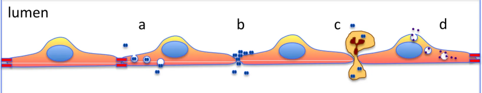
Host-‐pathogen interactions are complex multi-‐facet phenomena determining how host cells are colonized and how pathogens can disseminate (Figure1). Intracellular bacteria, viruses and microbial toxins penetrate into host cell after crossing plasma membranes. They can then proliferate and/or spread to invade host tissues. Extracellular pathogens, instead, need to cross the mechanical barrier constituted by skin, airways, gut, urinary, or genital tract epithelia to diffuse to the bloodstream and colonize organs. Crossing the first layer of epithelial cells and then endothelia can be achieved by different mechanisms. Pathogens can pass through these barriers via endocytosis at their apical (for epithelia) or luminal (for endothelia) side and then be shuttled inside vesicles to the basolateral side, a phenomenon known as transcytosis. They can also disseminate through the intercellular space between two adjacent cells via the so-‐called paracellular route. Finally, they can first infect blood cells, which are physiologically capable of crossing epithelia and endothelia (by diapedesis), and carry the hidden pathogen on the other side of the barrier like a Troy horse.
In all these cases, early steps of infection usually require pathogen adhesion to host cells via specific interaction with cell surface receptors. Then, pathogen binding to host cell receptors induces signaling events leading to important changes in cell metabolism, shape, organization, trafficking, which are exploited by the pathogen for proliferation and productive infection. On the other hand, pathogens must circumvent host cell responses, which induce signaling cascades leading to inflammation and other early mechanism of defense known as innate immunity. Thus, in the context of host-‐pathogen interaction, host cell receptors and signaling pathways are essential for both pathogens and the defending organism.
1. Beta arrestins are multi-‐task proteins, which regulate cell surface receptors and orchestrate signaling pathways in time and space
Beta arrestins 1 and 2 (βarr1 and βarr2, also named arrestin 2 and 3) are the ubiquitous isoforms of visual arrestin, which in the retinal tissue is responsible for the “arrest” of rhodopsin activation. It is not surprising therefore, that βarrs were originally identified as negative regulators of G protein-‐coupled receptor (GPCR) function in non-‐retinal tissues, because their binding promotes GPCR desensitization (Lohse et al. 1990). Indeed, the translocation of cytoplasmic βarrs to activated and phosphorylated receptors uncouples GPCRs from downstream G protein-‐dependent signaling pathways. βarrs were subsequently shown to play a role as adaptor proteins connecting activated and phosphorylated GPCRs to AP2 and clathrin, two components of the endocytic machinery (Goodman et al. 1996; Laporte et al. 1999). Thank to this molecular bridge GPCRs are recruited in clarthin-‐coated pits and subsequently internalized into endosomes. Successive investigations extended the spectrum of the roles of βarrs in receptor trafficking. Indeed, βarrs promote the recruitment of ubiquitin ligases and thus participate in the agonist-‐induced ubiquitylation of receptors, which impact on their subcellular localization and stability. Ubiquitination, in addition to its well-‐known function in soluble protein proteasomal degradation, serves as a signal to recruit ubiquitin-‐binding domain-‐containing proteins, for specific biological functions, such as endocytosis or sorting to lysosomes (Chen and Sun 2009). Mdm2 was the first E3 ligase recognized as a βarr partner; by ubiquitilating βarrs, it provides a signal necessary for the internalization of βarr-‐bound GPCRs (Shenoy et al. 2001). Other E3 ligases, such as NEDD4 or AIP4, were instead reported to provide a lysosome sorting-‐signal to internalized receptors (Shenoy et al. 2008; Marchese et al. 2003). βarrs were also found to participate in internalization or ubiquitylation (or both) of many non-‐GPCR receptors or plasma membrane
proteins: the type III transforming growth factor-‐β receptor, the insulin-‐like growth factor I receptor, voltage-‐dependent calcium channels, the Na(+)/H(+) exchangers NHE1 and NHE5, the vascular endothelial (VE) cadherin and Notch (reviewed in (Shukla et al. 2011)). Interestingly, recent studies have identified a larger and more ancient family of arrestin-‐fold proteins that display some structural similarity with βarrs and share their trafficking and down-‐regulating functions. This family of “α-‐arrestins” is conserved in eukaryotes (Alvarez 2008) and comprises ARRDC (Arrestin domain-‐containing) proteins (Nabhan et al. 2010; Patwari et al. 2011) in mice and humans and ARTs (arrestin-‐related trafficking adaptors) in yeast (Lin et al. 2008).
In addition to their role in receptor desensitization and trafficking, βarrs have a function of signaling adaptors and scaffolds. Assembling signaling proteins into molecular hubs (or signalosomes) constructed around scaffolding proteins is a common mechanism used by all cells to correctly deliver specific signals in space and time (Good et al. 2011). Since the first description of the Ste5 scaffold of the MAP kinase cascade in yeast (Choi et al. 1994), an increasing number of protein scaffolds have been identified, based on their ability of binding multiple signaling partners via direct protein–protein interaction, due to their high content of modular protein binding domains (Zeke et al. 2009). After the pioneering report describing the role of βarrs in organizing the oriented activation of MAP kinases in the cytoplasm (McDonald et al. 2000; Luttrell et al. 2001), many other effector pathways orchestrated by βarrs have been characterized (reviewed in (Shenoy and Lefkowitz 2005; Lefkowitz et al. 2006; Kovacs et al. 2009; Luttrell and Gesty-‐Palmer 2010)) illustrating the prominent role of βarrs in the control of cell signaling.
2. βarrs in the host-‐cell response to pathogens
Because of their central role as scaffolding proteins downstream of activated receptors, βarrs are involved in multiple pathways activated in host cells by pathogens. The important phenomenon of βarr-‐dependent regulation of cell motility and chemotaxis, via the control of actin polymerization and cytoskeletal rearrangements, will be treated in another chapter of this book (ref to K. DeFea contribution). Here we will summarize the principal established roles of βarrs in innate immunity, inflammatory response and apoptosis (Table I).
2.1. βarr involvement in leukocyte degranulation
The first evidence for a βarr involvement in innate immunity came from studies on chemoattractant-‐stimulated granule release in leukocytes (Barlic et al. 2000). Leukocyte granules contain several enzymes and non-‐enzymatic compounds that participate in bactericidal activity. The release of these granules is controlled by the activation of Fc receptors or GPCRs for chemoattractants. Interleukin 8 (IL-‐8) activation of the chemokine receptor CXCR1 was found to stimulate rapid formation of βarr complexes with the Src-‐ family tyrosine kinases Hck or c-‐Fgr. Hck association with βarrs activates the kinase and allows its targeting to granules. In case of expression of dominant-‐negative βarr mutant with altered polyproline-‐rich region (known to be critical for the interaction with the c-‐Src tyrosine kinase), granulocytes fail to release granules or activate tyrosine kinases in response to IL-‐8 stimulation. Thus, in this pathophysiological context, βarrs are important signaling molecules in the innate immune response.
2.2. βarr regulation of Toll-‐like receptor signaling
Pathogen-‐associated molecular patterns (PAMPs) (Janeway 1989) such as flagellin, the lipopolysaccharide (LPS) or the peptidoglycan of bacterial cell wall are recognized by specific host receptors known as pattern-‐recognition receptors (PRRs). During infection, PAMPs-‐mediated activation of PRRs initiates inflammatory reactions, which constitute the first line of defense and prepare the establishment of adaptative immune responses. Several classes of PRRs have been described, among which the Toll-‐like receptors (TLRs) are key initiators of the innate immune response (Medzhitov et al. 1997). Some TLRs (1, 2, 4, 5, and 6) operate primarily at the plasma membrane whereas other TLRs, mostly involved in the recognition of nucleic acids, are localized to late endosomes and lysosomes. Signal transduction mechanisms of TLRs are similar to those elicited by some interleukin receptors. TLRs contain a Toll interleukin-‐1 receptor homology (TIR) domain (O’Neill and Bowie 2007), which engage cytoplasmic TIR-‐domain-‐containing adaptors such as the myeloid differentiation primary response gene 88 (MyD88), or the TIR domain containing adaptor protein (TIRAP). These adaptors recruit members of the IRAK (IL-‐1 receptor–associated kinase) family of serine-‐threonine kinases that induce inflammatory cytokine expression. MyD88 and TIRAP promote the expression of nuclear factor NF-‐kB-‐dependent cytokines via the activation of NFκB and of mitogen-‐activated protein kinases, whereas other adaptors induce the expression of type I interferons (IFNs).
The first indication that βarrs can modulate TLR signaling was based on the observation that βarr2 (not βarr1) directly interacts with IkBα (Gao et al. 2004). The protein kinase IKK, activated by phosphorylation downstream of stimulated TLRs or the TNF receptor, phosphorylates IκBα that normally binds to the transcription factor NF-‐κB and
inhibits its nuclear translocation. Once phosphorylated, IκBα is ubiquitinated and targeted for degradation by the proteasome, releasing NF-‐κB. NF-‐κB-‐containing heterodimers then translocate into the nucleus and mediate the transcription of a vast array of proteins involved in immune and inflammatory responses. Interaction with βarr2 prevents the phosphorylation and degradation of IκBα. Interestingly, GPCR stimulation can enhance βarr2-‐IκBα interaction and consecutive stabilization of IκBα, leading to the inhibition of the NF-‐κB pathway. Supporting the hypothesis that βarrs are negative regulators of the innate immune activation via TLRs, it was reported that both βarr isoforms interact with TRAF6 preventing its auto-‐ubiquitination and subsequent activation of NF-‐κB (Wang et al. 2006). TRAF6 is a ring domain E3 ubiquitin ligase that it is involved in the activation of IKK downstream of TLRs and IL-‐1 receptor; it interacts with βarrs upon stimulation by IL1-‐β or gram-‐negative bacteria lipopolysaccharide. Consistently, endotoxin-‐treated βarr2-‐deficient mice had higher expression of pro-‐inflammatory cytokines were more susceptible to endotoxin shock than controls. A subsequent study comparing wild type and βarr2-‐deficient mice confirmed that βarr2 is a negative regulator of the inflammatory response in polymicrobial sepsis (Fan et al. 2010). However, the existence of different functional outputs in mice models investigated with diverse experimental approaches (Porter et al. 2010), indicate a more complex regulation of TLRs response by βarrs. Part of the explanation might be that βarr1 and βarr2 differentially regulate TLR signaling and pro-‐inflammatory gene expression. For example, one study reported that both βarrs negatively regulate LPS-‐induced NFκB, whereas only βarr2 mediates LPS-‐induced ERK 1/2 activation (Fan et al. 2007). Also, in a report examining adenovirus-‐vector-‐induced innate immune responses and involving TLR-‐ dependent pathways, βarr1 was found to be a positive regulator and βarr2 a negative regulator (Seregin et al. 2010). The functional output of the specific involvement of each
βarr isoform might also vary in different cell types. In macrophages, both βarr1 and the G protein receptor kinase GRK5 inhibit LPS-‐dependent signaling of the TLR4. More specifically, βarr1 (not βarr2) modulates the MAP kinase arm of TLR4 signaling by interacting with NFκB1 p105, which is the precursor of NFκB1 p50 and a cytoplasmic inhibitor of NF-‐κB: p105 functions as an IκB and retains associated p50 in the cytoplasm. As described in fibroblasts for βarr2, which directly interacts with IkBα preventing its phosphorylation and degradation (Gao et al. 2004), βarr1 stabilizes p105. Knockdown of βarr1 leads to enhanced LPS-‐induced phosphorylation and degradation of p105, enhanced MAP3K release, and enhanced MAP2K phosphorylation (Parameswaran et al. 2006).
In addition to its role in the inflammatory response via the NFκB and the MAP kinase pathways, TLR4 activation can promote apoptosis under certain conditions and in some cell types (Gay and Gangloff 2007). A recent study identified the glycogen synthase kinase-‐3b (GSK-‐3b) as an intermediate for TL4-‐mediated apoptosis (Li et al. 2010). Interestingly, the apoptotic cascade was attenuated by βarr2, likely via the stabilization of phospho-‐GSK-‐3b, an inactive form of GSK-‐3b.
2.3. βarr regulation of natural killer cells.
Natural killer (NK) cells are critical components of the innate immune system that recognize and kill tumor or virus-‐infected target cells. These cells express at their surface two sets of receptors. Activating receptors that are involved in the killing activity of NK cells, whereas inhibitory receptors contribute to tolerance to normal healthy cells. The association of the inhibitory receptor KIR2DL1 with with βarr2 was reported to induce the recruitment of the tyrosine phosphatases SHP-‐1 and SHP-‐2 to KIR2DL1, contributing to the inhibitory signaling. Cytotoxicity of NK cells is consequently higher in βarr2-‐deficient mice and inhibited
in animals overexpressing βarr2. The inhibitory effect of βarr2 is functionally relevant in vivo, as shown by decreased NK cell-‐dependent susceptibility to cytomegalovirus infection in βarr2-‐deficient mice (Yu et al. 2008).
3. Receptors and signaling pathways involving βarrs that are hijacked by microbes and toxins to penetrate into host cells or spread (Table II).
3.1. Bacteria
Streptococcus pneumoniae (pneumococcus), a gram-‐positive pathogen causing
pneumonia, sepsis and meningitis, is the first reported example of bacteria exploiting βarrs for tissue invasion. Pneumococci translocate across human endothelial cells through vesicular structures without intracellular multiplication (transcytosis). Early studies identified the receptor for platelet-‐activating factor (PAF) as the pneumococcus adhesion receptor in both epithelial and endothelial cells (Cundell et al. 1995; Ring et al. 1998). Pneumococcus binding to PAF receptors induces βarr translocation and endocytosis of pathogen-‐receptor complexes. Cytoplasmic activation of ERK, presumably mediated by βarrs, is required for pneumococcal endocytosis (Radin et al. 2005). Interestingly, instead of being directed to lysosomes or recycled to the cell surface as agonist-‐bound receptors, a significant proportion of bacteria-‐PAF receptor complexes are diverted to basolateral membranes, this proportion being enhanced by βarr overexpression. Thus, pneumococci subvert the βarr-‐dependent trafficking machinery of PAF-‐receptors to drive pathogen-‐containing vacuoles away from lysosomes and across endothelial cell barriers (Radin et al. 2005).
N. meningitidis (meningococcus) is a Gram-‐negative diplococcus causing
with peripheral vascular leakage, ischemic tissue damage and septic shock. The ability of meningococci to interact with endothelial cells is essential in meningococcal pathogenesis (Coureuil et al. 2012). After initial attachment, mediated by a still unidentified receptor, bacteria have the ability to resist blood flow, to multiply and form micro-‐colonies on the apical surface of endothelial cells. The stabilization of bacterial colonies depends on the formation of host cell protrusions, which occur in response to signaling cascades elicited by the pathogen in the endothelial cells. In addition, bacterial-‐induced signaling eventually results in the opening of intercellular junctions with subsequent meningeal colonization via the paracellular route (Coureuil et al. 2009). It has been established that signaling in host cells is provoked by polymeric filaments found on many Gram-‐negative bacteria, known as type IV pili, which correspond to the multimeric assembly of various pilin subunits (Miller et al. 2012). Recently, it was reported that N.meningitidis pilins allosterically stimulate a biased β2-‐adrenoceptor-‐βarr signaling pathway in endothelial cells, which ultimately traps βarrs and their interacting partners, such as the Src tyrosine kinase and junctional proteins VE-‐ cadherin and p120, under bacterial colonies (Coureuil et al. 2010). The cytoskeletal reorganization mediated by βarr-‐activated Src stabilizes bacterial adhesion to endothelial cells under permanent flow, whereas βarr-‐dependent delocalization of junctional proteins results in anatomical gaps between adjacent endothelial cells, which are used by bacteria to penetrate into tissues. The bacterial ligand, which activates the β2-‐adrenoceptor by interacting with the receptor N-‐terminal region, corresponds to two particular components of the pili, namely the pilins PilE and PilV.
N gonorrheae (gonococcus) a close relative of meningococcus, which most often cause isolated infection of the genito-urinary tract but can, in rare cases, spread into the bloodstream and colonize meninges (Martín et al. 2008), elicits similar signaling events as N
meningitidis in endothelial cells (Coureuil et al. 2010). In addition, many other bacteria take advantage of host cell signaling pathways involving Src activation and its substrate cortactin to invade tissues, as in the case of Neisseria species. Although not investigated yet, βarrs might well participate in the signaling pathways induced by these other pathogens.
3.2. Viruses
Marburg virus (MARV) and Ebola virus (EBOV), two members of the Filoviridae family, are the causative agents of a deadly infection, known as viral hemorrhagic fever (Schnittler and Feldmann 2003). Although several monocyte, macrophage, dendritic and endothelial cell surface proteins have been implicated in filovirus entry, a common receptor, the T-‐cell immunoglobulin and mucin domain 1 (TIM-‐1), was reported for both Ebola and Marburg viruses (Kondratowicz et al. 2011). Following viral glycoprotein (GP)-‐dependent receptor binding, filoviruses are internalized by clathrin-‐mediated endocytosis. The cellular endocytic machinery sorts internalized viruses to an acidic endosomal compartment, which is the site of virus-‐cell membrane fusion. A recent report examined the specific requirements for different components of the clathrin endocytic machinery in Ebola GP versus Marburg GP pseudovirion entry (Bhattacharyya et al. 2011). Whereas Ebola GP pseudovirions specifically required the adaptor proteins Eps15 and AP-‐2 to be connected to clathrin, Marburg GP pseudovirions specifically necessitated βarr1 and the adaptor protein AP-‐1 instead of AP-‐2. Knocking down βarr1 significantly delayed virus fusion with no evident virus-‐binding defect.
The endosomal sorting-‐complex required for transport (ESCRT) machinery comprises multiprotein complexes (ESCRT 0–III) that cooperate in a sequential and a coordinated manner to target ubiquitinated membrane cargo into vesicles that bud into late endosomes to form multivesicular bodies (MVBs) (Hurley and Emr 2006). In particular, internalized cell
surface receptors that are programmed for lysosomal degradation are delivered to MVBs via this machinery. Internalized ubiquitinated receptors in the endosomes are initially recognized by ESCRT-‐0, which subsequently recruits ESCRT-‐I to endosomal membranes, followed by recruitment of ESCRT II and III. The process terminates with receptor sorting into budding intra-‐endosomal vesicles (Raiborg and Stenmark 2009). For some receptors, such as the chemokine receptor CXCR4, βarr1 connects the ubiquitinated receptor with ESCRT-‐0 and regulates the amount of CXCR4 that is degraded (Malik and Marchese 2010). ESCRT machinery also plays a key role in the budding of many enveloped viruses, including HIV-‐1 and other retroviruses. Recently, it was reported that ARRDC (Arrestin domain-‐containing) proteins are involved in budding of murine leukemia virus or human T-‐cell leukemia virus type 1 by interacting with HECT ubiquitin ligases and promoting ESCRT-‐III recruitment (Rauch and Martin-‐Serrano 2011).
3.3. Toxins
Bacillus anthracis, the bacterium responsible for the anthrax disease produces the anthrax toxin, which is composed of three independent polypeptide chains. Two proteins have an enzymatic activity (calmodulin dependent adenylate cyclase and metalloprotease, claving the MAP kinase kinase, respectively) the third one being required for the translocation of the two enzymes into the cytoplasm where their activity produces the toxic effects (Young and Collier 2007). The protein involved in toxin translocation, known as the protective antigen (PA), interacts with the target cells. It is processed by host cell proteases, such as furin, leading to the formation of a 63kDa fragment that heptamerizes into a ring like structure. The complex between the heptamer and the two enzymes is internalized into endosomes where the heptamer forms a pore allowing the partially unfolded/activated enzymes to
cross the endosomal membranes and reach the cytosol (Collier 2009). Heptamerization of the 63kDa PA fragment leads to the activation of src-‐like kinases (Abrami et al. 2010b), which phosphorylate the cytoplasmic tail of capillary morphogenesis 2 (CMG2) a type-‐I membrane protein that serves as toxin receptor. Toxin receptors (CMG2 and also the tumor endothelial marker 8 TEM8) are ubiquitinated, via a process that requires βarr1. Receptor modification finally allows the recruitment of AP-‐1 adaptin and clathrin, leading to their internalization via clathrin-‐coated pits (Abrami et al. 2010a).
4. Conclusions and Perspectives
Facing the vast array of pathogen ligands and of potential host cell receptors that have been selected by pathogens to penetrate into host cells or to cross epithelial and endothelial barriers, the number of cellular pathways hijacked by pathogens or employed by host cells as primary defense line are relatively limited. Moreover, a restricted set of proteins such as kinases (Src family, MAP kinases), proteins involved in endocytosis and sorting, junctional proteins, signaling adaptors at the cross-‐road of various pathways downstream of TLRs or cytokine receptors, is constantly involved in these processes, whatever the type of the pathogen. Most of these proteins appear as direct or indirect interactors of arrestins, suggesting that our current knowledge of the role of arrestins in host-‐pathogen interactions only represents the tip of the iceberg.
A remarkable feature of βarrs, is the large number of cellular proteins they interact with, contrasting with limited amino-‐acid residues and areas of contact involved in individual interactions (Gurevich and Gurevich 2012). This feature might be exploited to develop very specific molecules capable of targeting signaling pathways at the appropriate level and with exquisite precision for therapeutic purposes. In this context, host-‐pathogen interactions
appear a particularly interesting area of investigation.
References
Abrami L, Bischofberger M, Kunz B, Groux R, van der Goot FG (2010a) Endocytosis of the anthrax toxin is mediated by clathrin, actin and unconventional adaptors. PLoS pathogens 6:e1000792.
Abrami L, Kunz B, van der Goot FG (2010b) Anthrax toxin triggers the activation of src-‐like kinases to mediate its own uptake. Proc Natl Acad Sci USA 107:1420-‐1424.
Alvarez CE (2008) On the origins of arrestin and rhodopsin. BMC Evol Biol 8:222.
Barlic J, Andrews JD, Kelvin AA, Bosinger SE, DeVries ME, Xu L, Dobransky T, Feldman RD, Ferguson SS, Kelvin DJ (2000) Regulation of tyrosine kinase activation and granule release through beta-‐arrestin by CXCRI. Nat Immunol 1:227-‐233.
Bhattacharyya S, Hope TJ, Young JA (2011) Differential requirements for clathrin endocytic pathway components in cellular entry by Ebola and Marburg glycoprotein pseudovirions. Virology 419:1-‐9.
Chen ZJ, Sun LJ (2009) Nonproteolytic functions of ubiquitin in cell signaling. Mol Cell:275-‐ 286.
Choi KY, Satterberg B, Lyons DM, Elion EA (1994) Ste5 tethers multiple protein kinases in the MAP kinase cascade required for mating in S. cerevisiae. Cell 78:499-‐512.
Collier RJ (2009) Membrane translocation by anthrax toxin. Mol Aspects Med 30:413-‐422. Coureuil M, Join-‐Lambert O, Lecuyer H, Bourdoulous S, Marullo S, Nassif X (2012)
Coureuil M, Lécuyer H, Scott MGH, Boularan C, Enslen H, Soyer M, Mikaty G, Bourdoulous S, Nassif X, Marullo S (2010) Meningococcus hijack a ß2-‐adrenoceptor-‐ß-‐arrestin pathway to cross brain microvasculature endothelium. . Cell 143:1149-‐1160
Coureuil M, Mikaty G, Miller F, Lecuyer H, Bernard C, Bourdoulous S, Dumenil G, Mege RM, Weksler BB, Romero IA, Couraud PO, Nassif X (2009) Meningococcal type IV pili recruit the polarity complex to cross the brain endothelium. Science 325:83-‐87. Cundell DR, Gerard NP, Gerard C, Idanpaan-‐Heikkila I, Tuomanen EI (1995) Streptococcus
pneumoniae anchor to activated human cells by the receptor for platelet-‐activating factor. Nature 377:435-‐438.
Fan H, Bitto A, Zingarelli B, Luttrell LM, Borg K, Halushka PV, Cook JA (2010) Beta-‐arrestin 2 negatively regulates sepsis-‐induced inflammation. Immunology 130:344-‐351.
Fan H, Luttrell LM, Tempel GE, Senn JJ, Halushka PV, Cook JA (2007) Beta-‐arrestins 1 and 2 differentially regulate LPS-‐induced signaling and pro-‐inflammatory gene expression. Molecular immunology 44:3092-‐3099.
Gao H, Sun Y, Wu Y, Luan B, Wang Y, Qu B, Pei G (2004) Identification of beta-‐arrestin2 as a G protein-‐coupled receptor-‐stimulated regulator of NF-‐kappaB pathways. Mol Cell 14:303-‐317.
Gay NJ, Gangloff M (2007) Structure and Function of Toll Receptors and Their Ligands. Annu Rev Biochem 76:141-‐165.
Good MC, Zalatan JG, Lim WA (2011) Scaffold proteins: hubs for controlling the flow of cellular information. Science 332:680-‐686.
Goodman OBJ, Krupnick JG, Santini F, Gurevich VV, Penn RB, Gagnon AW, Keen JH, Benovic JL (1996) ß-‐arrestin acts as a clathrin adaptor in endocytosis of the ß2-‐adrenergic receptor. Nature 383:447-‐450.
Gurevich VV, Gurevich EV (2012) Synthetic biology with surgical precision: targeted reengineering of signaling proteins. Cell Signal 24:1899-‐1908.
Hurley JH, Emr SD (2006) The ESCRT complexes: Structure and mechanism of a membrane-‐ trafficking network
. Annu Rev Biophys Biomol Struct 35:277–298.
Janeway CA (1989) Approaching the asymptote? Evolution and revolution in immunology. . Cold Spring Harbor Symposia on Quantitative Biology 54:1-‐13
Kondratowicz AS, Lennemann NJ, Sinn PL, Davey RA, Hunt CL, Moller-‐Tank S, Meyerholz DK, Rennert P, Mullins RF, Brindley M, Sandersfeld LM, Quinn K, Weller M, McCray PB, Jr., Chiorini J, Maury W (2011) T-‐cell immunoglobulin and mucin domain 1 (TIM-‐1) is a receptor for Zaire Ebolavirus and Lake Victoria Marburgvirus. Proc Natl Acad Sci USA 108:8426-‐8431.
Kovacs JJ, Hara MR, Davenport CL, Kim J, Lefkowitz RJ (2009) Arrestin development: emerging roles for beta-‐arrestins in developmental signaling pathways. Dev Cell 17:443-‐458.
Laporte SA, Oakley RH, Zhang J, Holt JA, Ferguson SS, Caron MG, Barak LS (1999) The beta2-‐ adrenergic receptor/betaarrestin complex recruits the clathrin adaptor AP-‐2 during endocytosis. Proc Natl Acad Sci U S A 96:3712-‐3717.
Lefkowitz RJ, Rajagopal K, Whalen EJ (2006) New roles for beta-‐arrestins in cell signaling: not just for seven-‐transmembrane receptors. Mol Cell 24:643-‐652.
Li H, Sun X, LeSage G, Zhang Y, Liang Z, Chen J, Hanley G, He L, Sun S, Yin D (2010) beta-‐ arrestin 2 regulates Toll-‐like receptor 4-‐mediated apoptotic signalling through glycogen synthase kinase-‐3beta. Immunology 130:556-‐563.
Lin CH, MacGurn JA, Chu T, Stefan CJ, Emr SD (2008) Arrestin related ubiquitin-‐ligase adaptors regulate endocytosis and protein turnover at the cell surface. Cell 135:714– 725.
Lohse MJ, Benovic JL, Codina J, Caron MG, Lefkowitz RJ (1990) beta-‐Arrestin: a protein that regulates beta-‐adrenergic receptor function. Science 248:1547-‐1550.
Luttrell LM, Gesty-‐Palmer D (2010) Beyond desensitization: physiological relevance of arrestin-‐dependent signaling. Pharmacol Rev 62:305-‐330.
Luttrell LM, Roudabush FL, Choy EW, Miller WE, Field ME, Pierce KL, Lefkowitz RJ (2001) Activation and targeting of extracellular signal-‐regulated kinases by beta-‐arrestin scaffolds. Proc Natl Acad Sci U S A 98:2449-‐2454.
Malik R, Marchese A (2010) Arrestin-‐2 Interacts with the Endosomal Sorting Complex Required for Transport Machinery to Modulate Endosomal Sorting of CXCR4. Mol Biol Cell 21:2529-‐2541.
Marchese A, Raiborg C, Santini F, Keen JH, Stenmark H, Benovic JL (2003) The E3 ubiquitin ligase AIP4 mediates ubiquitination and sorting of the G protein-‐coupled receptor CXCR4. Dev Cell 5:709-‐722.
Martín MC, Pérez F, Moreno A, Moral A, Alvarez MA, Méndez FJ, Vázquez F (2008) Neisseria gonorrhoeae meningitis in pregnant adolescent. Emerg Infect Dis 14:672-‐674.
McDonald PH, Chow CW, Miller WE, Laporte SA, Field ME, Lin FT, Davis RJ, Lefkowitz RJ (2000) Beta-‐arrestin 2: a receptor-‐regulated MAPK scaffold for the activation of JNK3. Science 290:1574-‐1577.
Medzhitov R, Preston-‐Hurlburt P, Janeway CA (1997) A human homologue of the Drosophila Toll protein signals activation of adaptive immunity. Nature 388:394–397.
Miller F, Lecuyer H, Join-‐Lambert O, Bourdoulous S, Marullo S, Nassif X, Coureuil M (2012) Neisseria meningitidis colonization of the brain endothelium and cerebrospinal fluid invasion. Cellular microbiology.
Nabhan JF, Pan H, Lu Q (2010) Arrestin domain-‐containing protein 3 recruits the NEDD4 E3 ligase to mediate ubiquitination of the β2-‐adrenergic receptor. EMBO Rep 11:605-‐ 611.
O’Neill LA, Bowie AG (2007) The family of five: TIR-‐domain containing adaptors in Toll-‐like receptor signalling. Nat Rev Immunol 7:353–364.
Parameswaran N, Pao CS, Leonhard KS, Kang DS, Kratz M, Ley SC, Benovic JL (2006) Arrestin-‐ 2 and G protein-‐coupled receptor kinase 5 interact with NFkappaB1 p105 and negatively regulate lipopolysaccharide-‐stimulated ERK1/2 activation in macrophages. The Journal of biological chemistry 281:34159-‐34170.
Patwari P, Emilsson V, Schadt EE, Chutkow WA, Lee S, Marsili A, Zhang Y, Dobrin R, Cohen DE, Larsen PR, Zavacki AM, Fong LG, Young SG, Lee RT (2011) The arrestin domain-‐ containing 3 protein regulates body mass and energy expenditure. Cell Metab 14:671-‐683.
Porter KJ, Gonipeta B, Parvataneni S, Appledorn DM, Patial S, Sharma D, Gangur V, Amalfitano A, Parameswaran N (2010) Regulation of lipopolysaccharide-‐induced inflammatory response and endotoxemia by beta-‐arrestins. J Cell Physiol 225:406-‐ 416.
Radin JN, Orihuela CJ, Murti G, Guglielmo C, Murray PJ, Tuomanen EI (2005) beta-‐Arrestin 1 participates in platelet-‐activating factor receptor-‐mediated endocytosis of Streptococcus pneumoniae. Infection and immunity 73:7827-‐7835.
Raiborg C, Stenmark H (2009) The ESCRT machinery in endosomal sorting of ubiquitylated membrane proteins. Nature 458:445-‐452.
Rauch S, Martin-‐Serrano J (2011) Multiple Interactions between the ESCRT Machinery and Arrestin-‐Related Proteins: Implications for PPXY-‐Dependent Budding. J Virol 85:3546-‐ 3556.
Ring A, Weiser JN, Tuomanen EI (1998) Pneumococcal trafficking across the blood-‐brain barrier. Molecular analysis of a novel bidirectional pathway. J Clin Invest 102:347-‐ 360.
Schnittler HJ, Feldmann H (2003) Viral hemorrhagic fever-‐-‐a vascular disease? Thromb Haemost 89:967-‐972.
Seregin SS, Appledorn DM, Patial S, Bujold M, Nance W, Godbehere S, Parameswaran N, Amalfitano A (2010) beta-‐Arrestins modulate Adenovirus-‐vector-‐induced innate immune responses: differential regulation by beta-‐arrestin-‐1 and beta-‐arrestin-‐2. Virus Res 147:123-‐134.
Shenoy SK, Lefkowitz RJ (2005) Seven-‐transmembrane receptor signaling through beta-‐ arrestin. Sci STKE 2005:cm10.
Shenoy SK, McDonald PH, Kohout TA, Lefkowitz RJ (2001) Regulation of receptor fate by ubiquitination of activated beta 2-‐ adrenergic receptor and beta-‐arrestin. Science 294:1307-‐1313.
Shenoy SK, Xiao K, Venkataramanan V, Snyder PM, Freedman NJ, Weissman AM (2008) Nedd4 mediates agonist-‐dependent ubiquitination, lysosomal targeting, and degradation of the beta2-‐adrenergic receptor. J Biol Chem 283:22166-‐22176.
Shukla AK, Xiao K, Lefkowitz RJ (2011) Emerging paradigms of b-‐arrestindependent seven transmembrane receptor signaling. Trends Biochem Sci 36:457-‐469.
Wang Y, Tang Y, Teng L, Wu Y, Zhao X, Pei G (2006) Association of beta-‐arrestin and TRAF6 negatively regulates Toll-‐like receptor-‐interleukin 1 receptor signaling. Nature immunology 7:139-‐147.
Young JA, Collier RJ (2007) Anthrax toxin: receptor binding, internalization, pore formation, and translocation. . Annu Rev Biochem 76:243–265.
Yu MC, Su LL, Zou L, Liu Y, Wu N, Kong L, Zhuang ZH, Sun L, Liu HP, Hu JH, Li D, Strominger JL, Zang JW, Pei G, Ge BX (2008) An essential function for beta-‐arrestin 2 in the inhibitory signaling of natural killer cells. Nature immunology 9:898-‐907.
Zeke A, Lukacs M, Lim WA, Remenyi A (2009) Scaffolds: interaction platforms for cellular signalling circuits. Trends Cell Biol 19:364-‐374.
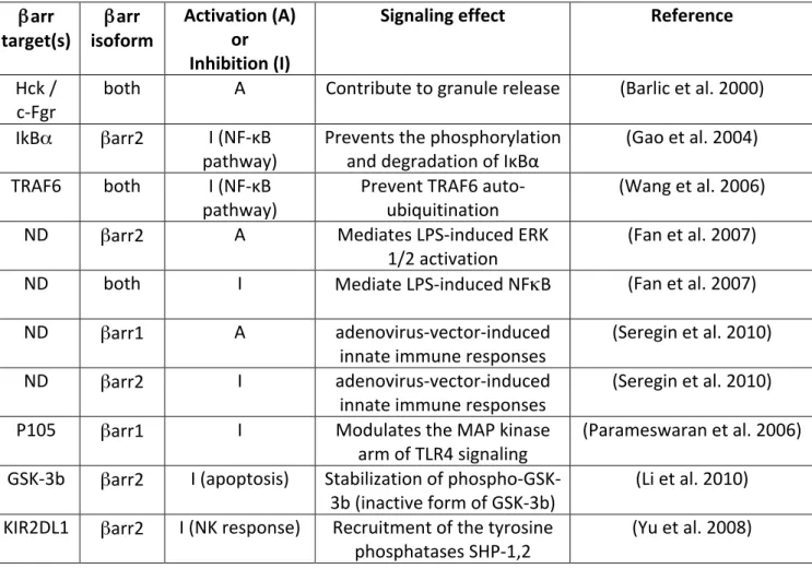
Table I. βarrestins in the host-‐cell response to pathogens
βarr
target(s) isoform βarr
Activation (A) or Inhibition (I)
Signaling effect Reference
Hck /
c-‐Fgr both A Contribute to granule release (Barlic et al. 2000) IkBα βarr2 I (NF-‐κB
pathway) Prevents the phosphorylation and degradation of IκBα (Gao et al. 2004) TRAF6 both I (NF-‐κB
pathway) Prevent TRAF6 auto-‐ubiquitination (Wang et al. 2006) ND βarr2 A Mediates LPS-‐induced ERK
1/2 activation (Fan et al. 2007) ND both I Mediate LPS-‐induced NFκB (Fan et al. 2007)
ND βarr1 A adenovirus-‐vector-‐induced
innate immune responses (Seregin et al. 2010) ND βarr2 I adenovirus-‐vector-‐induced
innate immune responses (Seregin et al. 2010)
P105 βarr1 I Modulates the MAP kinase
arm of TLR4 signaling (Parameswaran et al. 2006) GSK-‐3b βarr2 I (apoptosis) Stabilization of phospho-‐GSK-‐
3b (inactive form of GSK-‐3b) (Li et al. 2010) KIR2DL1 βarr2 I (NK response) Recruitment of the tyrosine
phosphatases SHP-‐1,2 (Yu et al. 2008)
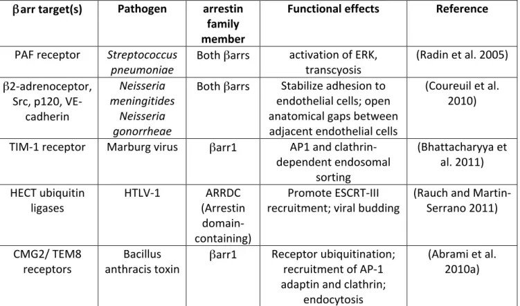
Table II: βarr-‐dependent pathways hijacked by microbes and toxins βarr target(s) Pathogen arrestin
family member
Functional effects Reference
PAF receptor Streptococcus
pneumoniae Both βarrs activation of ERK, transcyosis (Radin et al. 2005) β2-‐adrenoceptor, Src, p120, VE-‐ cadherin Neisseria meningitides Neisseria gonorrheae
Both βarrs Stabilize adhesion to endothelial cells; open anatomical gaps between adjacent endothelial cells
(Coureuil et al. 2010) TIM-‐1 receptor Marburg virus βarr1 AP1 and clathrin-‐
dependent endosomal sorting
(Bhattacharyya et al. 2011) HECT ubiquitin
ligases HTLV-‐1 (Arrestin ARRDC domain-‐ containing)
Promote ESCRT-‐III
recruitment; viral budding (Rauch and Martin-‐Serrano 2011) CMG2/ TEM8
receptors anthracis toxin Bacillus βarr1 Receptor ubiquitination; recruitment of AP-‐1 adaptin and clathrin;
endocytosis
(Abrami et al. 2010a)