THE JOURNAL OF INFECTIOUS DISEASES. VOL. ISS, NO.3· MARCH 1987 © 1987 by The University of Chicago. All rights reserved. 0022-1899/87/5504-0016$01.00
Role of Bacterial Exopolymers and Host Factors on Adherence. and
Phagocytosis of
Staphylococcus aureus
in Foreign Body Infection
Elisabetta Falcieri;* Pierre Vaudaux, Elzbieta Huggler, Daniel Lew, and Francis Waldvogel
From the Department of Morphology, University Medical Center, and the Division of infectious Diseases, Department of Medicine, University Hospital, Geneva, Switzerland Using a previously developed guinea pig model of foreign body infection, we examined
ultrastructural and functional surface alterations ofStaphylococcus aureus strain Wood 46 during the early phase of infection. Exopolymer-free bacteria were prepared and in-oculated into subcutaneously implanted tissue cages. After three hours, the bacteria showed abundant capsular and intercellular exopolymers, which were visualized by transmission electron microscopy. Exopolymers were also produced by S.aureus exposed in vitro to fluid from the tissue cage. In contrast, human serum albumin prevented exopolymer produc-tion by S.aureus. The influence of exopolymers on the susceptibility of S. aureus to in-gestion and phagocytic killing by neutrophils was tested in vitro and found to be negligi-ble. Furthermore, adherence of S.aureus to fibronectin-coated surfaces was unaffected by the presence or absence of exopolymers. Thus, in our experimental model, exopolymers are produced early during the onset of infection, but they have little impact on adherence and phagocytosis.
Clinical foreign body infections are characterized by their limited spread beyond the tissues in immediate contact with the implants, their poor response to an-tibiotic treatment, and their spontaneous cure after removal of the prosthesis [1, 2]. Correlating with these clinical observations, animal models of foreign body infections (primarily with staphylococci) have demonstrated the high infective power of a low in-oculum and confirmed the ineffectiveness of local host defense mechanisms against microorganisms colonizing the implants [3-5].
In addition to ineffective host defense mecha-nisms, other factors may explain the high suscepti-bility of foreign bodies to bacterial infections. Several reports [6-13] have described the production of bac-terial biofilms on the surface of implanted bio-materials. Scanning and transmission electron mi-croscopy showed these biofilms to be composed of extensive bacterial microcolonies embedded in a highly hydrated, fibrous matrix made of either pure polysaccharide or glycoproteins [11]. The biofilms
Received for publication 11 June 1986, and in revised form 29 August 1986.
This work was supported by grants 3.990-0.84 and 3.460.83 from the Swiss National Research Foundation.
Please address requests for reprints to Dr. Pierre Vaudaux, Di-vision of Infectious Diseases, Department of Medicine, Univer-sity Hospital, CH-1211 Geneva 4, Switzerland.
*Present address: Instituto di Anatomia Umana Normale, Universita di Bologna, Bologna, Italy.
524
probably promote bacterial adherence to the foreign surfaces [6-13] and reduce the susceptibility of em-bedded microorganisms to host clearance mecha-nisms and to treatment with antibiotics [6-15]. Var-ious terms have been used to designate the bacteria-produced biofilm: slime [6-9], glycocalyx [10-13], exopolysaccharide [16], or more simply, extracellu-lar polymeric substances (EPS) [17].
The presence of host-derived adhesins contribut-ing to the colonization of biomaterials by microor-ganisms has also been described recently. Various blood- or tissue-derived proteins or glycoproteins, such as fibrinogen [18], lan.inin [19], or fibronectin
[20-22], may interact with S. aureus. We have
re-cently shown [23] that fibronectin was deposited on biomaterials implanted subcutaneously into guinea pigs and contributed significantly to increased staphylococcal adherence. The characteristics of fibronectin adsorption onto polymethylmethacrylate
and the promotion of S. aureusadherence were also
studied in detail in a simplified in vitro system [24]. In this study we use a recently developed ex-perimental model of foreign body infection to ana-lyze the sequence of morphological changes occur-ring on the surface of staphylococci. Using a specifically designed procedure for injecting exopoly-mer-free bacteria into tissue cages implanted sub-cutaneously in guinea pigs, we monitored the produc-tion of staphylococcal exopolymers within the first 3 hr of experimental foreign body infection. With
simplified in vitro assay conditions, we also studied the role of exopolymers in the adherence properties of staphylococci and the susceptibility of staphyl-ococci to phagocytosis by neutrophils.
Materials and Methods
Bacterial strains. We usedStaphylococcus aureus
strain Wood 46, which lacks protein A [25], through-out this study. A total of 108 cfu from an overnight culture was incubated in 50 ml of Mueller-Hinton broth for 3 hr at 37 C, grown to a total amount
of 5 X 10⁹ cfu, washed by two centrifugations at
3,000gfor 10 min, and suspended at a final
concen-tration of 109 cfu/ml.
Animal model. Tissue cages made of poly-methylmethacrylate were implanted subcutaneously into guinea pigs as described previously [4]. After four weeks, samples of sterile fluid from the tissue cage (TCF) were obtained by percutaneous aspira-tion [4].
EPS. The EPS were removed from the washed
3-hr culture of S.aureusWood 46 by ultrasonic
treat-ment for 10 sec at 40 W with a B 12 Sonifier'" (Bran-son, Danbury, Conn); the removal of EPS was con-firmed by electron microscopy (see below). After
sedimentation for 10 min at 3,000g, the EPS-free
bacterial cells were suspended in PBS (GIBCO,
Pais-ley, Scotland) at a concentration of 108cfu/ml. The
EPS fragments present in the supernatant after the low-speed centrifugation were sedimented by
over-night ultracentrifugation at 150,000g.The EPS-free
bacteria were then injected into tissue cages at a con-centration of 108 cfu/ml. This high inoculum was selected to facilitate study of the bacterial cells by electron microscopy. Samples of TCF were obtained after either 1, 2, or 3 hr of infection, and the cells contained in the sample (including leukocytes and bacteria) were prepared for electron microscopy (see below). When EPS-free bacteria were studied for EPS production in vitro, they were incubated for 1-3 hr at 37 C in test tubes containing 1 ml of PBS sup-plemented with either 20070 TCF, 10% guinea pig se-rum, or 5 mg of human serum albumin/ml and pre-pared for electron microscopy.
Light microscopy. Capsular exopolymers were visualized by the India ink test, as previously de-scribed [26].
Transmission electron microscopy. After incu-bation under conditions affecting the production of
EPS, the I09-cfu portions of S.aureus were
thor-oughly washed to remove the protein-containing me-dia, fixed for 1 hr at 4 C with 2.5070 glutaraldehyde
in 0.1M phosphate buffer, rinsed, and postfixed with
2070 OS04 in Millonig phosphate buffer for 1 hr.
Af-ter a quick passage through 50070 alcohol, the bacte-ria were contrasted by using 2070 uranyl acetate in
50070 alcohol for 30 min in the dark. Specimens were
dehydrated with graded alcohols and embedded in Epon. Thin sections were collected on parlodion car-bon-coated copper grids and stained with uranyl ace-tate and lead citrate [27].
Preparation ofneutrophils. Acute peritoneal ex-udates rich in PMNLs (>95070 neutrophils) were ob-tained from guinea pigs after two intraperitoneal in-jections of sterile glycogen, as previously described [4].
Uptake of radiolabeled bacteria by neutrophils.
We used bacterial cells that expressed a constant amount of EPS to evaluate the specific contribution of EPS to bacterial uptake by neutrophils. The 3-hr
culture of S.aureus Wood 46 in Mueller-Hinton
broth was radiolabeled with pH]thymidine as pre-viously described [23, 24]. The cells were washed and sonicated as described above. One I08-cfu portion of the culture was immediately fixed for 18 hr with
0.5070 formaldehyde at 4 C, and the other portion
was grown for another 2 hr at 37 C in PBS sup-plemented with 20070 TCF to allow further produc-tion of EPS before fixaproduc-tion in cold formaldehyde. After fixation, EPS-free or EPS-rich bacteria were rinsed and suspended in PBS. For the uptake assay,
106neutrophils in a final volume of 1 ml were mixed
in a shaking water bath with 4
x
106 cfuequiva-lents of fixed bacteria (either EPS-free or EPS-rich) that had been preopsonized for 15 min with either 0.1 ml of pooled guinea pig serum or 0.2 ml of pooled TCE We removed OJ-ml portions of the phagocytic mixture at 0, 15, and 30 min to evaluate bacterial uptake by neutrophils. Uningested bacteria were re-moved by selective lysis in a solution containing lysostaphin (20 units/ml; Sigma, St. Louis) and pan-creatic DNase I (20 ug/ml; Sigma). Each OJ-ml por-tion of the phagocytic mixture was incubated with 0.9 ml of the lysostaphin-DNase solution in PBS
(supplemented with 1mMMgS04) for 20 min at 37
C with occasional shaking. Previous work [28, 29]
has demonstrated that S.aureus ingested by
neu-trophils are fully protected from lysostaphin-induced lysis. After the extracellular bacteria were digested by the lysostaphin-DNase solution, neutrophils
526
for 5 min and washed in PBS. The neutrophils were suspended in I ml of PBS supplemented with I mM
MgS04 , O.lltJo Triton X-IOO, lysostaphin (20 units/
ml), and DNase(20ug/rnl) and digested for 20min
at 37 C. We then added 1 mg of 3 x crystallized
trypsin (Serva Feinbiochemica, Heidelberg, Ger-many)/ml to the solution to allow complete recov-ery of the radioactivity associated with the ingested bacteria. The radioactivity was estimated by using a liquid scintillation counter (Beckman Instruments, Fullerton, Calif).
Adherence ofS.aureus to fibronectin-coated cov-erslips. Using previously described techniques [23, 24], we coated polymethylmethacrylate coverslips with purified fibronectin by incubating them for 1 hr at 37 C in PBS containing 40 ug of fibronectin/ml.
Fa/cieri et al.
Radiolabeled bacteria [23, 24] were incubated un-der conditions affecting EPS production by one of
the following procedures.(1)A portion of sonicated
bacteria was grown for 2 hr at 37 C in PBS
sup-plemented with either20ltJoTCF, 10ltJoguinea pig
se-rum, or 5 mg of human serum albumin/ml; rinsed; and suspended in PBS. After adjusting the cell
con-centration to 4
x
106cfu for each incubationmix-ture, we tested viable bacteria for adherence to fibronectin-coated coverslips as described previously [24]. (2) Another portion of sonicated bacteria was also grown for 2 hr as described under the first pro-cedure, but the bacteria were fixed for 18 hr with
0.5ltJoformaldehyde at 4 C, rinsed, and tested in the
adherence assay with fibronectin-coated coverslips. (3) A third portion of sonicated bacteria was
immedi-~"',!.
....
·'~.r.
.•.••..~
..
• ..
\.~.~.. " t
• • . . . , . ' I . .".~ . . . .•.•.•••••~;. . \ . ' ' ' : ; "~ft·
3·~, '.. ',. .:~. ,~..
..•...' JIt'...•....•.:•..."".•.. . . . .. ....•... ...•... .:,'~ ~""'~~'.;
..•" . " ' . ": :...•...•...:: ,.,...•... . : . ' ~ ;~~.'••."~'.'.'.' ...• ,1 ....•~' '-';.: -. .. ~' - ; ':.::....~;:::~. ~, ;::":.:-;:..' : ~...
.
"A~.,...,. .
...
..: .' ',. ...,.
'iii ' \ . . .. F ' . . • "..•... ":'i<~.::.. ,
~':;i~
.J1.~#'
. ..: ...'.:.~:~
..,.;i
...•
1\. -
,J~;,cl--: .j''"~'j.-
•.••...••.••
···Am./.j{•
..."'...•...•...":
~~
.. ".
~
..
\j:rtf.·i;;;.~""
....<.\.••..:
i••....
:..
·.i:..:.:.:.·.:···: :..: ' ".\.'.: .~
" . '
t·
4lIt
~ ~
'. .
•.
,"·;:.·:i
l'.'~:'"
.'.',.... ..•' ..",.
.'.:'/ • . ~.~.>.;:." f'., . ~.
.
<:.:
:~.:. ';;<1:''-, . : : . . • 'f" • i ,"'::~1I":'i,,:...:""~~'
A.:.\:~;:~.
..'"';~.
': .'
J.'
.".,'''"...~..
.:
.~.~
. , . . : ...~
~
.
~:::. • • , ' ' ; - ' - ' '.
~--~-.
...
~_·e·-t
ee••
-~
It
_e~
1._._
t86e.-.
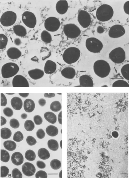
Figure 1. (A)Untreated control bac-teria of S. aureusWood 46 grown in Mueller-Hinton broth showing septa(s)
in dividing forms and EPS (arrows)
around the cells and in the intercellu-lar spaces. (B) EPS-free S. aureus
Wood 46 observed after the sonication treatment seem to be completely de-prived of EPS. (C) Concentration of the stripped-off EPS by ultracentrifu-gation, with one contaminating bac-terial cell. Bars = 0.5 urn.
ately fixed under EPS-free conditions for 18 hr with cold formaldehyde, rinsed, and incubated for a fur-ther 2 hr at 37 C in PBS supplemented with eifur-ther
200/0 TCF~ 100/0 guinea pig serum, or 5 mg of
hu-man serum albumin/ml; this incubation was fol-lowed by rinsing in PBS. The adherence assays used
4 x 10⁶cfu equivalents (as measured by
radioactiv-ity counts) with fibronectin-coated coverslips.
Results
Morphologic observations. We examined thin sections of bacterial cells from a 3-hr culture grown in a conventional broth medium (Mueller-Hinton broth) and found finely dispersed EPS either at-tached to the bacterial cell wall or located in the in-tercellular spaces.
To study EPS production by bacterial cells dur-ing the onset of experimental foreign body infection, we inoculated EPS-free bacteria into the TCE
Ac-cordingly, the 3-hr culture of S.aureusWood 46 was
treated by sonication (as indicated above) to produce EPS-free bacterial cells (figure 1, A). The EPS
sepa-rated from S. aureusby sonication and subsequent
low-speed centrifugation could be recovered as a pel-let after high-speed centrifugation, and we observed the EPS by transmission electron microscopy
(fig-ure 1, C). The EPS fragments stripped from S.aureus
by sonication and reincubated with EPS-free bacte-ria before the high-speed centrifugation did not reas-sociate with the bacterial cells (data not shown).
Under in vivo conditions of experimental foreign
body infection, EPS were rapidly produced by S.
au-reusWood 46 (figure 2). One hour (figure 2, A) or
•
•
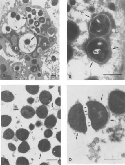
{",-Figure 2. (A) Neutrophilic granulo-cyte found in the infected TCF I hr af-ter inoculation of the EPS-free bacte-ria; ingested bacterial cells are sur-rounded by abundant electron-dense EPS(arrows)in the cytoplasmic vacu-oles(v);cells are surrounded by abun-dant electron-dense EPS(arrows) n =
neutrophil nucleus.(B)Abundant EPS (arrows) in bacterial cells ingested by a phagocyte 3 hr after the onset of in-fection. (C) In vitro production ofEPS byS. aureusWood 46 incubated with 20010 TCF for I hr; electron-dense ma-terial appears both around the cells and in the intercellular spaces(arrows). (D) S. aureusWood 46 incubated for 3 hr with TCF; EPS arc strongly attached to the original cell wall(arrows)but not to the newly formed wall of the septa of dividing cells(arrowheads).Bars =
528 Fa/cieri et 0/.
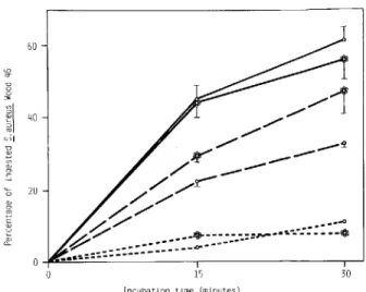
Figure 3. Phagocytic uptake ofS.aureus Wood 46 in-cubated with 106peritoneal exudate neutrophils/ml. Fixed,
radiolabeled EPS-free (0 ---0) or EPS-rich
( 0 - - 0) bacteria preopsonized for 15 min in 10070 guinea pig serum are compared with fixed, radiolabeled, EPS-free(0---0)or EPS-rich ( 0 - - 0 )bacteria preopsonized in 20070 TCE Control incubations were run in 10070 guinea pig serum and consisted of unopsonized, EPS-free(0- - - -0)and EPS-rich (0- - - -0) bac-teria. Data are mean (± SE) from three experiments.
cubated in either serum or TCF whose opsonic prop-erties had been destroyed by heating (figure 3).
Influence of EPS on the adherence ofS. aureus Wood 46 to fibronectin-coated covers/ips. When liveS.aureuswere tested in the adherence assay, EPS-rich bacteria preincubated in serum were 54% less
adherent(P
<
.01,n = 6) to fibronectin-coatedcov-erslips than were EPS-free S. aureus preincubated
in albumin. In contrast, EPS-rich bacteria precubated in TCF showed a slight, nonsignificant in-crease in adherence when compared with the free bacteria (figure 4, A). When rich or
EPS-free S.aureuswere fixed after preincubation (but
be-fore the adherence assay) with either TCF, serum, or albumin, the results were no different from those obtained with live bacteria (figure 4, B). As a final control experiment to insure that EPS were not directly involved in modifying adherence to fibro-nectin-coated coverslips, we fixed EPS-negative bac-teria before incubating them with either TCF, serum, or albumin; the differences in adherence were the same as those shown in figure 4, A and B (figure 4, C). Thus, adsorbing components from either
se-rum or TCF modified the adherence of S. aureus
Wood 46, but did not affect the production of EPS.
30
15
Incubation time (minutes)
/ / f
./' 1 / / ./'" --~11"
__
--/ '
/" / r
-/ -/
/ . / '
,..-:::. / ' . - • • - • • • ..0 ~Y ...-.-.--."....·~-·--·--o.f;;:-:.:::::::.:::::::..,..
-~..--_.
60 OJ to' C OJ OJ D OJ OJ 0' C '6 20three hours (figure 2, B) after EPS-freeS.aureuswas
inoculated into tissue cages, coated bacteria were uni-formly present in TCF aspirated from infected animals. The EPS-rich bacteria were present as free forms in TCF or were located inside phagocytic vesi-cles of neutrophils (figure 2, A). Particularly con-densed EPS together with the central bacterial nucleoid and the well-defined cell walls are shown
in figure 2, B.
To further define whether factors present in TCF contributed in a major way to production of EPS,
we used an in vitro system in which S. aureus was
incu bated in 20010 TCF in PBS, as indicated above. The initially EPS-free bacteria rapidly (i.e., after 1 hr and 3 hr) produced fresh EPS that were very simi-lar to those observed in TCF from experimentally infected animals (figure 2, C and D). An interesting finding of these in vitro experiments was the non-uniform attachment of EPS to the bacteria: in divid-ing cells (figure 2, D), EPS were attached to the origi-nal cell wall but not to the newly formed wall of the septa. This observation militates against the hypoth-esis that a surface coat may be passively adsorbed onto the bacteria from the surrounding medium. Light microscopy confirmed the presence of a true, trypsin-resistant capsule that excluded India ink par-ticles. In contrast to incubation with TCF, in vitro
incubation of S.aureuswith human serum albumin
did not lead to the production of EPS (data not shown). Thus we could produce bacteria that were either rich in EPS (by using TCF) or devoid of it (al-bumin).
Inf/uence ofEPS on the susceptibility ofS.aureus to phagocytic ingestion. Before the phagocytic as-say, bacteria were fixed with formaldehyde to pre-vent further production of EPS. Fixed, coated or un-coated cells were preopsonized for 15 min at 37 C with either 10010 serum or 20010 TCF and then tested for uptake by neutrophils. The percentage of ingested S.aureusWood 46 (figure 3) was higher for bacteria preopsonized with serum than for those
preop-sonized with TCE EPS-rich and EPS-freeS.aureus
were ingested at the same rate when preopsonized with serum, but EPS-rich bacteria were ingested more rapidly (P< .05) than EPS-free bacteria when both were preopsonized with TCE Thus, the produc-tion of EPS did not inhibit - but in fact moderately
increased - the uptake of S. aureus Wood 46 by the
phagocytic cells. EPS-rich or EPS-free bacteria showed low, albeit measurable, uptake when
in-Discussion
Figure 4. Adherence of S. aureus Wood 46 to fibronectin-coated polymethylmethacrylate coverslips. Ra-diolabeled bacteria were tested either(A)under live con-ditions after incubation in either human serum albumin (0), serum(E]),or TCF(~);or fixed with 0.5070 formal-dehyde overnight after(B)or before(C)preincubation in one of the three media. Data are mean(± SE) from six, three, and three experiments (A, B, C, respectively).
This study documents EPS associated with S.aureus
Wood 46 during growth in various media. The EPS was predominantly of a capsular type and, in some situations, was found in intercellular spaces as well. It was rapidly and abundantly produced in vivo dur-ing the onset of experimental foreign body infection. Such production could also be achieved by simple
in vitro exposure of S.aureusto TCE We also found
that bacteria grown in the presence of albumin did not produce EPS, a result providing us with an im-portant negative control. The role of these EPS in
the adherence properties of S.aureusand in its
sus-ceptibility to opsonophagocytosis by neutrophils was tested and found to be negligible by a variety of tech-niques. Our results suggest that some humoral fac-tors found in TCF are unrelated to the production
of EPS and may increase the virulence of S.aureus
Wood 46 in foreign body infections by enhancing both bacterial adherence and resistance to phagocy-tosis.
The relation between EPS, as described in our study, and materials referred to as "glycocalyx" or "slime" by other authors [6-16] is presently unclear, because no detailed chemical or antigenic composi-tion of any of these materials is presently available. Even the distinction made on a morphological ba-sis between capsular and intercellular exopolymers
may disappear when their chemical composition is elucidated. Also, the biosynthesis of exopolymers must be defined to find some target-specific anti-biotics.
We evaluated the role of EPS in modifying ad-herence to fibronectin and susceptibility to phago-cytosis under a variety of experimental conditions and comparatively tested fixed and live bacteria. Formaldehyde-fixed bacterial cells have been repeat-edly used to immunize animals against EPS [30, 31], with minimal changes in bacterial surface proper-ties. The surprising result from both adherence and phagocytosis experiments performed with fixed and non-fixed bacteria was the absence of any
signifi-cant role for EPS; EPS-rich and EPS-free S.aureus
had almost identical properties in these assays. The exact mechanisms allowing EPS-rich and EPS-poor S.aureusWood 46 to be opsonized properly and to interact with fibronectin are still unknown. We have
previously reported that S.au reusWood 46 was
op-sonized predominantly by the alternative pathway
of complement activation [32]. The capsule of S.
au-reus has been reported to be highly permeable to
macromolecules such as immunoglobulins or com-plement components [33, 34]. These observations militate against the hypotheses that EPS may be a physical barrier between the bacterial and neutrophil receptors or between fibronectin and the putative
fibronectin receptors of S.aureusWood 46. Other
strains of S.aureus, particularly the protein
A-bear-ing clinical isolates, may behave differently from strain Wood 46.
Although our results and conclusions concerning the role of EPS in foreign body infections conflict with previous reports, which described the signifi-cant virulence of slime-producing bacteria in this clinical context [6-13], there were important meth-odological differences between previous observations and our experimental system. First, we used short periods of incubation (limited to a few hours) as op-posed to one to several days [6-13]. Second, our sam-pling was limited to the fluid phase incubated with the foreign body, whereas most of the other observations concerned surface-adherent bacteria. Third, strain and species differences may be impor-tant in both the quantity and the chemical structure of EPS. This is particularly relevant for the strains
of S. epidermidis, which were among the highest
slime-producing gram-positive bacteria found in catheter-related sepsis [6-9]. Thus, our experimen-© ® ® T -I
T
Tr
h
r
... [.» '.:::::: :,:,' 1::-: -1x10⁵ cg. 8x10~?-II
6x10~ vol ~x1O~ CD 6 2x1O~ E530
tal protocol does not exclude the possibility that in long-standing infections, EPS may contribute sig-nificantly to the persistence of the disease. However, we can state that EPS does not protect invading S. au reus bacteria, during the initial steps of foreign body infection, from recognition by neutrophils, nor does it influence bacterial adherence to fibronectin.
Finally, one aspect of our study concerns the role of TCF, a protein-rich inflammatory fluid in con-tact with the implanted foreign body [4], which de-creased the susceptibility of S. aureus to phagocytic uptake by neutrophils (figure 4). This result may be linked with previous observations showing a progres-sive decrease in opsonic properties of TCF during experimental foreign body infection; these proper-ties could be improved by incubating the bacteria with serum, either in vitro or in vivo [4]. Together, these results give further evidence that host defense mechanisms are altered in the vicinity of a foreign body and thus explain its high susceptibility to bac-terial infections.
References
1. Hirshman HP, Schurman OJ. Deep infections following to-tal hip replacement. In: Remington JS, Swartz MN, eds. Current clinical topics in infectious diseases. Vol. 3. New York: McGraw-Hill, 1982:206-17
2. Maki DG. Infections associated with intravascular lines. In: Remington JS, Swartz MN, eds. Current clinical topics in infectious diseases. Vol 3. New York: McGraw-Hill, 1982:309-63
3. Noble We. The production of subcutaneous staphylococcal skin lesions in mice. Br J Exp Pathol 1965;46:254-62 4. Zimmerli W, Waldvogel FA, Vaudaux P, Nydegger UE.
Patho-genesis of foreign body infection: description and charac-teristics of an animal model. J Infect Dis 1982;146:487-97 5. Zimmerli W, Lew PO, Waldvogel FA. Pathogenesis of for-eign body infection. Evidence for a local granulocyte de-fect. J Clin Invest 1984;73:1191-200
6. Bayston R, Penny SR. Excessive production of mucoid sub-stance in Staphylococcus SIlA: a possible factor in coloni-zation of Holter shunts. Dev Med Child Neurol 1972; 14(Suppl 27):25-8
7. Christensen GO, Simpson WA, Bisno AL, Beachey EH. Ad-herence of slime-producing strains of Staphylococcus epidermidis to smooth surfaces. Infect Immun 1982;
37:318-26
8. Peters G, Locci R, PulvererG. Microbial colonization of prosthetic devices. Il. Scanning electron microscopy of nat-urally infected intravenous catheters. Zentralbl Bakteriol Mikrobiol Hyg [B) 1981;173:293-9
9. Peters G, Locci R, Pulverer G. Adherence and growth of coagulase-negative staphylococci on surfaces of intrave-nous catheters. J Infect Dis 1982;146:479-82
Falcieri et al.
to. Gristina AG, Costerton JW. Bacterial adherence to bio-materials and tissue. J Bone Joint Surg [Br] 1985;67-A: 264-73
11. Costerton JW, Irvin RT, Cheng K-J. The bacterial glycocalyx in nature and disease. Annu Rev Microbiol 1981;35:299-324 12. Costerton JW, Irvin RT, Cheng K-J. The role of bacterial
sur-face structures in pathogenesis. CRC Crit Rev Microbiol 1981;8:303-38
13. Marrie TJ, Nelligan J, Costerton JW. A scanning and trans-mission electron microscopic study of an infected en-docardial pacemaker lead. Circulation 1982;66:1339-41 14. Schwarzmann S, Boring JR III. Antiphagocytic effect of slime
from a mucoid strain ofPseudomonas aeruginosa. Infect
Immun 1971;3:762-7
15. Nickel JC, Ruseska I, Wright JB, Costerton JW. Tobramy-cin resistance ofPseudomonas aeruginosa cells growing
as a biofilm on urinary catheter material. Antimicrob Agents Chemother 1985;27:619-24
16. Sutherland IW. Bacterial exopolysaccharides-their nature and production. In: Sutherland I, ed. Surface carbohy-drates of the prokaryotic cells. London: Academic Press, 1977:27-96
17. Geesey GG. Microbial exopolymers: ecological and economic considerations. ASM News 1982;48:9-14
18. Hawiger J, Hammond OK, Timmons S. Human fibrinogen possesses binding site for staphylococci on Au and BI} poly-peptide chains. Nature 1975;258:643-5
19. Lopes JD, Dos Reis M, Brentani RR. Presence of laminin receptors inStaphylococcus aureus. Science 1985;229:275-7
20. Kuusela P. Fibronectin binds toStaphylococcus aureus.
Na-ture 1978;276:718-20
21. Mosher OF, Proctor RA. Binding and factor Xfl L-mediated cross-linking of a 27-kilodalton fragment of fibronectin to Staphylococcus aureus. Science 1980;209:927-9
22. Vercellotti GM, Lussenhop 0, Peterson PK, Furcht LT, McCarthy JB, Jacob HS, Moldow CF. Bacterial adher-ence to fibronectin and endothelial cells: a possible mech-anism for bacterial tissue tropism. J Lab Clin Med 1984; 103:34-43
23. Vaudaux P, Suzuki R, Waldvogel FA, Morgenthaler 11, Nydegger UE. Foreign body infection: role of fibronectin as a ligand for the adherence ofStaphylococcus aureus.
J Infect Dis 1984;150:546-53
24. Vaudaux PE, Waldvogel FA, Morgenthaler J J, Nydegger UE. Adsorption of fibronectin onto polymethylmethacrylate and promotion ofStaphylococcus aureus adherence.
In-fect Immun 1984;45:768-74
25. Kronvall G, Quie PG, Williams RC Jr. Quantitation of staphylococcal protein A: determination of equilibrium constant and number of protein A residues on bacteria. J Immunol 1970;104:273-8
26. Bayer ME, Thurow H. Polysaccharide capsule ofEscherichia coli: microscope study of its size, structure, and sites of
synthesis. J Bacteriol 1977;130:911-36
27. Reynolds ES. The use of lead citrate at high pH as an electron-opaque stain in electron microscopy. J Cell Bioi 1963; 17:208-12
28. Tan JS, Watanakunakorn C, Phair JP. A modified assay of neutrophil function: use of lysostaphin to differentiate
defective phagocytosis from impaired intracellular killing. J Lab Clin Med 1971;78:316-22
29. Vaudaux P, Waldvogel FA. Gentamicin antibacterial activ-ity in the presence of human polymorphonuclear leuko-cytes. Antimicrob Agents Chemother 1979;16:743-9 30. Brock JH, ReiterB.Chemical and biological properties of
extracellular slime produced byStaphylococcus aureus
grown in high-carbohydrate, high-salt medium. Infect Im-mun 1976;13:653-60
31. Caputy GG, Costerton JW. Morphological examination of the glycocalyces ofStaphylococcus aureus strains Wiley
and Smith. Infect Immun 1982;36:759-67
32. Lew PO, Zubler R, Vaudaux P, Farquet 11, Waldvogel FA,
Lambert P-H. Decreased heat-labile opsonic activity and complement levels associated with evidence of C3 break-down products in infected pleural effusions. J Clin Invest 1979;63:326-34
33. Wilkinson BJ, Sisson SP, Kim Y, Peterson PK. Localization of the third component of complement on the cell wall of encapsulated Staphylococcus aureus M: implications
for the mechanism of resistance to phagocytosis. Infect Immun 1979;26:1159-63
34. King BF, Wilkinson BJ. Binding of human immunoglobulin G to protein A in encapsulatedStaphylococcus aureus.