Nephrol Dial Transplant (2013) 28: 1031–1038 doi: 10.1093/ndt/gfs549
Advance Access publication 8 January 2013
Original Articles
Impact of graft loss among kidney diseases with a high risk of
post-transplant recurrence in the paediatric population
Karlijn J. Van Stralen
1,
Enrico Verrina
2,
Mirco Belingheri
3,
Jan Dudley
4,
Jiří Dušek
5,
Ryszard Grenda
6,
Marie-Alice Macher
7,
Zvonimir Puretic
8,
Jacek Rubic
6,
Sarunas Rudaitis
9,
Christoph Rudin
10and Franz Schaefer
11Kitty J. Jager
1on behalf of the ESPN/ERA-EDTA
Registry
1ESPN/ERA-EDTA Registry, Department of Medical Informatics, Academic Medical Center, Amsterdam, the Netherlands,
2Department of Paediatric Nephrology, G Gaslini Hospital, Genoa, Italy,
3Pediatric Nephrology Unit, IRCCS Policlinico Maggiore Hospital Foundation, Milan, Italy,
4Department of Paediatric Nephrology, Bristol Royal Hospital for Children, Bristol, UK,
5Department of Pediatrics, University Hospital Motol, Charles University, 2nd Medical School, Prague, Czech Republic, 6Department of Nephrology & Kidney Transplantation, The Children’s Memorial Health Institute, Warsaw, Poland,
7Pediatric Nephrology Unit, Robert Debré Hospital, Paris, France, 8Pediatric Dialysis Unit, University Hospital Centre Zagreb, Zagreb, Croatia,
9Clinic of Children Diseases, Hospital of Lithuanian University of Health Sciences Kaunas Clinics, Kaunas, Lithuania,
10Department of Paediatric Nephrology, University Children’s Hospital Basel, Basel, Switzerland and
11Department of Paediatric Nephrology, University of Heidelberg Center for Pediatrics and Adolescent Medicine, Heidelberg, Germany
Correspondence and offprint requests to: ESPN/ERA-EDTA Registry; E-mail: k.j.vanstralen@amc.uva.nl
Keywords: disease recurrence, graft survival, kidney trans-plantation, paediatrics
A B S T R AC T
Background. Some kidney diseases tend to recur in the renal allograft after transplantation. We studied the risk of graft loss among primary renal diseases known for their high risk of recurrence and compared it with that of patients with hy-poplasia and/or dysplasia.
Methods. Within the European Society of Paediatric Ne-phrology and European Renal Association and European Dialysis and Transplant Association (ESPN/ERA-EDTA) reg-istry, we studied children from 33 countries who received a
kidney transplant before the age of 20 between 1990 and 2009. Patients were censored after 5 years of follow-up and cumulative incidence competing risk analysis was used to cal-culate survival curves.
Results.Patients with focal and segmental glomerulosclerosis (FSGS), haemolytic uraemic syndrome (HUS), membrano-proliferative glomerulonephritis Type I or II (MPGN), IgA nephropathy or Henoch Schönlein Purpura (HSP/IgA) or systemic lupus erythomatosus (SLE) underwent pre-emptive transplantation significantly less often than patients with hy-poplasia and/or dysplasia. The rate of living donation was lower among patients with FSGS and SLE than in patients
with hypoplasia and/or dysplasia. In comparison with hypo-plasia and/or dyshypo-plasia patients with a risk of 14.4%, the 5-year risk of graft loss was significantly increased in patients with FSGS (25.7%) and MPGN (32.4%) while it was not sig-nificantly increased in children with HUS (18.9%), HSP/IgA (16.3%) or SLE (20.3%). One-year graft survival strongly im-proved among HUS patients from 17.1% in 1995–1999 to 3.6% in 2005–2009 and was not accompanied by a decrease in the number of transplantations.
Conclusion.The risk of graft loss is increased among specific causes of renal failure with a high risk of post-transplant re-currence. It seems likely that, due to anticipation of such risk, physicians perform less pre-emptive transplantation and provide fewer grafts from living related donors in patients with these conditions. Improved risk stratification by phys-icians, resulting in the identification of patients with HUS at higher or lower risk of recurrence, might explain the much improved graft survival rates.
INTRODUCTIO N
Renal transplantation is the treatment of choice for children and adults with end-stage renal disease. However, it is not always an ultimate solution, as some kidney diseases tend to recur in the renal allograft and disease recurrence is more fre-quent in children than in adults [1]. One could make a dis-tinction between kidney diseases that tend to have a ‘full blown’ recurrence leading to graft loss, and kidney diseases that recur without apparent increased risk of graft loss [1]. Although several studies reported an increased risk of graft loss among very rare specific kidney diseases, they were usually based on a very limited number of patients and were compared with the general risk of graft loss.
In the present paper we describe the real-life risk of graft loss among kidney diseases known to be associated with a high risk of disease recurrence and subsequent graft loss [idiopathic focal and segmental glomerulosclerosis (FSGS), haemolytic uraemic syndrome (HUS), membranoproliferative glomerulo-nephritis (MPGN) and mesangiocapillary glomeruloglomerulo-nephritis Type I and II (dense deposit disease)] together with those kidney diseases with a high risk of disease recurrence but in which the risk of graft loss is expected to be low(er), namely IgA nephropathy and Henoch Schönlein Purpura (HSP/IgA) and systemic lupus erythomatosus (SLE). We compared these risks with those of patients with hypoplasia or dysplasia (ex-cluding patients with obstructive uropathy or reflux), who do not suffer from disease recurrence. We wanted to determine (i) patient characteristics at the time of transplantation for individ-uals with specific kidney diseases; (ii) the risk of graft loss for these kidney diseases and (iii) subgroups that were at higher or lower risk and any recent time trend in the risk of graft loss.
M AT E R I A L S A N D M E T H O D S
Within the framework of the European Society of Paediatric Nephrology and European Renal Association and European
Dialysis and Transplant Association (ESPN/ERA-EDTA) Registry, countries collected individual patient data on the date of birth, gender, treatment modality at the start of renal replacement therapy (RRT) and changes in RRT modality [2]. Patients aged 20 years or less who received a transplant between 1 January 1990 and 31 December 2009 were in-cluded in the analysis. Austria, Belgium, Belarus, Bulgaria, Croatia, Czech Republic, Denmark, Estonia, Finland, France, FYR of Macedonia, Greece, Hungary, Iceland, Italy, Latvia, Lithuania, Montenegro, the Netherlands, Norway, Poland, Portugal, Romania, Russia, Serbia, Slovakia, Slovenia, Spain, Sweden, Switzerland, Turkey, Ukraine and the UK all con-tributed to the study. To avoid a selection bias among patients, we limited our analyses to time periods for which countries contributed complete data. The kidney diseases were coded according to the ERA-EDTA coding system [3]: FSGS (Code 11), HUS (Code 88), MPGN Type I (Code 15), MPGN Type II (Code 13), HSP/IgA (Code 12 and 85), SLE (Code 84), hypoplasia and/or dysplasia (Codes 60, 61, 63, and 66). In the ESPN/ERA-EDTA registry, insufficient data were available on the recurrence of the disease as the cause of graft loss. We assumed that any additional risk of graft loss among diseases known for their increased risk of disease re-currence, when compared with that among patients with hy-poplasia and/or dysplasia would be due to disease recurrence. Patients were censored after living with a transplant at the end of the observation period, reaching 31 December 2009, being transferred to a centre for treatment of adult patients without information on the follow-up or after death without prior dialysis (assumed with a functioning graft).
Analyses
Analyses were performed using SAS 9.2. Differences between categorical variables were calculated using the χ2 test, whereas differences between continuous variables were calculated using a t-test. The associations between patient characteristics at the time of transplantation and the likeli-hood of transplantation were calculated using logistic regression analyses, resulting in odds ratios (ORs) and 95% confidence intervals (95% CIs). To study 1- and 5-year graft survival and to determine the percentage of graft survival, survival curves were calculated taking the competing event death into account by using cumulative incidence competing risk analysis [4]. In this analysis, significance was determined using P-values calculated according to Pepe and Mori [5]. Trends over time were only calculated among those countries providing complete follow-up data between 1995 and 2009 and only those causes of renal failure were selected in whom at least 20 patients per era received a renal allograft. Cox-regression models were used to calculate the risk of graft loss at 1 and 5 years. The obtained hazard ratios (HRs) were ad-justed for age at the start of RRT, age at transplantation, gender and era of transplantation. For different subgroups, the Cox-regression analyses were repeated. A sensitivity ana-lyses was performed including adjustment for living versus deceased donation. P-values of <0.05 were considered as stat-istically significant.
ORIGINAL
R E S U LT S
In the ESPN/ERA-EDTA registry between 1 January 1990 and 31 December 2009, 5892 patients with end-stage renal failure received a renal transplant. The 3937 patients who suf-fered from other causes of renal failure than the ones under study were excluded. Therefore, 1955 patients were included in the present analyses. Patient characteristics are shown in Table1.
Characteristics at time of transplantation
Patients with a high risk of recurrence of their underlying condition underwent pre-emptive transplantation signifi-cantly less frequently than those with hypoplasia and/or dys-plasia, even after adjustment for age, gender and time period (Table 2). Compared with children with hypoplasia and/or dysplasia, patients with FSGS were 74% less likely, those with HUS 52% less likely, those with MPGN 80% less likely and those with HSP/IgA 68% less likely to receive a transplant pre-emptively when compared with receiving a transplant while being on dialysis for more than a year. However, when a patient started on dialysis, there were no differences between the different causes of renal failure with respect to receiving a transplant after short-term dialysis or dialysis for more or less than 1 year, except among patients with SLE. In addition, patients with FSGS and SLE received a transplant from a deceased donor significantly more often than from a living donor (OR 1.65, 95% CI 1.16–2.34, respectively, 3.50 95% CI 1.14–10.8) when compared with children with hypo-plasia and/or dyshypo-plasia.
Risk of graft loss
Patients with FSGS had the highest risk of graft loss in the first year after transplantation namely 14.7%, Table3. Patients with other kidney diseases lost their graft in 6.8% (HSP/IgA) and 10.1% (MPGN) of the cases, which was not significantly different from those with hypoplasia and/or dysplasia (6.4%), Figure1.
After 5 years, 32.4% of the patients with MPGN had suf-fered from a graft loss, which was considerably higher than among those with hypoplasia and/or dysplasia (14.4%), Table4, while the risk among patients with FSGS was 25.7%. The patterns of graft loss differed between the different causes of renal failure, while the risks of graft loss for example due to FSGS (Figure 1A) was very high in thefirst year and were more stable thereafter, the risk of graft loss in MPGN was consistently high over time (Figure1C).
Focal and segmental glomerulosclerosis
Patients with FSGS who started RRT during puberty (>12) had a significantly higher 5-year risk of graft loss (32.4%) when compared with those who started RRT before the age of 6 (17.1%), Figure1A. Patients with FSGS who started RRT between the ages of 6 and 12 had an intermediate, but signi fi-cantly increased risk of graft loss (27.7%). Relative risks for the different age groups when compared with their peers of the same age with hypoplasia and/or dysplasia resulted in
HRs of 1.4 for the youngest, 1.7 for those between 6–12 and 3.2 for the oldest age group (Table 4). Among patients with FSGS who received a pre-emptive transplantation the risks of graft loss were very similar (5.8% at 1 year and 22% at 5 years) when compared with the overall FSGS group; however, numbers were small.
Haemolytic uraemic syndrome
The 5-year risk of graft loss among patients with HUS was 18.9%. Patients with HUS who started RRT before the age of 6 had a higher risk (21.9%) of graft loss at 5 years than those who had started thereafter (16.5%), Figure1B. The risk of 1-year graft loss significantly decreased among patients over time (17.1% in 1995–1999 to 3.6% in 2005–2009, P = 0.03). This decrease was not associated with a change in the number of transplanted patients in absolute terms or relative to the number of patients with hypoplasia and/or dysplasia.
Membranoproliferative glomerulonephritis
When studying MPGN Type I and Type II separately, we found that the risk of 5-year graft loss was 23.5% for MPGN Type I and 67.5% among patients with Type II, Figure 1C. Although the rates between both types were sig-nificantly different from patients with hypoplasia and/or dys-plasia, they were not significantly different from each other.
Systemic lupus erythomatosus
Patients with SLE had a slightly but not significantly in-creased risk of graft loss (20.3% at 5 years). However, after adjustment for the lower rate of living donation and pre-emptive transplantation, surprisingly the differences between SLE and hypoplasia and/or dysplasia patients were signifi-cant, showing a 3-fold increased risk of graft loss for SLE patients when compared with those with hypoplasia and/or dysplasia (Table4).
HSP/IgA nephropathy
Patients with HSP/IgA had no increased risk of graft loss when compared with patients with hypoplasia and/or dyspla-sia. There were no differences between patients with HSP alone when compared with those with IgA nephropathy.
D I S C U S S I O N
Patients with FSGS and MPGN had a significantly higher risk of graft loss when compared with patients with hypopla-sia and/or dysplahypopla-sia. There were large differences in the pattern of graft loss; while patients with FSGS mainly suffered from graft loss shortly after transplantation, patients with MPGN had a consistently high rate of graft loss independent from time since transplantation. The risk of graft loss was not increased for patients with HSP/IgA nephropathy. All patients received a pre-emptive transplant less often than patients with hypoplasia and/or dysplasia, and patients with FSGS or SLE received an organ from a living donor less often in comparison with patients with hypoplasia and/or dyspla-sia. We will discuss the results separately for each disease.
O R IGIN AL AR TICL ES
Focal and segmental glomerulosclerosis
After 5 years, over a quarter of the patients with FSGS had lost their renal allograft, which is equal to the fraction re-ported in previous studies, and very close to that rere-ported also among adult patients [6, 7]. There was a strong differ-ence in graft survival between patients who were below the age of 6 at the start of RRT and those who started during puberty. Very young patients with FSGS more often have genetic or congenital forms of FSGS, which might less often lead to a recurrence. Unfortunately, no information on the genetic background of the patients was available. Other studies have also suggested higher risks of graft loss among
adolescents than among younger patients, with a lower risk again among the adult population [1,8].
Haemolytic uraemic syndrome
Patients with HUS had an increased risk of graft failure. However, this increased risk only reached significance in the 1994–1999 period. Since then there has been a big improve-ment in graft survival. Recommendations not to perform transplantations (from living related donors) in patients with atypical HUS [9] may have resulted in a selection towards more patients with typical HUS. Furthermore, genetic screen-ing of patients with atypical HUS for abnormalities in specific
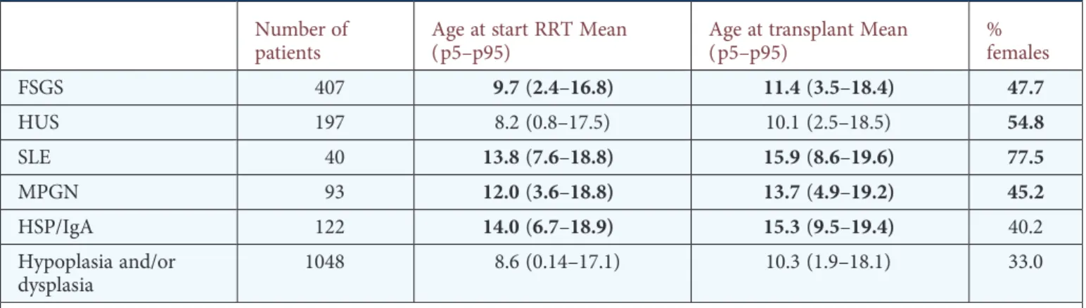
Table 1. Baseline characteristics
Number of patients
Age at start RRT Mean (p5–p95)
Age at transplant Mean (p5–p95) % females FSGS 407 9.7(2.4–16.8) 11.4(3.5–18.4) 47.7 HUS 197 8.2 (0.8–17.5) 10.1 (2.5–18.5) 54.8 SLE 40 13.8(7.6–18.8) 15.9(8.6–19.6) 77.5 MPGN 93 12.0(3.6–18.8) 13.7(4.9–19.2) 45.2 HSP/IgA 122 14.0(6.7–18.9) 15.3(9.5–19.4) 40.2 Hypoplasia and/or dysplasia 1048 8.6 (0.14–17.1) 10.3 (1.9–18.1) 33.0
Bold values denote significant difference from hypoplasia and/or dysplasia (P < 0.05).
FSGS, focal and segmental glomerulosclerosis; HUS, haemolytic uraemic syndrome; SLE, systemic lupus erythomatosus; MPGN, membranoproliferative glomerulonephritis type I or II; HSP/IgA, IgA nephropathy or Henoch Schönlein Purpura.
Table 2. Transplant characteristics for patients with different causes of renal failure, and odds
ratios for (i) receiving a transplant from a deceased versus a living donor; (ii) receiving a
transplant pre-emptively compared with >1 year on dialysis or (iii) within a year on dialysis
compared with >1 year on dialysis, adjusted for age at start of RRT, gender and period
Percentages Odds ratios % Living
donora
% Pre-emptive Tx
Deceased versus living donora,b
Pre-emptive txb Tx after dialysis <1 yearb FSGS 29.3 9.1 1.65(1.16–2.34) 0.26(0.18–0.39) 0.99 (0.76–1.33) HUS 34.7 12.2 1.22 (0.80–1.86) 0.48(0.29–0.77) 1.17 (0.81–1.68) SLE 16.7 10.0 3.50(1.14–10.8) 0.20(0.07–0.60) 0.36(0.16–0.82) MPGN 45.7 8.6 0.82 (0.44–1.53) 0.22(0.10–0.48) 0.89 (0.55–1.45) HSP/IgA 43.8 13.1 1.00 (0.60–1.66) 0.32(0.18–0.59) 1.04 (0.68–1.62) Hypoplasia and/or dysplasia 41.0 26.1 1 1 1
Bold values denote significant difference from hypoplasia and/or dysplasia (P < 0.05).
OR, odds ratio; FSGS, focal and segmental glomerulosclerosis; HUS, haemolytic uraemic syndrome; SLE, systemic lupus erythomatosus; MPGN, membranoproliferative glomerulonephritis Type I or II; HSP/IgA, IgA nephropathy or Henoch Schönlein Purpura.
aInformation on living versus deceased donation was available for 55.6% of the patients. bAdjusted for age, gender and time period.
ORIGINAL
complement regulating proteins is increasingly performed and a genetic disease cause can now be identified in 50–70% of cases [10,11]. Since the number of patients with HUS re-ceiving a transplantation did not change, current practice will have most likely led to a improved risk stratification, resulting in a lower number of patients with a high-risk profile receiv-ing a transplant and an increased number of transplants in those with a low-risk profile.
Unfortunately, our data did not allow the distinction between patients with typical and atypical HUS. We tried to distinguish between these groups by studying two different age groups as traditionally young age at onset was an indi-cator for typical HUS. However, we found a higher risk of graft loss among patients who started RRT before the age of 6, while the risk among those who were above 6 years was very similar to that among patients with hypo- or dysplasia. Recently, Geerdink et al. also showed that nearly half of the patients with atypical HUS first manifest before the age of 5, questioning age as a valuable marker for the type of HUS
[12]. Early onset atypical HUS might be an even more aggres-sive form, explaining the high risk of graft loss.
Systemic lupus erythomatosus
There was a tendency towards increased graft loss among patients with SLE, although it was not significantly different from that of patients with hypoplasia and/or dysplasia. Our results were similar to those found in adults, but these varied widely as 5-year graft survival has been shown to be between 69 and 91% [13–15]. Nevertheless, the tendency towards an increased risk of graft loss was worse than expected given the potential for current immunosuppressive regimens to main-tain remission [1].
Membranoproliferative glomerulonephritis
MPGN is usually divided into Type I and II on the basis of ultrastructural features [16, 17]. It has been suggested, however, that differences in recurrence rates may be more related to the severity of the disease than to the underlying
Table 3. One-year risk of graft loss for specific causes of renal failure
Number at risk at 1 year 1-year graft loss (%) HR—1-year graft
loss HR—1-year graftlossa HR—1-year graftlossa,b
FSGS 304 14.7 2.44(1.71–3.48) 2.46(1.72–3.55) 1.75 (0.98–3.13) ≤6 years at the start of RRT 84 11.7 1.71 (0.86–3.40) 1.79 (0.84–3.82) 1.37 (0.42–4.45) 6–12 years at the start of RRT 121 14.2 1.93(1.08–3.40) 1.88(1.04–3.40) 2.03 (0.74–5.58) >12 years at the start of RRT 101 17.4 4.38(2.28–8.43) 4.61(2.35–9.04) 2.18 (0.80–5.94) HUS 149 7.5 1.17 (0.66–2.09) 1.18 (0.66–2.11) 1.17 (0.52–2.60) ≤6 years at the start of RRT 71 9.4 1.33 (0.60–2.96) 1.47 (0.62–3.48) 2.05 (0.63–6.68) >6 years at the start of RRT 79 5.9 0.97 (0.41–2.28) 1.00 (0.42–2.38) 0.64 (0.15–2.77) SLE 31 10.2 1.74 (0.64–4.79) 1.94 (0.59–5.48) 1.80 (0.51–6.42) MPGN 74 10.1 1.61 (0.80–3.24) 1.70 (0.84–3.46) 1.01 (0.30–3.41) MPGN Type I 48 13.3 2.15(1.03–3.87) 2.64(1.23–5.66) 2.05 (0.60–7.07) MPGN Type II 26 4.7 0.54 (0.07–3.87) 0.60 (0.08–4.33) Not possible to
estimate
HSP/IgA 97 6.8 1.10 (0.59–2.29) 1.22 (0.57–2.59) 0.71 (0.21–2.12)
Hypoplasia and/or dysplasia
869 6.4 1 1 1
Bold values denote significant difference from hypoplasia and/or dysplasia (P < 0.05).
HR, Hazard Ratio; FSGS, focal and segmental glomerulosclerosis; RRT, renal replacement therapy; HUS, haemolytic uraemic syndrome; SLE, systemic lupus erythomatosus; MPGN, membranoproliferative glomerulonephritis Type I or II; HSP/IgA, IgA nephropathy or Henoch Schönlein Purpura.
aAdjusted for age at start of RRT, age at transplantation, gender and era of transplantation.
bAdditionally adjusted for living versus deceased donation, information on living versus deceased donation was available for 55.6% of the
patients. O R IGIN AL AR TICL ES
ultrastructural features [17]. We found a higher risk of graft loss at 5 years among patients with dense deposit disease which was similar to that found in previous studies per-formed in the 1980s [18]. The low graft loss among patients with MPGN Type I was similar to that reported in other studies among adult patients [6, 19]. Differences between MPGN Type I and II did not reach statistical significance due to the small number of patients with MPGN Type II. Unlike the other causes of renal failure, the rate of graft loss did not improve over time in the years since transplantation. Previous studies suggested a 50% reduced risk of graft loss among patients with an organ from a living-related donor [18], which possibly also explains the slightly higher proportion of patients who received an organ from a living donor. However, adjustment for living versus deceased donation did not affect the risk estimates suggesting a very low advantage of a living donation among patients with MPGN when com-pared with that among patients with hypoplasia and/or dysplasia.
IgA nephropathy or Henoch Schönlein Purpura
Even though previous studies have reported recurrence rates of around 33% (range 9–61%) for patients with HSP/ IgA nephropathy [16], many studies, among adult patients, have shown that this does not affect graft survival [6, 20]. Our data also show that the risk of graft loss (16.3%) was very similar to that among patients with hypoplasia and/or dysplasia. Little is known about the rate of actual graft loss in these children, but in adults 5-year graft loss rates have been reported of 16–30% [20–23].
L IM ITAT I O N S
In this study, we investigated whether the risk of graft loss was increased among children suffering from causes of renal failure with a high rate of recurrence of the primary disease in the transplantations. We compared those risks with the risk among patients with hypoplasia and/or dysplasia. Although patients with hypoplasia and/or dysplasia do not have higher risks of graft loss due to disease recurrence, there might be other factors affecting the risk of graft loss. Many patients with hypoplasia and/or dysplasia are diagnosed an-tenatally and disease progression is typically slower and more predictable than patients with glomerulopathy, making these conditions more amenable to pre-emptive transplantation. There was quite a large group of patients in whom infor-mation on whether the received kidney was from a living or a deceased donor was missing. This might have affected the results of the sensitivity analyses.
In this study, we cannot say whether an increased risk of graft loss is actually related to a recurrence of the disease or whether other factors are involved. Conversely, we do not have information on disease recurrence without graft loss. Nevertheless, we believe that most of the additional risk when compared with hypoplasia and/or dysplasia will have been caused by a recurrence of the disease.
Finally, children become adults. Although for some of the patients we had follow-up data into adulthood, this infor-mation was not available for all. There has been debate on the presence of an increased graft loss after transfer to adult-hood [23, 24], although opposed by others [25], and if this
F I G U R E 1 : Graft survival among specific causes of end-stage renal disease compared with patients with hypoplasia and/or dysplasia. The curves have been adjusted for competing risks, namely death. (A and B) Separate curves for those children starting on RRT according to different age groups for patients with FSGS (A) and HUS (B). (C) Separate curves for patients with MPGN Type I and II (dense deposit disease). (D) Curves for patients with HSP/IgA and SLE. FSGS, focal and segmental glomerulosclerosis; RRT, renal replacement therapy; HUS, haemolytic uraemic syndrome; SLE, systemic lupus erythomatosus; MPGN, membranoproliferative glomerulonephritis Type I or II; HSP/IgA, IgA nephropathy or Henoch Schönlein Purpura.
ORIGINAL
were the case we might have underestimated the actual risk of graft loss for all causes of renal failure.
CON CL U SIO N
Graft loss remains a major problem in children living with a renal allograft. This large study was able to determine the actual rate of graft loss for many of the rare diseases in whom graft loss is feared to be high. In dense deposit disease, MPGN Type I and FSGS requiring RRT after the age of 6 the risk of graft loss is extremely high. Conversely, we were able to show that among some of these causes of renal failure the rate of graft loss is not increased.
AC K N OW L E D G E M E N T S
We would like to thank the patients, their parents and the staff of all the dialysis and transplant units who have contrib-uted data via their national registries and contact persons. We
would also like to thank P. Cochat, R. Coppo, D. Haffner and J. Harambat for being members of the ESPN/ERA-EDTA Registry Committee, D. Shitza, R. Kramar, R. Oberbauer, S. Baiko, A. Sukalo, K. van Hoeck, F. Collart, J.M. des Grottes, D. Pokrajac, D. Roussinov, D. Batinić, M. Lemac, J. Slavicek, T. Seeman, K. Vondrak, J. Heaf, U. Toots, P. Finne, C. Grönhagen-Riska, C. Holmberg, C. Couchoud, M. Lasalle, J. Harambat, E. Sahpazova, G. Gersdorf, C. Scholz, C. Barth, C. Scholz, B. Tönshoff, L. Plotznicki, G. A. Ioannidis, A. Kapogiannis, N. Printza, C. Stefanidis, G. Reusz, S. Túri, L. Szabó, T. Szabó, E. Kis, Zs. Györke, R. Palsson, V. Edvardsson, B. Gianolgio, T. de Palo, C. Pecoraro, S. Picca, S. Testa, E. Vidal, A. Jankauskiene, B. Punziene, S. Pavićević, V. Said-Conti, T. Leivestad, A. Bjerre, A. Zurowska, I. Zagozdzon, C. Mota, M. Almeida, C. Afonso, G. Mircescu, L. Garneata, E.A. Molchanova, N.A. Tomilina, B.T. Bikbov, M. Kostic, A. Peco-Antic, D. Kruscic, S. Puric, B. Spasojevic-Dimitrijeva, G. Milosevski-Lomic, D. Paripovic, L. Podracka, G. Kolvek, J. Buturovic-Ponikvar, G. Novljan, N. Battelino, A. Alonso Melgar and the Spanish Pediatric Registry, S. Schön, K.G. Prütz, B. Rippe,
Table 4. 5-year risk of graft loss for specific causes of renal failure
Number atrisk at 5 years
5-year
graftloss (%) HR—5-yeargraftloss HR—5-yeargraftlossa HR—5-yeargraftlossa,b
FSGS 138 25.7 2.09(1.59–2.75) 2.04(1.54–2.70) 2.20(1.37–3.52) ≤6 years at the start of RRT 51 17.1 1.46 (0.80–2.67) 1.36 (0.70–2.64) 1.01 (0.34–3.02) 6–12 years at the start of RRT 65 27.7 1.58(1.04–2.40) 1.72(1.13–2.64) 2.43(1.11–5.29) >12 years at the start of RRT 22 32.4 3.51(2.15–5.72) 3.17(1.92–5.24) 3.75(1.61–8.75) HUS 55 18.9 1.28 (0.84–1.94) 1.39 (0.91–2.12) 1.90 (1.05–3.44) ≤6 years at the start of RRT 29 21.9 1.83(1.00–3.34) 1.96(1.01–3.83) 3.43(1.33–8.89) >6 years at the start of RRT 26 16.5 0.96 (0.53–1.77) 1.08 (0.58–1.98) 0.88 (0.31–2.56) SLE 9 20.3 1.84 (0.86–3.94) 1.58 (0.72–3.44) 3.21(1.19–8.69) MPGN 27 32.4 2.36(1.51–3.70) 2.15(1.36–3.40) 2.73(1.31–5.71) MPGN Type I 25 23.5 1.95(1.10–3.46) 1.91(1.06–3.44) 2.76(1.14–6.67) MPGN Type II 2 67.5 3.34(1.75–6.38) 2.68(1.38–5.23) 2.70 (0.91–7.98) HSP/IgA 28 16.3 1.12 (0.64–1.94) 1.08 (0.61–1.91) 0.90 (0.34–2.37) Hypoplasia and / or dysplasia 401 14.4 1 1 1
Bold values denote significant difference from hypoplasia and/or dysplasia (P < 0.05).
HR, hazard ratio; FSGS, focal and segmental glomerulosclerosis; RRT, renal replacement therapy; HUS, haemolytic uraemic syndrome; SLE, systemic lupus erythomatosus; MPGN, membranoproliferative glomerulonephritis Type I or II; HSP/IgA, IgA nephropathy or Henoch Schönlein Purpura.
aAdjusted for age at start of RRT, age at transplantation, gender, and era of transplantation.
bAdditionally adjusted for living versus deceased donation, information on living versus deceased donation was available for 55.6% of the
patients. O R IGIN AL AR TICL ES
M. Herthelius, L. Backmän, S. Rossi, E. Maurer, B. Schnarwyler, G. Laube, C.E. Kuenhi, A. Hoitsma, A. Hemke, and all centres participating in the RICHQ study, R. Topaloglu, O. Sollemezoglu, A. Duzova, D. Ivanov, T. Feest and C. Inward for contributing data to the ESPN/ ERA-EDTA Registry.
F UN D I NG
The ESPN/ERA-EDTA registry is funded by the European Renal Association and European Dialysis and Transplant Association (ERA-EDTA) and the European Society of Pae-diatric Nephrology (ESPN).
CON F LICT O F INT E REST STAT EMENT None declared.
R E F E R E N C E S
1. Cochat P, Fargue S, Mestrallet G et al. Disease recurrence in paediatric renal transplantation. Pediatr Nephrol 2009; 24: 2097–2108
2. Tizard EJ, Verrina E, van Stralen KJ et al. Progress with the European Society for Paediatric Nephrology (ESPN)/ERA-EDTA Registry for children with established renal failure (ERF). Nephrol Dial Transplant 2009; 24: 2615–2617
3. van Dijk PC, Jager KJ, de Charro F et al. Renal replacement therapy in Europe: the results of a collaborative effort by the ERA-EDTA registry and six national or regional registries. Nephrol Dial Transplant 2001; 16: 1120–1129
4. Verduijn M, Grootendorst DC, Dekker FW et al. The analysis of competing events like cause-specific mortality—beware of the Kaplan–Meier method. Nephrol Dial Transplant 2011; 26: 56–61
5. Pepe MS, Mori M. Kaplan–Meier, marginal or conditional prob-ability curves in summarizing competing risk failure time data? Stat Med 1993; 12: 737–751
6. Briganti EM, Russ GR, McNeil JJ et al. Risk of renal allograft loss from recurrent glomerulonephritis. N Engl J Med 2002; 347: 103–109
7. Vinai M, Waber P, Seikaly MG. Recurrence of focal segmental glomerulosclerosis in renal allograft: an in-depth review. Pediatr Transplant 2010; 14: 314–325
8. Moroni G, Gallelli B, Quaglini S et al. Long-term outcome of renal transplantation in adults with focal segmental glomerulo-sclerosis. Transpl Int 2010; 23: 208–216
9. Zimmerhackl LB, Besbas N, Jungraithmayr T et al. Epidemiol-ogy, clinical presentation, and pathophysiology of atypical and recurrent hemolytic uremic syndrome. Semin Thromb Hemost 2006; 32: 113–120
10. Malina M, Roumenina LT, Seeman T et al. Genetics of hemoly-tic uremic syndromes. Presse Med 2012; 41: e105–e114
11. Loirat C, Fremeaux-Bacchi V. Hemolytic uremic syndrome re-currence after renal transplantation. Pediatr Transplant 2008; 12: 619–629
12. Geerdink LM, Westra D, van Wijk JA et al. Atypical hemolytic uremic syndrome in children: complement mutations and clini-cal characteristics. Pediatr Nephrol 2012; 27: 1283–1291 13. Oliveira CS, Oliveira I, Bacchiega AB et al. Renal transplantation
in lupus nephritis: a Brazilian cohort. Lupus 2012; 21: 570–574 14. Azevedo LS, Romao JE, Jr, Malheiros D et al. Renal
transplan-tation in systemic lupus erythematosus. A case control study of 45 patients. Nephrol Dial Transplant 1998; 13: 2894–2898 15. Ghafari A, Etemadi J, Ardalan MR. Renal transplantation in
patients with lupus nephritis: a single-center experience. Trans-plant Proc 2008; 40: 143–144
16. Ponticelli C, Glassock RJ. Posttransplant recurrence of primary glomerulonephritis. Clin J Am Soc Nephrol 2010; 5: 2363–2372 17. Little MA, Dupont P, Campbell E et al. Severity of primary
MPGN, rather than MPGN type, determines renal survival and post-transplantation recurrence risk. Kidney Int 2006; 69: 504–511
18. Braun MC, Stablein DM, Hamiwka LA et al. Recurrence of membranoproliferative glomerulonephritis type II in renal allo-grafts: The North American Pediatric Renal Transplant Coop-erative Study experience. J Am Soc Nephrol 2005; 16: 2225–2233
19. Lorenz EC, Sethi S, Leung N et al. Recurrent membranoproli-ferative glomerulonephritis after kidney transplantation. Kidney Int 2010; 77: 721–728
20. Andresdottir MB, Haasnoot GW, Persijn GG et al. HLA-B8, DR3: a new risk factor for graft failure after renal transplantation in patients with underlying immunoglobulin A nephropathy. Clin Transplant 2009; 23: 660–665
21. Kanaan N, Mourad G, Thervet E et al. Recurrence and graft loss after kidney transplantation for Henoch-Schönlein Purpura ne-phritis: a multicenter analysis. Clin J Am Soc Nephrol 2011; 6: 1768–1772
22. Samuel JP, Bell CS, Molony DA et al. Long-term outcome of renal transplantation patients with Henoch-Schönlein Purpura. Clin J Am Soc Nephrol 2011; 6: 2034–2040
23. Samuel SM, Nettel-Aguirre A, Hemmelgarn BR et al. Graft failure and adaptation period to adult healthcare centers in pedi-atric renal transplant patients. Transplantation 2011; 91: 1380–1385
24. van den Heuvel ME, van der Lee JH, Cornelissen EA et al. Tran-sition to the adult nephrologist does not induce acute renal transplant rejection. Nephrol Dial Transplant 2010; 25: 1662–1667
25. Kramer A, Stel VS, Tizard J et al. Characteristics and survival of young adults who started renal replacement therapy during childhood. Nephrol Dial Transplant 2009; 24: 926–933
Received for publication: 4.9.2012; Accepted in revised form: 24.10.2012
ORIGINAL