In vitro studies of core peptide‐bearing immunopotentiating reconstituted influenza virosomes as a non‐live prototype vaccine against hepatitis C virus
12
0
0
Texte intégral
(2) 616 Primary CTL induction with HCV peptide-bearing virosomes lymphocytes or other immunocompetent cells (14), (ii) it mediates the membrane fusion between virosomes and endosomal membranes of target cells, thereby facilitating antigen delivery (15), and (iii) it serves as a `recognition antigen', since most humans are primed to HA due to prior exposure to either in¯uenza disease or vaccination (16,17). Finally, the protective role of the virosomes is by far the most important one: as stated by Widmann et al. (18), soluble synthetic peptides have differential stabilities due to the activity of proteases which may have important consequences for attempts to manipulate the development of an immune response in vivo. In addition, using virosomes also avoids the induction of toleranceÐa phenomenon that has been observed under certain conditions by immunizing mice i.p. with soluble peptide (19). Several different vaccine trials based on virosomes have been performed so far, mainly focusing on in¯uenza or hepatitis A virus (HAV) with the aim to induce protective, neutralizing antibodies (20±24). In contrast to HAV and HBV viruse where protective antibody titers can be obtained after immunization, the role of immunoglobulins against HCV remains to be de®ned. Envelope antibodies would be the prime candidates for virus neutralization, but their presence in chronically infected patients as well as the observation that chimpanzees can be infected repeatedly by the identical HCV strains argue against an ef®cient humoral virus neutralization in vivo (25,26). On the other hand, antibody-mediated immune pressure seems to be directly correlated with an evolution of viral escape mutants during the course of chronic infection (27,28). As cytotoxic T lymphocytes (CTL) seem to be of greater importance in HCV elimination and since the core protein is the most highly conserved structural protein in the HCV genome (29), we are focusing on vaccine development based on virosomes containing either core 35±44 (IRIV-core 35) or core 131±140 (IRIV-core 131) inside. The function of the virosome is, on the one hand, to protect its contents from early protease degradation and, on the other hand, to deliver the core peptides into the MHC class I pathway. In our study we were able to show that it is possible to lyse EBV-transformed B cells (JY) as well as T1 and T2 cells preincubated with HCV core peptide-containing virosomes in a standard Cr-release assay using core peptide-speci®c effector cell lines. We analyzed the method of peptide presentation in the virosome model in more detail based on peptide-stripping experiments and on the post-Golgi inhibitor Brefeldin A (BFA), and using the EBVtransformed B cell line JY, the transporter associated with antigen processing (TAP)-competent cell line T1 and its TAPde®cient counterpart T2. Furthermore, using virosomes as vehicles to channel peptides into the cytoplasm of peripheral blood mononuclear cells (PBMC) from HCV± healthy human blood donors, we showed that correct epitopes can be presented on the cell surface, ef®ciently enough to prime naive CTL against HCV epitopes in vitro. Although primary CTL induction in vitro is also possible based on soluble peptide, the protective effect of the virosomal carrier will be of great importance for the performance of in vivo vaccine trials.. Methods Synthetic peptides The following synthetic peptides were used: two HCV corederived peptides, core 131±140 (ADLMGYIPLV) (30,31) and core 35±44 (YLLPRRGPRL) (30,31), and one control peptide, in¯uenza matrix 58±66 (GILGFVFTL) (32), used for re-stimulation of memory CTL, all obtained by Neosystem (Strasbourg, France) or the Institute for Biochemistry (Epalinges, Switzerland). Peptides were dissolved in DMSO (20 mg/ml) and diluted in PBS to a stock solution (1 mg/ml). IRIV Preparation of peptide-IRIV was performed according to the following procedure (Swiss Serum and Vaccine Institute, Bern, Switzerland): 32 mg of phosphatidylcholine (Lipoid, Ludwigshafen, Germany) and 8 mg of phosphatidylethanolamine (R. Berchtold, Biochemical Laboratory, Bern, Switzerland) were dissolved in 2 ml of PBS containing 100 mM of octaethyleneglycol (PBS/OEG) (Fluka Chemicals, Buchs, Switzerland). In¯uenza A/Singapore HA was puri®ed as described previously (33). A solution containing HA (4 mg) was centrifuged at 100,000 g for 30 min and the pellet was dissolved in 1 ml of PBS/OEG. Then 5 mg of the hydrophobic peptides was dissolved in 100 ml of DMSO and 900 ml of PBS/ OEG were added. The phospholipids, the HA solution and the peptide solution were mixed and sonicated for 1 min. This mixture was then centrifuged at 100,000 g for 1 h and the supernatant sterile ®ltered (0.22 mm). Virosomes were then formed by detergent removal using BioRad (Glottbrugge, Switzerland) SM Bio-beads. Quanti®cation of the peptide incorporation rate was performed using radioactively labeled 14C-tracer core 131 peptide. The actual peptide concentration incorporated was measured as 166 mg peptide/ml and all experiments performed with peptide-bearing virosomes were based on that quanti®cation. If proceeded before having performed the quanti®cation, experiments were then adapted to that standard concentration retrospectively, thereby explaining the unusual concentrations of virosome-derived peptide in some cases. To make sure that there was no `contamination' of soluble peptide outside of the virosomes, some batches of peptidecontaining virosomes were puri®ed by means of a G10 column in addition. Whether or not this procedure was performed, there was no difference in the results obtained in our experiments. Via HPLC we obtained another proof for virosomal purity: in the virosome formulations there was no peak seen corresponding to free peptide outside of the constructs (data not shown). Recombinant vaccinia virus (rVV) rVV virus encoding the full-length HCV core protein and part of the E1 protein vv9A (C/5¢ end of E1; Met1±Arg 39) was kindly provided by Dr Michael Houghton (Chiron, Emeryville, CA). For rVV infection 1 3 106 JY EBV-transformed B cells were put into a 15 ml tube and centrifuged. Then, the virus was added to the cell pellet in 1 ml with a concentration of 10 m.o.i. (corresponding to 1 3 107 p.f.u.) with 1 h incubation on a shaker at room temperature. Cells were centrifuged again and.
(3) Primary CTL induction with HCV peptide-bearing virosomes 617 resuspended in 1 ml of fresh medium and then incubated at 37°C for later use in a standard Cr-release assay. Cell lines JY, an EBV-transformed B cell line (HLA-A2.1, -B7, -Cw7, DR4, -Drw6, -Dpw2; kindly provided by Dr F. V. Chisari), T1 cells and their TAP-de®cient counterpart T2 cellsÐboth originally produced by fusing the T leukemic line CEM with the TAP± mutant LCL 721.174 (34,35)Ðwere cultured in RPMI 1640 medium (Sigma, St Louis, MO) enriched with L-glutamine (2 mM), penicillin (50 U/ml), streptomycin (50 mg/ml) and HEPES (5 mM), and containing 10% (v/v) heat-inactivated FCS (FCS medium). The T1 hybrid has normal antigen-presenting function, and expresses the HLA-A1, -A30 and -B8 alleles of the CEM parent and the HLA-A2.1 and -B51 alleles of the 721.174 parent; the T2 hybrid was derived from T1 by selecting for loss of the CEM HLA locus and resembles its 721.174 parent in being TAPde®cient and defective in antigen presentation, with a 70±80% reduction in surface HLA-A2.1 expression compared to T1 and virtually no detectable surface HLA-B51 (36). Induction of primary and secondary (recall stimulation) CTL Buffy coats or lymphaphereses of HLA-A2+ healthy human blood donors (negative for HIV, HBV and HCV serology) were separated on Ficoll-Paque density gradients (Pharmacia Biotech, Uppsala, Sweden) and washed at least 3 times in 50 ml of PBS containing 10% FCS medium. Then, as described previously (37), 4 3 106 PBMC were incubated either with synthetic peptide i.e. HCV core 131, core 35 or in¯uenza matrix 58 or with virosomes IRIV-core 131, IRIVcore 35 and IRIV-¯u containing core 131, core 35 and in¯uenza matrix peptide respectively in RPMI 1640 medium supplemented with L-glutamine (2 mM), penicillin (50 U/ml), streptomycin (50 mg/ml) and HEPES (5 mM), and containing 10% (v/v) of heat-inactivated human AB serum (AB medium) in 24-well plates for 3 days. On day 3 and weekly thereafter, 1 ml of complete AB medium supplemented with rIL-2 (20 U/ml; EuroCetus, Amsterdam, The Netherlands) was added to each well. On day 7 and weekly thereafter, cultures were re-stimulated with 106 irradiated (7400 rad) autologous feeder cells in 1 ml of AB medium containing rIL-2 (20 U/ml) and the corresponding peptide or virosomes. Cytotoxicity (CTL activity) of the in vitro stimulated PBMC was then determined in a standard 4-h Cr-release assay between 2 and 7 weeks of in vitro stimulation as indicated above. Generation of peptide-speci®c cell lines CTL bulk cultures with a peptide speci®city of >80% at an E:T ratio of ~20:1 obtained in our primary in vitro inductions as described above were depleted of CD4+ cells by negative selection according to the manufacturers instructions (Dynabeads; Dynal, Oslo, Norway) and plated at 100 effector cells/well in 96-well plates in FCS medium enriched with phytoHA (1 mg/ml) and rIL-2 (30 U/ml) containing irradiated (10400 rad) allogeneic PBMC (106/ml) and irradiated (22000 rad), peptide-pulsed (10 mg/ml; 1 h) JY EBV-B cells (105 cells/ ml).. Peptide-speci®c wells were expanded in 48-, 24- or 12-well plates and the resulting peptide-speci®c cell lines could be kept in culture over several months. Cr-release assay (cytotoxicity assay) Target cells (JY, T1 or T2; 106 per condition) were incubated with peptide (10 mg/ml) or virosomes (IRIV) containing peptide (varying concentrations) in FCS medium at 37°C and 5% CO2 overnightÐor in case of rVV-infected target cells the protocol was followed as indicated above under `rVV'. Preincubated target cells were then labeled with 100 mCi of 51Cr (Amersham, Little Chalfont, UK) for 1 h at 37°C and washed 4 times with cold PBS containing 10% FCS medium to remove non-incorporated Cr. Cytolytic activity was determined in a standard 4-h Cr-release assay using U-bottom 96well plates containing 2500 target cells. Depending on the number of effector cells available, E:T ratios between 60:1 and 1:1 were selected. Percent cytotoxicity was determined using the formula: 100 3 [(experimental release ± spontaneous release)/(maximum release ± spontaneous release)]. Maximum release was determined by lysis of targets in 2 N HCl, spontaneous release by lysis of targets in FCS medium. Spontaneous release was usually ~12%, but always <25% of maximum release. Speci®c lysis was calculated as difference between lysis of targets with peptide (or peptide-bearing virosomes) and targets without peptide (or naked virosomes). All results were based on duplicates with the exception of the analysis of primary induction cultures, where we only performed unicates due to lack of cells. For the peptide preincubation time curves, targets cells (JY or T2) were only incubated for the times indicated, i.e. a few hours or even fractions of an hour instead of the overnight incubation. Peptide `stripping' by mild acid elution After overnight incubation of T2 or JY target cells with peptide or virosomes at 37°C and 5% CO2 they were washed once with cold PBS containing 10% FCS medium and then put on ice in 1 ml of an ice-cold citric acid±Na2HPO4 buffer at pH 3.0, the mixture of equal volumes of 0.263 M citric acid and 0.123 M Na2HPO4 containing 1% BSA (290 mosmol/kg H2O), for 2 min (38). Immediately after the acid treatment, cells were washed with an excess of FCS medium to neutralize the acid buffer. Cytolytic activity against peptide-stripped targets was either measured directly after the mild acid treatment or after another 5-h incubation of the targets at 37°C and 5% CO2. In the case of virosome-preincubated targets this 5-h incubation was either performed with or without the addition of new peptidebearing virosomes. BFA treatment of JY and T2 target cells Target cells (JY and T2) were preincubated with various BFA concentrations ranging from 3 mg/ml to 10 ng/ml at 37°C for 4 h, before adding either soluble core 131 or virosomes containing core 131 for overnight incubation. The next morning, cells were collected and prepared for Crrelease assay..
(4) 618 Primary CTL induction with HCV peptide-bearing virosomes. Fig. 1. Speci®c cell lysis of targets preincubated with core 131 or IRIV-core 131. (A) JY or T2 cells were pulsed with 1 or 10 mg/ml of soluble (black bars) or 1 or 2 mg/ml of virosome-derived (gray bars) peptide overnight and tested against several different peptidespeci®c effector cell lines at the individual E:T ratios indicated. (B) T2 cells were either pulsed with 1 mg/ml of core 131 (solid circles) or 1.66 mg/ml of IRIV-core 131 (open circles) for various incubation times between 30 min and 6 h to obtain the kinetics of T2 target cell sensitization. Speci®c target cell lysis was then determined in standard 4-h Cr-release assays based on effector cell line D97a and an E:T ratio of 30:1. (C) JY cells were pulsed with the different core 131 (solid circles and triangles for D98d and D99a respectively) or IRIV-core 131 (open circles and triangles respectively) concentrations indicated and analyzed in a CTL assay at an E:T ratio of 20:1.. Results Target cell sensitization of JY, T1 and T2 cells First, two different target cell lines, i.e. JY, an EBV-transformed B cell line, and T2, a TAP-de®cient cell line, were pulsed with either soluble core 131 (1 or 10 mg/ml) or IRIV-core 131 (1 or 2 mg/ml) overnight. Several peptide-speci®c effector cell lines were then used at various E:T ratios indicated to test the ef®ciency of the target cell sensitization in standard 4-h Crrelease assays (Fig. 1a). All ®ve cell lines (D68, D76, D97, D98 and D99) tested against JY and/or T2 could lyse targets incubated with soluble and virosome-derived peptide, although varying in their absolute amounts of speci®c cell lysis. Both targets were able to present virosome-derived peptide on MHC class I via a TAP-independent internal pathway that was analyzed in further experiments. There was a general tendency towards a higher lysis in the case of soluble peptide. Secondly, a time curve based on T2 target cell sensitization with either core 131 (1 mg/ml) or IRIV-core 131 (1.66 mg/ml) was obtained to analyze the kinetics of soluble versus virosome-derived peptide based on CTL line D97a at an E:T. ratio of 30:1 (Fig. 1b). After 30 min of incubation there was maximum target cell lysis in the case of soluble peptide, whereas for IRIV-core 131 preincubated targets half of the maximum cell lysis was obtained after ~3 h and a lysis nearly comparable to the one obtained with soluble peptide was obtained after 6 h. Between 6 and 12 h of target sensitization there was a constant cell lysis as analyzed in an additional experiment (data not shown). The two different time curves obtained for an ef®cient peptide presentation on the target cell surface of both T2 and JY targets (data not shown) are the ®rst hint that two independent pathways of exogenous (soluble peptide) versus endogenous (IRIV-core 131) target cell sensitization are used. Third, we obtained a concentration curve using soluble and virosome-derived peptide ranging between 2 mg/ml and 20 ng/ml (Fig. 1c) to give a direct comparison of the ef®ciency of these two different forms of peptide on target cell lysis based on an E:T ratio of 20:1. There is a remarkable difference between the resulting cell lysis obtained for core 131 and IRIVcore 131 preincubated targets (Fig. 1c): compared to the lysis obtained based on soluble peptide, virosome-derived peptide at a similar concentration of 0.2 mg/ml led to a target cell killing.
(5) Primary CTL induction with HCV peptide-bearing virosomes 619. Fig. 2. JY, T1 and T2 target cell sensitization in comparison. The three different targets were either pulsed with 10 mg/ml of core 131 or 0.66 mg/ml of IRIV-core 131 overnight. Then, a 4-h Cr-release assay was performed based (A) on effector cell line D98b or (B) on cell line D99a at various E:T ratios ranging from 30:1 to 1:1.. reduction of 39% for D98d and 79% for D99a respectively. The varying peptide concentrations necessary to obtain similar target cell lysis with both effector cell lines might be partly explained due to differences in the TCR±peptide±MHC class I af®nity. Finally, to focus on TAP-bearing and -de®cient targets more directly, we analyzed JY, T2 and its TAP-bearing parental cell line T1 in parallel (Fig. 2a and b). Targets pulsed with core 131 were generally slightly more ef®ciently killed than those incubated with IRIV-core 131. The CTL line of donor D98 (Fig. 2a) again showed a higher peptide af®nity than that of donor D99 (Fig. 2b), as demonstrated in the higher killing of core 131-pulsed targets at low E:T ratios. Interestingly, the D99a CTL line was not able to lyse IRIV-core 131-sensitized TAP-competent T1 and JY targets as ef®ciently as TAPde®cient T2 cells. In fact, it seems that the lower the peptide af®nity of the effector cells is, the bigger the discrepancy between the killing potential of TAP-de®cient versus TAPbearing IRIV-core 131 preincubated targets, also observed in other experiments not shown.. peptide stripping (Fig. 3a). It appears that the whole internal peptide pool derived from the virosomes was presented at once, since there was no ef®cient reloading of the HLA-A2 molecules during the ongoing 5-h incubation after having washed the present peptide from the surface of the T2 cells, except after the addition of new peptide-bearing virosomes which led to a recovery of target cell lysis to even a slightly higher amount than before the acid treatment, comparable to the killing obtained with core 131 after peptide stripping. In parallel, we also performed another peptide stripping experiment based on JY target cells and three different effector cell lines (D68, D98d and D99) speci®c for core 131 using very low peptide concentrations (0.1 mg/ml) for target cell pulsing (Fig. 3b). Based on all three cell lines, there was a reduction of speci®c cell lysis after the mild acid elution by 50± 100% depending on the particular effector cells used. There seems to be a clear correlation between the peptide amount for target cell pulsing and the ef®ciency of the peptide stripping, which explains the data obtained in Fig. 3(a) with soluble peptide after the mild acid treatment.. Peptide stripping of T2 and JY cells pulsed with soluble core 131 versus IRIV-core 131. BFA and its in¯uence on peptide-presentation. T2 cells were incubated in the presence of either core 131 peptide (1 mg/ml) or IRIV-core 131 (1.66 mg/ml) overnight. Then, cells were treated with a mild acid solution as described in Methods and incubated for another 5 h with or without the addition of new virosomes. Based on the effector cell line D97a, there was a high lysis of peptide-pulsed targets at all E:T ratios indicated, which could only be reduced to about three-quarters of the original lysis after the acid treatment. In the case of IRIV-core 131, however, the original cell lysis was only about half the amount obtained with soluble peptide and it could be reduced to zero after. To make sure that the virosome-derived core 131 was actually presented via an endogenous pathway and not directly binding to surface class I molecules after leakage of the virosomal peptide carrier, in addition to the time curves performed in parallel with core 131 and IRIV-core 131 (Fig. 1b) we performed a post-Golgi blocking study with different BFA concentrations ranging from 10 ng/ml to 3 mg/ml. Targets were therefore ®rst incubated with varying amounts of BFA for 4 h and then either soluble core 131 (1 mg/ml) or IRIV-core 131 (1 mg/ml) was added in addition. BFA did not alter the percentage of speci®c lysis of targets pulsed with soluble peptide, neither in JY and based on CTL.
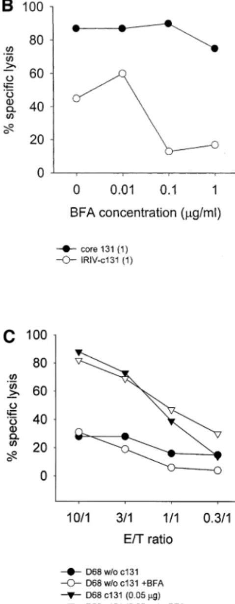
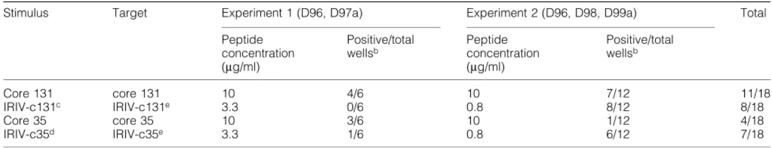
(6) 620 Primary CTL induction with HCV peptide-bearing virosomes. Fig. 3. In¯uence of mild acid treatment on target cell lysis. A peptide-stripping based on a mild acid treatment followed by another 5 h of incubation was performed with T2 cells either preincubated with 1 mg/ml of soluble core 131 or 1.66 mg/ml of IRIV-core 131 (A) or JY cells preincubated with 0.1 mg/ml of core 131 (B) overnight. (A) The Cr-release assay performed was based on CTL line D97a and E:T ratios ranging between 30:1 and 1:1 as indicated. Control cells pretreated with core 131 (solid circles) or IRIV-core 131 (solid squares) were compared to acid-treated T2 cells preincubated with core 131 (open circles) or IRIV-core 131 (open squares) and to acid-treated target cells that were incubated with new virosomes after the peptide stripping (open triangles) for another 5 h. (B) The assay was based on CTL lines D68, D98d and D99 using E:T ratios of 10:1, 3:1, and 1:1. Solid symbols represent control cells and open symbols acid-treated cells.. line D98d (Fig. 4a) nor in T2 cells and based on CTL line D68c (Fig. 4b). In the case of IRIV-core 131 preincubated targets both JY and T2 cells were less ef®ciently lysed in the presence of 0.1 mg/ml of BFA. As for JY targets (Fig. 4a) the cell lysis in the presence of 0.1 mg/ml of BFA decreased to half of the lysis in the absence of BFA and, with regard to T2 cells, this decrease was even more relevant (Fig. 4b). Higher BFA concentrations did not increase the negative effect on cell lysis. Importantly, even at very low peptide concentrations (0.05 mg/ml), BFA did not have any negative effect on cell killing based on soluble peptide (Fig. 4c). In conclusion, this is the proof that virosome-derived peptide is presented in part in a BFA-sensitive internal pathway, whereas exogenous soluble peptide is presented in a BFA-independent pathway. This, together with the results obtained from the time curve, indicates that the two pathways of peptide presentation for core 131 and IRIV-core 131 are independent. Primary induction based on core 131, core 35 and their virosome-derived counterparts PBMC obtained by four HCV± healthy blood donors (D96, D97, D98 and D99) were stimulated with soluble peptide (core 131 or core 35) or peptide-bearing virosomes (IRIV-core 131 or IRIV-core 35) in induction cultures of 4 3 106 cells for several weeks according to the protocol indicated. In a ®rst step, we compared the ef®ciency of soluble peptide (10 mg/ml) versus virosome-derived peptide (3.3 and 0.8 mg/ ml in experiments 1 and 2 respectively) based stimulation for primary induction in vitro. In Table 1 two experiments are. summarized as an overview. In experiment 1, four out of six cultures stimulated with core 131 were positive, whereas no positive culture could be obtained with IRIV-core 131 stimulation as determined in CTL assays. In the case of core 35, three out of six cultures were positive using the soluble peptide and one well was positive after IRIV-core 35 stimulation, shown in more detail in Fig. 5(a). In experiment 2, seven out of 12 cultures stimulated with core 131 were positive and eight out of 12 cultures were positive after IRIV-core 131 stimulation, whereas one single well was positive after core 35 stimulation and six of them were positive after IRIV-core 35 stimulus. The considerable variation in individual experiments and the differing numbers of positive cultures of various blood donors obtained is due to a very low precursor frequency of CTL speci®c for HCV epitopes in seronegative healthy human blood donorsÐa phenomenon that was already observed previously (37). Nevertheless, it was discovered that virosome-derived peptide can be as ef®cient as soluble peptide to prime CTL in vitro. Indeed, the importance of these results will be more obvious with respect to the situation in vivo focusing primarily on humans where the use of a suitable adjuvant and delivery system for a peptidebased vaccine is crucial. The effect of priming was dose dependent, since no positive cultures were obtained using 10 times lower IRIV-core 131 or IRIV-core 35 concentrations (data not shown). The kinetics of primary versus secondary stimulation is shown in Fig. 5(a and b). In vitro generation of CTL to in¯uenza peptide (secondary response) as well as to HCV core 131 or IRIV-core 131 and core 35 or IRIV-core 35 respectively are.
(7) Primary CTL induction with HCV peptide-bearing virosomes 621 presented in parallel (Fig. 5a and b). The data shown represent typical examples out of the results obtained in soluble peptide- or IRIV-peptide-induced cultures. As indicated, there is not a big difference between the time needed for a successful priming of an induction culture for a primary CTL response based on soluble and virosome-derived peptide in vitro. However, there is a big difference between a primary and a secondary in vitro stimulation, as shown in the curves obtained for ourin¯uenza control peptide that had its peak around week 3 or 4, which was 2±3 weeks earlier compared to the peak response obtained with core 35 or core 131. Primary induction compared to secondary stimulation in vitro based on virosomes In a second step, we compared the responses of primary induction cultures induced with IRIV-core 131 or IRIV-core 35 to the secondary responses induced with IRIV-¯u based on PBMC obtained by three different HCV± blood donors D84, D85 and D98 (Table 2). As for memory responses induced with IRIV-¯u stimulation, we measured CTL activities between 2 and 4 weeks of stimulation, whereas in the case of primary in vitro induction, we checked for lysis of targets between 4 and 6 weeks of stimulation. Concerning IRIV-¯u stimulation, all four replicates of all three donors were positive at the ®rst time point checked as expected for a secondary stimulation based on an in¯uenza virus-derived peptide that is well known by the immune system of most individuals. The following week, most of them had an even higher lysis potential which decreased after 1 month of in vitro stimulation. In the case of IRIV-core 131, all replicates of donor D98 showed peptide-speci®c lysis after 4 weeks of stimulation, reaching a maximum after 5 or 6 weeks and progressively loosing their speci®city again (data not indicated). Unfortunately, no positive cultures were obtained against IRIV-core 35 in the three donors evaluated in this assay, which has again to be attributed to a very low precursor frequency, thereby reducing the chances of bearing peptide-speci®c precursors in each individual induction culture well. After IRIV-core 131 and IRIV-¯u stimulation targets preincubated with soluble and virosome-derived peptide could be lysed ef®ciently, thereby pointing to the fact that the effector cells were really peptide speci®c. Then, we wanted to analyze if there was any correlation between different IRIV-¯u concentrations and the actual time of stimulation needed to obtain positive induction cultures based on PBMC from donor D96. As shown in Table 3, a higher IRIV-¯u amount led to positive cultures faster: stimulation of cultures with 1.66 mg/ml of IRIV-¯u led to three positive. Fig. 4. In¯uence of BFA on peptide presentation in (A) JY and (B) T2 target cells. Targets were preincubated either in the absence or the presence of various BFA concentrations ranging between 10 ng/ml and 3 mg/ml for 4 h and then either 1 mg/ml of core 131 (solid circles) or 1 mg/ml of IRIV-core 131 (open circles) was added for an overnight incubation. In another experiment (C), JY cells were pulsed with a very low peptide concentration of 0.05 mg/ml either in the presence of absence of 0.1 mg/ml of BFA. Percentage of cytotoxicity was determined in a standard Cr-release assay based on (A) CTL line D98d at an E:T ratio of 20:1, (B) CTL line D68c at an E:T ratio of 10:1 and (C) CTL line D68 at various E:T ratios indicated..
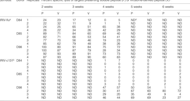
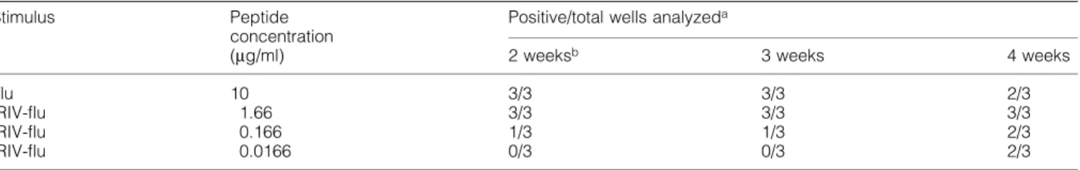
(8) 622 Primary CTL induction with HCV peptide-bearing virosomes Table 1. Primary in vitro induction of HCV peptide speci®c CTL with soluble versus virosome-derived peptide Stimulus. Core 131 IRIV-c131c Core 35 IRIV-c35d. Target. core 131 IRIV-c131e core 35 IRIV-c35e. Experiment 1 (D96, D97a). Experiment 2 (D96, D98, D99a). Peptide concentration (mg/ml). Positive/total wellsb. Peptide concentration (mg/ml). Positive/total wellsb. 10 3.3 10 3.3. 4/6 0/6 3/6 1/6. 10 0.8 10 0.8. 7/12 8/12 1/12 6/12. Total. 11/18 8/18 4/18 7/18. aDifferent. blood donors tested. was determined as a speci®c target cell lysis >15%. cIRIV-c131 = virosomes containing core 131 (ADLMGYIPLV). dIRIV-c35 = virosomes containing core 35 (YLLPRRGPRL). ePositive responses were also obtained against targets pulsed with core 131 or core 35 respectively. bPositive. out of three cultures after 2 weeks, whereas the lower amounts such as 0.166 and 0.0166 mg/ml led to one and zero positive out of three cultures respectively. Finally, after 4 weeks of stimulation, there were two positive cultures obtained after both of the lower stimuli. Recognition of endogenously processed core 131 peptide by epitope-speci®c CTL To determine whether in vitro primed CTL were not only able to lyse target cells preincubated with soluble core 131 or peptide-bearing virosomes, but could also recognize endogenously processed peptide, we performed Cr-release assays using rVV9A encoding the full-length HCV core protein. The experiment was based on both donor D76 and D98 effector cell lines and JY targets infected with rVV9A leading to speci®c target cell killing of nearly 40% at an E:T ratio of 40:1 based on donor D98 effector cell line (Fig. 6).. Discussion. Fig. 5. Primary in vitro induction using (A) core 131 or IRIV-core 131 and (B) core 35 or IRIV-core 35 respectively against secondary in vitro stimulation. PBMC (4 3 106) were stimulated with either the corresponding soluble or virosome-derived peptide. Percentage of cytotoxicity was then determined after various weeks of stimulation against JY targets. Both, (A) and (B) represent typical examples out of the results shown in Table 1.. The aim of this study was to analyze HCV core peptidebearing IRIV in vitro as a novel non-live vaccine approach to induce primary CTL against two well-characterized HLAA*0201 binding epitopes of core, the best conserved HCV protein representing also the main target of several other vaccine studies published so far (29,39±41). One central point was the characterization of the intracellular pathway with respect to TAP dependence, BFA sensitivity and mild acid treatment, and to check the remaining lysis after peptide stripping. Overnight incubation of JY and the TAP-de®cient cell line T2 in the presence of IRIV-core 131 resulted in the lysis of both targets by peptide-speci®c CTL effector cell lines (Fig. 1a). This led to two main questions: (i) how to rule out the possibility that virosomes are leaky and peptide is simply released into solution after a certain time of incubation (thereby binding from outside to MHC class I), and (ii) more generally, what are the possible different mechanisms involved in such a supposed TAP-independent pathway? Target cell sensitization time curves using core 131 and IRIV-core 131 in parallel showed an obvious difference in kinetics of a successful pulsing between soluble and.
(9) Primary CTL induction with HCV peptide-bearing virosomes 623 virosome-derived peptide (Fig. 1b). In addition, the virosomal pathway was BFA-sensitive (Fig. 4a and b), as the egress of MHC class I peptide-loaded molecules to the cell surface was partly inhibited (42), whereas BFA did not have any negative effect on external soluble peptide sensitization of MHC class I on the surface of target cells, even at a very low peptide concentration of 0.05 mg/ml (Fig. 4c). These ®ndings led to the assumption that leakiness of the virosomes could be de®nitely ruled out, especially as the virosome preparations had been puri®ed on a G10 column to eliminate any possible free peptide. In a dose curve based on various core 131 and IRIV-core 131 concentrations ranging between 2 mg/ml and 20 ng/ml there was a clear dose±response relationship for soluble and virosome-derived peptide (Fig. 1c). However, the two effector cell lines used required targets presenting different soluble and virosome-derived peptide concentrations for a comparable cell lysis, probably due to differences in the TCR± peptide±MHC class I af®nity. In a following study focusing in parallel on IRIV-core 131 sensitization of JY, T1 and T2 we compared the in¯uence of the presence or absence of TAP on virosome-derived peptide presentation (Fig. 2). Although both effectors cell lines, D98b (Fig. 2a) and D99a (Fig. 2b), were able to kill virosomepreincubated targets, there was a difference between TAPbearing and TAP-de®cient cell lines: especially in the case of. the weaker effector cell line D99a, JY and T1 cells incubated with IRIV-core 131 were less ef®ciently lysed than T2 cells (Fig. 2b). A ®nding con®rmed in other experiments not shown was that there is a positive correlation between a strong effector cell line and an ef®cient lysis of TAP-bearing targets sensitized with virosome-derived peptide. TAP de®ciency even seemed to be an advantage to obtain higher lysis especially if dealing with effector cells of lower killing potential. An explanation for this phenomenon could be that in T2 cells there is no real competition between endogenous TAP-transported peptide and core 131 as in TAP-competent cell lines. There are some well-known classical types of peptides which are TAP independent: those that are naturally derived from signal sequences (43,44) and that have direct access to the endoplasmic reticulum where they are liberated by signal peptidase or those arti®cially linked to N-terminal signal sequences (45±47), peptides of membrane-bound proteins, i.e. transmembrane proteins (36,48), or ®nally those from within secretory proteins (49). It should be mentioned here that our HCV core peptides are lipophilic, therefore possibly behaving like normal transmembrane proteins, which might be one reasonable explanation in favor of a TAP independence. However, we rather support the hypothesis that the TAP independence is somehow related to the virosomal pathway per se and not limited to particular epitopes as HCV core 131 or core 35.. Table 2. Primary in vitro induction versus secondary stimulation of peptide-speci®c CTL based on virosome-derived peptide (IRIV-core 131 and IRIV-¯u respectively) Stimulus. Donor Replicate Percent speci®c lysis of targets presenting soluble peptide (P) or virosome-derived peptide (V)c 2 weeks. IRIV-¯ua. D84. D85. D98. IRIV-c131b D84. D85. D98. aIRIV-¯u. 1 2 3 4 1 2 3 4 1 2 3 4 1 2 3 4 1 2 3 4 1 2 3 4. 3 weeks. 4 weeks. 6 weeks. P. V. P. V. P. V. P. V. P. V. 24 22 24 26 89 82 77 85 100 100 92 87 ND ND ND ND ND ND ND ND ND ND ND ND. 23 32 25 22 71 71 70 62 80 87 93 93 ND ND ND ND ND ND ND ND ND ND ND ND. 17 11 65 34 84 66 56 79 91 87 89 89 ND ND ND ND ND ND ND ND ND ND ND ND. 12 9 51 26 60 53 46 48 84 79 91 90 ND ND ND ND ND ND ND ND ND ND ND ND. 0 1 65 42 69 54 19 38 75 28 73 60 1 0 1 0 1 0 0 3 47 30 29 46. 5 1 38 29 40 41 23 26 72 34 73 83 7 0 0 0 3 0 0 0 57 41 20 44. NDd ND ND ND ND ND ND ND ND ND ND ND 0 0 0 0 0 0 0 0 50 67 35 69. ND ND ND ND ND ND ND ND ND ND ND ND 0 0 0 0 0 0 0 0 54 60 49 69. ND ND ND ND ND ND ND ND ND ND ND ND 0 0 0 0 0 0 0 2 1 80 9 23. ND ND ND ND ND ND ND ND ND ND ND ND 0 0 0 0 2 3 0 0 3 72 4 27. = virosomes containing in¯uenza matrix peptide (GILGFVFTL). = virosomes containing core 131 (ADLMGYIPLV). c After weeks (w) of in vitro stimulation. dNot determined. No positive results were obtained using IRIV-c35 (YLLPRRGPRL) in this experiment. bIRIV-c131. 5 weeks.
(10) 624 Primary CTL induction with HCV peptide-bearing virosomes Table 3. Comparison of secondary in vitro stimulation of in¯uenza-speci®c CTL at various IRIV-¯u concentrations Stimulus. Flu IRIV-¯u IRIV-¯u IRIV-¯u. Peptide concentration (mg/ml). Positive/total wells analyzeda 2 weeksb. 3 weeks. 4 weeks. 10 1.66 0.166 0.0166. 3/3 3/3 1/3 0/3. 3/3 3/3 1/3 0/3. 2/3 3/3 2/3 2/3. aPositive bAfter. was determined as a speci®c target cell lysis >15%. weeks of in vitro stimulation.. Fig. 6. Killing of endogenously processed peptide based on targets infected with rVV. JY target cells were incubated with rVV (at 10 m.o.i. or 1 3 107 p.f.u.) expressing the core protein for 3 h and then a Cr-release assay was performed based on donor D76 (solid circles) or D98 effector cell line (open circles) at E:T ratios of 40:1, 13:1, 4:1, and 1:1.. To obtain more information about the TAP-independent pathway, we focused on the in¯uence of peptide stripping based on a mild acid treatment (38,50,51) of T2 targets (Fig. 3a). Cells pulsed with virosomes and incubated for another 5 h after the acid treatment were supposed to be able to present new peptide on their surfaces derived from an internal pool in the cytoplasm. However, it became evident that IRIV-core 131pulsed cells could be ef®ciently peptide stripped, whereas core 131-pulsed T2 cells could not. Obviously, no MHC class I molecules could be reloaded with new peptide, except after the addition of new virosomes, leading to the assumption that IRIV-derived peptide was presented at once, not leaving any internal pool left. Evidently, in the case of soluble core 131, many more MHC molecules were presenting peptide than in the case of IRIV-core 131, which means that the acid treatment failed, as not enough peptide could be removed from the cell surface, explaining the high remaining lysis after mild acid treatment. This was con®rmed in additional peptide-stripping experiments performed at lower core 131 concentrations of 0.1 mg/ml and based on JY target cells (Fig. 3b), where the mild acid treatment could reduce target cell killing by 50± 100%, again depending on the killing potential of the corres-. ponding effector cells. Alternatively, exogenously added peptides may be transported by recirculating MHC class I complexes to an intracellular compartment, thus forming a protected intracellular pool. On the other hand, the loss of target cell killing in IRIV-core 131-incubated cells might be partly due to the fact that a certain percentage of the original peptide was degraded despite the protecting virosomal vehicle after the release of core 131 into the cytoplasm or else it could be that the general ef®ciency of the TAPindependent transport is limited. Although only ~200 MHC class I molecules need to present a certain epitope to induce target cell lysis by speci®c effector cells (52) due to the possibility of serial triggering of many TCR by only a few peptide±MHC complexes (53), there was obviously still not enough core 131 left on the cell surface. Interestingly, addition of new virosomes together with further incubation led to sensitivity and resulting target cell lysis again, even with a slightly higher ef®ciency than before the acid elution, which might be partly due to the accumulation of new virosomederived core 131 with remaining peptide. It may also be that acid treatment led to the availability of increased amounts of recycled empty class I molecules available for IRIV-derived peptides. Importantly, it was possible to induce a secondary stimulation in vitro based on IRIV-¯u which was dose dependent (Table 3), and even to prime CTL with IRIV-core 131 and IRIVcore 35 (Table 1) as successfully as based on the respective soluble peptides. According to the same protocol we were also able to prime CTL from spleen cells of BALB/c mice in vitro based on either core 133±142 (LMGYIPLVGA), IRIV-core 133 or in¯uenza NP peptide 147±155 (TYQRTRALV) (data not included). Although in vitro there is no real difference in the number of positive primary induction cultures obtained between soluble and IRIV-derived peptide, focusing on the importance of an in vivo model, the most striking fact becomes evident: while it is impossible to inject soluble peptide not protected from extracellular proteases into a human body, thereby obtaining CTL speci®c against the desired epitope, virosomes might be an important peptide carrier for the induction of a cellular immune response as based on our studies performed so far. It remains to be determined if the ef®cacy of such a proposed vaccine approach based on HCV core peptide-bearing IRIV will be successful in the HLAA*0201 transgenic mouse model, too. However, so far we could already show that in vitro primed CTL are able to speci®cally kill targets infected with rVV expressing HCV core.
(11) Primary CTL induction with HCV peptide-bearing virosomes 625 protein, demonstrating their capacity to lyse endogenously processed peptideÐwhich is of great value for the planned in vivo vaccine trials. Actually, there already exist different vaccines on the market based on virosomes and aimed at inducing protective antibodies in vaccinees. One of them, Epaxal Berna, is directed against HAV (9,11,20); a second one, In¯exal V Berna, against in¯uenza virus (21,24). The difference of our vaccine project is to focus primarily on CTL induction by incorporating HCV-derived peptides into the vehicles instead of linking them onto the virosomal surface. A possible combination of both, extra-virosomal and incorporated epitopes would be one solution to induce both humoral and cellular immunity in parallelÐthe optimal combination for a vaccine against viral infections.. 8 9. 10. 11 12 13. Acknowledgements This work was supported by the Swiss National Science Foundation (SNF 32-059564-99), the Commission for Technology and Innovation (KTI 4415.1 KTS), EU-QLK2-1999-00356, the Sandoz Foundation, the Helmut Horten Foundation, the Niels and DesireÂe Yde Foundation, the University of Bern, and the Central Laboratory Blood Transfusion Service of the Swiss Red Cross. We thank Dr P. Romero for the TAPcompetent T1 cell line. Many thanks also to Dr M. Groettrup for his assistance concerning the BFA experiments and his critical reading of the manuscript.. Abbreviations BFA CTL EBV HA HAV HBV HCV IRIV OEG PBMC rVV TAP. Brefeldin A cytotoxic T lymphocyte Epstein±Barr virus hemagglutinin hepatitis A virus hepatitis B virus hepatitis C virus immunopotentiating reconstituted in¯uenza virosome octaethyleneglycol peripheral blood mononuclear cell recombinant vaccinia virus transporter associated with antigen processing. References 1 Almeida, J. D., Edwards, D. C., Brand, C. M. and Heath, T. D. 1975. Formation of virosomes from in¯uenza subunits and liposomes. Lancet ii:899. 2 Ignatius, R., Mahnke, K., Rivera, M., Hong, K., Isdell, F., Steinmann, R. M., Pope, M. and Stamatatos, L. 2000. Presentation of proteins encapsulated in sterically stabilized liposomes by dendritic cells initiates CD8+ T-cell responses in vivo. Blood 96:3505. 3 Perrin, P., Sureau, P. and Thibodeau, L. 1985. Structural and immunogenic characteristics of rabies immunosomes. Dev. Biol. Stand. 60:483. 4 Cornet, B., Vandenbranden, M., Cogniaux, J., Giurgea, L., Dekegel, D. and Ruysschaert, J. M. 1990. Virosomes reconstituted from human immunode®ciency virus proteins and lipids. Biochem. Biophys. Res. Commun. 167:222. 5 Grimaldi, S., Giuliani, A., Ferroni, L., Lisi, A., Santoro, N. and Pozzi, D. 1995. Engineered liposomes and virosomes for delivery of macromolecules. Res. Virol. 146:289. 6 Stegmann, T., Morselt, H. W., Booy, F. P., van Breemen, J. F., Scherphof, G. and Wilschut, J. 1987. Functional reconstitution of in¯uenza virus envelopes. EMBO J. 6:2651. 7 Nerome, K., Yoshioka, Y., Ishida, M., Okuma, K., Oka, T., Kataoka, T., Inoue, A. and Oya, A. 1990. Development of a new type of. 14 15 16. 17. 18 19 20 21 22. 23. 24. 25. 26 27. 28. in¯uenza subunit vaccine made by muramyldipeptide-liposome: enhancement of humoral and cellular immune responses. Vaccine 8:503. Glueck, R. 1992. Immunopotentiating reconstituted in¯uenza virosomes (IRIVs) and other adjuvants for improved presentation of small antigens. Vaccine 10:915. Glueck, R., Mischler, R., Brantschen, S., Just, M., Althaus, B. and Cryz, S. J., Jr. 1992. Immunopotentiating reconstituted in¯uenza virus virosome vaccine delivery system for immunization against hepatitis A. J. Clin. Invest. 90:2491. Dijkstra, J., Bron, R., Wilschut, J., de Haan, A. and Ryan, J. L. 1996. Activation of murine lymphocytes by lipopolysaccharide incorporated in fusogenic, reconstituted in¯uenza virus envelopes (virosomes). J. Immunol. 157:1028. Mengiardi, B., Berger, R., Just, M. and Glueck, R. 1995. Virosomes as carriers for combined vaccines. Vaccine 13:1306. Nussbaum, O., Lapidot, M. and Loyter, A. 1987. Reconstitution of functional in¯uenza virus envelopes and fusion with membranes and liposomes lacking virus receptors. J. Virol. 61:2245. Bron, R., Ortiz, A., Dijkstra, J., Stegmann, T. and Wilschut, J. 1993. Preparation, properties, and applications of reconstituted in¯uenza virus envelopes (virosomes). Methods Enzymol. 220:313. Wiley, D. C. and Skehel, J. J. 1987. The structure and function of the hemagglutinin membrane glycoprotein of in¯uenza virus. Annu. Rev. Biochem. 56:365. Wharton, S. A., Martin, S. R., Ruigrok, R. W., Skehel, J. J. and Wiley, D. C. 1988. Membrane fusion by peptide analogues of in¯uenza virus haemagglutinin. J. Gen. Virol. 69:1847. Jackson, D. C., Crabb, B. S., Poumbourios, P., Tulip, W. R. and Laver, W. G. 1991. Three antibody molecules can bind simultaneously to each monomer of the tetramer of in¯uenza virus neuraminidase and the trimer of in¯uenza virus hemagglutinin. Arch. Virol. 116:45. Taylor, A. H., Haberman, A. M., Gerhard, W. and Caton, A. J. 1990. Structure±function relationships among highly diverse T cells that recognize a determinant from in¯uenza virus hemagglutinin. J. Exp. Med. 172:1643. Widmann, C., Maryanski, J. L., Romero, P. and Corradin, G. 1991. Differential stability of antigenic MHC class I-restricted synthetic peptides. J. Immunol. 147:3745. Aichele, P., Brduscha-Riem, K., Zinkernagel, R. M., Hengartner, H. and Pircher, H. 1995. T cell priming versus T cell tolerance induced by synthetic peptides. J. Exp. Med. 182:261. Loutan, L., Bovier, P., Althaus, B. and Glueck, R. 1994. Inactivated virosome hepatitis A vaccine. Lancet 343:322. Glueck, R., Mischler, R., Finkel, B., Que, J. U., Scarpa, B. and Cryz, S. J., Jr. 1994. Immunogenicity of new virosome in¯uenza vaccine in elderly people. Lancet 344:160. Poovorawan, Y., Theamboonlers, A., Chumdermpadetsuk, S., Glueck, R. and Cryz, S. J., Jr. 1995. Safety, immunogenicity, and kinetics of the immune response to a single dose of virosomeformulated hepatitis A vaccine in Thais. Vaccine 13:891. Holzer, B. R., Hatz, C., Schmidt-Sissolak, D., Glueck, R., Althaus, B. and Egger, M. 1996. Immunogenicity and adverse effects of inactivated virosome versus alum-adsorbed hepatitis A vaccine: a randomized controlled trial. Vaccine 14:982. Conne, P., Gauthey, L., Vernet, P., Althaus, B., Que, J. U., Finkel, B., Glueck, R. and Cryz, S. J., Jr. 1997. Immunogenicity of trivalent subunit versus virosome-formulated in¯uenza vaccines in geriatric patients. Vaccine 15:1675. Farci, P., Alter, H. J., Wong, D. C., Miller, R. H., Govindarajan, S., Engle, R., Shapiro, M. and Purcell, R. H. 1994. Prevention of hepatitis C virus infection in chimpanzees after antibody-mediated in vitro neutralization. Proc. Natl Acad. Sci. USA 91:7792. Prince, A. M. 1994. Challenges for development of hepatitis C virus vaccines. FEMS Microbiol. Rev. 14:273. Shimizu, Y. K., Hijikata, M., Iwamoto, A., Alter, H. J., Purcell, R. H. and Yoshikura, H. 1994. Neutralizing antibodies against hepatitis C virus and the emergence of neutralization escape mutant viruses. J. Virol. 68:1494. Farci, P., Shimoda, A., Wong, D., Cabezon, T., De Gioannis, D., Strazzera, A., Shimizu, Y., Shapiro, M., Alter, H. J. and Purcell, R..
(12) 626 Primary CTL induction with HCV peptide-bearing virosomes. 29. 30. 31. 32. 33 34. 35 36. 37 38. 39. 40 41. H. 1996. Prevention of hepatitis C virus infection in chimpanzees by hyperimmune serum against the hypervariable region 1 of the envelope 2 protein. Proc. Natl Acad. Sci. USA 93:15394. Hitomi, Y., McDonnell, W. M., Killeen, A. A. and Askari, F. K. 1995. Sequence analysis of the hepatitis C virus (HCV) core gene suggests the core protein as an appropriate target for HCV vaccine strategies. J. Viral Hepatol. 2:235. Cerny, A., McHutchison, J. G., Pasquinelli, C., Brown, M. E., Brothers, M. A., Grabscheid, B., Fowler, P., Houghton, M. and Chisari, F. V. 1995. Cytotoxic T lymphocyte response to hepatitis C virus-derived peptides containing the HLA A2.1 binding motif. J. Clin. Invest. 95:521. Battegay, M., Fikes, J., Di Bisceglie, A. M., Wentworth, P. A., Sette, A., Celis, E., Ching, W. M., Grakoui, A., Rice, C. M., Kurokohchi, K., et al. 1995. Patients with chronic hepatitis C have circulating cytotoxic T cells which recognize hepatitis C virusencoded peptides binding to HLA-A2.1 molecules. J. Virol. 69:2462. Gotch, F., Rothbard, J., Howland, K., Townsend, A. and McMichael, A. 1987. Cytotoxic T lymphocytes recognize a fragment of in¯uenza virus matrix protein in association with HLA-A2. Nature 326:881. Skehel, J. J. and Schild, G. C. 1971. The polypeptide composition of in¯uenza A viruses. Virology 44:396. De Mars, R., Chang, C. C., Shaw, S., Reitnauer, P. J. and Sondel, P. M. 1984. Homozygous deletions that simultaneously eliminate expressions of class I and class II antigens of EBV-transformed Blymphoblastoid cells. I. Reduced proliferative responses of autologous and allogeneic T cells to mutant cells that have decreased expression of class II antigens. Hum. Immunol. 11:77. Salter, R. D., Howell, D. N. and Cresswell, P. 1985. Genes regulating HLA class I antigen expression in T±B lymphoblast hybrids. Immunogenetics 21:235. Lee, S. P., Thomas, W. A., Blake, N. W. and Rickinson, A. B. 1996. Transporter (TAP)-independent processing of a multiple membrane-spanning protein, the Epstein±Barr virus latent membrane protein 2. Eur. J. Immunol. 26:1875. Cerny, A., Fowler, P., Brothers, M. A., Houghton, M., Schlicht, H. J. and Chisari, F. V. 1995. Induction in vitro of a primary human antiviral cytotoxic T cell response. Eur. J. Immunol. 25:627. Sugawara, S., Abo, T. and Kumagai, K. 1987. A simple method to eliminate the antigenicity of surface class I MHC molecules from the membrane of viable cells by acid treatment at pH 3. J. Immunol. Methods 100:83. Koziel, M. J., Dudley, D., Afdhal, N., Choo, Q. L., Houghton, M., Ralston, R. and Walker, B. D. 1993. Hepatitis C virus (HCV)speci®c cytotoxic T lymphocytes recognize epitopes in the core and envelope proteins of HCV. J. Virol. 67:7522. Lagging, L. M., Meyer, K., Hoft, D., Houghton, M., Belshe, R. B. and Ray, R. 1995. Immune responses to plasmid DNA encoding the hepatitis C virus core protein. J. Virol. 69:5859. Hitomi, Y., McDonnell, W. M., Baker, J. R., Jr and Askari, F. K.. 42. 43. 44 45. 46. 47. 48 49 50. 51. 52 53. 1995. High ef®ciency prokaryotic expression and puri®cation of a portion of the hepatitis C core protein and analysis of the immune response to recombinant protein in BALB/c mice. Viral Immunol. 8:109. Nuchtern, J. G., Bonifacino, J. S., Biddison, W. E. and Klausner, R. D. 1989. Brefeldin A implicates egress from endoplasmic reticulum in class I restricted antigen presentation. Nature 339:223. Henderson, R. A., Michel, H., Sakaguchi, K., Shabanowitz, J., Appella, E., Hunt, D. F. and Engelhard, V. H. 1992. HLA-A2.1associated peptides from a mutant cell line: a second pathway of antigen presentation. Science 255:1264. Wei, M. L. and Cresswell, P. 1992. HLA-A2 molecules in an antigen-processing mutant cell contain signal sequence-derived peptides. Nature 356:443. Anderson, K., Cresswell, P., Gammon, M., Hermes, J., Williamson, A. and Zweerink, H. 1991. Endogenously synthesized peptide with an endoplasmic reticulum signal sequence sensitizes antigen processing mutant cells to class I-restricted cell-mediated lysis. J. Exp. Med. 174:489. Bacik, I., Cox, J. H., Anderson, R., Yewdell, J. W. and Bennink, J. R. 1994. TAP (transporter associated with antigen processing)independent presentation of endogenously synthesized peptides is enhanced by endoplasmic reticulum insertion sequences located at the amino- but not carboxyl-terminus of the peptide. J. Immunol. 152:381. Yewdell, J. W., Snyder, H. L., Bacik, I., Anton, L. C., Deng, Y., Behrens, T. W., Bachi, T. and Bennink, J. R. 1998. TAPindependent delivery of antigenic peptides to the endoplasmic reticulum: therapeutic potential and insights into TAP-dependent antigen processing. J. Immunother. 21:127. Hammond, S. A., Bollinger, R. C., Tobery, T. W. and Silliciano, R. F. 1993. Transporter-independent processing of HIV-1 envelope protein for recognition by CD8+ T cells. Nature 364:158. Wood, P. and Elliott, T. 1998. Glycan-regulated antigen processing of a protein in the endoplasmic reticulum can uncover cryptic cytotoxic T cell epitopes. J. Exp. Med. 188:773. Storkus, W. J., Zeh, H. J. d., Salter, R. D. and Lotze, M. T. 1993. Identi®cation of T-cell epitopes: rapid isolation of class Ipresented peptides from viable cells by mild acid elution. J. Immunother. 14:94. van der Burg, S. H., Visseren, M. J., Brandt, R. M., Kast, W. M. and Melief, C. J. 1996. Immunogenicity of peptides bound to MHC class I molecules depends on the MHC±peptide-complex stability. J. Immunol. 156:3308. Christinck, E. R., Luscher, M. A., Barber, B. H. and Williams, D. B. 1991. Peptide binding to class I MHC on living cells and quantitation of complexes required for CTL lysis. Nature 352:67. Valitutti, S., Muller, S., Cella, M., Padovan, E. and Lanzavecchia, A. 1995. Serial triggering of many T-cell receptors by a few peptide±MHC complexes. Nature 375:148..
(13)
Figure
Documents relatifs