HAL Id: hal-03242443
https://hal.archives-ouvertes.fr/hal-03242443
Submitted on 31 May 2021
HAL is a multi-disciplinary open access
archive for the deposit and dissemination of sci-entific research documents, whether they are pub-lished or not. The documents may come from
L’archive ouverte pluridisciplinaire HAL, est destinée au dépôt et à la diffusion de documents scientifiques de niveau recherche, publiés ou non, émanant des établissements d’enseignement et de
Amorphous carbon nitride microband integrated in a
microfluidic device for DNA biosensors applications
Marie-Charlotte Horny, Florence Billon, Claude Deslouis, Mathieu Lazerges,
Vincent Dupuis, Jean-Michel Siaugue, Alain Pailleret, Jean Gamby
To cite this version:
Marie-Charlotte Horny, Florence Billon, Claude Deslouis, Mathieu Lazerges, Vincent Dupuis,
et al.. Amorphous carbon nitride microband integrated in a microfluidic device for DNA
biosensors applications. Journal of Electroanalytical Chemistry, Elsevier 2021, pp.115395. �10.1016/j.jelechem.2021.115395�. �hal-03242443�
Amorphous carbon nitride microband integrated in a microfluidic device for
DNA biosensors applications
Marie-Charlotte Hornya,b, Florence Billonb, Claude Deslouisb, Mathieu lazergesd, Vincent
Dupuisc, Jean-Michel Siauguec, Alain Pailleretb and Jean Gambya,*
a Centre de Nanosciences et de Nanotechnologies, C2N, CNRS, Université Paris-Saclay, UMR9001, 10
Boulevard Thomas Gobert, 91120 Palaiseau, France.
b Sorbonne Université, CNRS, Laboratoire Interfaces et Systèmes Electrochimiques (LISE, UMR 8235), 4
place Jussieu, F-75005, Paris, France.
c Sorbonne Université, CNRS, UMR 8234, Laboratoire PHysico-chimie des Electrolytes et Nanosystèmes
InterfaciauX (PHENIX), 4 place Jussieu, F-75005, Paris, France.
d Université Paris Descartes, Faculté des Sciences Pharmaceutiques et Biologiques, 4 avenue de
l’observatoire, 75006 Paris, France.
* Corresponding author (CNRS, Université Paris-Saclay, Centre de Nanosciences et de
Nanotechnologies): [email protected] Tel: +33 1 70 27 06 70
Abstract
This study presents the use of new kind of carbon electrode materials as ultramicroelectrodes (UMEs) in the field of electrochemical DNA biosensors which has already been proven to be effective in protocols to DNA sequences hybridization. In contrast to other carbon materials such as diamond like carbon, that are difficult to integrate in microfluidic devices due to their high temperature deposition, amorphous carbon nitride (a-CNx) is easily synthesized at room temperature on various materials using sputtering techniques. Here, we report a-CNx use as microband electrodes in Glass/PDMS microfluidic devices. a-CNx electrodes were activated and then biofuntionalized by covalent grafting of a DNA probe as self-assembled monolayer (SAM) with a view to future development of a detection platform targeting circulating DNA or RNA sequences in microfluidic channels.
Keywords: nucleic acids; channel microelectrode; microfluidics; Amorphous Carbon Nitride; Impedance Spectroscopy.
1. Introduction
Combining high specificity, low-cost, easiness to use, portability and compatibility with microfabrication processes, electrochemical DNA biosensors in microfluidics are excellent candidates for challenging methods using polymerase chain reaction (PCR) amplification in the frame of diagnosis of clinical interest. In this purpose, many recent works tackle to optimizing DNA biosensors in terms of cost, rapidity and limit of detection without an amplification stage. The choice of the immobilization technique depends on the biological element (DNA, RNA, miRNA), the transduction (electrochemical, optical, acoustic), the medium (serum, aqueous buffer), and the chemical stability of the probe [1]. Among a plethora of tested substrate materials, gold is widely used due to its facile deposition as thin film microelectrodes on various materials at room temperature, to its high conductivity and to numerous known functionalization methods such as physical adsorption, microcontact impression and polymerization. Physical adsorption is more attractive as it maintains the biological activity of the bioreceptors. Gold substrate provides an easy way to immobilize a DNA probe via the chemical adsorption, yielding to the formation of a self-assembled monolayer (SAM) using thiol-labelled probes [2-8].
However, SAMs on gold are known to be unstable architectures in time due to natural desorption of thiolated-DNA and gold dissolution in sodium chloride medium that impact negatively both the biosensor's robustness and sensitivity [9]. These drawbacks are especially critical for microfluidic devices due to the continuous convection flow [10],[11]. The need of stable biosensors in such microdevices is a key step for accurate real-time continuous monitoring of a wider range of bioanalytical applications. Among the many other alternative materials to gold, carbon-based surfaces for DNA sensing have also been of great interest [12]. In a non-exhaustive list, one can cite carbon paste [13], pencil lead [14], screen-printed carbon electrodes [15], carbon fiber [16], glassy carbon [17], or carbon materials resulting from pyrolysis of photoresists [18]. Additionally, carbon nitride based nanomaterials appears as promising new class for nanosensors. Among them, graphitic carbon nitride (g-C3N4) and ultra-thin graphitic carbon nitride
nanosheets (g-C3N4@Au NPs), or dots composite (g-C3N4/NCDS) can drastically enhance electrochemical
performances in photocatalysis or sensing applications [19-23].
One of the covalent grafting strategies of DNA probe widely used in the literature is the one exploiting carbodiimide covalent binding. On the activated surface [17] of electrochemically oxidized glassy carbon electrodes, N-hydrosulfosuccinimide (NHS) and water soluble 1-ethyl-3-(3-dimethylaminopropyl)-carbodiimide (EDC) reagents are known to activate carboxylic acid groups [24, 25], which favours strongly DNA grafting.
Despite their better performances than those of gold in terms of stability and selectivity, the main drawback of carbon materials, such as diamond like carbon (DLC) or boron-doped diamond (BDD), is their high deposition temperature which precludes to integrate them on other materials in MEMS or in microfluidic devices. In this work, we propose a-CNx thin film as an alternative carbon material deposited on transparent glass substrates using the DC cathodic reactive magnetron sputtering technique. The deposition involves a graphite target in the presence of an Ar/N2 gas
mixture. This technique presents a double advantage: first, the deposit can be easily produced at room temperature on a wide variety of polymeric or metallic substrates, and second, the possibility to tune the chemical surface composition [26] with a fine control of the atomic nitrogen content, x , being typically lower than 0.4 [27].
The control of nitrogen composition leads to a high variety of dangling bonds. In addition, as for DLC and BDD, a-CNx materials offer a large potential window typically between 3 V and 4 V depending on the supporting electrolyte, on the x value and on the pH. For example perchlorate offers a potential window close to 3.8V as shown in the I-V curves of references [28] or [29] for a-CHNx or a-CNx respectively with the same limits fo the cathodic or anodic waves. In reference [28] potential window is also shown for KCl : the cathodic wave starts at the same potential value as for perchlorate, the anodic wave starting 300 mV earlier than for perchlorate due to the oxidation wave of chloride. Therefore, in conditions closer to biological ones, the potential windows remains close
to 3.5 V. When x is equal to 0.16, high values of the electron transfer rate constant are measured for usual fast redox systems [30].
Recently, a study based on spectroscopic, microscopic and electrochemical characterizations of several ITO/a-CNx (0.12<x<0.30) disk electrodes led us to conclude why a-CN0.30, behaves both as
a dielectric material (low electronic conductivity) and as an ideally polarizable interface (high polarization resistance) [31],[32].
In this work, a-CN0.12 (lowest nitrogen content) thin films were synthesized and lithographied on
Ti/Pt substrates so as to produce planar (30 µm wide) microband electrodes. Then, an anodic electrochemical pre-treatment was performed to activate these microband electrodes. Finally, the formation of DNA probe based SAMs was achieved on pre-treated a-CN0.12 microelectrodes
opening the door to a future development of detection platform in the context of real time detection of circulating biomarkers such as circulating microRNAs in microfluidics.
2. Experimental
2.1. Reagents and chemicals
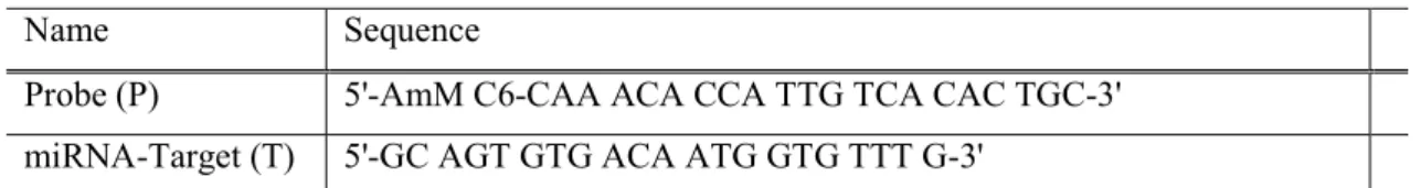
Sodium chloride (NormaPur), methylene blue (Alfa Aesar), potassium ferricyanide (III), and potassium ferrocyanide (II) (Sigma-Aldrich) were used in the experiments without further purification. The labelled DNA probe (P) and complementary miRNA-target (T) (Integrated DNA Technologies) sequences are shown in Table 1. The melting temperature (Tm) given by the supplier is 64°C and the molecular weight (MW) is 6500 g mol-1. The DNA probe oligonucleotides were
synthesized with a 5' amino modification and the target sequence, miRNA-122, is a fragment of interest for the diagnosis of liver cells in case of injury (hepatitis, alcoholism, obesity). The electrolyte used for the Electrochemical Impedance Spectroscopy (EIS) and Cyclic Voltammetry (CV) measurements was made of an equimolar (3 mM) aqueous solution of [Fe(III)(CN)6]3− and
[Fe(II)(CN)6]4− containing also 0.5 M NaCl and 10-8 M Methylene Blue (MB) [11],[33]. The
2.2. a-CNx microelectrode fabrication
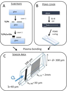
a-CNx thin layers similar to those used in this work have been described in a previous paper [31]. Instead of an ITO sublayer, a titanium / platinum (5 nm / 50 nm) bilayer is deposited on glass so as to promote a-CNx adhesion and to provide an electrical contact on glass. Then, a-CNx is deposited using a mask (see mask in Figure 1) as well as a DC magnetron sputtering setup (MP300S Plassys, deposition time: 20 min, power: 200 W, total pressure before injection of the gas mixture: Ptot(i)<2.10 -7 Torr) including a graphite target and containing a gas mixture composed of argon and nitrogen
(total pressure: Ptot = PAr + PN2 = 0.4 Pa, pressures ratio : 100xPN2/Ptot= 3 %). A 220 nm thickness
was measured with SEM for the resulting film as described in reference [31]. XPS data were used for determining the stoichiometric coefficient, x, of a-CNx thin films by considering the nitrogen atomic percentage in the film divided by the sum of nitrogen and carbon atomic percentages in the film, i.e. the N/(N+C) percentage ratio. The measured nitrogen atomic content was 0.12. Then, a classical PDMS fluidic circuit was microfabricated and pre-treated with nitrogen plasma to favor its adhesion onto the glass slide supporting the glass/Ti/Pt/a-CNx microelectrodes. The main steps of this microfabrication process are summarized in Figure 1.
2.3. Apparatus and Electrochemical measurements
The Electrochemical study was performed with a 2-electrode setup: 30 µm wide a-CNx microband working electrode and 2 mm wide Pt microband counter-electrode (see Figure 1C). The huge ratio of the CE area with respect to the WE one (~ 67) allowed to use the former one as reference electrode simultaneously. A potentiostat (Gamry 600+) was used in CV and EIS experiments. The scan rate for CV experiments was set to 10 mV.s-1 between -1 V and 1 V and the frequency range used for EIS measurements was varied from 1
MHz to 0.1 Hz. The sinusoidal AC signal excitation between the two microelectrodes was set to 10 mV peak to peak, since DC was fixed to 0 V. The EIS experimental data were simulated using Simad software developed at LISE laboratory.
The microfluidic setup was connected to a NEMESYS programmable syringe pump allowing a flow rate in the range from 0.01 μL.s−1 to 5 μL.s−1.
3. Results and discussion
3.1. a-CNx microelectrodes pre-treatment
Usually, as grown a-CNx thin films prepared by using the magnetron sputtering technique show
moderate electrochemical reactivity. Depending upon the intended goal, this latter can be changed by electrochemical pretreatments in either acidic or basic aqueous solutions, impacting either on wettability, or on the sign of the electronic affinity resulting from the nature of the dangling bonds thus formed. For example, a cathodic pre-treatment carried out in a 0.5 M H2SO4 aqueous solution
[30] substantially improved their surface reactivity resulting in high values of the standard electron transfer rate constant (k0 = 5x10⁻²-10-1 cm.s-1 for [Ru(NH3)6]3+/2+, [Fe(CN)6]3−/4− and [IrCl6]2−/3−).
Conversely, an anodic pretreatment in KOH allowed a better peak separation for detection of ascorbic acid and dopamine because it shifted in opposite directions the oxidation peaks of these two components [34],[35].
Here, an anodic activation in a 0.1 M KOH aqueous solution was performed because it favours an increase of oxygenated dangling bonds at the surface, mostly as carboxyl groups [35],[36] which are expected to facilitate amino-modified DNA strands grafting onto a-CN0.12 substrate surface. The
optimized microband electrode pre-treatment consists in filling the microfluidic device with a 0.1 M KOH solution under a 0.5 µL.s-1 flow rate and applying a 2.7x10⁻⁵ mA constant current for the
anodic activation.
The hydrodynamic voltammograms are carried out on the as-grown microelectrode (layout A, Figure 2) and after electrochemical pre-treatment (layout B, Figure 2) in an equimolar solution of [Fe(III)(CN)6]3−/[Fe(II)(CN)6]4− (3 mM each) containing NaCl (0.5 M). In both cases, the
steady-state currents shown in Figure 2 are read as experimental current plateau values. The limiting current ratio between experimental current, Iexp, and theoretical current, Ith, as a function of the 1/3 power
of the volumetric flow rate (layout C, Figure 2) for the channel electrode can be estimated by using the Levich theory, for which Compton has proposed the following practical expression (see Equation 1) [37-39].
𝐼 = 0.925 𝑛 𝐹 𝐶 𝐷 ⁄ 𝑥 ⁄
𝑤 4 𝑄
ℎ 𝑑 (1)
where n is the number of electrons exchanged, F, the faraday constant, D and C0, the diffusion coefficient
and the bulk concentration of electroactive species, w and xe, the width and the length of the WE, d and h,
the width and height of the channel and Q the flow rate. w is equal to d (see Figure 1C).
The Iexp/ Ith current ratio plotted as a function of the 1/3 power of the flow rate (layout C, Figure 2)
approaches a value of 1, indicating that it matches rather well the Levich prediction [11],[40]. However, the a-CN0.12 pre-treatment seems to influence the current kinetics shape as it appears on
the displayed straightened slopes. A more quantitative analysis of steady-state voltammograms was carried out according to the procedure proposed by Mirkin et al. [41] that allows the extraction of the standard rate constant, k0. This latter was found equal to 5.6x10⁻⁴ cm.s-1 on as-grown a-CN
0.12
electrodes and to 1.3x10⁻³ cm.s-1 after activation, i.e. on pre-treated a-CN
0.12 electrodes. This is
consistent with previous observations related to activation of glassy carbon [42] and amorphous carbon nitride [43] electrodes.
3.2. DNA probe immobilization and miRNA-122 target detection
The single strand DNA-NH2 probe (P) immobilization for detecting the miR-122 sequence in a
microfluidic device was performed via the protocol illustrated in Figure 3A. First, carboxyl groups on the pre-treated a-CN0.12 surface were activated by introducing a 2.10-7 M EDC and 2.10-7 M NHS
solution mixture (diluted in bi-distilled water) inside the chip for 20 minutes. Then, a 0.15 µM DNA-NH2 probe dissolved in a 0.5 M NaCl aqueous solution was circulated, then the flow was stopped
for three hours. The flushing was carried out by using deionized water (2 µL.s-1) and the SAM
stability test was checked in 0.5 M NaCl, (0.5 µL.s-1) for 30 minutes. Hybridization of miR-122
target in 0.5 M NaCl was performed during 30 minutes under the same flow conditions.
The EIS experiments displayed in Nyquist plot (Figure 3B) were performed in the same electrolyte conditions to check each protocol step from as-grown a-CN0.12 until target hybridization test (Figure
3A). All the experiments were performed in an equimolar ferri-ferrocyanide electrolytic solution around 0 V, the equilibrium potential. An appropriate Randles circuit (Figure 3C) was used here to simulate the impedance spectrum for each step. The electrolyte resistance or ohmic resistance, R
(only dependent on the electrolyte conductivity and on the channel microelectrode size) is the limit at high frequency of the real part (ZRE) in the Nyquist plot, while the sum of R, charge transfer
resistance Rct (semi-circle diameter) and the diffusion impedance, ZD (f-> 0), is the limit at low
frequency. Therefore, instead of a traditional double layer capacitance Cdl in parallel with the Rct, a
constant-phase element CPE (Qdl and parameters) was preferred due to the surface heterogeneity
and/or the presence of 2-D distribution (of current and potential on the working electrode [44-46]. The analytical expression used for simulation that corresponds to the selected Randles circuit presented in Figure 3C is given by
Z(f) = R + (𝑅 + 𝑍 (𝑓))
1 + (𝑗2𝜋𝑓) 𝑄 (𝑅 + 𝑍 (𝑓)) (2)
where the diffusion-convection impedance ZD was approximated by the expression for a finite
diffusion layer thickness, x,
Z (f) = Z (0)𝑡𝑎𝑛ℎ 𝑗2𝜋𝑓𝜏
𝑗2𝜋𝑓𝜏 (3)
τ = 𝛿
𝐷 (4)
Therefore, equation (1) is valid if the diffusion layer which develops from the leading edge until the leaving edge of the microelectrode remains very small, i.e. for 0<x<xe. The local diffusion layer
thickness is then defined as [47],[48]
δ = 3⁄ Γ 𝐷𝑥
𝑆 (5)
where is the Gamma function.
This assumption could be justified in our case for Q = 5x10⁻⁴ cm3.s-1 corresponding to the imposed
constant volumetric flow rate, and with D = 10-5cm2.s-1, x
e = 30 m, h = 40 m and d = 300 m.
With these values, xe is estimated to about 2 m, that leads to D values in the expected order of
magnitude, i.e. a few milliseconds.
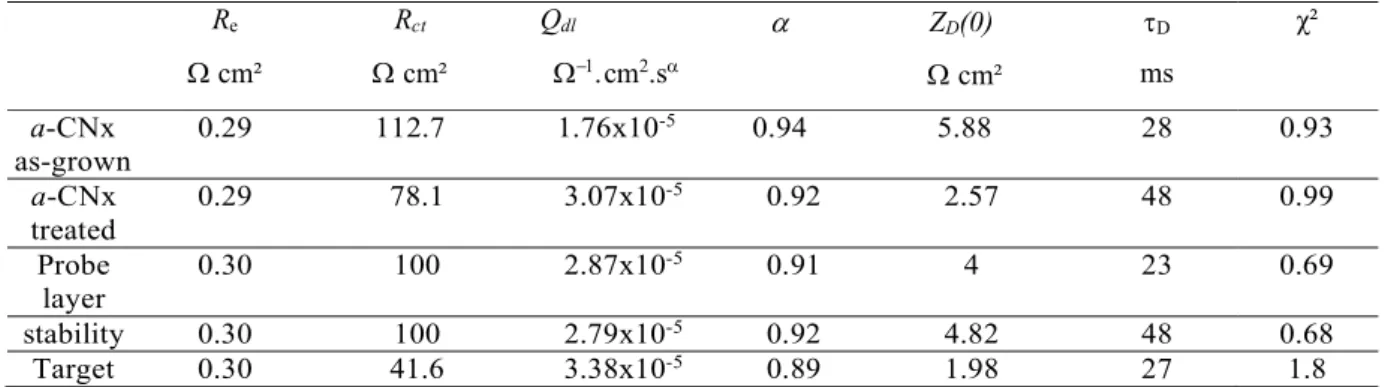
The parameter values used for simulation are listed in Table 2. As expected, the R values are similar
between each step. The double layer capacitance is also not sensitive to the protocol, its value being in the same order of magnitude as for glassy carbon electrode (i.e. 20 µF.cm-2) [42]. Before
pre-treatment, the Rct value is about 112.7 cm2, then it decreases to 78.1 cm2 after a-CN0.12 surface
pre-treatment. Therefore, a DNA modified with a -NH2 ending group was selected as DNA probe
for the miR-122 target detection. These ones are covalently grafted to the a-CN0.12 carboxyl groups
(Figure 3A) that were previously activated with the EDC/NHS procedure [24]. After the immobilization step, Rct increases again and reaches 100 cm2. This behaviour is related to the
grafted DNA probes which are negatively charged, hindering therefore the diffusion of the [Fe(III)(CN)6]3−redox probe towards the microelectrode. The stability of the grafted SAM was also
checked by flushing the channel with deionized water (2 µL.s-1) and then with a blank solution (0.5
M NaCl at 0.5 µL.s-1 for 30 minutes). As shown in Figure 3B (enlarged area) the low difference
observed on the raw data (< 0.9 Ω.cm-2) after the DNA stability test underlines that no SAM
consistent with a robust and covalent grafting conversely to SAM on gold where a probe desorption stage was often observed.
The target hybridization experiment follows the protocol described by Kelley S. O. et al. [5] with the use of methylene blue (MB) as a redox intercalant to enhance the response sensitivity. MB is a redox intercalator that enhances electron transfer across double-stranded DNA. As illustrated in Figure 3A, MB reduction step is reversible with a redox potential that is lower than those allowing [Fe(III)(CN)6]3- reduction. The
electrons are conducted from the a-CN0.12 microelectrode to the intercalated MB thanks to a hopping process
and are then accepted by the ferrocyanide ions dissolved in solution. The oxidized form of MB is regenerated for a further electrochemical reduction. As obtained with the experiment, the hybridization with target miR-122 decreases the Rct value (blue curve) down to 41.6 cm2, a value lower than
the one measured in the presence of the DNA probe only (green curve, 100 cm2), underlining the
success of hybridization.
3.2.2. Density of DNA-NH2 molecules immobilized onto pre-treated a-CNx electrodes
The influence of increasing concentrations of DNA probes from 10 to 104 µg.mL-1 (i.e. from 1.54
µM to 1.54 mM) on the grafting density is shown in Figure 4A. The plot of the difference between Rct (as grown a-CNx-DNA probe) and Rct (pre-treated a-CNx) reaches a plateau that may indicate the
saturation of the COOH groups available for grafting. As illustrated in Figure 3A, the active surface, i.e. uncovered surface by the DNA-NH2 probes (ligand) is accessible for electroactive redox couple.
In this case, the fraction of a-CNx microelectrode surface grafted with ligands can be estimated as follows
𝜃 = 1 −𝑅
𝑅 (6)
The experimental evolution of the surface grafting is plotted in Figure 4A according to equation (6) and the experimental curve is modelled with the Hill-Langmuir equation [49],[50] , as follows
𝜃 = 1
1 + (𝐾 [𝐷𝑁𝐴 − 𝑁𝐻 ]⁄ ) (7)
where KA represents DNA-NH2 concentration that produces a half-maximal response, and n is the
Hill coefficient related to the cooperativity degree between the first ligands and the following ones throughout the immobilization step: if n <1, cooperativity tends to decrease, if n=1, there is no cooperativity or independent, if n>1, cooperativity tends to increase.
The result of the fitting procedure using equation (7) is illustrated in Figure 4B. The obtained values of KA and n are (100.25 ± 9.65) µg.mL-1 and 2.41 ± 0.65, respectively. This latter value confirms an
excellent cooperativity between ligands during the immobilization procedure. The observed plateau (max=0.52± 0.16) in the high concentration range (103-104 µg.mL-1) suggests that the maximum
COOH sites (available for EDC/NHS activation procedure) on pre-treated a-CN0.12 is reached and
represents only 52% of the active surface of the microelectrode.
The maximal area covered by the DNA probes was estimated to Amax =4.68x10⁻⁵ cm2 by taking into
account that the WE electrode surface (SWE) is about 9x10⁻⁵ cm2. The cross-section of a DNA
molecule was currently modelled as a square of 5 nm per side [51] leading to an occupied surface area of ADNA-NH2 = 2.5x10⁻¹³ cm2 per DNA-NH2 molecule. The maximum number, Nmax, of
DNA-NH2 immobilized represents the ratio between Amax and ADNA-NH2 i.e. 1.87x10⁸ molecules and the
binding site density was deduced as the ratio between Nmax and SWE i.e. 2.08x10¹² molecules.cm-2.
This latter value is lower than the maximum amount of DNA attached on a surface and it is close to the value obtained on other materials in the literature [4],[11],[51],[52].
4. Conclusion
Amorphous carbon nitride (a-CNx) thin films were developed as working electrode materials for the elaboration of a DNA biosensor. They were proved to be an alternative to gold or other carbon
and diamond materials like carbon (DLC) or BDD. To this end, a-CN0.12 thin films were
synthesized and lithographied on Ti/Pt substrates so as to produce planar 30 µm width microband electrodes. Then, an electrochemical pre-treatment was performed inside a microfluidic device to activate these microband electrodes, leading to a drastic increase of the charge transfer kinetics. Finally, a biofunctionalization protocol (EDC/NHS) leading to the formation of a robust DNA probe SAM was achieved. In future works, our goal is to compare the performances (LOD, specificity and sensitivity) of the miR-122 target capture (synthetic DNA target mimicking the liver-specific micro-ribonucleic acid 122 (miRNA122) on two different materials: as-grown a-CN0.12, and pre-treated a-CN0.12 microelectrodes.
Acknowledgements
This research was supported by the LabEx MiChem part of French state funds managed by the ANR within the Investissements d'Avenir program under reference ANR-11IDEX-0004-02, and partly supported by the French RENATECH network.
Declaration of Competing Interest
The authors declare that they have no known competing financial interests or personal relationships that could have appeared to influence the work reported in this paper.
Credit author statement
Marie-Charlotte Horny: Conceptualization, Methodology, Investigation, Formal analysis, Software, Visualization. Florence Billon: Conceptualization, Methodology, Investigation. Claude Deslouis: Conceptualization, Methodology, Formal analysis. Mathieu lazerges: Conceptualization, Methodology. Vincent Dupuis: Conceptualization, Methodology. Jean-Michel Siaugue: Conceptualization, Methodology. Alain Pailleret: Conceptualization, Methodology, Investigation. Jean Gamby: Conceptualization, Methodology, Investigation, Formal analysis, Supervision, Visualization, Validation, Resource, Project administration, Writing- Original draft, Review and Editing.
References
[1] V. Perumal, U. Hashim, Advances in biosensors: Principle, architecture and applications, Journal of Applied Biomedicine, 12 (2014) 1-15.
[2] C.D. Bain, J. Evall, G.M. Whitesides, Formation of monolayers by the coadsorption of thiols on gold: variation in the head group, tail group, and solvent, Journal of the American Chemical Society, 111 (1989) 7155-7164.
[3] C.E.D. Chidsey, D.N. Loiacono, Chemical functionality in self-assembled monolayers: structural and electrochemical properties, Langmuir, 6 (1990) 682-691.
[4] A.B. Steel, T.M. Herne, M.J. Tarlov, Electrochemical Quantitation of DNA Immobilized on Gold, Analytical Chemistry, 70 (1998) 4670-4677.
[5] S.O. Kelley, E.M. Boon, J.K. Barton, N.M. Jackson, M.G. Hill, Single-base mismatch detection based on charge transduction through DNA, Nucleic Acids Res., 27 (1999) 4830-4837.
[6] K.M. Koo, L.G. Carrascosa, M.J.A. Shiddiky, M. Trau, Poly(A) Extensions of miRNAs for Amplification-Free Electrochemical Detection on Screen-Printed Gold Electrodes, Analytical Chemistry, 88 (2016) 2000-2005.
[7] K.M. Koo, L.G. Carrascosa, M.J.A. Shiddiky, M. Trau, Amplification-Free Detection of Gene Fusions in Prostate Cancer Urinary Samples Using mRNA–Gold Affinity Interactions, Analytical Chemistry, 88 (2016) 6781-6788.
[8] M.N. Islam, M.K. Masud, M.H. Haque, M.S.A. Hossain, Y. Yamauchi, N.-T. Nguyen, M.J.A. Shiddiky, RNA Biomarkers: Diagnostic and Prognostic Potentials and Recent Developments of Electrochemical Biosensors, Small Methods, 1 (2017) 1700131.
[9] L.Y.S. Lee, R.B. Lennox, Electrochemical Desorption of n-Alkylthiol SAMs on Polycrystalline Gold: Studies Using A Ferrocenylalkylthiol Probe, Langmuir, 23 (2007) 292-296.
[10] S. Choi, J.J.M. Chae, Nanofluidics, A regenerative biosensing surface in microfluidics using electrochemical desorption of short-chain self-assembled monolayer, Microfluidics and Nanofluidics, 7 (2009) 819.
[11] M.C. Horny, M. Lazerges, J.M. Siaugue, A. Pallandre, D. Rose, F. Bedioui, C. Deslouis, A.M. Haghiri-Gosnet, J. Gamby, Electrochemical DNA biosensors based on long-range electron transfer: investigating the efficiency of a fluidic channel microelectrode compared to an ultramicroelectrode in a two-electrode setup, Lab Chip, 16 (2016) 4373-4381.
[12] F. Lucarelli, G. Marrazza, A.P.F. Turner, M. Mascini, Carbon and gold electrodes as electrochemical transducers for DNA hybridisation sensors, Biosensors and Bioelectronics, 19 (2004) 515-530.
[13] J. Wang, E. Palecek, P.E. Nielsen, G. Rivas, X. Cai, H. Shiraishi, N. Dontha, D. Luo, P.A.M. Farias, Peptide Nucleic Acid Probes for Sequence-Specific DNA Biosensors, Journal of the American Chemical Society, 118 (1996) 7667-7670.
[14] J. Wang, A.-N. Kawde, E. Sahlin, Renewable pencil electrodes for highly sensitive stripping potentiometric measurements of DNA and RNA, Analyst, 125 (2000) 5-7.
[15] G. Marrazza, G. Chiti, M. Mascini, M. Anichini, Detection of Human Apolipoprotein E Genotypes by DNA Electrochemical Biosensor Coupled with PCR, Clinical Chemistry, 46 (2000) 31.
[16] D.J. Caruana, A. Heller, Enzyme-Amplified Amperometric Detection of Hybridization and of a Single Base Pair Mutation in an 18-Base Oligonucleotide on a 7-μm-Diameter Microelectrode, Journal of the American Chemical Society, 121 (1999) 769-774. [17] K.M. Millan, A.J. Spurmanis, S.R. Mikkelsen, Covalent immobilization of DNA onto glassy carbon electrodes, 4 (1992) 929-932. [18] S. Ranganathan, R. McCreery, S.M. Majji, M. Madou, Photoresist-Derived Carbon for Microelectromechanical Systems and Electrochemical Applications, Journal of The Electrochemical Society, 147 (2000) 277.
[19] M.L. Yola, N. Atar, Development of molecular imprinted sensor including graphitic carbon nitride/N-doped carbon dots composite for novel recognition of epinephrine, Composites Part B: Engineering, 175 (2019) 107113.
[20] H. Medetalibeyoglu, M. Beytur, O. Akyıldırım, N. Atar, M.L. Yola, Validated electrochemical immunosensor for ultra-sensitive procalcitonin detection: Carbon electrode modified with gold nanoparticles functionalized sulfur doped MXene as sensor platform and carboxylated graphitic carbon nitride as signal amplification, Sensors and Actuators B: Chemical, 319 (2020) 128195.
[21] M.L. Yola, N. Atar, Amperometric galectin-3 immunosensor-based gold nanoparticle-functionalized graphitic carbon nitride nanosheets and core–shell Ti-MOF@COFs composites, Nanoscale, 12 (2020) 19824-19832.
[22] C. Pelin Böke, O. Karaman, H. Medetalibeyoglu, C. Karaman, N. Atar, M. Lütfi Yola, A new approach for electrochemical detection of organochlorine compound lindane: Development of molecular imprinting polymer with polyoxometalate/carbon nitride nanotubes composite and validation, Microchemical Journal, 157 (2020) 105012.
[23] M.L. Yola, Sensitive sandwich-type voltammetric immunosensor for breast cancer biomarker HER2 detection based on gold nanoparticles decorated Cu-MOF and Cu2ZnSnS4 NPs/Pt/g-C3N4 composite, Microchimica Acta, 188 (2021) 78.
[24] S. Sam, L. Touahir, J. Salvador Andresa, P. Allongue, J.N. Chazalviel, A.C. Gouget-Laemmel, C. Henry de Villeneuve, A. Moraillon, F. Ozanam, N. Gabouze, S. Djebbar, Semiquantitative Study of the EDC/NHS Activation of Acid Terminal Groups at Modified Porous Silicon Surfaces, Langmuir, 26 (2010) 809-814.
[25] K.M. Millan, S.R. Mikkelsen, Sequence-selective biosensor for DNA based on electroactive hybridization indicators, Analytical Chemistry, 65 (1993) 2317-2323.
[26] H. Cachet, C. Debiemme-Chouvy, C. Deslouis, A. Lagrini, V. Vivier, Correlation between electrochemical reactivity and surface chemistry of amorphous carbon nitride films, Surface and Interface Analysis, 38 (2006) 719-722.
[27] P. Tamiasso-Martinhon, H. Cachet, C. Debiemme-Chouvy, C. Deslouis, Thin films of amorphous nitrogenated carbon a-CNx: Electron transfer and surface reactivity, Electrochim. Acta, 53 (2008) 5752--5759.
[28] A. Lagrini, C. Deslouis, H. Cachet, M. Benlahsen, S. Charvet, Elaboration and electrochemical characterization of nitrogenated amorphous carbon films, Electrochemistry Communications, 6 (2004) 245-248.
[29] H. Cachet, C. Deslouis, M. Chouiki, B. Saidani, N.M.J. Conway, C. Godet, Electrochemistry of Nitrogen-Incorporated Hydrogenated Amorphous Carbon Films, Journal of The Electrochemical Society, 149 (2002) E233.
[30] A. Benchikh, C. Debiemme-Chouvy, H. Cachet, A. Pailleret, B. Saidani, L. Beaunier, M.H. Berger, C. Deslouis, Influence of electrochemical pre-treatment on highly reactive carbon nitride thin films deposited on stainless steel for electrochemical applications, Electrochimica Acta, 75 (2012) 131-138.
[31] M. Faure, F. Billon, A.M. Haghiri-Gosnet, B. Tribollet, C. Deslouis, A. Pailleret, J. Gamby, Influence of the atomic nitrogen content in amorphous carbon nitride thin films on the modulation of their polarizable interfaces properties, Electrochimica Acta, 280 (2018) 238-247.
[32] M. Faure, F. Billon, I. Le Potier, A.M. Haghiri-Gosnet, B. Tribollet, A. Pailleret, C. Deslouis, J. Gamby, Improvement of electrochemical detection of transthyretin synthetic peptide and its amino acids on carbon electrodes: Glassy carbon versus amorphous carbon nitride a-CNx, Electrochimica Acta, 296 (2019) 251-258.
[33] A. Nassi, F.X. Guillon, A. Amar, B. Hainque, S. Amriche, D. Maugé, E. Markova, C. Tsé, P. Bigey, M. Lazerges, F. Bedioui, Electrochemical DNA-biosensors based on long-range electron transfer: optimization of the amperometric detection in the femtomolar range using two-electrode setup and ultramicroelectrode, Electrochimica Acta, 209 (2016) 269-277.
[34] M. Benlahsen, H. Cachet, S. Charvet, C. Debiemme-Chouvy, C. Deslouis, A. Lagrini, V. Vivier, Improvement and characterization of the electrochemical reactivity of amorphous carbon nitride electrodes, Electrochemistry Communications, 7 (2005) 496-499. [35] R.A. Medeiros, A. Benchick, R.C. Rocha-Filho, O. Fatibello-Filho, B. Saidani, C. Debiemme-Chouvy, C. Deslouis, Simultaneous detection of ascorbic acid and dopamine with electrochemically pretreated carbon nitride electrodes: Comparison with boron-doped diamond electrodes, Electrochem. Commun., 24 (2012) 61-64.
[36] R.A. Medeiros, R. Matos, A. Benchikh, B. Saidani, C. Debiemme-Chouvy, C. Deslouis, R.C. Rocha-Filho, O. Fatibello-Filho, Amorphous carbon nitride as an alternative electrode material in electroanalysis: Simultaneous determination of dopamine and ascorbic acid, Anal. Chim. Acta, 797 (2013) 30-39.
[37] V.G. Levich, Physicochemical Hydrodynamics, Prentice-Hall, 1962.
[38] R.G. Compton, A.C. Fisher, R.G. Wellington, P.J. Dobson, P.a. Leigh, Hydrodynamic voltammetry with microelectrodes: channel microband electrodes; theory and experiment, The Journal of Physical Chemistry, 97 (1993) 10410-10415.
[39] C. Amatore, N. Da Mota, C. Sella, L. Thouin, Theory and Experiments of Transport at Channel Microband Electrodes Under Laminar Flow. 3. Electrochemical Detection at Electrode Arrays under Steady State, Analytical Chemistry, 82 (2010) 2434-2440. [40] M. Faure, A. Pallandre, S. Chebil, I. Le Potier, M. Taverna, B. Tribollet, C. Deslouis, A.-M. Haghiri-Gosnet, J. Gamby, Improved electrochemical detection of a transthyretin synthetic peptide in the nanomolar range with a two-electrode system integrated in a glass/PDMS microchip, Lab Chip, 14 (2014) 2800-2805.
[41] M.V. Mirkin, A.J. Bard, Simple analysis of quasi-reversible steady-state voltammograms, Analytical Chemistry, 64 (1992) 2293-2302.
[42] I.-F. Hu, D.H. Karweik, T. Kuwana, Activation and deactivation of glassy carbon electrodes, Journal of Electroanalytical Chemistry and Interfacial Electrochemistry, 188 (1985) 59-72.
[43] P. Tamiasso-Martinhon, H. Cachet, C. Deslouis, V. Vivier, Amorphous carbon nitride a-CNx microelectrode: Fabrication and characterization, Electrochemistry Communications, 12 (2010) 1074-1076.
[44] B. Hirschorn, M.E. Orazem, B. Tribollet, V. Vivier, I. Frateur, M. Musiani, Constant-Phase-Element Behavior Caused by Resistivity Distributions in Films: II. Applications, Journal of The Electrochemical Society, 157 (2010) C458-C463.
[45] B. Hirschorn, M.E. Orazem, B. Tribollet, V. Vivier, I. Frateur, M. Musiani, Constant-Phase-Element Behavior Caused by Resistivity Distributions in Films: I. Theory, Journal of The Electrochemical Society, 157 (2010) C452-C457.
[46] M. Musiani, M.E. Orazem, N. Pébère, B. Tribollet, V. Vivier, Constant-Phase-Element Behavior Caused by Coupled Resistivity and Permittivity Distributions in Films, Journal of The Electrochemical Society, 158 (2011) C424-C428.
[47] M. Lévêque, A,, Les lois de la transmission de chaleur par convection, Ann. Mines, 13 (1928) 283-287.
[48] S.C. Ling, Heat Transfer From a Small Isothermal Spanwise Strip on an Insulated Boundary, Journal of Heat Transfer, 85 (1963) 230-235.
[49] A.V. Hill, PROCEEDINGS OF THE PHYSIOLOGICAL SOCIETY: January 22, 1910, in, 1910, pp. i-vii.
[50] I. Langmuir, THE ADSORPTION OF GASES ON PLANE SURFACES OF GLASS, MICA AND PLATINUM, Journal of the American Chemical Society, 40 (1918) 1361-1403.
[51] B. Sun, P.E. Colavita, H. Kim, M. Lockett, M.S. Marcus, L.M. Smith, R.J. Hamers, Covalent Photochemical Functionalization of Amorphous Carbon Thin Films for Integrated Real-Time Biosensing, Langmuir, 22 (2006) 9598-9605.
[52] Z. Lin, T. Strother, W. Cai, X. Cao, L.M. Smith, R.J. Hamers, DNA Attachment and Hybridization at the Silicon (100) Surface, Langmuir, 18 (2002) 788-796.
Tables Table1
Name Sequence
Probe (P) 5'-AmM C6-CAA ACA CCA TTG TCA CAC TGC-3'
miRNA-Target (T) 5'-GC AGT GTG ACA ATG GTG TTT G-3'
Table 2 Re cm² Rct cm² Qdl cm2.s ZD(0) cm² D ms χ² a-CNx as-grown 0.29 112.7 1.76x10⁻⁵ 0.94 5.88 28 0.93 a-CNx treated 0.29 78.1 3.07x10⁻⁵ 0.92 2.57 48 0.99 Probe layer 0.30 100 2.87x10 -5 0.91 4 23 0.69 stability 0.30 100 2.79x10⁻⁵ 0.92 4.82 48 0.68 Target 0.30 41.6 3.38x10⁻⁵ 0.89 1.98 27 1.8 (*) Values of the resulting 2 statistics were obtained with errors of fitted values, =0.01, for frequency ranging from 1 MHz to 1 Hz.
Figures Figure 1
Fig. 1: Microfabrication key steps for a-CNx sensor microelectrode integration. A. Glass slides spin-coated with AZ5214 resin followed by an inverted lithography step and then metallized with a 200 nm thick a-CNx layer on the top of a 5/50 nm bilayer of titanium/platinum by two classical lift-off processes. B. Fluidic circuit master mold made with SU-8 on silicon wafer. PDMS layer is poured onto the SU-8 master mold to get a negative replica. The PDMS and the glass substrates with the electrodes are washed, dried and treated with nitrogen plasma for bonding. C. Detection area and fluidic channel microelectrodes with their dimensions. WE stands for a-CNx working electrode (width, w=300 μm, length, xe=30 μm), CE stands for
platinum counter electrode (w=300 μm, xe=2 mm), and h and d represent the fluidic channel height and
width, respectively. Note that here w = d. Q represents the flow rate and the arrow the flow direction. CE was purposely placed downstream of WE to avoid a parasitic additional current due to the species formed on the former one in the opposite configuration.
Figure 2
Fig. 2: CV experiments recorded in an aqueous solution containing Fe(CN)64- / Fe(CN)63- (3 mM) and NaCl
(0.5 M) under various flow rates : static (i), 0.2 (j), 0.5 (k), 1.0 (l), 1.5 (m), µL.s-1. Scan rate : 10 mV.s-1. A.
at a as-grown a-CN0.12 microelectrode, B. at a pre-treated a-CN0.12 microelectrode. C. Plot of the limiting
current ratio between experimental current, Iexp, and Levich equation current, ILevich, as a function of the
cubic root of the volumetric flow rate. The dotted line displays the mean ratio for experimental values to the theoretical ones. 2.0 1.5 1.0 0.5 0.0
I
exp/
I
th 0.12 0.08 0.04 0.00 Q1/3 / (cm3s-1)1/3 -4x10⁻⁴ 0 4 J ( A cm -2 ) -1.0 -0.5 0.0 0.5 1.0 E (V) i j k l m -4x10⁻⁴ 0 4J (
A
cm
-2 ) -1.0 -0.5 0.0 0.5 1.0 E (V) KOH activation A B CFigure 3
Fig.3: A. Hybridization protocol on a microchannel containing a a-CN0.12 electrode sensor for a working
flow of 0.5 µL.s-1 and in presence of [Fe(III)(CN)
6]3−/[Fe(II)(CN)6]4− (3 mM) + MB (10-8 M) in 0.5 M NaCl
aqueous solution. B. EIS response of as grown a-CN0.12 (), a-CN0.12 pre-treated () electrodes. Probe
immobilized by circulating of 100 µg.mL-1 DNA probe sequence diluted in a 0.5 M NaCl aqueous solution
C
O
O
H DNA-NH2EDC/NHS miR-122MB
FeIIIFeII
FeII
FeIII FeIII FeII
MB+ LB+
FeII FeIII a. pre-treated b. DNA probe c. miR Target A 140 120 100 80 60 40 20 0 -ZIM / Ω c m 2 120 80 40 0 ZRE / Ω cm2 B 1 MHz 200 Hz 0.1 Hz 100 Hz 40 Hz 10 Hz10 Hz 100 Hz 100 Hz 10 Hz 0.1 Hz a-CNx_as grown a-CNx pre-treated a-CNx - DNA probe a-CNx - DNA probe stability a-CNx - DNA probe // miR target fitting with analytical expression (eqs. 2-3)
12 8 4 0 110 100 90 ZRE / Ω cm2 10 Hz 0.1 Hz 0.9 cm-2 2 Hz
(). Stability test of DNA probe layer after 30 minutes in a 0.5 M NaCl aqueous solution ().Target hybridization by introducing 10-18 M of miR-122 (). EIS curves simulated (dashed line) with analytical
expressions given in equations 2 and 3. C. Equivalent circuit (Randles adapted) used to simulate the experimental impedance data.
Figure 4
Fig.4: A. a-CN0.12 microelectrode functionalization response in EIS subjected to increasing concentrations
of DNA probe. B. Experimental evolution of the surface grafting simulated with the Hill-Langmuir equation according to equation (7) where parameters KA, n, and max are equal to (100.25 ± 9.65) µg mL-1, (2.41 ±