394
CLINICAL INFECTIOUS DISEASE ARTICLES
Bacterial Brain Abscesses: Factors Influencing Mortality and Sequelae
Ch. Seydoux and P. Francioli
From the Department of Internal Medicine. Division of InfectiousDiseases. Centre Hospitalier Universitaire Vaudois. Lausanne. Switzerland
Thirty-nine cases of brain abscess diagnosed since the advent of the computed tomographic (CT) scan were analyzed for factors influencing the outcome. The mortality rate was 13%, and severe sequelae were present in 22% of the survivors. The mean delay between occurrence of the first symptoms and hospitalization was significantly shorter for the 12 patients with poor out-come (death or severe sequelae) than for the 25 who recovered (fully or with moderate sequelae). Moreover, severely impaired mental status and neurological impairment at admission were asso-ciated with a poor outcome in terms of both mortality and sequelae. In all cases with fatal outcome or severe sequelae, the diagnosis was made and treatment was initiated within 24 hours of admission. There was no apparent correlation between the outcome and the presence or type of predisposing factors, the radiological, biological, or microbiological findings, or the treatment modalities. Thus, with the advent of the CT scan and the possibility of early diagnosis and treatment, the prognosis of brain abscess appears to be mainly determined by the rapidity of progression of the disease before hospitalization and the patient's mental status on admission.
Since the advent ofantimicrobial agents, the mortality rate associated with brain abscess has remained between
20%
and50%,
and sequelae have been reported for30%
to70%
of survivors[1-8].
Recently, substantial improvement in the prognosis of brain abscess with a mortality rate between5%
and20%,
has been reported[9-11].
This has been attributed to the advent of more-reliable methods of identification of microorganisms, more-effective antibiotic regimens, new surgical techniques, and, most important, the availability of the computed tomographic (CT) scan [12]. Magnetic reso-nance imaging (MRI) was recently shown to be more sensi-tive in the early detection of cerebritis in animal models, but evidence of its superiority over the CT scan in terms of clini-cal benefit for patients with cerebral abscess has not yet been established[13-15].
The purpose of the present study was to review the cases diagnosed at our institution since the intro-duction of the CT scan and before the introintro-duction ofMRI in order to delineate the clinical, biological, and therapeutic characteristics that are of importance for the diagnosis and outcome of brain abscess.Materials and Methods
PatientsThirty-nine cases of brain abscesses were diagnosed at the Centre Hospitalier Universitaire Vaudois (Lausanne,
Swit-Received 6 March 1992; revised 18 May 1992.
Reprints and correspondence: Dr. Patrick Francioli, Division of In-fectious Diseases. Centre Hospitalier U niversitaire Vaudois. 10 II Lau-sanne-CHUV, Switzerland.
Clinical Infectious Diseases 1992;15:394-401
©1992by The University of Chicago. All rights reserved.
1058-4838/92/1503-0002$02.00
zerland) from January
1977
to December1987.
All patients had been hospitalized in either the Department of Internal Medicine or the Department of Neurosurgery. The diagnoses were retrospectively traced by the hospitals computerized system. Brain abscess was defined as a localized lesion visible on CT scan that was located in the cerebrum, cerebellum, or midbrain and associated with at least one of the three follow-ing criteria: positive blood culture, positive culture of intra-cerebral material, or histology suggesting an acute brain ab-scess. Materials obtained from brain abscesses were cultured for aerobic and anaerobic bacteria, fungi, and mycobacteria with use of standard methods. Patients who tested positive for the human immunodeficiency virus and those with sus-pected Toxoplasma gondii encephalitis, Listeria monocyto-genes meningoencephalitis, or brain abscess due to fungi (inone case, Cryptococcus) were excluded.
Statistical Analysis
Comparison of groups with continuous variable distribu-tion (age, duradistribu-tion of symptoms) was conducted with use of an unpaired Student'sz-test.Comparison of groups with dis-crete variable distribution (predisposing factors, neurological signs, meningismus, microbiological cultures) was per-formed by means of the X2test with Yates' correction. A
P
value of~.05was considered significant. Data are given as mean± SD unless stated otherwise.
Results
Demographic Data
There were 26 males and
13
females (male/female ratio:2/1;
age: mean, 44 years; and range,5-80
years) in the studyClD 1992; 15 (September) Brain Abscesses: Outcome Factors 395
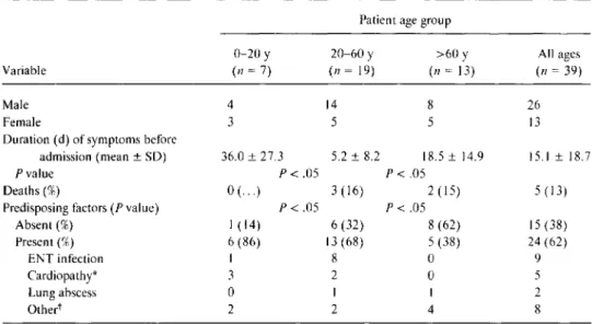
Table 1. Clinical characteristics of 39 patients with brain abscess, analyzed according to age group.
Patient age group
0-20 y 20-60 Y >60 Y All ages
Variable (n= 7) (n= 19) (n= 13) (n= 39)
Male 4 14 8 26
Female 3 5 5 13
Duration (d) of symptoms before
admission (mean ± SO) 36.0 ± 27.3 5.2 ± 8.2 18.5± 14.9 15.1 ± 18.7
P value P< .05 P< .05
Deaths(%) 0( ... ) 3 (16) 2 (15) 5 (13)
Predisposing factors (P value) P< .05 P< .05
Absent(%) I (14) 6 (32) 8 (62) 15 (38) Present(%) 6 (86) 13 (68) 5 (38) 24 (62) ENT infection I 8 0 9 Cardiopathy" 3 2 0 5 Lung abscess 0 I I 2 Othert 2 2 4 8
NOTE. Data are given as numbers of patients except for duration of symptoms before admission (no. of days) andPvalues. Percentages are based on data in each column. ENT = ear, nose. and throat.
* Cyanotic cardiopathy; three of these patients also had ENT infection.
tHead injury (2), steroid therapy (2), skin infection with bacteremia (2). or endocarditis (2).
group. Most of the brain abscesses diagnosed were in adults aged >30 years (31 of 39; 79%), but six (15%) were observed in children
<
15 years old (table I).Overall, there were 3.6 cases diagnosed per year, i.e., ..., I case per 12,000 hospitalizations (39 of 460,000). Twenty-two patients (56%) were transferred from other hospitals, while 17 were admitted directly to our hospital.
PresentingSymptoms and Physical Findings
The duration of symptoms before hospitalization ranged from several hours to 44 days, with a mean of 15.1 days. It
was significantly less for the patients between 20 and 60 years of age than for those in other age groups (table I).
On admission, 56% of the patients (22 of 39) had a head-ache, the most common symptom. Fever was present in only 41 % of the cases (16 of 39) and was not correlated with the presence of predisposing factors or the location of abscess. General seizures ( II patients) or focal seizures ( 3 patients) prompted hospitalization of 14 patients. On physical exami-nation, all patients presented with neurological signs sugges-tive of brain lesions: 19 (49%) had focal neurological signs (hemiparesis or focal seizure), 13 (33%) had diffused neuro-logical dysfunction (coma, general seizures, or behavioral disturbances), and 7 (18%) had a hemisyndrome and a gen-eral seizure. Abscesses in an occipital or temporal location were more frequently accompanied by diffuse signs than were frontal, parietal, or multiple abscesses (table 2).
Meningismus was observed in nine patients (23%) and was associated with fever in six (67%) of them. Five patients also had focal neurological signs, three had neuropsychological disturbances, and one was comatose. Abscesses in an occipi-tal or temporal location were more frequently associated
with meningismus (50%) than frontal or parietal (II%) or multiple abscesses (27%) (table 2). Funduscopic examina-tion was performed on 18 patients: 6 of them (33%) had a papilledema (3 bilateral and 3 unilateral) that was not re-lated to the location of the abscess and did not appear to have prognostic value.
On admission, 19 patients (48%) had a normal mental sta-tus (mental stasta-tus A), 17 (44%) presented with confusion (mental status B), I (3%) was stuporous (mental statusC), and 2 (5%) were comatose (mental status D).
Predisposing Factors and Location of theAbscesses
Twenty-four patients (62%) presented 27 predisposing fac-tors. Fifteen (63%) had an infectious focus (9 otic or sinus
Table2. Location of brain abscess compared witheNS signs at admission and the presence of meningismus in 39 patients.
No.(%) of patients with indicated abscess(es)
FIP OIT Multiple Total
Variable (n= 18) (n= 6) (n== 15) (n==39) CNS signs* Focal 8 (44) 2 (33) 9 (60) 19 (49) Diffuse 6 (33) 3 (50) 4 (27) 13 (33) Complex 4 (22) I (17) 2 (13) 7 (18) Meningismus (P value) P< .05 P< .05 Present 2 (II) 3 (50) 4 (27) 9 (23) Absent 16 (89) 3 (50) II (73) 30 (77)
NOTE. F= frontal; P= parietal;
a
=occipital; T= temporal locations of all single abscess(es). Percentages are based on data in each column.396 Seydoux and Francioli ClD 1992; 15 (September)
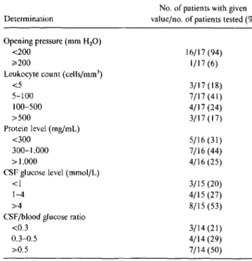
Table 3. CSFfindings for 17patients with brain abscess.
Microbiological Findings
A microbiological diagnosis was established in 32 cases (82%), by means of culture of brain abscess material in 28 cases, of blood in 3 cases, and of both in 1 case.
determine if ventriculitis or abscess rupture had occurred. Twelve of the 17 patients (71 %) had aspecific and mildly alterated CSF perturbations compatible with a CNS process, and two patients (12%) with a frontal abscess had entirely normal CSF findings. Eight patients had received antibiotics 2-8 days before the LP; their CSF findings were not different from those observed for the nine patients who had not been treated before the LP. All CSF cultures remained sterile and were not valuable to microbiological diagnosis.
CT Scan Findings
For all patients, a CT scan was performed: 30 (77%) during the first 3 days of hospitalization, 8 (21 %) between days 4 and l
O,
and 1 (2%) on day 30. All the scans were analyzed for evidence of sinusitis and mastoiditis. Plain films revealed one or several hypodense lesions in 34 cases (87%), hyper-dense lesions in 3 cases (8%), and findings consistent with the presence of gas in 2 cases (5%). Surrounding edema was present in 29 patients (74%): 24 had focal neurogical signs or meningismus. One patient had diffused cerebral edema. Contrast material was injected in 25 cases (64%): nodular enhancement in 7 patients and ring-enhancing lesions in 18 patients were noted. There was no correlation between the radiological characteristics of the lesions and the time elapsed between the appearance of the first symptoms and the performance of the CT scan.5/16 (31) 7/16 (44) 4/16 (25) 3/17 (18) 7/17 (41) 4/17 (24) 3/17 (17) 3/15 (20) 4/15 (27) 8/15 (53) 3/14 (21) 4/14 (29) 7/14 (50) 16/17 (94) 1/17 (6) No. of patients with given value/no. of patients tested(%) Determination
Opening pressure (mm H20)
<200 ~200
Leukocyte count (cells/rum") <5 5-100 100-500 >500 Protein level (rng/rnl.) <300 300-1,000 >1.000
CSF glucose level (mrnol/L) <I
1-4 >4
CSF/blood glucose ratio <0.3
0.3-0.5 >0.5
Laboratory Findings
Peripheral white blood cell (WBC) counts were> 10,000/ mrrr' in 49% of the cases. Anemia was noted in three cases and thrombopenia
«
10,000 blood platelets/rum") in one case. These findings did not influence the clinical presenta-tion or the outcome.A lumbar puncture (LP) was performed on 17 patients (44%), always before the CT scan. No complication oc-curred, although 3 patients had papilledema. The decision to perform an LP was mainly related to a clinical suspicion of bacterial meningitis: six patients (35%) had true meningis-mus with fever and two ( 12%) were highly confused or in a coma with fever. For two patients (12%) with perforated sin-usitis, the LP was performed to rule out meningitis. Seven patients (41 %) with hemisyndrome (five of whom had fever) also underwent an LP, although restrospective analysis did not disclose a definite indication for the procedure. Analysis of CSF (table 3) revealed bacterial meningitis (criteria: a WBC count of>500/mm3and a CSF/blood glucose ratio of <0.3) in only three patients (18%), who had 37,000, 2,470, and 597 WBCs/mm3and CSF/blood glucose ratios of 0.02, 0.06, and 0.1, respectively. These 3 patients were febrile and 2 of them had meningismus: 1 died, 1 fully recovered, and 1 had moderate sequelae at follow-up. It was not possible to infections, 2 skin infections with bacteremia, 2 cases of endo-carditis, and 2 pulmonary infections), 5 (21 %)had a congeni-tal cyanotic cardiopathy (3 of them associated with an ear, nose, and throat infection), 2 (8%) were receiving corticoste-roids, and 2 (8%) had had a prior head injury. Predisposing factors were present in 73% (19 of 26) of the patients <60 years old but in only 38% (5 of 13) of those aged >60 years. Otogenic or sinus infections were all observed in patients <50 years old. Five of the 7 patients >20 years old had either a cyanotic cardiopathy (3 patients) or a prior head trauma (2 patients) (table 1). No patients had undergone a dental or surgical procedure during the 3 months before the diagnosis of brain abscess. Neither the occurrence nor the type of pre-disposing factors seemed to influence the clinical presenta-tion or the durapresenta-tion between the appearance of the first symptoms and hospitalization. For 38% (15 of 39) of the patients, no predisposing condition was documented. These patients did not differ in terms of the clinical manifestation, location of abscess, or type of isolated organisms.
In cases of a single abscess (24 of 39; 62%), it was mostly located in the frontal lobe (12 patients) or parietal lobe (6 patients) rather than in the occipital (4 patients) or temporal (2 patients). The sites were independent from the presumed origin of infection, except for abscesses caused by an ear or sinus infection (7 of 12 patients [58%] with such abscesses presented with a frontal brain abscess). In the 15 patients (38%) with multiple abscesses, the parietal lobe was involved in all but 3 cases, the occipital lobe in 6 cases, the frontal lobe in 7 cases, and the temporal lobe in 5 cases.
CID 1992; 15 (September) Brain Abscesses: Outcome Factors 397
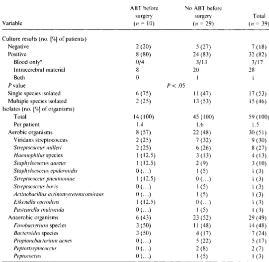
Table 4. Microorganisms isolated in cultures of intracerebral material (35 patients), in blood cul-tures only (3 patients), or in both (l patient) for 39 patients with brain abscess, in relation to administration of antibiotic therapy(ABT)before surgery.
ART before No ART before
surgery surgery Total
Variable (n= 10) (n=29) (n=39)
Culture results (no.[%]of patients)
Negative 2 (20) 5 (27) 7 (18) Positive 8 (80) 24 (83) 32 (82) Blood only* 0/4 3/13 3/17 Intracerebral material 8 20 28 Both 0 I I P value P< .05
Single species isolated 6 (75) II (47) 17 (53)
Multiple species isolated 2 (25) 13 (53) 15 (46)
Isolates (no. [%]of organisms)
Total 14 (100) 45 (100) 59 (100) Per patient 1.4 1.6 1.5 Aerobic organisms 8 (57) 22 (48) 30 (51) Viridans streptococcus 2 (25) 7 (32) 9 (30) Streptococcus miller! 2 (25) 6 (26) 8 (27) Haetnophilus species I (12.5) 3 (13) 4 (13) Staphylococcus aureus I (12.5) 2 (9) 3 (10) Staphylococcus epidermidis 0( ... ) I (5) I (3) Streptococcus pneumoniae I (12.5) 0( ... ) I (3) Streptococcus bovis 0( ... ) I (5) I (3) Actinobacillus actinotnvcetcnicomitans 0( ... ) I (5) I (3) Eikenella corrodens I (12.5) 0( ... ) I (3) Pasteurella niultocida 0( ... ) I (5) I (3) Anaerobic organisms 6 (43) 23 (52) 29 (49) Fusobacterium species 3 (50) II (48) 14 (48) Bacteroides species 3 (50) 4 (17) 7 (24) Propionebacterium acnes 0( ... ) 5 (22) 5 (17) Peptostreptococcus 0( ... ) 2 (8) 2 (7) Peptococcus 0( ... ) I (5) I (3)
* Blood cultures were performed for 17 patients; the three patients whose cultures were positive had no surgery.
A total of 59 microorganisms were isolated (table 4). There were
30
aerobic and 29 anaerobic bacteria. Viridans streptococci (9 cases) and Streptococcus milleri (8 cases) were the most frequently isolated aerobes, while Fusobacterium species were the most frequently isolated anaerobes (14 cases). A single organism was isolated from 17 patients (53%); it was aerobic in 12 cases and anaerobic in 5 cases (in 4 of these, Bacteroides species). Multiple organisms were re-covered in IScases (47%), with a mean of2.8 bacterial spe-cies per patient. Anaerobes, mainly Fusobacterium spespe-cies, were present in almost all these polymicrobial cases (13 of15; 87%).
Blood cultures were performed for 17 patients and were positive for four. None of these four had had prior antibiotic therapy; two had multiple abscesses. Three patients did not undergo further neurosurgical procedures and fully recov-ered with medical therapy alone; one was infected with
Staphylococcus aureus, one with Actinobacillus
actinomyce-temcomitans.and one with both S. aureus and Streptococcus
bovis.In the two cases in which S. aureus was isolated, the abscesses originated from skin infections. In the latter case,
cultures of both blood and abscess material were positive for
Bacteroides fragilis. In two cases (in which A.
actinomycetem-comitansand S. aureus were isolated), the brain abscess was associated with infectious endocarditis.
Culture of intracerebral material remained sterile for seven of the 36 surgical patients (19%). In two cases, preoper-ative antibiotic therapy (for 16 and 28 days) had been ad-ministered, but the gram stain was positive for gram-positive and gram-negative cocci in both cases. In four cases, the diagnosis was based on histologic examination of excised brain material that revealed meningoencephalitis with nu-merous inflammatory cells and necrotic tissue highly sugges-tive of an acute abscess, but direct examination was negasugges-tive for bacteria or mycobacteria. None ofthese four patients had had prior antibiotic therapy. Only two of these patients re-ceived antibiotics after diagnosis, but all four recovered. The last patient presented with a subdural empyema and a brain lesion characteristic of an abscess. He recovered after drain-age of the empyema and administration of antibiotics (direct examination was negative for the presence of bacteria, and cultures remained sterile).
398 Seydoux and Francioli CID 1992; 15 (September)
Surgical Treatment
Surgery was performed on 36 patients (92%). The median delay between hospital admission and surgical intervention was 2 days (0-30 days). A delay exceeding I week was ob-served in only five cases and was related to intercurrent medi-cal problems. It was decided that the early performance of surgery on all these patients was necessary for diagnostic and microbiological purposes.
In 26 patients (72%), the lesions were partially or totally excised. In 10 cases (28%), lesions were aspirated for the main purpose of obtaining material for culture. The surgical technique used did not depend on the location but on the depth of the lesions: excision was attempted only for superfi-cial lesions and stereotaxic aspiration for deeper ones.
Medical Treatment
Thirty-seven patients(95%) were treated with antibiotics. The median duration of total treatment (intravenous and oral), which was precisely defined for28 of the 34 antibiotic-treated patients who lived, was 47 days (range, 24-180 days). All these patients were treated parenterally for at least the first 18 days of treatment.
In 10 cases, antibiotic therapy was started before surgical intervention. The duration of treatment before surgery was 2-8 days for the eight patients whose cultures were positive and 16 and 28 days, respectively, for the two patients whose cultures were negative. For one patient of the positive-cul-ture group, antibiotic therapy had to be modified because of the resistance pattern of the isolated microorganisms(s. aur-eus). In the other
7
cases, initial treatment was either simpli-fied (4 cases) or continued without change (3 cases).In 29 cases, therapy with broad-spectrum antibiotics was started only after surgery(26 patients) or after blood culture (3 patients). The initial choice of antibiotics was appropriate in regard to the sensitivity of the microorganisms isolated from the 24 patients who had positive cultures. However, the antibiotic regimen was simplified for eight patients according to the sensitivity of the microorganisms.
Three of four patients who had positive blood cultures recovered with antibiotic therapy alone. The positivity ofthe blood cultures enabled diagnosis in these three cases (two cases of endocarditis and one case of bacteremia originating from the skin). All four patients had multiple abscesses and medical conditions precluding surgical intervention (e.g., co-agulation disorders).
All aerobic isolates were sensitive to penicillin, amoxicil-lin, or ceftriaxone except the three isolates of S.aureus
(sen-sitive only to a ,8-lactamase-resistant penicillin) and Eiken-ella corrodens (sensitive to chloramphenicol). All anaerobic
isolates were sensitive to chloramphenicol or metronidazole, and most were also sensitive to penicillin. Thus, the most frequent antibiotic regimen included administration of a
,8-lactam agent (penicillin, amoxicillin, or ceftriaxone) with chloramphenicol or metronidazole. Administration of a (3-lactamase-resistant penicillin (flu cloxacillin) was initiated only in the cases due to S.aureus infection.
Corticosteroids were administered to 28 patients (72%). In 16 cases, administration was restricted to the perioperative period (up to 6 days). For 12 patients, corticosteroids were administered for6-90 days (median, 21 days). There was no detectable difference in the outcome for patients who had and had not received corticosteroids.
Outcome
Five patients (two males and three females; 13%)died dur-ing hospitalization (table 1). Their mean age was 57 years. Predisposing factors were prior head trauma, tetralogy of Fal-lot, and corticosteroid use (one case each); in two cases, no predisposing factor was detected. In all five cases, a CT scan was performed within 24 hours of admission and surgical intervention was attempted during the first
2
days ofhospital-ization. For four patients, cultures of brain abscess speci-mens were positive (for viridans streptococcus and S.milleri, either alone or with Fusobacterium species), and they weretreated with an appropriate antibiotic regimen. The fifth pa-tient's abscess specimen was negative on culture. Four of these five patients were hospitalized a few hours after the first symptoms were noted. Moreover, all had severe neuropsy-chological conditions or mental impairments on admission:
2presented with general seizure and mental statusB, 1with a hemisyndrome and mental status B, and 2 with mental statusD. No other clinical, biological, radiological, or micro-biological factors could be correlated with fatal outcome (ta-ble 5).
Follow-up neurological examination was performed on 32 of the 34 survivors (94%). The median follow-up period was 12 months (range, 1-84 months). Sequelae were present in 44% (14 of 32) of the cases. In 7 cases, these sequelae were considered severe since they had led to modification of social and professional activities (5 patients had seizures and 2 had hemiparesis). Seven patients had only moderate sequelae not impairing daily life. Seventy-five percent (12 of 16) of the patients between20 and 60 years old had moderate to severe sequelae, as compared with none of the six patients younger than20 years old and 20% (2 of 10) of the patients older than 60 years of age.
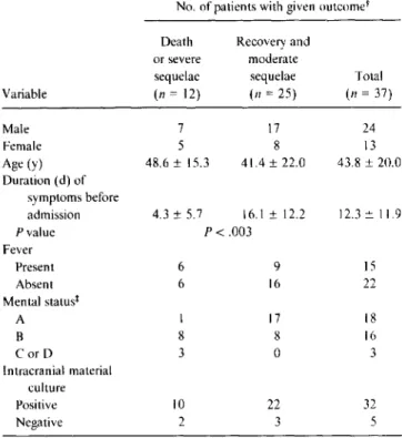
The mean delay between the appearance of the first symp-toms and hospitalization for the 37 patients evaluable at fol-low-up was 12.3 days. This delay was significantly shorter for the 12 patients who died or had severe sequelae than for the 25 patients for whom the outcome was better (4.3 and 16.1 days, respectively). The presence of sequelae was correlated with the severity of the mental status on admission, since most of the patients who recovered (fully or with moderate sequelae) were of mental status A(17 of 25; 68%) and the
CID 1992; 15 (September) Brain Abscesses: Outcome Factors 399
Table5. Outcome for 37 evaluable patients* with brain abscess as compared with clinical characteristics.
No. of patients with given outcome" Death Recovery and
or severe moderate
sequelae sequelae Total
Variable (n=12) (n=25) (11 =37) Male 7 17 24 Female 5 8 13 Age (y) 48.6 ± 15.3 41.4 ± 22.0 43.8 ± 20.0 Duration (d) of symptoms before admission 4.3 ± 5.7 16.1 ± 12.2 12.3± 11.9 Pvalue P< .003 Fever Present 6 9 15 Absent 6 16 22 Mental status! A I 17 18 B 8 8 16 Cor 0 3 0 3 Intracranial material culture Positive 10 22 32 Negative 2 3 5
*Two patients lost at follow-up.
tExcept categories of age and duration of symptoms before admission. which show data as mean±SO.
tSee definitions in text.
three patients with the worst mental status (namely, C or D) died (table 5). There was no correlation with microbiological findings, antibiotic regimens, or type of surgical interven-tion.
Discussion
Although brain abscess is a rare disease, its prognosis is poor. In most series mortality rates of
30%
to50%
were re-ported, but two recent reports have documented rates of4%
to20% [9, 10],
figures that are similar to the13%
rate ob-served in the present series. This improvement has been at-tributed to several factors, among which early diagnosis by means of CT scan was thought to be the most important [2,10, 16-19].
Hence, in a series that compared periods before and after the advent of the CT scan, the excessive mortality associated with brain abscess observed in the first period could be explained entirely by delay in the diagnosis[10].
The increased sensitivity ofMRI observed in the diagnosis of experimental brain abscess appears to be of limited value in humans because there is already a lesion obvious on CT scan when the patient presents[20].
Neurological sequelae which occurred in46%
of our cases, were severe enough to alter social or professional capacities of half of those patients.Death and severe sequelae were mostly observed in patients who presented with a severe neurological impairment on ad-mission and were associated with a significantly shorter delay from the onset of symptoms to admission. Moreover, all pa-tients who died had undergone a CT scan within the first
24
hours after admission, and none of the factors analyzed (lo-cation and number of the abscesses, presence of a predispos-ing factor, type of microorganisms isolated, and type of sur-gery performed) influenced the outcome except the patient's initial clinical impairment and a slightly older mean age, al-though the latter was not a significant factor. These data indicate that, although CT scan is likely to have contributed to the decreased mortality observed in recent series, further improvement in the prognosis of cerebral abscess will now mainly depend on earlier admission and better management of predisposing conditions.Predisposing factors, present in
62%
of our patients, were not equally distributed in the different age groups, in con-trast with other studies[3, 18, 21-24],
and seem to be more frequent in patients<50
years old. Direct spread from an otic or sinusal infectious focus was the most frequent source of brain abscess in young adults in our series, although glob-ally less frequent than in other series[2,
7,10, 22],
and was never noted in patients older than60
years. The incidence of brain abscess due to congenital heart disease(12%
of our cases) can be expected to be lower in the future, thanks to recent advances in cardiovascular surgery[7, 21, 22, 25, 26].
Skin infections (with or without endocarditis) and pulmo-nary abscesses (often related to bronchiectasis) should be recognized as other potential sources of infection. With early and appropriate management of predisposing conditions, the overall incidence of brain abscess should decrease; however, the proportion of patients without predisposing conditions(38%)
or with less-well-defined predisposing conditions (e.g., head injury or steroid therapy) remains a difficult clinical problem, particularly among the elderly.Short duration of symptoms before hospitalization charac-terized patients of ages
20-60
years and those with a poorer outcome: it identified a patient subgroup with an important clinical impairment at admission. All patients presented with symptoms pointing to involvement of theeNS,
but only49%
had focal neurological manifestations; these patients had multiple abscesses slightly more frequently. Presence offever does not appear to be a diagnostic clue. Presence of menin-gismus seems to be more frequently related to an occipital or temporal location of abscess. Our series highlights the diag-nostic difficulties in regard to patients with nonspecific clini-cal presentation and indicates that a high index of suspicion should be maintained even in the absence of the classical manifestations of brain abscess[4, 27-29].
The only definite means of diagnosis remains the determi-nation of the etiologic agents. LP was not helpful in this respect since all the CSF specimens remained sterile in cul-ture. Moreover, this procedure is risky
[18,23, 30],
although400 Seydoux and Francioli CIO 1992; 15 (September)
there were fortunately no complications in our series. CSF analysis offers only nonspecific evidence of CNS involve-ment; it reveals signs of mild to severe inflammation in al-most all cases, a finding that has no prognostic value. Even the presence of biological signs suggestive ofbacterial menin-gitis in three patients (none of whom had focal neurological signs) was not related to the type of preexisting condition, the location of the abscess, or the presence of microorgan-isms in the intracerebral material. The use of LP should ini-tially be restricted only to patients with true meningismus and diffuse CNS signs, as a means of ruling out bacterial meningitis. For patients with focal CNS signs or without meningismus, initiation of antibiotic therapy (after culture of blood and a specimen from any suspected focus of infection) followed by a CT scan will lead to the detection of a focal intracranial process with minimal iatrogenesis.
Surgery is the only procedure allowing optimal microbio-logical documentation and should not be delayed. Antibiotic pretreatment for
<
1°
days does not reduce the rate of posit iv-ity of cultures of intracerebral material. The precise identifi-cation of the isolated microorganisms led to simplifiidentifi-cation of the antibiotic regimen for 12 patients in our series and to modification of the treatment because of the resistance ofS.
aureus
in one case. The choice between excision or stereotac-tic aspiration was mainly dictated by the depth of the lesion. There has been no detectable difference in outcome for the patients who have undergone either one of these two proce-dures, although some authors have observed an increased rate of sequelae, mainly epilepsy, after complete or partial excision [2, 10, 31, 32]. In very highly selected patients, non-surgical treatment with antibiotics alone can be successful. Several authors recommend medical therapy, particularly in cases in which the abscess is <2 em in diameter, when the lesion is of high density (suggesting cerebritis [28, 30, 33, 34]) or when multiple abscesses are present [24, 35-37]. This conservative approach appears to be most appropriate when the responsible microorganisms have been identified by blood culture, which may occasionally be positive (4 of 17 in our series). Such cultures should therefore always be performed, in particular when no prior treatment has been given.The combination of a {J-lactam agent with chlorampheni-col or metronidazole generally has been recommended as standard treatment for bacterial brain abscesses. In contrast with findings in other studies, the type of microorganisms isolated was not related to the origin of the infection in our study. The recognition of an infectious focus as a potential source is not helpful in choosing an antibiotic regimen, ex-cept when a cutaneous source is a possibility, in which case a {J-Iactamase-resistant {J-lactam agent should be used to cover S. aureus. Although all anaerobes we discovered were sensi-tive to {J-Iactam antibiotics, patients may benefit from the good penetration of chloramphenicol or metronidazole into the CNS [24, 35, 36]. Overall duration of treatment should
be 2 months, with the parenteral route of administration used for at least the first 2 weeks, as usually recommended [34-36]. When cultures are negative but histologic evidence of an acute inflammatory process or the presence of organ-isms on gram stain is noted, treatment of patients with a standard combination (a {J-lactam agent with chlorampheni-color metronidazole) or a {J-lactamase-resistant {J-lactam agent (in cases of skin infection) is recommended. However, some patients can recover without any medical treatment.
Twenty-eight patients received corticosteroids. No detect-able difference in outcome was observed between the pa-tients who had received corticosteroids and those who did not. This finding is in accordance with those in other studies [2, 29, 38]. Therapy with corticosteroids appears to be indi-cated only in cases of massive symptomatic cerebral edema. In conclusion, our study confirms that the mortality rate associated with brain abscess might be as low as 13%. Presen-tations at admission are very heterogenous, and no clinical or biological elements are diagnostic. Initial LP should be per-formed only in cases in which bacterial meningitis is highly suspected on account of meningismus and diffuse CNS signs. In other cases, a CT scan will determine the location of a focal brain process. Even though a brain abscess can be suc-cessfully treated with antibiotics alone in some cases, surgery (mainly stereotaxic aspiration) permits a precise microbiolog-ical documentation, thus allowing establishment of the best antibiotic regimen.
The only factor that definitely influences mortality and sequelae is the clinical presentation at admission. Efforts should be directed toward better recognition and manage-ment of predisposing conditions, actions that seem to be the only means of improving outcome for patients with bacterial brain abscess.
Acknowledgments
The authors thank Dr. Pascal Meylan and Dr. Heidi Decrey for their help in initiating this study, Prof. Nicolas de Tribolet for his useful comments, and Ms. Hanny Muller for her typing assistance.
References
I. Carey ME, Chou SN, French LA. Experience with brain abscesses. J Neurosurg 1972;36: 1-9.
2. Chun CH, Johnson JD, Hofstetter M, RatfMJ. Brain abscess: a study of 45 consecutive cases. Medicine (Baltimore) 1986;65:415-31. 3. Le Beau J, Creissard P, Harispe L, Redondo A. Surgical treatment of
brain abscess and subdural empyema. J Neurosurg 1973;38: 198-203.
4. McClelland CJ. Craig BF, Crockard HA. Brain abscesses in Northern Ireland: a 30 year community review. J Neurol Neurosurg Psychiatry 1978;41: 1043-7.
5. Krayenbuhl HA. Abscess of the brain. Clin Neurosurg 1966; 14:25-44. 6. Mathisen GE, Meyer RD, George WL, Citron DM, Finegold SM. Brain
CID 1992:15(September) Brain Abscesses: Outcome Factors 401
7. Morgan H. Wood MW. MurpheyF. Experience with 88 consecutive cases of brain abscess. J Neurosurg1973;38:698-704.
8. Van Alphen HAM. Dreissen JJR. Brain abscess and subdural em-pyema. J Neurol Neurosurg Psychiatry1976;39:481-90.
9. KaplanK.Brain abscess. Med Clin North Am1985;69:345-60. 10. Mampalam TJ. Rosenblum ML. Trends in the management
ofbacte-rial brain abscesses: a review of102 cases over 17 years. Neurosur-gery1988;23:451-8.
II. Yildizhan A. Pasaoglu A, Kandemir B. Effect of dexamethasone on various stages of experimental brain abscess. Acta Neurochir 1989;96: 141-8.
12. Gower D. McGuirt WF. Intracranial complications of acute and chronic infectious ear disease: a problem still with us. Laryngoscope 1983;93: 1028-33.
13. Brant-Zawadzki M, Enzmann DR, Placone RC Jr. et al. NMR imaging of experimental brain abscess: comparison with CT. AJNR 1983;4:250-3.
14. Runge VM. Clanton JA, Price AC, et al. Evaluation of contrast-en-hanced MR imaging in a brain-abscess model. AJNR1985;6: 139-47.
15.Davidson HD, Steiner RE. Magnetic resonance imaging in infections of the central nervous system. AJNR1985;6:499-504.
16. Alderson D. Strong AJ. Ingham HR. Selkon JB. Fifteen-year review of the mortality of brain abscess. Neurosurgery1981 ;8: 1-6.
17. Britt RH. Enzmann DR, Yeager AS. Neuropathological and computer-ized tomographic findings in experimental brain abscess. J Neuro-surg1981;55:590-603.
18. Spanos A. Harrell FE Jr. Durack DT. Differential diagnosis of acute meningitis: an analysis of the predictive value of initial observations. JAMA1989;262:2700-7.
19. Rosenblum ML, HoffJT, Norman D. Weinstein PRo Pitts L. Decreased mortality from brain abscesses since advent ofcomputerized tomogra-phy. J Neurosurg1978;49:658-68.
20. Wispelwey B. Dacey RG Jr. Scheid WM. Brain abscess. In: Scheid WM. Whitley RJ. Durack DT. eds. Infections of the central nervous system. New York: Raven Press,1991:457-86.
21. Brewer NS. MacCarty CS. Wellman WE. Brain abscess: a review of recent experience. Ann Intern Med 1975;82: 571-6.
22. Nielsen H. Gyldensted C, Harmsen A. Cerebral abscess: aetiology and pathogenesis. symptoms. diagnosis and treatment: a review of200 cases from 1935-1976. Acta Neurol Scand 1982;65:609-22.
23. Schliamser SE. Backman K, Norrby SR. Intracranial abscesses in adults: an analysis of 54 consecutive cases. Scand J Infect Dis 1988;20: 1-9.
24. Wispelwey B. Scheid WM. Brain abscess. Clin Neuropharmacol 1987; I0:483-51 O.
25. Nielsen H. Hamsen A. GyldenstedC.Cerebral abscess: a long-term follow-up. Acta Neural Scand 1983;67:330-7.
26. Samson OS. Clark K. A current review of brain abscess. Am J Med 1973;54:20 I-I O.
27. Ariza J. Casanova A. Fernandez Viladrich P. et al. Etiological agent and primary source of infection in42 cases of focal intracranial sup-puration. J Clin Microbiol1986;24:899-902.
28. Fevrier MJ. Nguyen JP. Brunbuisson C, Lepresle E. Abces cerebraux: attitude therapeutique. Presse Med 1987;16: 1222-5.
29. Yang SY. Brain abscess: a review of 400 cases. J Neurosurg 1981;55:794-9.
30. Rosenblum ML. Hoff JT. Norman D. Edwards MS. Berg BO. Nonoper-ative treatment of brain abscesses in selected high-risk patients. J Neurosurg1980;52:217-25.
31. KourtopoulusH.Holm SE, West KA. The management of intracranial abscesses: comparative study between two materials with signifi-cantly different rates of mortality. Acta Neurochir1981;56: 127-8. 32. Rousseaux M. Lesoin F. Destee A. Jomin M. Petit H. Long term
se-quelae of hemispheric abscesses as a function of the treatment. Acta Neurochir1985;74:61-7.
33. Garvey G. Current concepts of bacterial infections of the central ner-vous system. J Neurosurg1983;59:735-44.
34. Weisburg LA. Nonsurgical management of focal intracranial infection. Neurology1981;31:575-80.
35. Heineman HS. Braude A\, Osterholm JL. Intracranial suppurative dis-ease: early presumptive diagnosis and successful treatment without surgery. JAMA1971;218:1542-7.
36. Kamin M. Biddle D. Conservative management of focal intracerebral infection. Neurology1981;31: 103-6.
37. Gleason CA. Wise BL. Feinstein B. Stereotactic localization (with com-puterized tomographic scanning), biopsy. and radiofrequency treat-ment of deep brain lesions. Neurosurgery1978;2:217-22. 38. Quartey GR. Johnston JA. Rozdilsky B. Decadron in the treatment of
cerebral abscess: an experimental study. J Neurosurg 1976;45:301-10.