O R I G I N A L A R T I C L E
Missense mutations in
TENM4, a regulator of axon
guidance and central myelination, cause essential
tremor
Hyun Hor
1,2,3,4,
†,
*, Ludmila Francescatto
5,
‡
, Luca Bartesaghi
6,7,
‡
,
Sara Ortega-Cubero
8,
‡
, Maria Kousi
5
, Oswaldo Lorenzo-Betancor
8
,
Felix J. Jiménez-Jiménez
9
, Alexandre Gironell
10,11
, Jordi Clarimón
11,12
,
Oliver Drechsel
1,2
, José A. G. Agúndez
13
, Daniela Kenzelmann Broz
15
,
Ruth Chiquet-Ehrismann
15
, Alberto Lleó
11
, Francisco Coria
16
,
Elena García-Martin
14
, Hortensia Alonso-Navarro
9
, Maria J. Martí
17
,
Jaume Kulisevsky
10,12
, Charlotte N. Hor
1,2,3,4
, Stephan Ossowski
1,2
,
Roman Chrast
6,7,¶
, Nicholas Katsanis
5,¶
, Pau Pastor
8,§,
*
,
††
and
Xavier Estivill
1,2,3,4,18,§,
*
1
Bioinformatics and Genomics Program, Centre for Genomic Regulation (CRG), Barcelona, Spain,
2Universitat
Pompeu Fabra (UPF), Barcelona, Spain,
3Hospital del Mar Medical Research Institute (IMIM), Barcelona, Spain,
4CRG CIBER de Epidemiología y Salud Pública (CIBERESP), Barcelona, Catalonia 08003, Spain,
5Center for Human
Disease Modeling, Duke University, Duke University Medical Center, Durham NC 27710, USA,
6Department of
Medical Genetics, University of Lausanne, Lausanne 1005, Switzerland,
7Department of Neuroscience and
Department of Clinical Neuroscience, Karolinska Institutet, Stockholm 171 77, Sweden,
8Neurogenetics
Laboratory, Division of Neurosciences, Center for Applied Medical Research (CIMA), and Department of
Neurology, Clínica Universidad de Navarra, University of Navarra School of Medicine and Centro de Investigación
Biomédica en Red Enfermedades Neurodegenerativas (CIBERNED), Pamplona, Navarra 31008, Spain,
9Section of
Neurology, Hospital Universitario del Sureste, Arganda del Rey, Madrid 28030, Spain,
10Movement Disorders Unit,
Neurology Department, Hospital de Sant Pau, Barcelona, Spain,
11Sant Pau Biomedical Research Institute,
Barcelona, Spain,
12Universitat Autònoma de Barcelona and CIBERNED, Barcelona, Catalonia 08026, Spain,
13Department of Pharmacology,
14Department of Biochemistry and Molecular Biology, University of Extremadura,
Cáceres 10071, Spain,
15Faculty of Sciences and Department of Biomedicine, Friedrich Miescher Institute of
Biomedical Research, Novartis Research Foundation and University of Basel, Basel 4058, Switzerland,
16Clinic for
†Present address: Department of Internal Medicine, Lausanne University Hospital (Centre hospitalier universitaire vaudois– CHUV), Rue du Bugnon 46, 1011 Lausanne, Switzerland
††Present address: Memory and Movement Disorders Units, Department of Neurology, University Hospital Mutua de Terrassa, Terrassa, Barcelona, Spain ‡These authors contributed equally to this work
¶These authors contributed equally to this work §These authors contributed equally to this article Received: May 29, 2015. Revised and Accepted: July 13, 2015
© The Author 2015. Published by Oxford University Press. All rights reserved. For Permissions, please email: [email protected] doi: 10.1093/hmg/ddv281
Advance Access Publication Date: 17 July 2015 Original Article
Nervous Disorders, Service of Neurology, Son Espases University Hospital, Palma de Mallorca 07120, Spain,
17
Movement Disorders Unit, Neurology Service, Hospital Clinic, CIBERNED and Institut d’Investigacions
Biomèdiques August Pi i Sunyer (IDIBAPS), Barcelona, Catalonia 08036, Spain and
18Dexeus Women
’s Health,
University Hospital Quiron-Dexeus, Barcelona, Catalonia 08028, Spain
*To whom correspondence should be addressed. Email: [email protected] (H.H); [email protected] (X.E.); [email protected] (P.P.)
Abstract
Essential tremor (ET) is a common movement disorder with an estimated prevalence of 5% of the population aged over 65 years. In spite of intensive efforts, the genetic architecture of ET remains unknown. We used a combination of whole-exome sequencing and targeted resequencing in three ET families. In vitro and in vivo experiments in oligodendrocyte precursor cells and zebrafish were performed to test our findings. Whole-exome sequencing revealed a missense mutation in TENM4 segregating in an autosomal-dominant fashion in an ET family. Subsequent targeted resequencing of TENM4 led to the discovery of two novel missense mutations. Not only did these two mutations segregate with ET in two additional families, but we also observed significant over transmission of pathogenic TENM4 alleles across the three families. Consistent with a dominant mode of inheritance, in vitro analysis in oligodendrocyte precursor cells showed that mutant proteins mislocalize. Finally, expression of human mRNA harboring any of three patient mutations in zebrafish embryos induced defects in axon guidance, confirming a dominant-negative mode of action for these mutations. Our genetic and functional data, which is corroborated by the existence of a Tenm4 knockout mouse displaying an ET phenotype, implicates TENM4 in ET. Together with previous studies of TENM4 in model organisms, our studies intimate that processes regulating myelination in the central nervous system and axon guidance might be significant contributors to the genetic burden of this disorder.
Introduction
Essential tremor (ET [MIM 190300]) is a common hyperkinetic movement disorder with an estimated prevalence of 1% in the population. The prevalence increases with age, and 5% of indivi-duals over 65 years of age are affected (1). ET is typically charac-terized by rhythmic, involuntary shaking of one or more parts of the body, and occurs exclusively during voluntary movements (action tremor) or in positions against gravity ( postural tremor) (2). The phenotypic severity of ET is variable, as evidenced by the existence of both highly disabling and milder forms of the disease. ET is considered a complex disorder with a strong genet-ic component (3,4), since more than half of affected individuals have a positive family history.
Thefirst genetic locus associated with ET was near the leucine-rich repeat and lg domain containing nogo receptor-interacting protein 1 gene (LINGO1 [MIM 609791]), identified by a genome-wide association study (GWAS) in the Icelandic popula-tion (5) and replicated in other populations (6). Other studies, in-cluding several candidate gene association studies and a recent GWAS (7), revealed potential associations that nonetheless failed to replicate (4). Additionally, three linkage studies conducted in large ET families displaying an autosomal-dominant pattern of inheritance identified only putative candidate loci, but did not find any underlying disease causing gene(s) (8–10). Factors that have confounded the identification of causal genes include non-penetrance of ET; a considerable number of misdiagnoses in the absence of reliable biomarkers; and the existence of pheno-copies, as distinct sporadic cases may occur within the same family given the high prevalence in the general population (11). In this regard, numerous ET families have been reported that are characterized by an overrepresentation of affected individuals clearly exceeding the expected 50–50% ratio, which is clearly beyond the classical pattern of autosomal inheritance (11–13).
Thefirst published exome-sequencing study in ET identified a causative nonsense mutation in fused in sarcoma (FUS [MIM 137070]) that was segregating with ET in a large pedigree (14).
However, only two additional cases carried other mutations in the same gene, while independent replication studies uncovered few additional mutations, suggesting that FUS is a rare cause of ET (15–18). More recently, a rare variant in HtrA serine peptidase 2 (HTRA2 [MIM 610297]) was shown to cause ET in a large six-generation family. Interestingly, signs of Parkinson’s disease (PD) additionally appeared in the middle age in homozygous car-riers of this variant (19).
Given the large proportion of familial ET cases without causal mutations, and the low-to-moderate effect size of the identified variant near LINGO1, a large fraction of the heritability of ET re-mains unexplained. We sought to identify new genes involved in the pathogenesis of ET by studying orphan ET families by exome sequencing. Here we describe the identification of a new gene for ET by the combination of exome and targeted resequen-cing as well as functional testing using in vitro assays in oligoden-drocytes precursor cells and in vivo assays in a zebrafish model.
Results
Exome sequencing
The clinical diagnosis of ET was made initially in the index case (II-5) of a four-generation family (TEF-6) of Spanish origin (Fig.1A). The phenotypic severity varied among affected family members, ranging from a highly disabling to mild tremor (Sup-plementary Material, Table S1). Given such common phenotypic variance and transmission of the disease across three genera-tions, we postulated an autosomal-dominant pattern of inherit-ance in this family. The available data did not allow us to perform linkage analysis, mainly due to limited participation and de-ceased family members. We therefore performed whole-exome sequencing in four affected family members (II-5, III-2, III-4 and IV-1) assuming that the disease causing variant is located in the protein coding region, cognizant of the fact that a mutation in a regulatory region not captured by this technology cannot be excluded. After alignment and variant calling, we first 5678 | Human Molecular Genetics, 2015, Vol. 24, No. 20
evaluated variants in genes associated previously with auto-somal-dominant ET and PD (in particular FUS [MIM 137070], SNCA [MIM 163890], LRRK2 [MIM 609007]). Despite adequate coverage in these regions, we did notfind any candidate patho-genic mutations. We nextfiltered for single nucleotide variants (SNVs) and indels that (a) were novel and rare [minor allele fre-quency (MAF) below 0.5%]; (b) were shared by all four individuals sequenced and (c) were predicted computationally to be damaging by Polyphen-2, PROVEAN and CADD (20–22). This yielded a list of rare alleles in four candidate genes, namely RCOR3, CYTL1 [MIM 607930], E2F8 [MIM 612047] and TENM4 [MIM 610084], all of which segregated with the disease phenotype in our pedigree. To avoid bias, we mined expression data for all four genes. We found that RCOR3, CYTL1 and E2F8 are not expressed appreciably in neuronal tissues, while subsequent literature searches intimate an involvement of these transcripts in hepatitis, chondrogenesis and hepatocellular carcinoma (3,23,24). In contrast, TENM4 is expressed primarily in the brain and has been detected in several transcriptomes derived from neuronal tissues (see Materials and Methods). We also noted that Suzuki and colleagues (25) generated a mouse Tenm4 knockout model recently that displays hypomyeli-nation of small-diameter axons in the CNS and a severe action tre-mor phenotype (as seen in video by Suzuki et al., http://www. youtube.com/watch?v=bcmcjLuB_bs). Taken together, this evi-dence brought our focus to the missense mutation in TENM4 (c.4100C>A) that leads to a threonine-to-asparagine amino acid substitution (p.T1367N).
The c.4100C>A mutation, which has never been detected before (absent in 1000 Genomes and EVS), was present in all affected individuals. We found two healthy offspring in the fourth generation (IV-2 and IV-3) to be mutation carriers as well. However, these subjects are <40 years old and are thus likely presymptomatic carriers that may develop ET, although reduced
penetrance remains a possible scenario. copy number variants (CNVs) analysis did not yield any genomic rearrangements that were shared by all four subjects.
Targeted resequencing of TENM4 and segregation analysis in additional ET families
As afirst test of the candidacy of TENM4, we asked whether there might be additional ET families with segregating mutations in this gene. To this end, we performed targeted resequencing of TENM4 in 299 index cases of familial ET of Spanish origin (the mean age of onset = 45 ± 22 years). We recorded all novel mis-sense, splice-site or nonsense variants with a MAF below 0.1% according to the EVS and identified 12 missense mutations, of which 11 were predicted to be damaging by Polyphen-2 (20) as well as by PROVEAN and CADD (21,22) (Table1). DNA samples were available from additional family members of two mutation carriers. One of the two families was FET-3, a four-generation family comprising 11 affected family members (Fig.1B; Supple-mentary Material, Table S2). Having identified the predicted dam-aging variant c.4324 G>A ( p.A1442T) in II-1, we evaluated its segregation in all available family members. Strikingly, 9 out of 11 affected carried this variant. Targeted resequencing of TENM4 in III-1 and III-3 excluded other TENM4 mutations, sug-gesting that these two individuals are potential phenocopies, a phenomenon observed frequently in ET (13). We also detected c.4324 G>A in one unaffected offspring in the third generation (III-7) and another in the fourth generation (IV-2); although both subjects were reported to be healthy, they might still develop the disease at a later age (current age: 42 and 5, respectively). Nevertheless, reduced penetrance cannot be excluded at this stage. The second family was a three-generation pedigree, TEF-9, with five affected family members (Fig. 1C; Supplementary
Figure 1. Pedigrees of Spanish essential tremor families TEF-6 (A); FET-3 (B) and TEF-9 (C). (Square) male family members. (Circle) female family members, (filled symbol) individuals with ET. (*) exome-sequenced individuals. (Dashed symbol) deceased individual. TENM4 genotype status: TEF-6—TENM4 c.4100C>A (C/A = heterozygous mutant allele); FET-3: TENM4 c.4324 G>A (G/G = homozygous reference allele, G/A = heterozygous mutant allele; TEF-9: TENM4 c.3412G>A (G/G = homozygous reference allele, G/A = heterozygous mutant allele). If the genetic status is not indicated, the corresponding DNA samples were unavailable.
Material, Table S3), for three of which we had DNA available. We found the c.3412G>A variant ( p.V1138M) in two of these family members (II-4 and III-1). The third individual, III-2, did not carry this mutation, however, in contrast to her siblings, she was not self-aware of her tremor, which was mild and observed by the clinician only. Taken together, the segregation and phenotyping of individuals from three multigenerational ET pedigrees provided suggestive evidence of involvement of TENM4 mutations. As an alternative means to test the candidacy of TENM4, we interrogated the transmission behavior of the discovered alleles. The null hypothesis (TENM4 alleles transmit randomly) was rejected: among the 15 parent-affected offspring scorable transmissions across the three pedigrees, the TENM4 mutant allele was transmit-ted 12 times, a significant departure from 50:50 chance (χ2P < 0.02). In vitro testing of TENM4 mutations
Our genetic data suggested a causal link between TENM4 variants and ET. However, because the pathogenic potential of the discov-ered alleles was predictive, we sought direct evidence. We thus evaluated the impact of the missense mutations segregating with ET in our two largest ET pedigrees on subcellular localiza-tion of TENM4 protein. We transfected Oli-neu cells with expres-sion vectors containing full-length human TENM4 (wild-type), negative control c.7933G>A and TENM4 carrying the c.4100C>A, c.4324 G>A and c.3412G>A mutations. In cells transfected with wild-type TENM4 and the negative control c.7933G>A, an allele that has been found in homozygosity in 2/6342 individuals in EVS, the protein localized homogeneously at the cell membrane. In contrast, c.4100C>A ( p.T1367N), c.4324 G>A ( p.A1442T) and c.3412G>A ( p.V1138M) showed clustered TENM4 membrane localization (Fig.2).
Modeling of TENM4 in zebrafish
As a second test of allele pathogenicity, as well as a means to de-termine the direction of effect, we turned to an in vivo zebrafish model. Given previousfindings of central nervous system demye-lination in a homozygous null Tenm4 mouse model (25), wefirst assayed myelin levels in the brain by RNA in situ hybridization. In-jection of either a translation blocking morpholino (tbMO) or a
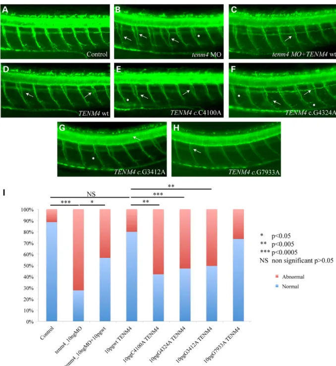
splice-blocking morpholino (sbMO) (Supplementary Material, Fig. S1) against the sole endogenous copy of zebrafish tenm4 caused modest reduction in intensity of myelin in the brain, likely reflective of the fact that most, but not all, endogenous tenm4 was suppressed by our MO (data not shown). In contrast, we observed a significant increase in the number of aberrant small-diameter neuronal axons along the notochord of morphants (either tb or sbMOs) at 2 days post fertilization (2dpf, Fig.3A and B). The lesions observed involved extension and pathfinding errors, as well as ab-errant branching of the remaining axons, in a pattern reminiscent of the pathfinding defects reported in a Drosophila mutant of the teneurin/tenascin family (26). The number or neuronal somata dorsal to the notochord was also compared across control and morphant embryos, but revealed no differences among the two groups of animals, suggesting that it is not the number or neurons that is affected but the innervation pattern along the peripheral nervous system (PNS). The innervation of the truncal muscula-ture as determined by the neuronal axon aberrations was rescued by co-injection of human TENM4 mRNA, demonstrating that the phenotype was specific (Fig.3). These data were encouraging but, by themselves, did not constitute proof of TENM4 causality, since they showed only that this transcript/protein is necessary for axonal guiding functions in the PNS. They did, however, offer the opportunity to test the effect of the discovered patient alleles on protein function. We therefore engineered each of the three mutations and injected them into wild-type zebrafish embryos. Injection of TENM4 mRNA carrying the mutations iden-tified in TEF-6, FET-3 and TEF-9 (c.4100C>A, c.4324 G>A, c.3412G>A and c.7933G>A), but not of the wt construct nor the negative con-trol high frequency allele c.7933G>A resulted in a phenotype that phenocopied the one induced by the MO. These data suggested that these alleles adversely impact the function of the protein and exert a likely dominant-negative effect (Fig.3), supporting our earlier in vitro observations.
Discussion
Here we report pathogenic mutations in TENM4 by exome se-quencing in a family with ET, followed by the identification of mutations in two additional families. Although the segregation of the candidate alleles indicates that penetrance is high, in
Table 1 Novel variants (MAF <0.001) found among 300aindex cases diagnosed for familial essential tremor
Chromosomal position Nucleotide change Type of mutation Ref.-allele Mut.-allele MAF in EVS (Caucasians) Number of occurrences in 300 cases Amino acid change Polyphen-2 prediction 11:78780832 c.158G>C M C G NA 1 p.R53P 2 11:78567058 c.1421C>A M G T NA 1 p.A474D 2 11:78565277 c.1553G>A M C T 0.000943 1 p.R518Q 2 11:78443446 c.3053G>A M C T NA 1 p.R1018H 2 11:78440445 c.3382G>A M C T NA 1 p.V1128M 2 11:78437262b c.3412G>A M C T NA 2 p.V1138M 2 11:78419515c c.4100C>A M G T NA 1 p.T1367N 2 11:78413334d c.4324G>A M C T 0 1 p.A1442T 2 11:78413055 c.4603A>C M T G NA 1 p.K1535Q 1 11:78412763 c.4895G>A M C T 0.00048 1 p.R1632H 1 11:78387406 c.5287G>A M C T 0.000942 1 p.G1763R 2 11:78380037 c.7353G>A M C T 0.0006 1 p.M2451I 1
aIncluding thefirst mutation identified by exome sequencing in TEF-6.
The variant at chromosomal position marked with‘c’ was segregating in family TEF-6. The variant at chromosomal position marked with ‘d’ was segregating in family FET-3. The variant at chromosomal position marked with‘b’ was segregating in family TEF-9. Type of mutations: M = missense. Polyphen-2 prediction: 1 = possibly damaging, 2 = probably damaging. NA = not applicable. All variants are heterozygous.
that we did not observe any healthy individuals above the me-dian age of ET with these mutations, we did identify three likely phenocopies across two families. This observation is consistent with recent discussions on the genetic architecture of ET (11–13), but also of other neurological disorders such as restless legs syndrome (27), where the presence of phenocopies has been highlighted as a major confounder in searching for causal alleles in large multiplex families. At the same time, the term ‘phenocopies’ might also be misleading, as the rate of misdiag-nosis is known to be high in ET and many other reported families are characterized by an overrepresentation of affected indivi-duals within one pedigree, raising the question whether a rather more complex genetic architecture should be considered in the pathogenesis of ET including di- or multigenetic inheritance as well as epigenetics (11–13). Indeed, the identification of a single causal variant in all six affected siblings in the third generation of family FET-3 seems unlikely. We also draw attention to the fact that in other neurological disorders, such as PD, which has some clinical overlap with ET, the presence of phenocopies is also a known phenomenon, especially in familial PD caused by mutations in LRRK2, a well-established cause of this condition (28). In fact, in∼14.4% of families with LRRK2 driver mutations, additional affected members carry the reference alleles only (29). The Teneurin family of genes (TENM1–TENM4) encodes trans-membrane proteins that are expressed predominantly in neu-rons (30). TENM4 is the only member also expressed in the white matter of the cerebellum of adult mice (31) and was identi-fied recently as a regulator of oligodendrocyte maturation and myelination of small-diameter axons in the CNS (25). Interesting-ly, fractional anisotropy neuroimaging has shown, though not consistently, white matter abnormalities in different parts of the brain, particularly in the cerebellum, in ET patients (32). Add-itionally, teneurins play a major role in motor neuron guidance
and neuronal synapse organization in Drosophila (26) and to affect neurite outgrowth in neuroblastoma cells (33). Our experiments in Oli-neu cells as well as our zebrafish experiments concord with these previous findings. Notably, the two of the three TENM4 point mutations assayed in cells (c.4100C>A, c.4324 G>A), are both located in the NHL-repeat/β propeller domain, which has recently been shown to be crucial for the homophilic interaction between TENM1 and TENM2 (34). Changes in TENM4 localization may contribute to defects in TENM4-mediated regu-lation of the phosphoryregu-lation of the focal adhesion kinase, which activates CDC42 and RAC1, two Rho-GTPases that are required for oligodendroglial process outgrowth (35). Furthermore, our sup-pression and overexsup-pression experiments in zebrafish show neuronal pathfinding defects, aberrant branching and extension defects of the small-diameter axons that innervate the truncal musculature. These are concordant with the aforementioned studies performed in neuronal cultures and the fruitfly, denoting that TENM4 is essential for the integrity of the axonal outgrowth and neuronal innervation patterns. Furthermore, the disease as-sociation of three of the familial TENM4 mutations, but not the benign c.7933G>A allele, is supported by the demonstration of a dominant-negative effect of these alleles. These data also recon-cile an apparent discrepancy between homozygous (the presence of tremor) and heterozygous (the absence of tremor) Tenm4 knockout mice: the dominant-negative effect of the discovered mutations is expected to be more severe than haploinsufficiency, but not as severe as the null phenotype. Similar observations have been made recently in other disorders and their corresponding mouse models (36). We are aware that our in vivo functional data provide only a limited insight into an adult onset phenotype. Nonetheless, in addition to our zebrafish studies being concordant with the mouse model, this model organism has been used suc-cessfully to model other adult onset disorders, including limb
Figure 2. In vitro characterization of the cellular distribution of human TENM4 mutants. Localization of wild-type (wt) and mutated forms of TENM4 in Oli-neu cells. Wild-type TENM4 as well as the control allele G7933A (c.7933G>A) showed a homogeneous membrane distribution. In contrast, the mutated forms C4100A (c.4100C>A), G4324A (c.4324 G>A) and G3412A (c.3412G>A) showed clustered localization at the membrane. TENM4 is in red; DAPI nuclear dye is in blue.
girdle muscular dystrophy (37) and, more relevant to the current phenotype, restless legs syndrome (38).
In conclusion, we provide several lines of evidence for a new gene for ET. First, we have identified three families of Spanish ori-gin, in which three distinct pathogenic mutations in TENM4 both segregated with the phenotype and were over-transmitted in parent–offspring analyses. Second, we showed both in vitro and
in vivo a dominant-negative effect of the identified mutations. Third, the existence of a TENM4 knockout mouse displaying a striking and severe ET phenotype with hypomyelination con-firms our findings that abolished or strongly reduced activity of TENM4 drives the development of ET by affecting key cellular processes in the nervous system, and substantiates the identifi-cation of a major gene in the disease pathogenesis of ET.
Figure 3. Suppression of tenm4 in zebrafish causes defects in motor axon pathfinding, outgrowth and branching. (A–H) Lateral views of zebrafish embryos at 2dpf stained with acetylated tubulin. In wild-type (wt) embryos (A), motor axons have completed migration from the periphery, along the common path, and extended to the myotome. In tenm4 morphants (B), secondary axons either fail to migrate along their path (white arrows), exit the periphery but fail to extend, or branch abnormally (asterisks). Suppression of tenm4 can be partially rescued by co-injection with human wt RNA (C). Overexpression of human Tenm4 wt causes mild pathfinding errors (D), suggesting dose sensitivity for TENM4, Human Tenm4 mutants C4100A (c.4100C>A), G4324A (c.4324 G>A) and G3412A (c.3412G>A), when overexpressed are significantly more severe than TENM4 wt and have similar effects to suppression of tenm4 by MO knockdown (E–G). Overexpression of control allele G7933A (c.7933G>A) causes mild pathfinding errors like overexpression of wt allele (H). (I) Percentage of normal versus abnormal embryos under the conditions being evaluated above.
We also show that, given the apparent genetic and phenotyp-ic confounders in ET, coupling in vitro and in vivo studies with gen-omics approaches is critical to improving our ability to interpret the pathogenic effect of rare candidate alleles. The genetic het-erogeneity and complex architecture mandate that candidates such as TENM4 should be investigated in additional families and larger cohorts. It is worthwhile to note that LINGO1, the first gene found to be consistently associated with ET, is also known to play an important role in oligodendrocyte maturation and central myelination. Together with thisfinding, our data sug-gest that further studies of oligodendrocyte and axonal guidance dysfunction might extend the catalog of genetic drivers of ET and help elucidate the genetic architecture of the disease.
Subjects, Materials and Methods
Study design and subjects
All participants included in the study were Europeans of Spanish origin and provided written informed consent. The study proto-col was approved by the local ethics committees of the different hospitals providing the samples. Each patient and unaffected relative of the three families described here were examined by movement disorders specialists (the same neurologist for each family) and diagnoses were made according to the guidelines of the Consensus Statement of the Movement Disorders Society (2). Tremor severity was rated by the Fahn, Tolosa and Marin as well as the Glass scale (39,40). Moreover, the families were pre-screened for known ET and PD mutations (FUS [MIM 137070], SNCA [MIM 163890], LRRK2 [MIM 609007], VPS35 [MIM 601501], PINK1 [MIM 608309] and PARKIN [MIM 602544]).
Exome sequencing
We prepared sequencing libraries using the TruSeq DNA library preparation kit (Illumina) followed by exome capturing with the Nimblegen SeqCap EZ Human Exome Library v3.0 kit according to the manufacturers’ protocols. Five single-indexed libraries were pooled before capturing. We sequenced the multiplexed cap-tured libraries on a HiSeq2000 (Illumina) resulting in afinal mean coverage ranging from 39 to 43-fold and aligned the paired-end reads (100 bp) to the Human Genome (UCSC hg19) using bwa (v0.6.2) (41). We then called SNV and indels using three different software packages (GATK (42), SAMtools (43) and SHORE (44)), annotated the variants with ANNOVAR (45) and intersected the output of the three calling programs to obtain a reliable set of variants. After selecting for variants shared by all affected individuals, and establishing population frequency for each variant from the 1000 Genomes database and the Exome Variant Server (46,47), we applied the followingfiltering steps to obtain afinal list of candidate variants: we focused on non-synonymous, splice-site, nonsense and frameshift variants, and performed in silico predictions of the damaging potential of each candidate variant using Polymorphism Phenotyping ver-sion 2 (Polyphen-2), Protein Variation Effect Analyzer (PROVEAN) and Combined Annotation Dependent Depletion (CADD) (20–22). We also analyzed the exome data for CNV with Conifer (48).
Targeted resequencing
We applied a recently published method based on the design and usage of molecular inversion probes (MIP) which allows high-throughput resequencing of loci of interest at low cost (49). MIPs were designed to target the coding region of TENM4 [MIM 610084] ( probe sequences available on request). After pooling
and 5′-phosphorylation, MIPs were hybridized to genomic DNA, followed by gap-filling and circularization. Capture circles were amplified by polymerase chain reaction (PCR) where the Illumina sequencing adaptor and one of 384 sample specific barcodes were appended. After cleanup and pooling of up to 384 single-indexed samples, multiplexed libraries were sequenced on a MiSeq (Illu-mina) using 150 bp paired-end reads. Paired-end reads were col-lapsed to one single read with FLASH (50) to reduce potential sequencing errors. Alignment, variant calling andfiltering fol-lowed the same pipeline as exome sequencing. Identified novel variants (MAF <0.001) were validated by Sanger sequencing ( pri-mer sequences available on request).
Production of anti-TENM4 antibodies
A recombinant fusion protein between the Tenascin-C signal peptide and the human teneurin-4 EGF-like repeats was gener-ated by splicing by overlap extension. The resulting PCR fragment was cloned into the expression vector pCEP4 (Invitrogen) and transfected into HEK293 EBNA cells. The recombinant protein was purified from the tissue culture medium using the Probond Purification System (Invitrogen) and sent for injection into rab-bits to generate polyclonal antibodies. The antibodies were tested to recognize the recombinant protein as well as the endogenous TENM4 protein in tissue lysates. Using human glioblastoma pro-tein lysates on western blots, the anti-TENM4 antiserum de-tected a single band above 200 kDa, which was not detectable anymore after preabsorption of the antiserum with the recom-binant TENM4 protein used for immunization (not shown).
TENM4 cloning and transfection
We PCR-amplified TENM4 from a human brain cDNA library (Clonetech) and cloned the product into pcDNA 3.1 (Invitrogen). Sanger sequencing validation of the vector uncovered eight synonymous changes and seven point mutations that we retro-mutated using site-directed in vitro mutagenesis (Agilent). We gen-erated TENM4 c.4100C>A, c.4324 G>A, c.3412G>A and c.7933G>A mutants using the same strategy. Murine oligodendroglial precur-sor cells (Oli-neu) were transfected with wild-type and mutant TENM4 as described (51). Cells werefixed in phosphate-buffered saline containing 4% paraformaldehyde, permeabilized with 0.1% Triton and blocked with 5% goat serum. Cells were then incubated overnight with the primary antiserum (rabbit anti-hTENM4) in 2.5% goat serum followed by goat anti-rabbit-biotin-conjugated and by goat anti-rabbit-streptavidin-Cy3 (Jackson Immunoresearch Laboratories). Nuclei were counterstained with 4’,6-diamidino-2-phenylindole (DAPI) and preparations were analyzed with a Leica SP5 AOBS confocal microscope.
In silico analysis of ET candidates
Expression profiles for RCOR3, CYTL1 [MIM 607930], E2F8 [MIM 612047] and TENM4 [MIM 610084] were obtained from the BioGPS database (52). Information on the specific tissues in which each of these genes is detected was retrieved from AceView (53).
Zebrafish functional assay
We used reciprocal BLAST against the Danio rerio genome and identified a sole zebrafish ortholog of TENM4 (ENSDARG00000 034264, 87% similarity, 77% identity). To determine the effect of tenm4 suppression in zebrafish embryos, two morpholinos (MO) were designed and obtained from Gene Tools, LLC (Philomath, OR, USA), tenm4 tbMO (AGGGTCTGCGTTCCTTGACTTCCAT),
targeting the translation site and tenm4 sbMO (ATGCGGTTTCA-TACTAACCGATTGC), targeting the splice junction at the 3′ end of exon 10. To determine MO efficiency, total mRNA was ex-tracted from control and MO-injected embryos, reverse-tran-scribed to produce cDNA and the site targeted by the MO was PCR amplified using the following primers: GGAGGTCG-CAGGTCTTTATTG and CTGCAGTCGGGTCCTCTGAAGC, as de-scribed (54).
For zebrafish injections, capped human TENM4 mRNA was produced by cloning wild-type TENM4 (NM_001098816.2) cDNA and the four mutant constructs (c.4100C>A, c.4324 G>A, c.3412G>A and c.7933G>A) into the pCS2+ expression vector and reverse transcribing each construct in vitro with the T7 Mes-sage Machine kit (Ambion).
All experiments were carried out with the approval of the In-stitutional Animal Care and Use Committee. Zebrafish were maintained and mated according to standard procedures (55). For knockdown and rescue experiments, we injected 10 ng of sbMO, and/or 10 pg human mRNA into zebrafish embryos at 1–4 cell stage. Injected embryos werefixed at 2 dpf and were stained with anti-α acetylated tubulin primary antibody as described (56). The integrity of the PNS was evaluated by scoring embryos for motor neuron abnormalities involving pathfinding errors, abnor-mal branching and failure to extend. All the experiments were re-peated three times and a Pearson’s chi-square test (χ2) was used
to determine the significance.
For in situ analysis, a myelin basic protein (mbp) probe was de-signed using the following primers: AATTAACCCTCACTAAAG GGGCCACTGCAAGCACCTCTGG and TAATACGACTCACTATAG GACGAGGAGAGGACACAAAGC. RNA in situ hybridization was performed as described (57).
Acknowledgments
We thank patients and families for their participation in this study. We thank Beth K. Martin and Jay Shendure for technical advice regarding MIP sequencing as well as Elena Lorenzo and Elena Alonso for technical assistance and Jacqueline Trotter for providing us with Oli-neu cells. We also thank our Spanish colla-borators Maria A. Pastor, Mario Riverol, Julian Benito-Leon, Ruben Fernandez-Santiago, Mario Ezquerra, Francesc Valldeoriola, Yar-oslau Compta and Eduard Tolosa for providing additional DNA samples of ET cases.
Conflict of Interest statement. None declared.
Funding
This work was supported by the Spanish Plan Nacional [SAF2008-00357 (NOVADIS) to X.E., SAF2006-10126 (2006–2009) and SAF2010-22329-C02-01 (2011–2013) to P.P. and RD12/0013/0002 (2013–2017) to J.A.G.A.]; the Generalitat de Catalunya (AGAUR 2009 SGR-1502 to X.E.); the European Commission 7th Framework Program [Project No. 261123 (GEUVADIS) and Project No. 262055 (ESGI) to X.E.]; the UTE project Foundation for Applied Medical Research (FIMA) to P.P.; by the Swiss National Science Foundation (PBLAP3-136962 to H.H., 31003A_135735/1 to R.C.); by seed funding from the Center for Human Disease Modeling, Duke University and by P50 MH094268 to N.K. N.K. is a Distinguished Brumley Professor.
References
1. Louis, E.D. and Ferreira, J.J. (2010) How common is the most common adult movement disorder? Update on the
worldwide prevalence of essential tremor. Mov. Disord., 25, 534–541.
2. Deuschl, G., Bain, P. and Brin, M. (1998) Consensus statement of the Movement Disorder Society on Tremor. Ad Hoc Scien-tific Committee. Mov. Disord., 13(Suppl. 3), 2–23.
3. Deng, Q., Wang, Q., Zong, W.Y., Zheng, D.L., Wen, Y.X., Wang, K.S., Teng, X.M., Zhang, X., Huang, J. and Han, Z.G. (2010) E2F8 contributes to human hepatocellular carcinoma via regulat-ing cell proliferation. Cancer Res., 70, 782–791.
4. Jimenez-Jimenez, F.J., Alonso-Navarro, H., Garcia-Martin, E., Lorenzo-Betancor, O., Pastor, P. and Agundez, J.A. (2013) Up-date on genetics of essential tremor. Acta Neurol. Scand., 128, 359–371.
5. Stefansson, H., Steinberg, S., Petursson, H., Gustafsson, O., Gudjonsdottir, I.H., Jonsdottir, G.A., Palsson, S.T., Jonsson, T., Saemundsdottir, J., Bjornsdottir, G. et al. (2009) Variant in the sequence of the LINGO1 gene confers risk of essential tre-mor. Nat. Genet., 41, 277–279.
6. Jimenez-Jimenez, F.J., Garcia-Martin, E., Lorenzo-Betancor, O., Pastor, P., Alonso-Navarro, H. and Agundez, J.A. (2012) LINGO1 and risk for essential tremor: results of a meta-analysis of rs9652490 and rs11856808. J. Neurol. Sci., 317, 52–57.
7. Thier, S., Lorenz, D., Nothnagel, M., Poremba, C., Papengut, F., Appenzeller, S., Paschen, S., Hofschulte, F., Hussl, A.C., Hering, S. et al. (2012) Polymorphisms in the glial glutamate transporter SLC1A2 are associated with essential tremor. Neurology, 79, 243–248.
8. Gulcher, J.R., Jonsson, P., Kong, A., Kristjansson, K., Frigge, M. L., Karason, A., Einarsdottir, I.E., Stefansson, H., Einarsdottir, A.S., Sigurthoardottir, S. et al. (1997) Mapping of a familial es-sential tremor gene, FET1, to chromosome 3q13. Nat. Genet., 17, 84–87.
9. Higgins, J.J., Pho, L.T. and Nee, L.E. (1997) A gene (ETM) for es-sential tremor maps to chromosome 2p22-p25. Mov. Disord., 12, 859–864.
10. Shatunov, A., Sambuughin, N., Jankovic, J., Elble, R., Lee, H.S., Singleton, A.B., Dagvadorj, A., Ji, J., Zhang, Y., Kimonis, V.E. et al. (2006) Genomewide scans in North American families reveal genetic linkage of essential tremor to a region on chromosome 6p23. Brain, 129, 2318–2331.
11. Ma, S., Davis, T.L., Blair, M.A., Fang, J.Y., Bradford, Y., Haines, J. L. and Hedera, P. (2006) Familial essential tremor with appar-ent autosomal dominant inheritance: should we also con-sider other inheritance modes? Mov. Disord., 21, 1368–1374. 12. Testa, C.M. (2013) Key issues in essential tremor genetics
re-search: Where are we now and how can we move forward? Tremor Other Hyperkinet. Mov. (N. Y.), 3, tre-03-105-1843-1. 13. Zimprich, A. (2012) Phenocopies in families with essential
tremor and restless legs syndrome challenge Mendelian laws. Epigenetics might provide answers. Parkinsonism Relat. Disord., 18, 711–716.
14. Merner, N.D., Girard, S.L., Catoire, H., Bourassa, C.V., Belzil, V. V., Riviere, J.B., Hince, P., Levert, A., Dionne-Laporte, A., Spie-gelman, D. et al. (2012) Exome sequencing identifies FUS mu-tations as a cause of essential tremor. Am. J. Hum. Genet., 91, 313–319.
15. Bentley, D.R., Balasubramanian, S., Swerdlow, H.P., Smith, G. P., Milton, J., Brown, C.G., Hall, K.P., Evers, D.J., Barnes, C.L., Bignell, H.R. et al. (2008) Accurate whole human genome se-quencing using reversible terminator chemistry. Nature, 456, 53–59.
16. Ortega-Cubero, S., Lorenzo-Betancor, O., Lorenzo, E., Alonso, E., Coria, F., Pastor, M.A., Fernandez-Santiago, R., Marti, M.J., 5684 | Human Molecular Genetics, 2015, Vol. 24, No. 20
Ezquerra, M., Valldeoriola, F. et al. (2013) Fused in Sarcoma (FUS) gene mutations are not a frequent cause of essential tremor in Europeans. Neurobiol. Aging, 34, e2449–e2441. 17. Rajput, A., Rajput, A.H., Rajput, M.L., Encarnacion, M.,
Ber-nales, C.Q., Ross, J.P., Farrer, M.J. and Vilarino-Guell, C. (2013) Identification of FUS p.R377W in essential tremor. Eur. J. Neurol., 21, 361–363.
18. Zheng, W., Deng, X., Liang, H., Song, Z., Gao, K., Yang, Y. and Deng, H. (2013) Genetic analysis of the fused in sarcoma gene in Chinese Han patients with essential tremor. Neurobiol. Aging, 34, 2078 e2073–2074.
19. Unal Gulsuner, H., Gulsuner, S., Mercan, F.N., Onat, O.E., Walsh, T., Shahin, H., Lee, M.K., Dogu, O., Kansu, T., Topalo-glu, H. et al. (2014) Mitochondrial serine protease HTRA2 p. G399S in a kindred with essential tremor and Parkinson dis-ease. Proc. Natl. Acad. Sci. U. S. A., 111, 18285–18290.
20. Adzhubei, I.A., Schmidt, S., Peshkin, L., Ramensky, V.E., Gera-simova, A., Bork, P., Kondrashov, A.S. and Sunyaev, S.R. (2010) A method and server for predicting damaging missense mu-tations. Nat. Methods, 7, 248–249.
21. Choi, Y., Sims, G.E., Murphy, S., Miller, J.R. and Chan, A.P. (2012) Predicting the functional effect of amino acid substitu-tions and indels. PLoS One, 7, e46688.
22. Kircher, M., Witten, D.M., Jain, P., O’Roak, B.J., Cooper, G.M. and Shendure, J. (2014) A general framework for estimating the relative pathogenicity of human genetic variants. Nat. Genet., 46, 310–315.
23. Kim, J.S., Ryoo, Z.Y. and Chun, J.S. (2007) Cytokine-like 1 (Cytl1) regulates the chondrogenesis of mesenchymal cells. J. Biol. Chem., 282, 29359–29367.
24. Xue, J.H., Zheng, M., Xu, X.W., Wu, S.S., Chen, Z. and Chen, F. (2011) Involvement of REST corepressor 3 in prognosis of human hepatitis B. Acta Pharmacol. Sin., 32, 1019–1024. 25. Suzuki, N., Fukushi, M., Kosaki, K., Doyle, A.D., de Vega, S.,
Yoshizaki, K., Akazawa, C., Arikawa-Hirasawa, E. and Yama-da, Y. (2012) Teneurin-4 is a novel regulator of oligodendro-cyte differentiation and myelination of small-diameter axons in the CNS. J. Neurosci., 32, 11586–11599.
26. Mosca, T.J., Hong, W., Dani, V.S., Favaloro, V. and Luo, L. (2012) Trans-synaptic Teneurin signalling in neuromuscu-lar synapse organization and target choice. Nature, 484, 237–241.
27. Winkelmann, J., Polo, O., Provini, F., Nevsimalova, S., Kem-link, D., Sonka, K., Hogl, B., Poewe, W., Stiasny-Kolster, K., Oertel, W. et al. (2007) Genetics of restless legs syndrome (RLS): state-of-the-art and future directions. Mov. Disord., 22 (Suppl 18), S449–S458.
28. Ross, O.A., Soto-Ortolaza, A.I., Heckman, M.G., Aasly, J.O., Abahuni, N., Annesi, G., Bacon, J.A., Bardien, S., Bozi, M., Brice, A. et al. (2011) Association of LRRK2 exonic variants with susceptibility to Parkinson’s disease: a case-control study. Lancet Neurol., 10, 898–908.
29. Klein, C., Chuang, R., Marras, C. and Lang, A.E. (2011) The curi-ous case of phenocopies in families with genetic Parkinson’s disease. Mov. Disord., 26, 1793–1802.
30. Tucker, R.P. and Chiquet-Ehrismann, R. (2006) Teneurins: a conserved family of transmembrane proteins involved in intercellular signaling during development. Dev. Biol., 290, 237–245.
31. Zhou, X.H., Brandau, O., Feng, K., Oohashi, T., Ninomiya, Y., Rauch, U. and Fassler, R. (2003) The murine Ten-m/Odz genes show distinct but overlapping expression patterns dur-ing development and in adult brain. Gene Expr. Patterns, 3, 397–405.
32. Passamonti, L., Cerasa, A. and Quattrone, A. (2012) Neuroima-ging of Essential Tremor: what is the evidence for cerebellar involvement? Tremor Other Hyperkinet. Mov. (N Y), 2, 02-67-421-3.
33. Suzuki, N., Numakawa, T., Chou, J., de Vega, S., Mizuniwa, C., Sekimoto, K., Adachi, N., Kunugi, H., Arikawa-Hirasawa, E., Yamada, Y. et al. (2014) Teneurin-4 promotes cellular protrusion formation and neurite outgrowth through focal adhesion kinase signaling. FASEB J., 28.3, 1386–1397. 34. Beckmann, J., Schubert, R., Chiquet-Ehrismann, R. and
Muller, D.J. (2013) Deciphering teneurin domains that facili-tate cellular recognition, cell-cell adhesion, and neurite out-growth using atomic force microscopy-based single-cell force spectroscopy. Nano Lett., 13, 2937–2946.
35. Hoshina, N., Tezuka, T., Yokoyama, K., Kozuka-Hata, H., Oyama, M. and Yamamoto, T. (2007) Focal adhesion kinase regulates laminin-induced oligodendroglial process out-growth. Genes Cells, 12, 1245–1254.
36. Cross, S.H., Macalinao, D.G., McKie, L., Rose, L., Kearney, A.L., Rainger, J., Thaung, C., Keighren, M., Jadeja, S., West, K. et al. (2014) A dominant-negative mutation of mouse Lmx1b causes glaucoma and is semi-lethal via LBD1-mediated dimerisation. PLoS Genet, 10, e1004359.
37. Sarparanta, J., Jonson, P.H., Golzio, C., Sandell, S., Luque, H., Screen, M., McDonald, K., Stajich, J.M., Mahjneh, I., Vihola, A. et al. (2012) Mutations affecting the cytoplasmic functions of the co-chaperone DNAJB6 cause limb-girdle muscular dystrophy. Nat. Genet., 44, 450–455, S451–452.
38. Schulte, E.C., Kousi, M., Tan, P.L., Tilch, E., Knauf, F., Lichtner, P., Trenkwalder, C., Hogl, B., Frauscher, B., Berger, K. et al. (2014) Targeted resequencing and systematic in vivo function-al testing identifies rare variants in MEIS1 as significant contri-butors to restless legs syndrome. Am. J. Hum. Genet., 95, 85–95. 39. Fahn, S., Tolosa, E. and Concepcion, M. (1993) Clinical rating scale for tremor. In: Jankovic, J. and Tolosa, E. (eds), Parkin-son’s Disease and Movement Disorders. 2nd ed. Williams and Wilkins, Baltimore, pp. 271–280.
40. Gironell, A., Martinez-Corral, M., Pagonabarraga, J. and Kulisevsky, J. (2010) The Glass scale: a simple tool to deter-mine severity in essential tremor. Parkinsonism Relat. Disord., 16, 412–414.
41. Li, H. and Durbin, R. (2009) Fast and accurate short read align-ment with Burrows–Wheeler transform. Bioinformatics, 25, 1754–1760.
42. McKenna, A., Hanna, M., Banks, E., Sivachenko, A., Cibulskis, K., Kernytsky, A., Garimella, K., Altshuler, D., Gabriel, S., Daly, M. et al. (2010) The genome analysis toolkit: a MapReduce framework for analyzing next-generation DNA sequencing data. Genome Res., 20, 1297–1303.
43. Li, H., Handsaker, B., Wysoker, A., Fennell, T., Ruan, J., Homer, N., Marth, G., Abecasis, G. and Durbin, R. (2009) The sequence alignment/map format and SAMtools. Bioinformatics, 25, 2078–2079.
44. Ossowski, S., Schneeberger, K., Clark, R.M., Lanz, C., Warthmann, N. and Weigel, D. (2008) Sequencing of natural strains of Arabidopsis thaliana with short reads. Genome Res., 18, 2024–2033.
45. Wang, K., Li, M. and Hakonarson, H. (2010) ANNOVAR: func-tional annotation of genetic variants from high-throughput sequencing data. Nucleic Acids Res., 38, e164.
46. Abecasis, G.R., Altshuler, D., Auton, A., Brooks, L.D., Durbin, R. M., Gibbs, R.A., Hurles, M.E. and McVean, G.A. (2010) A map of human genome variation from population-scale sequencing. Nature, 467, 1061–1073.
47. Callinan, P.A., Wang, J., Herke, S.W., Garber, R.K., Liang, P. and Batzer, M.A. (2005) Alu retrotransposition-mediated deletion. J. Mol. Biol., 348, 791–800.
48. Krumm, N., Sudmant, P.H., Ko, A., O’Roak, B.J., Malig, M., Coe, B.P., Quinlan, A.R., Nickerson, D.A. and Eichler, E.E. (2012) Copy number variation detection and genotyping from exome sequence data. Genome Res., 22, 1525–1532.
49. O’Roak, B.J., Vives, L., Fu, W., Egertson, J.D., Stanaway, I.B., Phelps, I.G., Carvill, G., Kumar, A., Lee, C., Ankenman, K. et al. (2012) Multiplex targeted sequencing identifies recur-rently mutated genes in autism spectrum disorders. Science, 338, 1619–1622.
50. Magoc, T. and Salzberg, S.L. (2011) FLASH: fast length adjust-ment of short reads to improve genome assemblies. Bioinfor-matics, 27, 2957–2963.
51. Hor, H., Bartesaghi, L., Kutalik, Z., Vicario, J.L., de Andres, C., Pfister, C., Lammers, G.J., Guex, N., Chrast, R., Tafti, M. et al. (2011) A missense mutation in myelin oligodendrocyte glyco-protein as a cause of familial narcolepsy with cataplexy. Am. J. Hum. Genet., 89, 474–479.
52. Wu, C., Orozco, C., Boyer, J., Leglise, M., Goodale, J., Batalov, S., Hodge, C.L., Haase, J., Janes, J., Huss, J.W. 3rd et al. (2009) BioGPS:
an extensible and customizable portal for querying and organ-izing gene annotation resources. Genome Biol., 10, R130. 53. Thierry-Mieg, D. and Thierry-Mieg, J. (2006) AceView: a
com-prehensive cDNA-supported gene and transcripts annota-tion. Genome Biol., 7(Suppl 1), S12, 11–14.
54. Niederriter, A.R., Davis, E.E., Golzio, C., Oh, E.C., Tsai, I.C. and Katsanis, N. (2013) In vivo modeling of the morbid human genome using Danio rerio. J. Vis. Exp., e50338.
55. Westerfield, M. (2000) The Zebrafish Book: A Guide for the Labora-tory use of Zebrafish (Danio Rerio). University of Oregon Press, Eugene, OR.
56. Margolin, D.H., Kousi, M., Chan, Y.M., Lim, E.T., Schmah-mann, J.D., Hadjivassiliou, M., Hall, J.E., Adam, I., Dwyer, A., Plummer, L. et al. (2013) Ataxia, dementia, and hypogonado-tropism caused by disordered ubiquitination. N. Engl. J. Med., 368, 1992–2003.
57. Zaghloul, N.A., Liu, Y., Gerdes, J.M., Gascue, C., Oh, E.C., Leitch, C.C., Bromberg, Y., Binkley, J., Leibel, R.L., Sidow, A. et al. (2010) Functional analyses of variants reveal a signifi-cant role for dominant negative and common alleles in oligo-genic Bardet–Biedl syndrome. Proc. Natl. Acad. Sci. U. S. A., 107, 10602–10607.