Purification of a factor from human peritoneal fluid that
is able to immobilize spermatozoa
G.A.Soldati, A.Piffaretti-Yanez, G.Medici,
U.Eppenberger
2and M.Balerna
1Andrology Laboratory, Endocrinological Gynaecology Unit, 'La Carita' Hospital, 6600 Locarno and 2Laboratories of the University Women's Hospital, 4031 Basel, Switzerland 'To whom correspondence should be addressed
Human peritoneal fluid has been claimed to influence sperm motility. This report gives evidence for the presence in mid-cycle peritoneal fluid of a protein-bound, lipidic (hydrophobic) component able to immobilize spermatozoa as a function of time. This component was extracted from molecular weight-sieving and ion-exchange/high pressure liquid chromato-graphy (HPLC)-purified peritoneal fluid fractions by either chloroform/methanol or charcoal treatments; resuspension of the chloroform/methanol extract with BWW-buffer and subsequent testing on spermatozoa resulted in sperm immobilization. Sequential or step-down chromatographic procedures (molecular weight-sieving—cation-exchange— anion-exchange HPLC separations of native peritoneal fluid) and extensive dialysis against double distilled water allowed the purification of the sperm immobilizing factor, as evidenced by the shorter incubation times necessary for sperm immobilization. Furthermore, the active fraction was found to immobilize spermatozoa without affecting its viability. Separation of the chloroform/methanol extracted immobiliz-ing fraction on thin layer chromatography under conditions for phospholipid detection allowed the identification of a characteristic band which, after re-extraction, was found to be the sperm immobilizing substance. This factor does not contain choline, ethanolamine or serine. These results suggest that some lipidic peritoneal fluid components may influence sperm motility.
Key words: albumin/human sperm motility/lipids/peritoneal fluid
Introduction
In the infundibular portion of the Fallopian tubes, there is no selective membrane separating the tubal fluid and the peritoneal fluid. Although oviductal fluid normally flows from the oviducts to the peritoneal cavity, it is also possible that peritoneal fluid or its constituents may enter the oviducts (Leese, 1988). Furthermore, at ovulation, the waving action of the fimbriae creates fluid currents which help to capture and transport the released ovum from the peritoneal cavity towards the Fallopian tubes. These currents not only serve to transport the ovum but
may also be responsible for the introduction into the oviducts of some volume of peritoneal and follicular fluid. Therefore, the micro-environment in which gamete interactions occur in vivo is not only constituted of tubal secretions but may also contain peritoneal and follicular fluids (Syrop and Halme, 1987; Harper,
1988).
Several studies have shown that peritoneal fluids from fertile and infertile women may influence sperm motility (Oak et al., 1985; Dorez et al. ,1985; Stone and Himsl, 1986). We recently reported that in-vitro peritoneal fluids can influence sperm motility and that spermatozoa from a given semen sample were affected in a different and characteristic way when exposed to native peritoneal fluids obtained from different women (Soldati
etal., 1989). Although there are many studies concerning
follicular fluid components and their effect(s) on sperm motility and/or acrosome reaction (Yanagimachi, 1970; Suarez et al., 1986; Rait et al., 1991), much less is known about the possible effects of peritoneal fluid constituents. The aim of the research reported here was to isolate and characterize the factor(s) in peritoneal fluids responsible for the previously observed negative effect on sperm motility.
We report herein the isolation and partial characterization of a lipidic substance in peritoneal fluid, which completely immobilized spermatozoa in < 1 h. We also give evidence to support the hypothesis that a protein-bound molecule is probably responsible for this effect.
Materials and methods
Native peritoneal fluids
Peritoneal fluids were obtained from the Douglas pouch of women undergoing laparoscopy during the pre-ovulatory phase for infertility work-up or tubal sterilization. The fluids were aspirated into plastic sterile tubes (Falcon, Oxnard, CA, USA) and immediately transported to the laboratory. Only blood-free peritoneal fluids were centrifuged (10 min at 600 g at room temperature). The supernatant was aspirated, filtered into plastic sterile tubes (0.22 (im, Swinnex Millipore filters, Milford, MA, USA) and stored in aliquots of 1 ml either at 4°C for early analyses or at — 20°C for prolonged storage.
Semen specimens
Fresh semen specimens were used in all the sperm interaction tests. Sperm donors either enrolled in our artificial insemination by donor programme (AID; n = 5) or were patients consulting our infertility unit (n = 10). Ejaculates were submitted after 3—5 days of abstinence. The ejaculates were collected by masturbation
into sterile, plastic-capped, glass containers and allowed to liquefy for at least 30 min at room temperature. Only sperm samples having the following characteristics were considered: > 80 X 106 spermatozoa/ml and >40% progressive motility at 30 min. These parameters were assessed according to the guidelines issued by the World Health Organization (WHO, 1987). Motile spermatozoa were harvested in B2-Menezo culture medium
(Bio-Merieux, Marcy l'Etoile, France) using the swim-up technique (1 h at 37°C, 5% CO2). Sperm concentration was
adjusted to 150 x 106 spermatozoa/ml by centrifuging the swim-up supernatant for 10 min at 600 g at room temperature and removing the excess medium.
Peritoneal fluid fraction—sperm interaction test
The experimental protocol previously described by Soldati et al. (1989) was used to observe the in-vitro effect of the various isolated peritoneal fluid fractions on the proportion of motile spermatozoa, and on the velocity distribution pattern of spermatozoa as a function of time. Briefly, in paired experiments, 25 /A of the motile-rich sperm suspension were mixed in a sterile Eppendorf tube with 100 /il of BWW-HEPES supplemented with 0.3% human serum albumin (HSA [control]; Sigma, St Louis, MO, USA), or 100 /tl of native peritoneal fluid or the isolated peritoneal fluid fraction. Throughout the test, the suspensions were maintained at 37°C and in 5% CO2.
Three objective assessments of the proportion of motile spermatozoa as well as of the velocity distribution (in 10 /tm/s classes) were performed at time (/) = 0, 2.5 and 5 h after mixing. Briefly, sperm suspensions were examined visually in a Makler chamber (X200; Sefi-Medical Instruments, Haifa, Israel) and photographs taken with a Polaroid camera (Polaroid Corporation, Cambridge, MA, USA) by means of a multiple exposure device. Six exposures per second (shutter speed: 22 jts) were obtained with an electromagnetic-driven shutter system on Polaroid film (type 667; 3000 ASA). Prints were photographically magnified by a positive/negative procedure and the final print ( x 675) was analysed on a dedicated computer (Spermamat, Softool S.A., Losone, Switzerland). This analysis provided the following data: sperm count ( x 106 spermatozoa/ml), total counted elements, percentage of motile spermatozoa, mean and median velocity, motility index and velocity distribution pattern in classes of
10 jtm/s (Soldati et at., 1989).
To minimize experimental variation, the motility assessments were done at the designated times on the same sperm suspension. The pH values of the various suspensions were checked with a microelectrode (MI-410, Microelectrodes Inc., Londonderry, NH, USA) just before each motility assessment.
Isolation and purification procedures
1. Molecular weight-sieving, low-pressure liquid chromatography (LPLC)
A sample of 250 /tl of native peritoneal fluid was applied at 4°C to a chromatography column (100 cm long) packed with Sephacryl S-300 Superfine (Pharmacia, Uppsala, Sweden) equilibrated with 145 mM NaCl, 10 mM NaH2PO4 (pH 7.4)
and having a molecular weight (MW) working range of 104 - 1 . 5 X 10⁶ kDa. The protein concentration of each fraction was
determined according to the method of Lowry et al. (1951). The eluted fractions were separated and pooled in the following MW ranges: > 140 kDa, fraction F l ; 25-140 kDa, fraction F2a; <25 kDa, fraction F2b. All fractions were dialysed separately at 4°C against deionized, double distilled water (3 X 8 h x 2 1), lyophilized and stored at 4°C until analysis. The lyophilized material was usually resuspended in BWW-HEPES (pH 7.5) before analysis or further purification procedures.
2. High-pressure liquid chromatography (HPLC)
To isolate and purify the immobilizing factor(s) in peritoneal fluids, three different types of HPLC separations were performed using a Waters 501 HPLC system (Millipore Corp., Milford, MA, USA).
2a. MW-sieving on a Protein-Pak 300 column (Millipore Corp., Milford, MA, USA)
A sample of 20 /d of peritoneal fluid was applied to the column
and separated under isocratic conditions (50 mM NaH2PO4,
300 mM NaCl, pH 7.4). The eluted fractions (n = 6) were subsequently dialysed and lyophilized as described above. Aliquots of the F3 fraction, containing the sperm immobilizing factors(s), were either further fractionated using the same column in order to determine more accurately the molecular weight of the factor, or separated by the cation-exchange chromatography procedure described below.
2b. Cation-exchange (SP-HPLC) on an HRLC MA7S column (7.8 X 50 mm; Bio-Rod, Richmond, CA, USA)
The column was first washed with the high-salt buffer B (10 mM MES, 1 M NaCl, pH 6.0) and then equilibrated with the low-salt buffer A (10 mM MES, 10 mM NaCl, pH 6.0). The specially tailored, non-linear gradient ranged from 100% A to 50% B for complete separation. A sample (200 /il) of the F3-HPLC fraction obtained in section 2a was injected and four distinct fractions were collected, dialysed, lyophilized and tested for their effect on sperm motility.
2c. Anion-exchange (DEAE-HPLC) on a DEAE 5-PW HPLC column (75 X 7.5 mm; Millipore Corp., Milford, MA, USA
A sample of 100 ;tl of the sperm immobilizing fraction obtained
in section 2b was separated by DEAE-HPLC using a non-linear salt gradient resulting from the mixture of buffer A and buffer B (see above). Six fractions were identified, dialysed, lyophilized and tested on spermatozoa.
Assessment of sperm viability and acrosome reaction
The eosin-nigrosin test (Eliasson, 1977) was used to assess the integrity of the sperm plasma membranes at t = 0, 2.5 and 5 h after mixing with the immobilizing fraction or BWW/0.3% HSA (control). The immunofluorescent technique described by Hinrichsen et al. (1985) was used to determine the percentage of acrosome reacted spermatozoa at t = 0, 2.5 and 5 h in sperm suspension from the control and the immobilizing fraction. The polyclonal antibody suspension against the outer acrosomal membrane of boar spermatozoa has been shown to cross-react with human outer acrosomal membrane antigens (Sanchez et al.,
1991).
Isoelectrofocusing on polyacrylamide gels (PAGIEF)
Wide-pH PAGIEF gels were prepared by adding Ampholine solutions (336 /d pH 4 - 6 ; 336 jd pH 5 - 7 ; 624 y\ pH 9 - 1 1 ; 2016 Ail pH 3 . 5 - 1 0 ; LKB, Bromma, Sweden) to 10 ml of acrylamide/bisacrylamide solution (C = 5.4) and to 4 ml of glycerol. The solution was then brought to a final volume of 60 ml with double distilled water. Polymerization was started by adding 360 /tl of a freshly prepared 100 mg/ml solution of ammonium peroxide. The mixture was poured into a 200 X 100 X 1.5 mm gel mould and allowed to polymerize overnight at room temperature. The gels were subsequently placed on a Multiphor II system (LKB, Bromma, Sweden) and kept at 5°C by means of a cooling system.
The samples (25 /xl) were deposited on the corresponding absorbant paper strips (5 x 10 mm) placed at 2 cm from the cathode edge of the gel. Migration was performed at constant power (15 W). The pH was measured over the entire length of the gel using a Microcombination MI-410 pH-probe. The gels were fixed and stained with a solution of 40% methanol, 4% formaldehyde and 0.15% Coomassie G250 for 3 h. The destaining step included several washings in 10% methanol aqueous solutions (Trah and Schleyer, 1982).
Sodium dodecylsulphate —polyacrylamide gel electrophoresis (SDS-PAGE)
The SDS-PAGE separations of peritoneal fluids and fractions were performed by the discontinuous method of Laemmli and Favre (1973). The stacking gel (0.75 mm thick, 2 cm height) had a concentration of 6% acrylamide whereas the running gel (0.75 mm thick, 8 cm height) had a 5 - 2 0 % polyacrylamide gradient. The acrylamide/N-N'-methylene-bisacrylamide ratio was C = 5.4 for both gels (Hjerten, 1962).
The samples were denatured and reduced by SDS and 0-mercaptoethanol (final concentrations: 1 and 5%, respectively) and by heating at 95 °C for 10 min. Depending on the protein content, 2 - 1 2 0 jtl of the denatured sample (total protein 50 /ig) was applied to the gel. Coomassie Blue staining was performed as recommended by Fairbanks et al. (1971) and 10% acetic acid was used for destaining.
Lipid extractions and thin layer chromatography (TLC)
Lipid extractions were performed by the chloroform/methanol and the charcoal adsorption methods. In the former, chloroform was mixed with methanol at a 2:1 ratio (v/v) and 1 ml of concentrated HC1 was added to 300 ml of the chloroform/methanol mixture. The extraction procedure consisted in mixing one volume of the peritoneal fluid fraction with 19 volumes of the working chloroform/methanol mixture and allowing the two sol-vent layers to settle out (3 min at room temperature). The isolated organic phase was placed under a N2 stream for 3 h at 37 °C
and the resultant residue was resuspended in BWW-HEPES (pH 7.5). The aqueous phase was centrifuged (3000 g for 10 min at room temperature) and the supernatant was aspirated, lyophilized and also resuspended in the BWW-HEPES buffer. The final osmolarity and pH were measured and adjusted (1 N NaOH) before testing on spermatozoa.
Charcoal extraction was performed following the procedure
% motile spermatozoa
t-2 5 t-5 Time (h)
I Control M F 1 : > 140 kDa IH)F2a: 140-25 kDa C D F2b: < 25 kDa
100% motile spermatozoa % viable spermatozoa100
t - o t = 5 - 20 0 I HSA 0.3% t • 2.5 Time (h)
3 F2a: 1 4 0 - 2 5 kDa - * - HSA viability • * " F2a viability
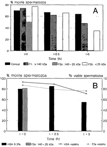
Fig. 1. (A) Effect on sperm motility of the three fractions obtained by separation of native peritoneal fluid (PF) on S-300 Sephacryl column (1 x 100 cm). F l , > 140 kDa; F2a, 140-25 kDa; and F2b, <25 kDa). Fraction F2a containing - 7 0 % of the eluted proteins was found to immobilize spermatozoa in 2.5 h when compared to the control incubations in BWW/0.3% human serum albumin (HSA) or native PF. Figure 1A represents a typical graph obtained in n = 20 experiments performed with different PFs and semen samples. (B) Percentage of viable cells as function of time with respect to sperm motility at t = 0, 2.5 and 5 h after incubation in BWW/0.3% HSA or native PF or F2a low pressure liquid chromatography (LPLC) fraction. Note that sperm motility in the F2a fraction was completely lost at 2.5 h of incubation, while sperm viability was maintained even after 5 h. Figure IB represents a typical graph obtained in n = 5 experiments performed with different PFs and semen samples.
described by Chen (1967). Briefly, peritoneal fluid fractions were mixed 20:1 (v/w) with charcoal (Merck AG, Darmstadt, FRG) and the pH was lowered to 3.0 with 1 N HC1. The suspension was stirred at 4 ° C for 1 h in an ice bath and then centrifuged at 20 000 g for 20 min at 2 ° C . The supernatant was removed, its pH re-adjusted to 7.5 (1 N NaOH) and finally filtered on 0.22 fim sterile Millipore filters. Aliquots were stored at 4°C until used in the sperm interaction test.
TLC was performed in vertical chambers (Merck AG, Darmstadt, FRG) with an eluent phase composed of chloroform, methanol and water in the ratio of 8:6:1 (v/v/v). The Kieselgel F2 5 4 plates (Merck AG, Darmstadt, FRG; 20 x 20 cm) with concentrating zone were pre-conditioned in the eluent phase and heated at 100°C for 5 min before starting the separation. The controls (Bachem Feinchemikalien AG, Bubendorf, Switzerland)
100
O.4O O.6O
K 101 Mi n u t * s
0.50 1.00
Fig. 2. Profiles of the sequential high pressure liquid chromatography (HPLC) separations of a given peritoneal fluid (PF) sample. (A)
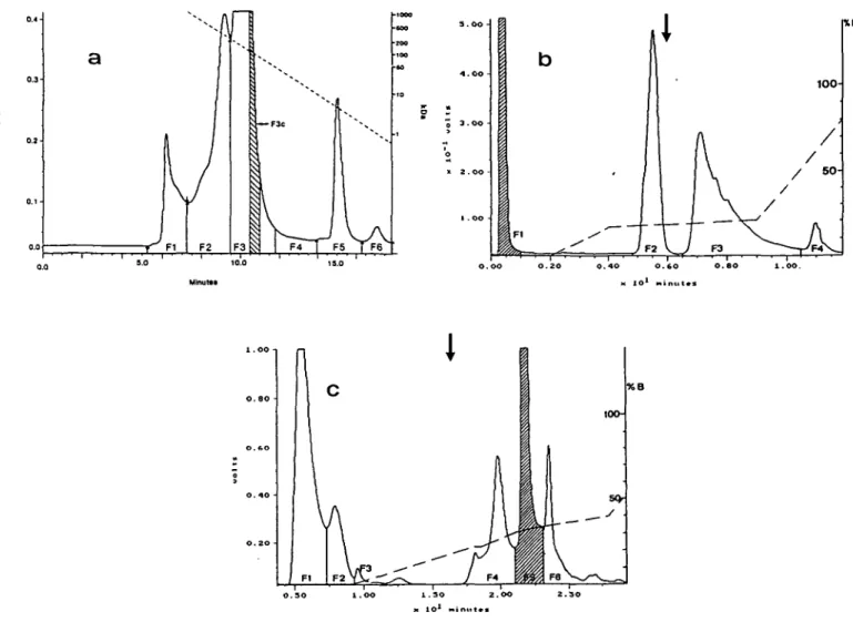
Elution pattern of molecular weight (MW)-sieving HPLC on a Protein Pak 300 column. Elution buffer, 350 mM phosphate buffer; pH 7.4; elution rate of 1 ml/min at room temperature. Fractions were collected as shown in the figure. The greatest sperm immobilizing activity was found in fraction F3c (MW-range: 51—73 kDa). For subsequent separations the entire fraction F3 was pooled. Molecular weight calibration was performed using Bio-Rad markers (cat. number 151-1901; MW-range, 670-1.35 kDa). (B) Cation-exchange (SP) and (C) anion-exchange (DEAE) separations of the immobilizing fractions. Immobilizing activity is shown as hatched areas. Left ordinate: absorbance in volts at 280 nm; right ordinate: MW in kDa (A) or percentage of B-buffer in the final elution mixture (B,C). The shapes of the gradients used during elution from the SP- and DEAE-columns are given as dashed lines. Note that the sperm immobilizing activity eluted without retention from the SP-HPLC column (B), whereas it was retained in the DEAE—HPLC column (C). Protein concentrations were as follows: native peritoneal fluid, 35.2 mg/ml; pooled F3 fraction after MW-sieving HPLC, 4.12 mg/rnl; active fraction after cation-exchange separation, 3.06 mg/ml; and the final active fraction obtained after anion-exchange chromatography, 1.96 mg/ml. Sample volumes used in the various chromatographic procedures are given in text.
and samples (chloroform/methanol extracts) were applied using an automatic positive displacement pipette (Gilson Medical Electronics, Villiers, France) under a stream of cold air. Running time was 1.5 h after which the plates were dried, stained with Coomassie Blue (0.5% in 20% methanol solution) for 20 min and destained in 20% methanol for 5 min (Nakamura and Handa, 1984). Other plates, prepared in the same manner, were stained for the detection of choline (Dragendorff reagent) or ethanolamine and serine (Ninhydrin reagent) (Kates, 1972). Eventually, the plates were scraped and the collected matrix was re-extracted in chloroform, methanol and HC1 as described above, resus-pended in BWW—HEPES and tested for sperm immobilization.
Effect of butylated hydroxytoluene on the immobilizing fraction
Butylated hydroxytoluene (BHT; Sigma, St Louis, MO, USA) was prepared as a 2.5 M stock solution in benzene following a modification of the method proposed by Jones and Mann (1977). The final concentration of the working dilution was 0.55 mg/ml in benzene. This solution (100 /tl) was dispensed in glass tubes pre-washed with benzene and the solvent was evaporated under a N2-flux for 1 h. The tubes were then closed
and stored at 4°C until analysis. The sperm contact test was performed by incubating spermatozoa with (i) BWW/0.3% HSA, (ii) BWW/0.3% HSA7BHT (2 mM final concentration), (iii) the
1 0 0% motile spermatozoa
t • 0 t • 1 hr t =• 2.5 h r s t • 5 hrs Time (h)
|HSAO.3% B H F 3 PPak C D SP from PPak ^•DEAEfromSP
Fig. 3. Comparison of the sperm motility effect of the various
active fractions obtained during the chromatographic purification steps of a given peritoneal fluid sample. Samples of 100 iA of the fractions eluted from the MW-sieving and ion-exchange columns shown in Figure 2 were tested for their sperm immobilizing activity and compared to a sperm preserving medium (BWW/0.3% HSA) as a function of time (n = 3 experiments; figure is a typical graph). Note that the more a fraction was purified, the shorter was the immobilization time.
immobilizing fraction and (iv) the immobilizing fraction + BHT (2 mM final concentration). Sperm motility was assessed at t = 0 and 2.5 h as described above.
Statistical analyses
Statistical evaluation of the acrosome reaction was done using the chi-square test.
Results
Three fractions of decreasing molecular weight (Fl, > 140 kDa; F2a, 25-140 kDa; and F2b, < 25 kDa) were separated by LPLC (MW-sieving), pooled and tested for their effect on sperm motility (Figure IA). Only fraction F2a (MW-range: 140-25 kDa) containing ~70% of the proteins eluted was found completely to immobilize spermatozoa after 2.5 h of incubation (Figure 1 A). The effects on spermatozoa were almost immediate (at t = 0) and constant (t = 5 h).
Testing the immobilizing fraction F2a in parallel for its effect on sperm motility and viability (Figure IB), spermatozoa were found to have competent membranes even after having interacted for 2.5 h with the F2a fraction and having been completely immobilized. In immunofluorescent experiments (« = 5; data not shown) performed with a specific anti-outer acrosomal
PAGIEF
SDS-PAGE
pH
4_
1 2 3 4 5 6 1 2 3 4 5 56 -7_ 8-9-MW
kDa
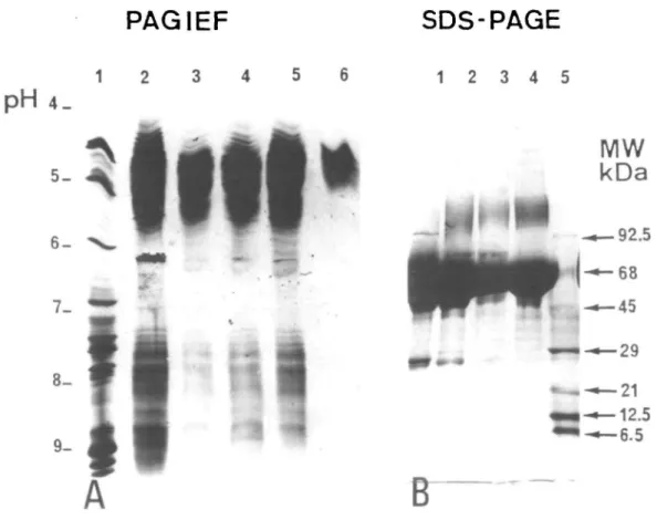
• 92.5 68 •45 • 2 9 •21 •12.5 -6.5Fig. 4. Electrophoretic studies: (A) Polyacrylamide gel isoelectrofocusing (PAGIEF) and (B) Polyacrylamide gel electrophoresis
(SDS—PAGE) of native and chromatographically purified peritoneal fluid (PF) fractions. Markers are respectively in lanes 1 (A) and 5 (B), native peritoneal fluid in lanes 2 (A) and 1 (B), sperm immobilizing fraction from high pressure liquid chromatography (HPLC) MW-sieving in lanes 3 and 4 (A) and lane 2 (B), sperm immobilizing fraction from cation exchange (SP)-HPLC in lane 5 (A) and sperm immobilizing fraction from DEAE—HPLC in lanes 6 (A) and 4 (B).
membrane antibody that cross-reacts with human outer acrosomal antigens, a significant increase in the percentage of acrosomally reacted spermatozoa was observed between the F2a and control suspensions (P < 0.05).
Figure 2A shows the elution profile of native peritoneal fluid on a gel filtration HPLC column. The F3 fraction was found to possess the immobilizing capacity. Sub-fractionation of this fraction using the same column allowed a more accurate determination of the MW of the immobilizing fraction. The greatest immobilizing activity was observed in the F3c sub-fraction (dashed area) having a MW range of 51.5-73.1 kDa. Because all the F3 subtractions exhibited immobilizing activity, the whole F3 fraction was used in the subsequent cation-exchange procedure. The immobilizing factor contained in the F3-HPLC fraction was found to elute without retention from the cation exchange SP-HPLC column (Figure 2B), whereas it was retarded on the anion exchange DEAE—HPLC column (Figure 2C). As shown in Figure 3, the fraction obtained after the first chromato-graphic step (Figure 2A) required 5 h to reduce sperm motility by 90% (Figure 3). Separation of the F3-HPLC fraction on the SP column resulted in the isolation of a fraction (Figure 2B; Fl) that reduced the immobilization t'tne when compared to the gel filtration separation. After 2.5 h only 20% of the sperm were motile against 62% in the gel filtration-sperm suspension. The maximum effect was observed with the F5-DEAE fraction (Figure 2C) which totally immobilized spermatozoa within 1 h. The immobilizing fractions after each purification step were also analysed by PAGIEF and SDS-PAGE (Figure 4A and B). SDS—PAGE under reducing conditions indicated the presence of a complex series of proteins dominated by a 68 kDa band (Figure 4B, lane 4). Because the 68 kDa protein (having the same MW as albumin) was found always to be present in all the immobilizing fractions eluted from the aforementioned sequential chromatographic steps, control experiments were conducted to ascertain if commercial albumin (HSA) treated with our experimental procedure could immobilize spermatozoa. The results obtained from these experiments demonstrated this was not the case (data not shown). PAGIEF showed that the DEAE fraction consisted of a multiple series of acidic bands focusing between pH 4.6 and 5.0 (Figure 4A, lane 6).
In order to determine if the immobilizing activity was due to a lipid peroxidation process, a series of experiments was conducted to observe the effect of BHT, a potent antioxidant agent, on the immobilizing action of the peritoneal fluid fraction. When BHT was added to the active fraction at a final concentra-tion of 2.0 mM, sperm immobilizaconcentra-tion was still observed (data not shown).
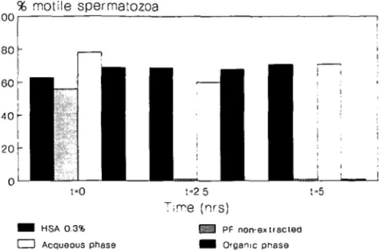
Lipid extraction of the sperm-immobilizing fraction by chloroform, methanol and HC1 (Figure 5; n = 3) or by charcoal under acidic conditions (data not shown) resulted in the complete removal of the sperm-immobilizing activity from the F3-HPLC fraction. Interestingly, however, resuspension of the chloro-form/methanol extract with BWW-medium resulted in a total recovery of the immobilizing activity (Figure 5; organic phase), whereas the aqueous phase did not show any effect on sperm motility even after 5 h of incubation.
Separation of the extracted active fraction on TLC plates is
% motile spermatozoa uu 80 60 40 20 - I >\ o —
• •
CD§-F
1
!•
|
t»0 HSA 0-3% Acqueous phasel l
l l
t-2 5 "Time (nrs) j i U PF non-• non-• Organic 1•
1
L
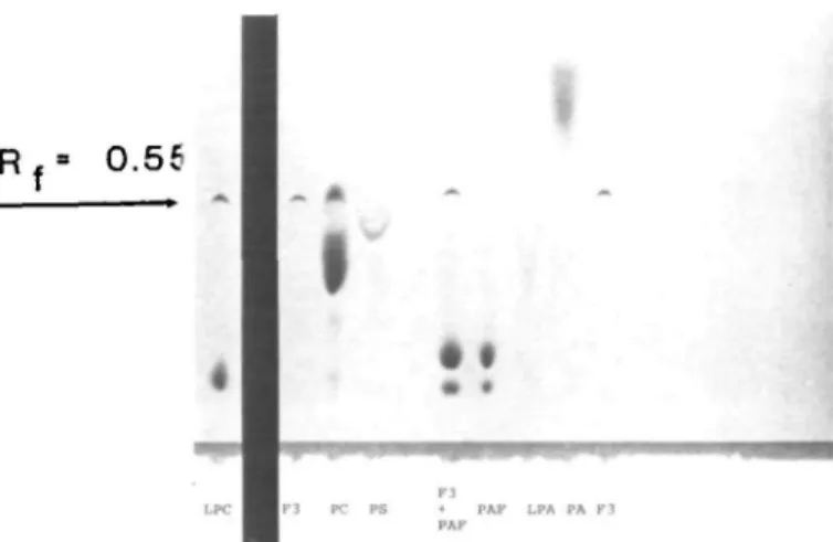
i i ] , t-5 axtracted phase Fig. 5. Effect of chloroform/methanol (n = 3) extraction of F3-high pressure liquid chromatography (HPLC) fraction on sperm motility. BWW-buffer supplemented with 0.3% human semen albumin (HSA) (positive) and non-extracted peritoneal fluid fraction (negative) were introduced as internal controls. Sperm motility could be preserved by extracting the immobilizing fraction by chloroform/methanol. The sperm immobilizing activity, recovered from the organic phase by resuspension of the chloroform/methanol extract in BWW-buffer, was observed at t = 5 h, whereas sperm motility was maintained in both the aqueous fraction of the chloroform/methanol extraction and in the supernatant of the charcoal extraction (data not shown).shown in Figure 6, lanes 2 and 9. Under the experimental conditions used, a characteristic curved band was demonstrated by Coomassie Blue staining (Rf = 0.55). Scraping, extracting
and testing this band on sperm motility confirmed the presence of the sperm immobilizing factor. Differential staining showed that this substance does not contain choline, ethanolamine or serine (data not shown). Interestingly, commercially available lysophosphatidylcholine (LPC) possessed a secondary band which, under these experimental conditions, migrated with the same retention factor and had a similar curved shape as that seen for the immobilizing factor (Figure 6, lane 1). Furthermore, the extracted band when tested on spermatozoa caused sperm immobilization (data not shown).
Discussion
Peritoneal fluid in women is not only the product of peritoneal transudation and secretion but also of ovarian exudation (Koninckx et al., 1980; Bouckaert et al., 1986). As there is no barrier to impede the entrance of peritoneal fluid or its constituents into the Fallopian tubes, peritoneal as well as follicular fluid may be present in the tubal micro-environment in which gamete interactions occur in vivo (Syrop and Halme, 1987; Harper, 1988; Leese, 1988). Soluble components of peritoneal fluid along with substances secreted by cells contained in this fluid, e.g. macrophages, have been claimed to influence sperm motility (Muscato et al. ,1982; Hill et al., 1987). We have also reported on the effect of peritoneal fluids on sperm motility in vitro, showing in particular that peritoneal fluids from different women may either enhance or decrease sperm motility (Soldati et al.,
Rf= 0.55
I
• •
PAF LPA PA FJ
Fig. 6. Separation of the chloroform/methanol extracted, active fraction (F3-HPLC; see Figure 2A) on a silica gel thin layer chromatography (TLC) plate. The fraction (F3; lanes 2 and 9) was deposited at the bottom of the plate and permitted to migrate in chloroform/methanol/water 8:6:1 (v/v/v) after activation at 100°C for 5 min. The plate was stained and destained as described in the text. Controls were lysophosphatidylcholine (LPC, lane 1); phosphatidylcholine (PC); phosphatidylserine (PS); F3 + platelet activating factor (F3+PAF); platelet activating factor (PAF); lysophosphatidic acid (LPA) and phosphatidic acid (PA). For the F3 sample, a sharp curved band with Rf = 0.55 was identified
after the Coomassie Blue staining. This band, when extracted from the gel with chloroform/methanol and resuspended in
BWW-HEPES buffer, was found readily to immobilize spermatozoa.
1989). In view of these findings, it is possible to hypothesize that peritoneal fluid or peritoneal fluid-related substances may in fact influence the approach of spermatozoa towards the oocyte in the upper part of the female genital tract and may be involved in certain cases of infertility. To the best of our knowledge, however, no work until now has dealt with the characterization of peritoneal fluid components that are possibly involved in the observed effects.
Another reason for the need to study the effect of peritoneal fluid on sperm function is the advent of assisted reproductive procedures, such as intraperitoneal insemination, in which spermatozoa may come into direct contact with peritoneal fluid. The success of such procedures may depend on the effect exerted by the peritoneal fluid of the patient on the spermatozoa of the spouse or donor (Menard et al., 1990).
In the present study we found that a given fraction purified from native peritoneal fluids by various chromatographic techniques was able to inhibit sperm motility. Interestingly, all the peritoneal fluids analysed herein contained a fraction with immobilizing activity. At first this finding was difficult to correlate with the few but significant motility enhancing effects described by Soldati et al. (1989) and Menard et al. (1990) with unfractionated peritoneal fluids (see also Figure 1A). A possible answer to these seemingly contrasting observations may be that native peritoneal fluids contain different factors able to influence sperm motility, some of which may inhibit sperm motility, and others which could stimulate or preserve it. The presence of the latter may in turn inhibit or neutralize the effect of the former, creating a sort of dynamic balance. Ovarian exudation contributes
434
to the formation of peritoneal fluid (Koninckx et al., 1980; Bouckaert et al., 1986) and follicular fluids appear to preserve or enhance sperm motility in vitro (Falcone etal, 1991). Therefore the peritoneal fluid factor stimulating or preserving sperm motility may be similar to the low MW, dialysable substance that Yanagimachi (1970) isolated from follicular fluid with the ability to stimulate hamster sperm motility. In view of the fact that the repeatedly isolated F3-HPLC fractions were able to immobilize spermatozoa only after dialysis, we believe that this step induced an enhancement of the motility-inhibiting potential of the chromatographic fractions due to the loss of a low MW substance; the latter may be responsible for the reported motility-preserving or -enhancing effects in native peritoneal fluids (Yanagimachi, 1970).
The various experiments conducted to characterize the immobilizing factor(s) yielded valuable information concerning its nature. Firstly, the heat-treatment experiments (37, 56 and 95 °C for 1 h; data not shown) indicate that the immobilizing factor is not a component of the complement system (56 °C did not alter the immobilizing capacity of the fraction) or a protein (95 °C caused only a temporary loss of the immobilizing capacity of the fraction). Secondly, the chloroform/methanol lipid-extraction experiments strongly suggest that the immobilizing factor may be a lipid or a hydrophobic substance. Furthermore, charcoal extraction under acidic conditions (Chen, 1967; Lui and Meizel, 1977) is known to remove lipids from a given solution and, in our experiment, the charcoal extraction completely removed the immobilizing potential from the dialy sed, active fractions. Thus we hypothesize that the immobilizing factor may be either a free fatty acid (see also Hong et al., 1986) or another hydrophobic low MW substance. Thirdly, the electrophoretic (SDS-PAGE) and the isoelectric (PAGIEF) separations demonstrating the constant presence of albumin in all of the active fractions from the MW-sieving and ion-exchange HPLC purification procedures seem to suggest that the immobilizing factor is in some way associated with albumin (Dow and Bavister, 1989; Davis et al., 1979, 1980; Davis 1980). Because albumin is commonly added to sperm survival media to induce capacitation and/or sperm motility activation, and no sperm immobilization occurred in the control experiments when commercial HSA was dialysed using the same procedure as for the peritoneal fluid fractions (data not shown), albumin as such is unlikely to be responsible for the observed effects. The lipid extraction experiments not only confirmed this hypothesis but also showed that the immobilizing capacity of the extracted factor did not depend on the presence of albumin. Therefore, we may postulate that the immobilizing factor (probably a lipidic or a low MW hydrophobic substance) might have a strong binding capacity to albumin but that its immobilizing potential does not depend on the presence or absence of albumin.
The biochemical or cellular processes that are initiated by the interaction of spermatozoa with the peritoneal fluid immobilizing factor and result in the inhibition of sperm motility are yet to be elucidated. The following observations made during this study may, however, lead the way to an understanding of these processes.
Firstly, the immobilizing effect does not seem to be due to membrane peroxidation. In our experimental approach, the
spermatozoa incubated with the various peritoneal fluid fractions were seminal plasma-free and therefore unprotected against oxidative agents (Jones and Mann, 1977). In view of the fact that hydroperoxides have been repeatedly claimed to be responsible for the depression of sperm motility and viability (Jones and Mann, 1976, 1977; Aitken and Clarkson, 1988), one could hypothesize that the immobilizing factor affected sperm motility by disrupting the plasma membrane. This is highly unlikely, since the eosin—nigrosin test showed that spermatozoa had competent membranes even when immobilized. Furthermore, the incorporation of BHT, a potent antioxidant agent, in the incubation suspensions did not inhibit the immobilizing capacity of the peritoneal fluid fraction.
Secondly, the peritoneal fluid factor does not seem to cause sperm immobilization via activation of the phospholipase A2-activity. Cellular phospholipases are able to initiate the
'arachidonate cascade', leading to the synthesis of arachidonic acid which has already been demonstrated to be detrimental for sperm motility (Hong et al., 1986). Furthermore, phospholipase A2 is associated with the sperm head membrane and its
activation by a calcium influx may cause the fusion of the outer acrosomal membrane with the overlying plasma membrane (Llanos et al., 1982). Therefore, the hypothesis that the immobilizing fraction may activate sperm membrane phospho-lipases, in particular phospholipase A2, should be explored. We
can report that incubation of spermatozoa with p-BPB, a phospholipase A2-inhibitor, did not prevent sperm
immobiliza-tion (data not shown). However, no attempts were made to test if phospholipases A1; C and D activities were involved.
Thirdly, the lipidic or hydrophobic immobilizing factor may bind to specific areas of sperm plasma membrane (in particular to those thought to contain enzymes involved in the regulation of sperm motility, such as the midpiece and/or the flagellum), causing a destabilization of the membrane. Indeed, the immobilizing fractions resulted in a significant increase of acrosome reactions. However with our present laboratory technology, we cannot demonstrate if this phenomenon occurs during or after immobilization. Nevertheless, we did observe that the immobilization by the peritoneal fluid factor in its purest form is extremely fast, and precedes by hours the onset of the acrosome reaction. Therefore, it is possible that the membrane of the midpiece and/or flagellum (motility organelles) are influenced directly by the peritoneal fluid factor, while the head membrane transformations, (i.e. acrosome reaction) may be an indirect result.
Linoleic, oleic and linolenic acids have been shown to inhibit sperm motility (Siegel et al., 1986). Furthermore, lysophos-phatidylcholine, arachidonic acid and docosahexaenoic acid (all products of phosphatidylcholine hydrolysis) have been found to have a dose-dependent inhibitory effect on the motility of human spermatozoa, while phosphatidylcholine does not (Hong et al., 1986). Since we used chromatography conditions specific for phospholipids or their derivatives, we can exclude the immobilizing factor being a free fatty acid. Thin-layer chromatography separations and specific staining procedures performed in the present study have not so far permitted matching of the immobilizing factor to any of the phospholipid standards used, but it has shown that the immobilizing factor does not
contain choline, ethanolamine or serine. However, we did observe that a secondary band of the lysophosphatidylcholine (LPC) not only possessed the same Rf value (0.55) as that of
the immobilizing factor but was also capable of immobilizing spermatozoa in vitro. Therefore, the immobilizing factor might be a degradation product of LPC or a substance co-purifying with LPC during its preparation.
In conclusion, the results presented indicate that human peritoneal fluids contain a heat-resistant, protein-bound, lipidic component able rapidly to immobilize spermatozoa and to increase acrosome reactions without apparently disrupting the internal sperm plasma membrane. In view of the results so far obtained, our research is now directed toward the cellular characterization of this factor, in particular the mechanisms involved in sperm binding and sperm motility inhibition.
Acknowledgements
The authors gratefully acknowledge the partial financial support of the Swiss National Science Foundation (grant 3.939-087 to M.B.). The polyclonal anti-outer acrosomal membrane antibody used for the detection of acrosomally reacted spermatozoa was a generous gift of Dr E.Topfer-Petersen (Munich, FRG). The authors express their warmest appreciation to Drs J.Stamm and M.Zeeb as well as Miss B.Sangiorgio for procuring the peritoneal fluid samples used in the present study.
References
Aitken.R.J. and Clarkson,J.S. (1988) Significance of reactive oxygen species and antioxidants in defining the efficacy of sperm preparation techniques. J. Androl., 9, 367—376.
Bouckaert,P.X.J.M., Evers,J.L.H., Doesburg,W.H., Schellekens,L.A., Brombacher,P.H. and Rolland,R. (1986) Patterns of changes in proteins in the peritoneal fluid and of women during the periovulatory phase of the menstrual cycle. J. Reprod. Fertil., 77, 329—336. Chen,R.F. (1967) Removal of fatty acids fnom serum albumin by
charcoal treatment. J. Biol. Chem., 242, 173-181.
Davis,B.K. (1980) Interaction of lipids with the plasma membrane of sperm cells. I. The antifertilization action of cholesterol. Arch. Androl., 5, 249-254.
Davis,B.K., Byrne,R. and Hangund,B. (1979) Studies on the mechanism of capacitation n. Evidence for lipid transfer between plasma membrane of rat sperm and serum albumin during capacitation in vitro. Biochem. Biophys. Ada, 558, 257—266.
Davis,B.K., Bryne,R. and Bedigian,K. (1980) Studies on the mechanism of capacitation: albumin mediated changes in plasma membrane lipids during in vitro incubation of rat sperm cells. Proc. Nat I. Acad. Sci. USA, 77, 1546-1550.
Dorez,F., Wahl,P., Bajolle,F. and Qu6reux,C. (1985) Immobilisation des spermatozoides par le liquide peritoneal de femmes steiiles. / . Gynecol. Obstet. Biol. Reprod., 14, 295-299.
Dow,M.P.D. and Bavister,B.D. (1989) Direct contact is required between serum albumin and hamster spermatozoa for capacitation in vitro. Gamete Res., 23, 171-180.
Eliasson,R. (1977) Supravital staining of human spermatozoa. Fertil. Sterii, 28, 1257-1260.
Fairbanks.G., Steck,T.L. and Wallach.D.F.H. (1971) Electrophoretic analysis of the major polypeptides of the human erythrocyte membrane. Biochemistry, 10, 2606—2617.
Falcone,L., Soldati.G., Piffaretti-Yanez,A., Marchini,M., Eppenberger,U. and Balerna,M. (1991) Follicular fluid enhances sperm motility and velocity in vitro. Fertil. Sterii., 55, 619—623.
Harper.M.J.K. (1988) In Knobil.E. and Neill.J.D. (eds), The Physiology of Reproduction. Raven Press, New York, pp. 103 — 134. Hill,J.A., Haimovici.F., Politch.J.A. and Anderson.D.J. (1987) Effect
of soluble products of activated lymphocytes and macrophages (lymphokines and monokines) on human sperm motion parameters. Fertil. Sterii, 47, 460-465.
Hinrichsen,A.C, Toepfer-Petersen.E., Dietl.T., Schmoeckel,C. and Schill.W.B. (1985) Immunological approach to the characterization of the outer acrosomal membrane of boar spermatozoa. Gam. Res.,
11, 143-155.
Hjerten.S. (1962) 'Molecular sieve' chromatography on polyacrylamide gels, prepared according to a simplified method. Arch. Biochem. Biophys. (Suppl. 1), 147-151.
Hong.C.Y., Shieh,C.C, Wu,P., Huang,J.J. and Chiang.B.N. (1986) Effect of phosphatidylcholine, lysophosphatidylcholine, arachidonic acid and docosahexaenoic acid on the motility of human sperm. Int. J. Androl, 9, 118-122.
Jones,R. and Mann.T. (1976) Lipid peroxides in spermatozoa; formation, role of plasmalogen, and physiological significance. Proc. R. Soc. Lond. B, 193, 317-333.
Jones,R. and Mann,T. (1977) Damage to ram spermatozoa by peroxidation of endogenous phospholipids. J. Reprod. Fertil, 50, 261-268.
Kates,M. (1972) Separation of lipid mixtures. In Work.T.S. and Work.E. (eds), Techniques ofLipidology. North Holland Publishing Company, Amsterdam, pp. 393-469.
Koninckx,P.R., Ranaer,M. and Brosens,I.A. (1980) Origin of peritoneal fluid in women: An ovarian exudation product. Br. J. Obstet. Gynecol, 87, 177-181.
Laemmli,U.K. and Favre.M. (1973) Maturation of the head of bacteriophage T4. I. DNA packaging event. J. Mot. Biol., 80, 575-599.
Leese,H.J. (1988) The formation and function of oviduct fluid. J. Reprod. Fertil., 82, 843-856.
Llanos,M.N., Lui,C.W. and Meizel,S. (1982) Studies of phospholipase A2 related to the hamster sperm acrosome reaction. J. Exp. Zool.,
221, 107-117.
Lowry.O.M., Rosenbrough.N.J., Farr.A.L. and Randall.R.J. (1951) Protein measurement with the pholin phenol reagent. J. Biol. Chem.,
193, 265-275.
Lui,C.W. and Meizel.S. (1977) Biochemical studies of the in vitro acrosome reaction inducing activity of bovine serum albumin. Differentiation, 9, 59—66.
Menard.A., Wittemer,C, Moreau,L. and Dellenbach,P. (1990) Evalua-tion and preparaEvalua-tion of spermatozoa for direct intraperitoneal insemina-tion. In Acosta,A.A., Swanson.R.J., Ackerman.S.B., Kruger,T.F., van Zyl,J.A. and Menkveld,R. (eds), Human Spermatozoa in Assisted Reproduction. Williams & Wilkins, Baltimore, MD, pp. 292-309. Muscato,J.J., Haney,A.F. and Weinberg.J.B. (1982) Sperm phagocytosis by human peritoneal macrophages: A possible cause of infertility in endometriosis. Am. J. Obstet. Gynecol., 144, 503—510.
Nakamura,K. and Handa.S. (1984) Coomassie brilliant blue staining of lipids on thin layer plates. Anal. Biochem., 142, 406—410. Oak,M.K., Chantler.E.N., Vaughan,C.A., Elstein,W. and Elstein.M.
(1985) Sperm survival studies in peritoneal fluid from infertile women with endometriosis and unexplained infertility. Gin. Reprod. Infert., 3, 297-303.
Ralt,D., Goldenberg,M., Fetterolf.P., Thompson,D., Dor,J., Mashiach,S., Garbers.D.L. and Eisenbach,M. (1991) Sperm attrac-tion to a follicular factor(s) correlates with human egg fertilizability. Proc. Natl. Acad. Sci. USA, 88, 2840-2844.
Sanchez.R., Toepfer-Petersen.E., Aitken,R.J. and Schill.W.B. (1991) A new method for evaluation of the acrosome reaction in viable human spermatozoa. Andrologia, 23, 197-203.
Siegel,!., Dudkiewicz.A.B., Friberg.J., Suarez,M. and Gleicher,N.
(1986) Inhibition of sperm motility and agglutination of sperm cells by free fatty acids in whole semen. Fertil. Sterii., 45, 273—279. Soldati,G., Piffaretti-Yanez,A., Campana,A., Marchini,M., Luerti,M.
and Balerna.M. (1989) Effect of peritoneal fluid on sperm motility and velocity distribution using objective measurements. Fertil. Sterii., 52, 113-119.
Stone,S.C. and Himsl.K. (1986) Peritoneal recovery of motile and non-motile sperm in the presence of endometriosis. Fertil. Sterii., 46, 338-339.
Suarez,S.S., Wolf,D.P. and Meizel.S. (1986) Induction of the acrosome reaction in human spermatozoa by a fraction of human follicular fluid. Gam. Res., 14, 107-121.
Syrop.C.H. and Halme.J. (1987) Peritoneal fluid environment and in-fertility. Fertil. Sterii., 48, 1-9.
Trah.T.J. and Schleyer,M. (1982) Formaldehyde fixation of proteins and small polypeptides after isoelectric focusing in polyacrylamide gels. Anal. Biochem., 127, 326-329.
Yanagimachi,R. (1970) In vitro acrosome reaction and capacitation of golden hamster spermatozoa by bovine follicular fluid and its frac-tions. J. Exp. Zool, 170, 269-280.
WHO (1987) Laboratory Manual for the Examination of Human Semen and Semen-Cervical Mucus Interaction. 2nd edn. Cambridge University Press, Cambridge.