Mutagenesis vol.9 no.4 pp.377-381, 1994
Characterization of the trp5-27 allele used to monitor drug-induced
mitotic gene conversion in the Saccharomyces cerevisiae tester
strain D7
Ann E.Ehrenhofer-Murray, Friedrich E.Wurgler and Christian Sengstag1
Institute of Toxicology, Swiss Federal Institute of Technology and University of Zurich, Schorenstrasse 16, CH-8603 Schwerzenbach, Switzerland
'To whom correspondence should be addressed
Mitotic gene conversions, among other recombinagenic events, can play an important role in the multistep process of carcinogenesis. The ability of chemicals to induce such gene conversions can easily be monitored in the Saccharomyces
cerevisiae tester strain YHE2, a derivative of strain D7. For
the detection of drug-induced gene conversions, two mutations in the TRP5 locus are used, trp5-12 and trp5-27. Here we report on the characterization of the stable allele trp5-27. Our analysis revealed two relevant mutations in trp5-27: (a) a transition C to T at position 121 after ATG that results in an amber stop codon and abolishes gene expression and (b) a transversion A to T at position 1555 that creates an ochre stop codon. Simultaneous amber and ochre suppression with the suppressors SUP3 and SUP11, respectively, was capable of relieving the tryptophan-requiring phenotype of strains carrying the trpS-27 allele. These findings have implications on the length of gene conversion tracts in conversion events between trpS-12 and trp5-27: conversion tracts can cover several Idlobases, if the site of the mutation hi trpS-12 lies outside of the positions mutated hi trp5-27. Conversely, the maximal length is limited to 1435 bp, if the mutation in
trp5-12 is located between the positions mutated hi trpS-27.
Introduction
Mitotic gene conversion is a process of nonreciprocal transfer of genetic information between homologous chromatic! regions of chromosomes. In cells that are heterozygous for two alleles of a marker gene on the two homologous chromosomes, gene conversion within the region of the marker gene can result in the loss of heterozygosity, and a clone can develop that is homozygous for one of the marker alleles (Wurgler, 1992). If in proliferating tissue a cell turns homozygous for a pre-existing mutation in a tumor suppressor gene that leads to the loss of gene function, this loss of heterozygosity will enable the phenotypical manifestation of the mutation and may result in increased proliferation of the cell (Sengstag, 1994). Thereby, gene conversions, among other recombinagenic events, represent a possible step in the multistep process of carcinogenesis (Fearon and Vogelstein, 1990). Chemical compounds that raise gene conversion frequencies are, therefore, thought to contribute con-siderably to the development of neoplasms. In addition to their effect on cancer induction, promotion and progression, gene conversion events leading to loss of heterozygosity can also affect other biological functions that influence health (Wurgler, 1992). During the last few years, the awareness of the importance of such processes has substantially increased, and various test systems have been designed to identify the recombinational
properties of substances which might be indicative of their carcinogenic potential (Sengstag, 1994). One of these test systems, the Saccharomyces cerevisiae tester strain D7 (Zimmermann
et al., 1975), allows the detection of gene conversions
simultaneously with reversions and mitotic recombinations. As a unicellular eukaryote, S. cerevisiae provides a readily manipulable, yet genetically complex model organism suitable for the study of recombinational events (Zimmermann, 1992), and since its introduction the tester strain D7 has been extensively used for genotoxicity testing (Zimmermann et al., 1984). As the yeast cells lack certain enzymatic activities that can be crucial for the activation of procarcinogens, various human enzymes have been heterologously expressed in a yeast strain derived from D7 (Eugster et al., 1990, 1991, 1992) and are capable of conferring such metabolic competence to yeast (Eugster et al., 1993; Eugster and Sengstag, 1993; Sengstag et al., 1994). This approach has proved promising in broadening the applicability of the test system. The process of gene conversion can easily be followed in heteroallelic diploid yeast strains that carry two defective alleles of the same gene locus (Zimmermann, 1975). hi strain D7 (Zimmermann et al., 1975), two non-complementing alleles of the tryptophan synthase gene TRP5 (Zalkin and Yanofsky, 1982) are combined. Taken on their own, the two alleles trp5-12 and
trp5-27 are completely stable (trp5-27) or exert low reversion
frequencies (trp5-12, reversion frequency 10"6) that do not
interfere with gene conversion (Zimmermann and Schwaier, 1967). Conversely, the tryptophan-requiring heteroallelic diploid D7 generates tryptophan nonrequiring convertants at a frequency of approximately 10~5. The conversion frequency can be
increased by treatment of the cells with DNA-damaging agents (Zimmermann et al., 1975). For our genotoxicity testing, we are working with a ura3 derivative of D7, YHE2 (Eugster et al., 1990). We were interested to characterize the trp5 mutations present in our tester strain in order to gain more insight into the gene conversion events taking place between the two trp5 alleles. Here we report the characterization of one of the alleles, trp5-27.
Materials and methods
Yeast and bacterial strains, media and culture conditions
The construction of the S.cerevisiae strains YHE2 (a/a, ade2-40/ade2-l 19,
ilvl-92/ilvl-92, trp5-27/trp5-12, ura3b5/ura3b5), and YHE1 (a, ade2-40, ilvl-92, trp5-27, um3A5), has previously been described (Eugster et al., 1990).
Strain YAE76 (a, ade2-119, itvl-92, trp5-lll, ura3A5) was constructed in this laboratory. Strain JRY1631 (a, ade2-110, cyh2, his3A200, leul, fys2S01, trp5,
uru3-52) was kindly provided by Dr J.Rine. Yeast transformations were performed
as described by Klebe et al. (1983). The bacterial strain DH5aF' was used for propagation of recombinant plasmids.
Yeast strains were grown in standard media. Minimal medium YM [0.67% yeast nitrogen base without amino acids (Difco), 2% glucose] was appropriately supplemented with amino acids as described by Sherman (1991). Escherichia
coli was grown in LB medium which was supplemented with 150 ^g/ml
ampicillin where appropriate.
Plasmid constructions
Piasmid pAE158 is a derivative of YEp352 (Hill et al., 1986) and contains in its BamHl site the TRP5 gene from pYASl (Dohmen a al., 1989) as a 3.3 kb
BamHl fragment. In pAE158, TRP5 has the same orientation as lacZ. Plasmid
pAE268 is analogous to pAE158, but contains the trp5-27gent which was cloned 377
from YHEl (see Results). Plasmids pAE470, pAE471, pAE475 and pAE491 are derived from pAE268 and have their BglUISall, Spell Sail, Ncol anc
BstEDJSall trp5-27 fragments, respectively, replaced by the corresponding TRP5 sequence. Plasmids pRS313SUP3, pRS316SUP3 and pUN60SUP3 wen
constructed by introducing the 137 bp SUP3 BamHl fragment of mWJ64 (Guc and Wu, 1982) into the BamHl sites of pRS313, pRS316 (Sikorski and Hieter, 1989) and pUN60 (Hledge and Davis, 1988), respectively. To obtain pAE492, the blunt-ended trp5-27 BamHl fragment of pAE268 was introduced into the
XbaVNotl cleaved, blunt-ended plasmid pRS316SUP3 to give pAE342.
Subsequently, the PvuU fragment of pAE342 containing trp5-27 and SUP3 was cloned in the PvuR cut vector pRS313. Plasmid pAE496 was constructed by doning the SacUSall trp5-27fragment of pAE268 in the SacUSall cleaved vector pRS313.
Colony hybridization and detection
Bacterial colonies representing the &mHI//#mlIH sublibrary of YHEl in pRS316 was transferred to Pall Brodyne A transfer membranes, lysed and their DNA bound to the filters as described by Sambrook ex al. (1989). For the detection of 77W.5-containing colonies, the 3.3 kb BamHl fragment of pAE158 was digoxygenin-labelled with the DIG DNA Labeling and Detection Kit from Boehringer. Labelling, hybridization and chemiluminescent detection were performed according to the supplier's protocols.
DNA sequence analysis
Fragments to be sequenced were subcloned in pBLKS and pBLSK (Stratagene). DNA sequence analysis was performed on single-stranded DNA by the dideoxy chain termination method (Sanger et al., 1977) using the Sequenase Version 2.0 DNA Sequencing Kit (US Biochemical Corporation) according to the manufacturer's specification. [a-^SJdATP was purchased from DuPont.
Results
Cloning of the trpS-27 gene
In order to clone the trp5-27 gene, a genomic sublibrary of the
S.cerevisiae trp5-27strain YHEl was constructed in the E.coli/ S.cerevisiae shuttle vector pRS316. To this end, total DNA
isolated from YHEl was double-digested with BamHl and
HindUI, which divides the trp5-27gene in two fragments of 1815
and 1516 bp length. DNA fragments in a size range of 1.4 — 1.9 kb were purified from an agarose gel and ligated to the
BamHl and HindM cleaved vector pRS316. Four hundred and
fifty individual ampicillin resistant E. coli transformants were used for a colony hybridization experiment (see Materials and methods). As a probe, the 3.3 kb digoxigenin-labelled TRP5
BamHl fragment of pAE158 was taken. To confirm the identity
of the 11 clones that gave rise to positive signals, their plasmid DNA was subjected to restriction analysis. By this procedure, two clones representing the 1.8 and 1.5 kb fragments of the
trp5-27 were chosen for further characterization. Subsequendy,
the complete trp5-27 gene was reconstituted in the
E.coli/S.cerevisiae shuttle vector YEp352 by triple-ligating the
two individual trp5-27BamHUHindni fragments to die BamHl cleaved YEp352 to give pAE268.
The phenotype conferred by the trp5-2 7 plasmid pAE268 was determined by transforming strain YHEl to uracil prototrophy with pAE268 and, as a control, with pAE158, which is a YEp352 derivative and carries the wild-type TRP5 gene. YHEl pAE268 and YHEl pAE158 were streaked on supplemented minimal medium lacking uracil or uracil and tryptophan, and the plates were incubated for 3 days at 30cC (Figure 1A). In contrast to
YHEl transformed with the 7RPJ-carrying plasmid, YHEl containing the trp5-2 7 plasmid was unable to grow on medium lacking tryptophan. Thus, plasmid pAE268 does not confer tryptophan prototrophy to YHEl and, therefore, presumably contains a mutation in its trp5-27 allele that distinguishes it from pAE158.
Mapping and sequencing of an amber mutation in trp5-27
Since knowledge of the underlying mutation(s) in the trp5-2 7 and
trp5-12 alleles present in the 5. cerevisiae strains D7 and YHE2
would give us a more detailed insight into the gene conversion
YHEl
pAEI58 pAE268 pAEI58 pAE268YHEl
-ura -trp YHEl pRS3l6 YHEl pRS316SUP3 YAE76pRS316 YAE76pRS3l6SUP.:i -ura -trp
Fig. 1. (A) Comparison of the tryptophan phenotypes conferred to YHEl by plasmids carrying TRP5 (pAE158) and trp5-27 (pAE268). YHEl pAE158 and YHEl pAE268 were streaked on supplemented minimal medium lacking uracil (—ura) or uracil and tryptophan (-ura -trp) and incubated for 3 days at 30°C. (B) Amber suppression in YHEl (trp5-27) and YAE76
(trp5-lll). The strains transformed with a vector control (pRS316) or an
amber suppressor containing plasmid (pRS316SUP3) were pregrown in supplemented minimal medium to stationary phase and diluted to an absorbance at 600 nm of 0.3. 5 /il of these cell suspensions and of 1:10, 1:100 and 1:1000 dilutions were placed on supplemented minimal medium lacking uracil (—ura) or uracil and tryptophan (-ura -trp). Plates were incubated for 3 days at 30°C.
Trp suppression phenotype with SUM
pAE158 —£ Bt Bt Bp N nrs Bt I r - • ATO- - » - T A A PAE268 —{•....;:w..:.:r::...:...: • p A E 4 7 0 ESS pAE471 EE3 pAE491 E S pAE475 E Z + + n.a.
Fig. 2. Replacement of plasmid-borne trp5-27 segments by wild-type TRP5 and determination of the phenotype conferred by these plasmids. The white and grey boxes represent the TRP5 and trp5-27 sequences, respectively. The phenotypes conferred by the plasmids were determined in strain YHEl and are indicated as (+) for tryptophan prototrophy and ( - ) for tryptophan auxotrophy. By testing the tryptophan phenotype of JRY1631 cotransformed with respective trpS plasmids and pRS313SUP3, novel trp5 alleles derived from trp5-27 were identified whose tryptophan conferring phenotype was (+) or was not (—) amber suppressible (n.a., not applicable). The direction and approximate length of the TRPS open reading frame are indicated by the arrow. Selected restriction shes are indicated (Ba = BamHl, Bg = BglU, Bs = flirEn, N = Ncol, Sp = SpeV).
events, we were interested in localizing the site of the mutation(s) in the trp5-27 allele. Therefore, we replaced fragments of increasing length of the trp5-27 plasmid pAE268 by the corresponding TRP5 fragment and tested the tryptophan phenotype conferred by the new constructs (Figure 2). For this purpose, YHEl was transformed with each novel construct and Ura+
transformants were streaked on supplemented minimal medium lacking uracil or tryptophan and uracil. A tryptophan non-requiring phenotype was only observed when trp5-2 7 sequences downstream of the BstER she were replaced by the corresponding wild-type sequences, while replacement of the sequences down-stream of the Spel site was not sufficient to confer tryptophan
Characterization of the trpS-27 allele JRY1631
pRS316 pRS313SUP3
pAE158 pRS313
pAE268 pRS313SUP3
pAE470 pRS313
pAE470 pRS313SUP3
-ura -his -ura -his -irp
Fig. 3. Amber suppression of various trpS alleles in JRY1631. JRY1631 cotransformed with the indicated plasmids was grown to stationary phase in liquid medium and diluted to 3 x 10⁶ cells/ml; 5 fd of these and of 1:10, 1:100 and 1:1000 dilutions were spotted on supplemented minimal medium lacking uracil and histidine (—ura —his) or uracil, histidine and tryptophan (-ura - h i s -trp). The plates were incubated for 2 days at 30°C.
prototrophy. This allowed us to map the site(s) of the mutation(s) in trp5-27 to a 380 bp BstEWSpel fragment. To elucidate the exact nature of the mutation(s) assumed to lie in this DNA segment, the DNA sequence of the corresponding trp5-27 fragment was determined. The fragment was cloned into pBLKS and pBLSK, single-stranded DNA was prepared and sequenced according to the dideoxy method. Comparison of the trp5-27 sequence with the wild-type TRPS sequence revealed a transition C to T at nucleotkle position 121 after ATG. This mutation results in an amber stop codon TAG in trp5-27and represents a potential cause for the Trp~ phenotype observed for the trp5-27 allele.
Attempted suppression of the trp5-27 amber mutation
To determine whether the amber mutation found on trp5-27 plasmid pAE268 was the only mutation responsible for the tryptophan auxotrophic phenotype conferred by the plasmid, we attempted to suppress this Trp" phenotype with the amber suppressor SUP3. In order to achieve this objective, S.cerevisiae strain JRY1631 with the relevant genotype trp5, his3A200,
ura3-52 was cotransformed with either YEp352 or pAE268 as URA3 plasmids and either pRS313 or pRS313SUP3 as HIS3
plasmids. Ura+/His+ transformants were grown in liquid
culture to stationary phase. Aliquots of serial dilutions were spotted on supplemented minimal medium lacking uracil and histidine or tryptophan, uracil and histidine, and the plates were incubated at 30°C for 2 days (Figure 3). The trp5 mutation of JRY1631 was not suppressible with SUP3 as shown by the inability of JRY1631 pRS316 pRS313SUP3 to grow without tryptophan. Surprisingly, JRY1631 carrying pAE268 and the SWJ-containing plasmid was also unable to grow without tryptophan, indicating that die Trp" phenotype conferred by pAE68 was not amber-suppressible.
To make sure that the inability to amber-suppress JRY1631 pAE268 was not due to a cloning artefact on pAE268, but that the cloned trp5-27 allele was identical with the allele present in YHE1, we attempted to suppress the tryptophan auxotrophic phenotype of YHE1 with the amber suppressor SUP3. YHEI was transformed with the control vector pRS316 and with pRS316SUP3. In parallel, strain YAE76, which has an amber-suppressible tryptophan-requiring phenotype (A.Ehrenhofer-Murray and C.Sengstag, unpublished), was transformed with die same plasmids in order to confirm functional expression of SUP3 in pRS316SUP3. Ura+ transformants were grown in liquid culture
to stationary phase and diluted to 3 x 10° cells/ml. Aliquots of serial dilutions were spotted on supplemented minimal medium lacking uracil or tryptophan and uracil, and the plates were
incubated at 30°C for 3 days (Figure IB). Only the amber suppression control strain YAE76 containing the SUP3 plasmid could grow without tryptophan, while trp5-27 strain YHEI containing SUP3 was unable to grow. Therefore, as found for the trp5-27 allele on pAE268, the trp5-27 mutation of YHEI is not suppressible with the amber suppressor SUP3. These results suggested either that SUP3 was not the appropriate amber suppressor for trp5-27, or that there was a second mutation present in trp5-27 that restricted its amber suppressibility.
Amber suppression of novel trpS alleles and identification of a second mutation in trp5-27
To rule out the possibility that the inability of SUP3 to suppress the Trp~ phenotype of trp5-27 was due to die nature of this suppressor, we attempted to amber-suppress novel trp5 alleles derived from trp5-27. This was achieved by exchanging fragments of pAE268 for the corresponding wild-type TRPS fragment, cotransforming JRY1631 with die new constructs and either pRS313 or pRS313SUP3 to uracil and histidine prototrophy and determining die tryptophan phenotype of die transformants by streaking diem on medium lacking uracil and histidine or tryptophan, uracil and histidine (Figure 2; c.f. also Figure 3). A trpS allele present on pAE470 widi the sequence downstream of the BglU site replaced by wild-type sequences was suppressible widi SUP3, while substitution of the trpS-27 sequences downstream of the Ncol site conferred no suppressible phenotype. These results demonstrated diat me suppressor SUP3 was in fact capable of suppressing certain trpS alleles derived from trpS-27. In addition, these results strongly suggested die presence of a further mutation widiin a 606 bp BglWNcol fragment oitrpS-27 diat interferes widi amber suppression. In order to identify tiiis potential second mutation, die trpS-27 BglWNcol fragment was subjected to DNA sequence analysis. This revealed a transversion A to T at nucleotide position 1555 after ATG. This mutation creates an ochre stop codon TAA in trpS-27 diat presumably was responsible for the inability to amber-suppress trpS-27.
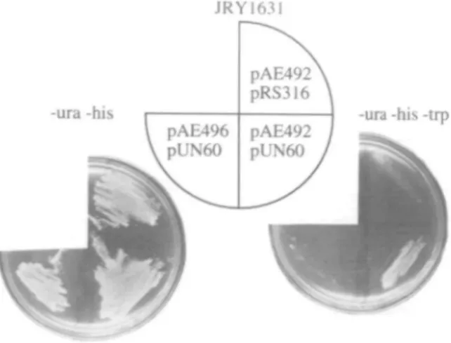
Simultaneous amber and ochre suppression of trp5-27
To confirm diat die amber and ochre mutations of trpS-27 at positions 121 and 1555 after ATG were the only mutations responsible for its tryptophan-requiring phenotype, we undertook experiments to suppress bodi mutations simultaneously. To diis end, die HIS3 plasmids pAE496 and pAE492 were constructed diat contain tire trp5-27gznt from YHEI alone or with the amber suppressor SUP3, respectively. JRY1631 was cotransformed with pAE496 or pAE492 as HIS3 plasmids and pRS316 or pUN60 as URA3 plasmids. Plasmid pUN60 carries die ochre suppressor
SUP11 (Elledge and Davis, 1988). Histidine and uracil
proto-trophic transformants were subsequendy streaked on supplemented minimal medium lacking histidine and uracil or tryptophan, histidine and uracil, and die plates were incubated for 3 days at 30°C (Figure 4). The Trp" phenotype of JRY1631 was not suppressed with SUP3 or SUP11 alone or in combination (data not shown). Similarly, a JRY1631 transformant containing die
trpS-2 7 plasmid and eidier SUP3 or SUP 11 was unable to grow
widiout tryptophan, whereas a transformant expressing trpS-27 and both SUP3 and SUP 11 showed a tryptophan prototrophic phenotype. This result clearly demonstrated that simultaneous amber and ochre suppression was necessary and sufficient to cure die Trp" phenotype of trp5-27. Similarly, die Trp" phenotype of YHEI was suppressed by a plasmid containing both suppressors
JRYI63I
-ura -his -ura -his -tip
\ r
Fig. 4. Simultaneous amber and ochre suppression of trp5-27. JRY1631 transformants were streaked on supplemented minimal medium lacking uracil and histidine (—ura —his) or uracil, histidine and tryptophan (—ura - h i s -trp). The plates were incubated at 30°C for 3 days.
Discussion
The trp5-27 allele, one of the two defective alleles of the TRP5 locus (Zalkin and Yanofsky, 1982) in the diploid S.cerevisiae genotoxicity strain D7 (Zimmermann et al., 1975), has found extensive use in the detection and quantification of drug-induced gene conversion events. Despite its broad application, the mutation in trp5-27 so far has not been characterized and, thus, little is known about the nature of gene conversion events in D7 that produce tryptophan prototrophic convertants.
In this study, we have identified two relevant mutations in
trpS-2 7 that are responsible for its stable tryptophan auxotrophic
phenotype. At nucleotide position 121 after ATG, trp5-27 contained a transition C to T that results in an amber stop codon. This stop codon presumably terminates translation, giving rise to a truncated and probably abortive Trp5 gene product. The fact that suppression of this mutation with the amber suppressor SUP3 (Guo and Wu, 1982) did not suffice to cure the Trp~ phenotype was not due to inherent characteristics of SUP3, since other, novel
trp5 alleles derived from trp5-27 were in fact suppressible with SUP3. It rather indicated the presence of a second mutation in
the trp5-27 gene. It was subsequently detected as a transversion A to T at nucleotide position 1555 after ATG and creates an ochre stop codon. Ochre suppression alone with SUP 11 (Elledge and Davis, 1988) in trp5-27did not result in tryptophan prototrophy. Conversely, simultaneous amber and ochre suppression was capable of relieving the tryptophan requirement of strains carry-ing trpS-27. Therefore, the two stop codons were the mutations relevant for the Trp~ phenotype of trp5-27.
This finding has implications concerning the length of gene conversion tracts in conversion events between trp5-27 and
trp5-12 that are successful in forming Trp+ convertants in D7.
In S.cerevisiae, gene conversion tracts are usually continuous, information being transferred as a single block from one chromosome to another (Borts and Haber, 1989). If transfer is to be directed from trp5-12 to np5-27 (Figure 5A), a contiguous DNA stretch has to cover both mutations of trp5-27 and thus requires a minimal length of 1435 bp. Consequently, the assumed mutation in trp5-12 would lie outside of the positions mutated in trp5-27, and therefore would be located either upstream of position 121 or downstream of position 1555, presumably within the TRP5 open reading frame that ends at position 2619. In such a case, the conversion tract could expand over several kilobases
trp5-27 trpS-12 trpS-27
I
trp5-12 trp5-27Fig. 5. Schematic representation of possible gene conversion events between
trp5-27 and trp5-12 that result in Trp+ convertants in D7. The approximate site of the relevant mutations in trp5-27 and one possible location of a mutation in trp5-12 are designated by crosses (X). The directions of information transfer during gene conversion are indicated by arrows. Conversion tracts can cover several kilobases, if the she of the mutation in
trp5-12 lies outside of the positions mutated in trp5-27 (A and B).
Conversely, the maximal tract length is limited to 1435 bp, if the mutation in trp5-12 is located between die positions mutated in trp5-27 (C).
downstream or upstream of the trp5-12 mutation, respectively. Likewise, if information transfer proceeds from trp5-27 to a
trp5-12 allele whose mutant site lies outside of the positions
mutated in trpS-27, the conversion tract could also span several kilobases (Figure 5B). Conversely, its size would be limited to a maximum of 1435 bp, if the mutation in trp5-12 was located between the positions mutated in trp5-27 (Figure 5 Q . Judd and Petes (1988) measured the tract length of mitotic conversions of the ura3-3 allele at the URA3 locus and reported a minimum average conversion tract lengtii of 0.9 ± 0.8 kb. In 40% of the cases, though, the average tract length was between 4 and 10 kb. We therefore favour the model of the mutated site in trp5-12 ly-ing upstream of position 121 or downstream of position 1555 after ATG (Figure 5A and B), also since the conversion frequency of D7 (10~5; Zimmermann et al., 1975) compares well to the
frequency in the ura3-3/URA3 strain (4 X 10~6; Judd and
Petes, 1988).
In light of these results, the characterization of the trp5-12 allele of D7 represents an important task and will give more precise information on the direction and extent of information transfer during gene conversion between the two alleles.
Acknowledgements
We wish to thank Drs H.P.Eugster, P.Niederberger, J.Rine and J.Thomer for generously sharing yeast strains and plasmids. This study was supported by a grant from rhe Swiss Cancer League to A.E.
References
Borts.R.H. and Haber^.E. (1989) Length and distribution of meiotic gene conversion tracts and crossovers in Saccharomyces cerevisiae. Genetics, 123, 6 9 - 8 0 .
Dohmen.R.J., Strasser.A.W., Zitomer.R.S. and HoUenberg.C.P. (1989) Regulated overproduction of alpha-amylase by transformation of the amylolytk yeast Schwanniomyces ocddenwlis. OUT. Genet., 15, 319—325. Elledge,S.J. and Davis.R.W. (1988) A family of versatile centromeric vectors
designed for use in the sectoring-shuffle mutagenesis assay in Saccharomyces
cerevisiae. Gene, 70, 303-312.
Eugster.H.P., Bartsch.S., WOrgler.F.E. and Sengstag.C. (1992) Functional co-expression of human oxidoreductase and cytochrome P450 1A1 in
Saccharomyces cemisiae results in increased EROD activity. Biochem. Biophys. Res. Convram., 185, 641-647.
Eugster.H.P., Probst.M., Wurgler.F.E. and Sengstag.C. (1993) Caffeine, estradiol, and progesterone interact with human CYP1A1 and CYP1A2.
Characterization of the trp5-27 alkie
Evidence from cDNA-directed expression in Saccharomyces cerevisiae. Drug
Metab. Dispos. Biol. Fate Chan., 21, 4 3 - 4 9 .
Eugster.H.P. and Sengstag.C. (1993) Saccharomyces cerevisiae: an alternative source for human microsomal liver enzymes and its use in drug interaction studies. Toxicology, 82, 6 1 - 7 3 .
Eugster.H.P., Sengstag.C., Hinnen.A., Meyer.U.A. and Wurgler.F.E. (1991) Heterologous expression of human microsomal epoxide hydrolase in
Saccharomyces cerevisiae. Study of the valpromide-carbamazepine epoxide
interaction. Biochem. Pharmacol., 42, 1367-1372.
Eugster.H.P., Sengstag.C., Meyer.U.A., Hinnen.A. and WOrgler,F.E. (1990) Constitutive and inducible expression of human cytochronie P450IA1 in yeast
Saccharomyces cerevisiae: an alternative enzyme source for in vitro studies. Biochem. Biophys. Res. Commun., 172, 737-744.
Fearon.E.R. and Vogelstein,B. (1990) A genetic model for colorectal tumorigenesis. Cell, 61, 759-767.
Guo.L.H. and Wu,R. (1982) New rapid methods for DNA sequencing based in exonuclease III digestion followed by repair synthesis. Nucleic Adds Res., 10, 2065-2084.
Hill.J.E., Meyers.A.M., Koerner.T.J. and Tzagoloff.A. (1986) YeasxJE.coU shuttle vectors with multiple unique restriction sites. Yeast, 2, 163-167. Judd.S. and Pttes.T. (1988) Physical lengths of meiotic and mitotic gene conversion
tracts in Saccharomyces cerevisiae. Genetics, 118, 401—410.
Klebe.RJ., HarrissJ.V., Sharp.Z.D. and Douglas.M.G. (1983) A general method for polyemylene-glycol-induced genetic transformation of bacteria and yeast.
Gene, 25, 333-341.
SambrookJ., Fritsch.E.F. and Maniatis,T. (1989) Molecular Cloning; a
Laboratory Manual, 2nd edn. Cold Spring Harbor Laboratory Press, Cold
Spring Harbor, NY.
Sanger.F., Niclden.S. and Coulson.A.R. (1977) DNA sequencing with chain-terminating inhibitors. Proc. Natl. Acad. Sci. USA, 74, 5463-5467. Sengstag.C., Eugster.H.P. and Wurgler.F.E. (1994) High promutagen activating
capacity of yeast microsomes containing human cytochrome P-450 1A and human NADPH-cytochrome P-450 reductase. Cardnogenesis, 15, 837-843. Sengstag.C. (1994) The role of mitotic recombination in carcinogenesis. Crit.
Rev. Toxicol., in press.
Sherman.F. (1991) Getting started with yeast. Methods Enzymol., 194, 3 - 2 1 . Sikorski.R.S. and Hieter.P. (1989) A system of shuttle vectors and yeast host strains designed for efficient manipulation of DNA in Saccharomyces cerevisiae.
Genetics, 122, 19-27.
Wurgler.F.E. (1992) International Commission for Protection Against Environmental Mutagens and Carcinogens. Recombination and gene conversion.
Mutat. Res., 284, 3 - 1 4 .
Zalkin.H. and Yanofsky.C. (1982) Yeast gene TRP5: structure, function, regulation. J. Biol. Cherru, 257, 1491-1500.
Zimmermann.F.K. (1975) Procedures used in the induction of mitotic recombination and mutation in the yeast Saccharomyces cerevisiae. Mutat. Res., 31, 7 1 - 8 6 .
Zimmermann.F.K. (1992) Tests for recombinagens in fungi. Mutat. Res., 284, 147-158.
Zimmermann.F.K., Kern.R. and Rasenberger.H. (1975) A yeast strain for simultaneous detection of induced mitotic crossing over, mitotic gene conversion and reverse mutation. Mutat. Res., 28, 381-388.
Zimmermann.F.K. and Schwaier.R. (1967) Induction and mitotic gene conversion with nitrous acid, l-methyl-3-nitroguanidine and other alleviating agents in
Saccharomyces cerevisiae. Mot Gen. Genet., 100, 63—76.
Zimmermann.F.K., von Borstel.R.C, von Halle.E.S., ParryJ.M., Siebert.D., Zetterberg.G., Barale.R. and Loprieno.N. (1984) Testing of chemicals for genetic activity with Saccharomyces cerevisiae: a report of the U.S. Environmental Protection Agency Gene-Tox Program. Mutat. Res., 133, 199-244.