Glyco-Forum section
Structure and function of laminin 1 glycans;
glycan profiling
Fan Jin, Roger Chammas1, Jurgen Engel2 and
Vernon Reinhold
Division of Biological Sciences, Harvard School of Public Health, Boston, MA, USA, 'Ludwig Institute for Cancer Research, Sao Paulo, Brazil and
2Biozentrum der Universitat Basel CH
Instrumental advances are progressing at a rapid pace and nowhere are the effects more apparent than in carbohydrate mass spectrometry (MS). Problems with sample gasification, for decades a major obstacle, have now been overcome with the introduction of electrospray (ES) ionization (Fenn etai, 1989). This has provided for the first time molecular ion profiles in the absence of fragmentation and fidelity in glycoform distribu-tions with the potential for quantification in a single analysis. These techniques are being matched with developments in MS analysers where ion trap technology brings unprecedented gains in sensitivity and resolution.
Capturing this technology to unravel biopolymer problems is not always straightforward and frequently demands modifica-tions in both instrumental operation and analyte preparation. Such is the case for carbohydrate samples which electro-spray better from methanol solutions when prepared as metal adducted lipophilic analytes. Molecular weight profiling by these techniques provides a measure of composition to assess glycotypes (complex, hybrid, high mannose) and their concentration (glycoform distribution). Further detailed studies may proceed on any selected glycan by periodate oxidation, reduction and methylation (ORM), collision induced dissoci-ation (CID), and tandem mass spectrometry (MS/MS). As we proceed to study glycoconjugates of greater complexity, comprising epitopes of diverse function and vanishing concentration, molecular profiling by these techniques will be a great asset.
As an example, consider the glycan complexity of laminins (LN). These glycoproteins constitute a family of basement membrane molecules (~1 000 000 Da) (Timpl and Brown, 1994), that in quaternary structure exist in three heterotrimeric chains which were previously classified as A, B1, and B2, and are now referred to as a, p, and y, respectively (Burgeson et al., 1994). Engelbreth-Holm-Swarm (EHS) tumour was the first source of laminin, now referred to as laminin 1, and it remains the proto-typical molecule for studies in development and maintenance of tissue architecture, as well as in tumour biology. To date, six additional laminin molecules have been isolated from animal cells and all chains share structural similarities with distinct domains containing globular regions, cysteine-rich repeats and helical structures. These structures are involved in selected aspects of morphogenesis, cell growth, and differentiation, neur-itogenesis and cell migration (Engel, 1993).
Functional activity appears to be associated with different domains of the molecule and is mediated upon binding to cellular receptors which have been identified as integrins (Hynes, 1992), P(l->4) galactosyltransferase (Shur, 1993), galectins (Hughes, 1994), and dystroglycan (Ervasti and Campbell, 1993; Gee etal, 1993). The structural bases of these interactions vary within the models studied; for example, Pi integrins interact with peptide domains, while p(l—»4) galactosyltransferase and galectins interact with carbohydrate
residues. Interestingly, in a role reversal, dystroglycans-laminin 1 interactions appear to involve carbohydrate residues from dystroglycan (since interactions are abolished with periodate treatment of the receptor). This is analogous to the interactions between laminin 1 and gp 120/140, a glyco-conjugate immunochemically related to a6pl integrins from melanoma cells (Chammas et al., 1993).
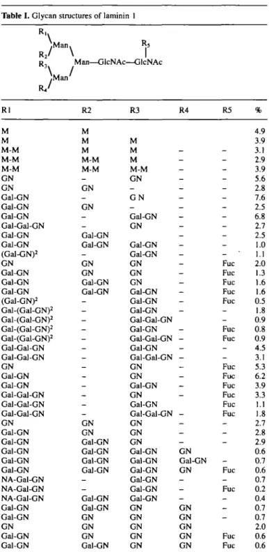
The glycans of laminin 1 comprise almost one-fifth of the molecular weight, all of which are N-linked. Structural studies (Arumugham et al., 1986; Fujiwara et al., 1988; Knibbs et al., 1989) have revealed unique patterns of glycosylation such as: (i) the presence of terminal galactosyl residues; (ii) expression of polylactosamines (necessary for galectin interactions); and (iii) terminal N-acetylglucosaminyl residues (ligands for P(l—>4) galactosyltransferase recognition). This last feature was only reported in the first structural study of laminin 1 glycans (Arumugham etai, 1986).
In an effort to correlate the structural variations with func-tion, among different laminin 1 preparations, we initiated a pro-file analysis of laminin glycans using ES-MS. The glycans were released from the protein enzymatically or with hydrazine, methylated, and the extract directly infused into the MS (Rein-hold et al., 1994). Further structural details were obtained by ORM (Linsley et al., 1994). Glycan tables are assembled on the premise that glycotypes follow known motifs with monomers and linkage patterns as previously established. Application of this technology to laminin 1 identified over 30 different glycan structures (Table I). All major glycotypes were identified with the high-mannose structures comprising -19%, hybrids 1.2%, and complex -80%, dispersed between -30 biantennary struc-tures. The majority of antennae terminate with N-acetylglucos-amine (-50% of all the glycans), with - 3 % displaying lactosN-acetylglucos-amine repeating units. These structures indicate the summed presence of terminal a-galactosyl residues at -59%. The profile suggests a form of incomplete processing, which may be due to the tumoral origin of the laminin 1.
In a general sense, these profiles support the role of galectins and P(l—»4) galactosytransferase in laminin 1 mediated func-tions. The detection of abundant high-mannose structures also upholds the recently described finding of oligomannoside-dependent spreading of melanoma cells on laminin (Chandra-sekaran et al., 1994). Although these analyses suggest glycan topology in a general sense, the question remains whether these glycans are present in non-tumoral laminins, how such inter-actions play a role in vivo, and what are their site specificity and detailed structural relationships to other family members. ES-MS provides an opportunity to rapidly profile indigenous structures at considerable sensitivity which may be followed by peptide mapping for site specificity relationships and tandem mass spectrometry for a detailed linkage and branching analysis.
Acknowledgements
We wish to thank Keyes Linsley for careful review of the data and gratefully acknowledge the support and helpful discussions with Dr R.Brentani. This work was partly supported by a NIH grant to V.N.R. (GM 45701).
References
Arumugham.R.G., Hsieh.T.C.Y., Tanzer.M. and Laine.R.A. (1986) Structures of the asparagine-linked sugar chains of laminin. Biochim. Biophys. Ada, 883, 112-126.
Glyco-Forum section
Table I. Glycan structures of laminin 1
.Man. R2/ \ R , Man—GlcNAc—GlcNAc \ /^Man' Rl M M M-M M-M M-M GN GN Gal-GN Gal-GN Gal-GN Gal-Gal-GN Gal-GN Gal-GN (Gal-GN)2 GN Gal-GN Gal-GN Gal-GN (Gal-GN)2 Gal-(Gal-GN)2 Gal-(Gal-GN)2 Gal-(Gal-GN)2 Gal-(Gal-GN)2 Gal-Gal-GN Gal-Gal-GN GN Gal-GN Gal-GN Gal-Gal-GN Gal-Gal-GN Gal-Gal-GN GN Gal-GN Gal-GN Gal-GN Gal-GN Gal-GN NA-Gal-GN NA-Gal-GN NA-Gal-GN Gal-GN Gal-GN GN Gal-GN Gal-GN R2 M M M M-M M-M _ GN _ GN _ _ Gal-GN Gal-GN — GN GN Gal-GN Gal-GN -_ — -_ _ -_ -GN GN Gal-GN Gal-GN Gal-GN Gal-GN _ -Gal-GN Gal-GN GN GN GN Gal-GN R3 M M M M-M GN _ G N -Gal-GN GN Gal-GN Gal-GN GN GN GN Gal-GN Gal-GN Gal-GN Gal-Gal-GN Gal-GN Gal-Gal-GN Gal-GN Gal-Gal-GN GN GN Gal-GN GN Gal-GN Gal-Gal-GN GN GN GN Gal-GN Gal-GN Gal-GN Gal-GN Gal-GN Gal-GN GN GN GN GN GN R4 -_ _ -— _ _ _ _ -_ — _ -— -— -_ _ _ -— _ _ -GN Gal-GN GN -— _ GN GN GN GN GN R5 -_ _ -_ -Fuc Fuc Fuc Fuc Fuc _ -Fuc Fuc -Fuc Fuc Fuc Fuc Fuc Fuc _ _ -_ Fuc _ Fuc _ _ _ Fuc Fuc % 4.9 3.9 3.1 2.9 3.9 5.6 2.8 7.6 2.5 6.8 2.7 2.5 1.0 1.1 2.0 1.3 1.6 1.6 0.5 1.8 0.9 0.8 0.9 4.5 3.1 5.3 6.2 3.9 3.3 1.1 1.8 2.7 2.8 2.9 0.6 0.7 0.6 0.7 0.2 0.4 0.7 0.7 2.0 0.6 0.6
Burgeson.R.E., Chiquet,M., Deutzmann.R., Ekblom.P, EngeU., Kleman.H., Martin.G., Meneguzzi.G., SanesJ., Timpl.R., Tryggvason.K., Yamada,Y. and YurchencoJ5. (1994) A new nomenclature for the laminins. Matrix BioL,
14,209-211.
Chammas.R., Veiga,S.S., Travasos.L.R. and Brentani,R.R. (1993) Functionally distinct roles for glycosylation of a and (3 integrin chains in cell-matrix inter-actions. Proc. NatlAcad. Sci. USA, 90, 1795-1799.
Chandrasekaran.S., Tanzer.M.L. and Giniger,M.S. (1994) Oligomannosides initiate cell spreading of laminin-adherent murine melanoma cells. J. Biol.
Chem., 269,, 3356-3366.
EngelJ. (1993). Structure and function of laminin. In Rohrbach,D. and Timpl, R. (eds), Molecular and Cellular Aspects of Basement Membranes. Academic Press Inc., San Diego, pp. 177-187.
ErvastiJ.M. and Campbell.K.P. (1993) A role for the dystrophin-glycoprotein complex as a transmembrane linker between laminin and actin. J. Cell Biol.,
1212, 809-823.
FennJ., Mann,M., Meng.C.K., Wong.S.F. and Whitehouse.C.M. (1989) Electrospray ionization for mass spectrometry of large biomolecules.
Science, 246, 64-71.
Fujiwara,S., Shinkai.H., Deutzmann,R., Paulsson.M. and Timpl,R. (1988) Structure and distribution of N-linked oligosaccharide chains on various domains of mouse tumor laminin. Biochem. J., 252, 453—461.
Gee.S.H., Blacher,R.W., Douville,P.J., Provost.P.R., Yurchenco.P.D. and Carbonetto,S. (1993) Laminin-binding protein 120 from brain is closely related to the dystrophin-associated glycoprotein, dystroglycan, and binds with high affinity to the major heparin binding domain of laminin. /. Biol.
Chem., 268, 14972-14980.
Hughes.R.C. (1994) Mac-2: a versatile galactose-binding protein of mam-malian tissues. Glycobiology, 4, 5-12.
Hynes.R. (1992) Integrins: versatility, modulation and signalling in cell adhesion. Cell, 69, 11-25.
Knibbs.R.N., Perini.F. and Goldstein.U. (1989) Structure of the major con-canavalin A reactive oligosaccharides of the extracellular component laminin.
Biochemistry, 28, 6379-6392.
Linsley.K.B., Chan.S., Chan.S., Reinhold,B.B., Lisi.PJ. and Reinhold.V.N. (1994) Applications of electrospray mass spectrometry to ervthropoietin N- and O-linked glycans. Anal. Biochem., 219, 207-217.
Reinhold.V.N., Reinhold.B.B. and Chan.S. (1994) Sequence analysis of glycoprotein glycans. In Matsuo.T., Seyama,Y., Caprioli.R.C. and Gross,M.L. (eds), Biological Mass Spectrometry Present and Future. John Wiley & Sons, pp. 403-434.
Shur.B. (1993) Glycosyltransferases as cell adhesion molecules. Curr. Opin.
Cell Biol., 5, 854-863.
Timpl.R. and BrownJ.C. (1994) The laminins. Matrix Biol., 14, 275-281.
Meeting Announcements
First EuroConference on Carbohydrate Mimics
Strasbourg, France May 15-18, 1995
The prominent role of carbohydrates in many biological pro-cesses is now well recognized. This has promoted numerous studies in the new field of analogues, often so-called 'mimics' of biologically significant carbohydrates. The purpose of this new series of two Euroconferences on Carbohydrate Mimics is to summarize the different aspects of this new expanding field of glycobiology. The first of these meetings will be held in May 1995 in France and will deal with the following topics: chem-ical synthesis, biologchem-ical evaluation, conformational aspects of carbohydrate analogues, including mono- and oligosaccharides, cyclitols, nucleosides and nucleotides.
Biochemical Society Glycobiology Group
Colloquia
Manchester July 18-19, 1995
Two day meeting on 'Mucins'; information from Dr J.Sheehan,
Department of Biochemistry, University of Manchester. Dublin (University College) September 13, 1995
One day meeting on 'Carbohydrate Recognition Proteins'; information from Dr M.Taylor, Glycobiology Institute, Department of Biochemistry, University of Oxford. 158