Publisher’s version / Version de l'éditeur:
Canadian Journal of Microbiology, 58, 2, pp. 124-131, 2012-01-19
READ THESE TERMS AND CONDITIONS CAREFULLY BEFORE USING THIS WEBSITE.
https://nrc-publications.canada.ca/eng/copyright
Vous avez des questions? Nous pouvons vous aider. Pour communiquer directement avec un auteur, consultez la première page de la revue dans laquelle son article a été publié afin de trouver ses coordonnées. Si vous n’arrivez pas à les repérer, communiquez avec nous à PublicationsArchive-ArchivesPublications@nrc-cnrc.gc.ca.
Questions? Contact the NRC Publications Archive team at
PublicationsArchive-ArchivesPublications@nrc-cnrc.gc.ca. If you wish to email the authors directly, please see the first page of the publication for their contact information.
NRC Publications Archive
Archives des publications du CNRC
This publication could be one of several versions: author’s original, accepted manuscript or the publisher’s version. / La version de cette publication peut être l’une des suivantes : la version prépublication de l’auteur, la version acceptée du manuscrit ou la version de l’éditeur.
For the publisher’s version, please access the DOI link below./ Pour consulter la version de l’éditeur, utilisez le lien DOI ci-dessous.
https://doi.org/10.1139/W11-116
Access and use of this website and the material on it are subject to the Terms and Conditions set forth at
Involvement of cytochrome c CymA in the anaerobic metabolism of
RDX by Shewanella oneidensis MR-1
Perreault, Nancy N.; Crocker, Fiona H.; Indest, Karl J.; Hawari, Jalal
https://publications-cnrc.canada.ca/fra/droits
L’accès à ce site Web et l’utilisation de son contenu sont assujettis aux conditions présentées dans le site LISEZ CES CONDITIONS ATTENTIVEMENT AVANT D’UTILISER CE SITE WEB.
NRC Publications Record / Notice d'Archives des publications de CNRC:
https://nrc-publications.canada.ca/eng/view/object/?id=841e1d7a-a1fe-4697-9012-3343f6f63e2b https://publications-cnrc.canada.ca/fra/voir/objet/?id=841e1d7a-a1fe-4697-9012-3343f6f63e2b
Involvement of cytochrome c CymA in the
anaerobic metabolism of RDX by Shewanella
oneidensis MR-1
Nancy N. Perreault, Fiona H. Crocker, Karl J. Indest, and Jalal Hawari
Abstract: Hexahydro-1,3,5-trinitro-1,3,5-triazine (RDX) is a cyclic nitramine explosive commonly used for military applica-tions that is responsible for severe soil and groundwater contamination. In this study, Shewanella oneidensis MR-1 was shown to efficiently degrade RDX anaerobically (3.5 µmol·h–1·(g protein)–1) via two initial routes: (1) sequential N-NO2
re-ductions to the corresponding nitroso (N-NO) derivatives (94% of initial RDX degradation) and (2) denitration followed by ring cleavage. To identify genes involved in the anaerobic metabolism of RDX, a library of ~2500 mutants of MR-1 was constructed by random transposon mutagenesis and screened for mutants with a reduced ability to degrade RDX compared with the wild type. An RDX-defective mutant (C9) was isolated that had the transposon inserted in the c-type cytochrome gene cymA. C9 transformed RDX at ~10% of the wild-type rate, with degradation occurring mostly via early ring cleavage caused by initial denitration leading to the formation of methylenedinitramine, 4-nitro-2,4-diazabutanal, formaldehyde, ni-trous oxide, and ammonia. Genetic complementation of mutant C9 restored the wild-type phenotype, providing evidence that electron transport components have a role in the anaerobic reduction of RDX by MR-1.
Key words:RDX, explosive, nitramine, biotransformation, Shewanella.
Résumé : La nitramine cyclique hexahydro-1,3,5-trinitro-1,3,5-triazine (RDX) est un explosif couramment utilisé pour des applications militaires et qui cause une contamination sévère de sols et eaux souterraines. Shewanella oneidensis MR-1 a dé-gradé efficacement le RDX en condition anaérobique (3,5 µmol·h–1·(g protéine)–1) via deux routes initiales : (1) la réduction
séquentielle du N-NO2en dérivés nitroso correspondants (N-NO) (94 % de la dégradation initiale du RDX); (2) dénitration
suivie du clivage de l’anneau du RDX. Afin d’identifier des gènes impliqués dans le métabolisme anaérobique du RDX, une banque d’environ 2500 mutants MR-1 fut construite par mutagenèse aléatoire par transposition et criblée à la recherche de mutants présentant une diminution de l’activité de dégradation du RDX par rapport à la souche sauvage. Un mutant (C9) fut isolé avec un transposon inséré dans le gène du cytochrome c cymA. C9 transformait le RDX à ~10 % du taux de dégra-dation de la souche sauvage. La dégradégra-dation s’effectuait principalement par dénitration suivie du clivage de l’anneau et conduisait à la formation de méthylènedinitramine, 4-nitro-2,4-diazabutanal, formaldéhyde, oxyde nitreux et ammoniac. La complémentation génétique du mutant C9 a permis de restaurer le phénotype sauvage et d’ainsi confirmer que des compo-sants de la chaîne de transport d’électrons ont un rôle à jouer dans la réduction anaérobique du RDX par MR-1.
Mots‐clés : RDX, explosif, nitramine, biotransformation, Shewanella. [Traduit par la Rédaction]
Introduction
The cyclic nitramine hexahydro-1,3,5-trinitro-1,3,5-triazine (RDX) is a major component of military high explosives and is used in a wide variety of munitions. The toxicity of RDX necessitates that contaminated soil and groundwater be reme-diated, preferably by low cost, environmentally friendly bio-logical processes. Some aerobic bacteria of the order Actinomycetales, such as Rhodococcus rhodochrous 11Y (Seth-Smith et al. 2002) and Rhodococcus sp. strain DN22 (Fournier et al. 2002), are able to use RDX as the sole
nitro-gen source for growth, while RDX appears to be used as a source of energy, carbon, and nitrogen for the growth of Wil-liamsia sp. strain KTR4 and Gordonia sp. strain KTR9 (Thompson et al. 2005). RDX was also used as the sole N source for the anaerobic growth of Desulfovibrio spp. (Boopathy et al. 1998). RDX-degrading Shewanella strains have been isolated from RDX-contaminated marine sediments (Zhao et al. 2005, 2006). Shewanella halifaxensis was shown to degrade RDX anaerobically, under trimethylamine N-oxide (TMAO)-respiring conditions. TMAO is a naturally occurring osmolyte found in marine environments that serves as a
ter-Received 3 September 2011. Revision received 1 November 2011. Accepted 2 November 2011. Published at www.nrcresearchpress.com/cjm on 19 January 2012.
N.N. Perreault and J. Hawari. Biotechnology Research Institute, National Research Council Canada, 6100 Royalmount Avenue, Montréal, QC H4P 2R2, Canada.
F.H. Crocker and K.J. Indest. US Army Engineer Research and Development Center, 3909 Halls Ferry Road, Vicksburg, MS 39180, USA.
Corresponding author: Jalal Hawari (e-mail: jalal.hawari@cnrc-nrc.gc.ca).
Can. J. Microbiol. 58: 124–131 (2012) doi:10.1139/W11-116 Published by NRC Research Press
Can. J. Microbiol. Downloaded from nrcresearchpress.com by National Research Council of Canada on 02/27/12
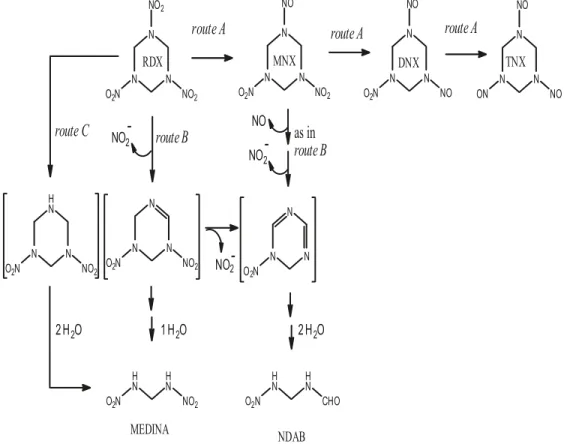
minal electron acceptor in various bacteria (Barrett and Kwan 1985). Under these conditions, S. halifaxensis degraded RDX via a major route consisting of the sequential reduction of the N-NO2 groups to produce the corresponding mono-, di-, and tri-nitroso derivatives hexahydro-1-nitroso-3,5-dinitro-1,3,5-triazine (MNX), hexahydro-1,3-dinitroso-5-nitro-1,3,5-tria-zine (DNX), and hexahydro-1,3,5-trinitroso-1,3,5-triahexahydro-1,3-dinitroso-5-nitro-1,3,5-tria-zine (TNX), respectively (Fig. 1, route A), and a minor route in-volving denitration followed by ring cleavage (Fig. 1, route B) to produce methylenedinitramine (MEDINA) and its de-composition products formaldehyde (HCHO) and nitrous ox-ide (N2O) (Halasz et al. 2002). A cytochrome c-552 purified from S. halifaxensis was shown to degrade RDX anaerobi-cally via initial denitration followed by ring cleavage to pro-duce MEDINA (Zhao et al. 2008), but other enzymes or cytochrome c proteins responsible for nitrosation have not been identified.
In a recent paper, the authors showed that polymerase chain reaction (PCR) failed to detect genes associated with RDX degradation (including the cytochrome P-450 gene xplA, hydrogenase gene hydA, organic nitrate reductase gene onr, and Pseudomonas flavoprotein xenobiotic reductases genes xenA and xenB) from RDX-contaminated groundwater samples (Fuller et al. 2010). Despite progress made towards understanding the degradation routes of RDX, the genetic de-terminants dictating the fate of RDX, particularly under anae-robic conditions, are still unclear. By analyzing the genomes
of 15 Shewanella strains, Zhao et al. (2010) observed that the RDX-degrading strains had a higher number of genes for cytochromes and nitrate or nitrite reductases. The iron-reduc-ing bacterium Shewanella oneidensis MR-1 (Myers and Neal-son 1988) has a complex electron transfer network and can utilize a variety of electron acceptors for anaerobic respira-tion, including Fe(III), Mn(IV), fumarate, nitrate, TMAO, and a variety of metals (Nealson and Saffarini 1994; Burns and DiChristina 2009). Because of its exceptional metabolic versatility and its potential use for the bioremediation of en-vironmental contaminants, MR-1 has been extensively studied and its genome has been sequenced (Heidelberg et al. 2002). Although MR-1 was not isolated from an RDX-contaminated environment, it was recently proposed that MR-1 can also use RDX as an electron acceptor (Kwon and Finneran 2008) but the pathway for RDX degradation was not determined. Other Fe(III)-reducing bacteria such as Geo-bacter metallireducens and Geobacter sulfurreducens were shown to directly reduce RDX to its nitroso derivatives (Kwon and Finneran 2006). Additionally, cyclic nitramine transformation mediated by extracellular electron shuttles, such as humic substances or humic substances analog anthra-quinone-2,6-disulfonate, is ubiquitous among the keystone Fe (III)-reducing microbial genera (Kwon and Finneran 2008, 2010). Electron shuttling compounds were shown to mediate RDX degradation via initial nitro group reduction (Bhushan et al. 2006; Kwon and Finneran 2006).
Fig. 1. Schematic representation of the proposed pathway for anaerobic transformation of RDX. Route A consists of the sequential reduction of the N-NO2groups to produce the corresponding mono-, di-, and tri-nitroso derivatives MNX
(hexahydro-1-nitroso-3,5-dinitro-1,3,5-tria-zine), DNX (hexahydro-1,3-dinitroso-5-nitro-1,3,5-tria(hexahydro-1-nitroso-3,5-dinitro-1,3,5-tria-zine), and TNX (hexahydro-1,3,5-trinitroso-1,3,5-tria(hexahydro-1-nitroso-3,5-dinitro-1,3,5-tria-zine), respectively. Route B con-sists of one or two denitration steps followed by ring cleavage to produce MEDINA (methylenedinitramine) or NDAB
(4-nitro-2,4-diazabutanal), respectively. Route C involves denitrohydrogenation leading to the formation of the unstable 3,5-dinitro-1,3,5-triazacyclohexane (in brackets) whose decomposition leads to the formation of MEDINA. Adapted from Fournier et al. (2002) and Bhushan et al. (2002).
Published by NRC Research Press
Can. J. Microbiol. Downloaded from nrcresearchpress.com by National Research Council of Canada on 02/27/12
This study was undertaken to identify genes involved in the anaerobic metabolism of RDX by S. oneidensis MR-1. Transposon insertion mutants of strain MR-1 were isolated that had a reduced rate of RDX transformation compared with the wild-type MR-1 strain. The gene that was inter-rupted by the transposon insertion as well as the rate of RDX transformation and product distribution in the mutant were determined.
Materials and methods Chemicals and culture media
RDX (99% pure), TNX, and MEDINA were provided by Defense Research and Development Canada, Quebec. MNX (98% pure), DNX, and 4-nitro-2,4-diazabutanal (NDAB) were purchased from SRI International, Menlo Park, Califor-nia. All other chemicals were of reagent grade. Shewanella oneidensis MR-1 was purchased from the American Type Culture Collection (ATCC 700550). MR-1 was routinely maintained on Luria–Bertani (LB) medium. The minimal me-dium MMR2 used in this study was a modification of the medium described by Tang et al. (2007) and consisted of (per L of distilled water): 1.34 mmol·L–1 KCl, 5 mmol·L–1 NaH2PO4, 0.7 mmol·L–1Na2SO4, 1 mmol·L–1MgSO4·7H2O, 0.2 mmol·L–1 CaCl
2·2H2O, 50 mmol·L–1NaCl, 20 mmol·L–1 PIPES, a vitamin mixture (Tang et al. 2007), and trace ele-ments SL-10 (Widdel et al. 1983). MMR2 contained 28 mmol·L–1 NH4Cl and 25 mmol·L–1sodium lactate as the nitrogen and carbon source, respectively; TMAO (30 mmol·L–1) was used as the terminal electron acceptor. Fumarate, Fe3+(FeCl
3), and nitrate (NaNO3) at 15 mmol·L–1 were also tested individually as the sole electron acceptor. The pH was adjusted to 7.0 before sterilization through a 0.2 µm filter.
Construction and screening of the transposon library A transposon library of MR-1 was generated by random mutagenesis using the EZ-Tn5 <R6Kgori/KAN-2> Tnp Transposome kit (Epicentre) according to the manufacturer’s instructions. Briefly, freshly prepared MR-1 cells were trans-formed with 1 µL of the Tnp transposome. Transformants were initially selected on LB agar containing 50 µg kanamy-cin·mL–1 (Kan 50) and then grown aerobically overnight in 96-well plates containing 1 mL of LB Kan 50. Glycerol was added at 15% v/v final concentration and the cultures were flash-frozen and kept at –80 °C until used. For the pheno-typic screening, a MULTI-BLOT Replicator (V&P Scientific) was used to transfer culture inocula to a new 96-well plate filled with 1.5 mL of LB supplemented with 90 µmol·L–1 RDX. Controls consisted of two wells inoculated with the wild-type MR-1 and two noninoculated wells. The plates were sealed with sterile breathable membranes and placed in anaerobic jars. The jars were purged with oxygen-free N2and incubated for 48 h at 30 °C in the dark. After centrifugation at 4000g for 10 min, the supernatants were transferred to 1 mL glass vials for RDX quantification. Kanamycin-resistant MR-1 strains showing a reduced RDX degradation activity were further characterized by identification of the disrupted gene, by RDX degradation rate, and by product distribution (described below).
Determination of the transposon insertion site
Genomic DNA was extracted from the mutant strain using the QIAGEN DNeasy Blood & Tissue kit. Five micrograms of purified DNA was added to 100 µL of Tris–EDTA (pH 8.0) and submitted to 200 passages through a 30-gauge hypodermic needle. DNA fragments of 6–8 kb in length were purified from agarose gel using the illustra GFX PCR DNA and Gel Band Purification kit (GE Healthcare) and eluted in MilliQ water. One microgram of purified DNA was blunt ended and 5′-phosphorylated using the it DNA End-Repair kit (Epicentre). The repaired DNA was self-ligated at 14 °C overnight with the T4 DNA ligase (Promega). Ligation products were transformed into freshly prepared electrocom-petent Escherichia coli EC100D pir-116 (Epicentre) and plated on LB agar Kan 50. The transposon insertion sites from a number of kanamycin-resistant rescue clones were amplified by PCR using the transposon-specific primers KAN-2 FP-1 and R6KAN-2 RP-1 (Epicentre). Sequencing of the amplicons was performed at the McGill University Ge-nome Quebec Innovation Centre (Montréal, Quebec, Can-ada). The identity of the DNA sequences was determined by a search of the NCBI nonredundant database using the DNA–DNA BLAST (blastn). The search was limited to re-cords matching the genome of S. oneidensis MR-1 (genome AE014299, taxid 211586).
Growing cell assay
Bacterial growth and RDX degradation were followed in MMR2 medium supplemented with 90 µmol·L–1 RDX. The medium was distributed in sterile glass serum bottles sealed with rubber septa and aluminum crimp caps. The bottles were made anaerobic by degassing for 30 min and then per-forming five cycles of degassing and recharging with argon. Bacterial cells were inoculated at an initial optical density of 0.04 at 600 nm (OD600). Abiotic controls containing RDX in uninoculated culture medium and controls containing bacteria in medium without RDX were conducted in parallel. The bot-tles (in triplicate) were incubated for a period of 72 h in the dark at 25 °C with agitation at 150 r·min–1. Growth was fol-lowed by monitoring the increase in OD600. Samples were collected over time for analysis of RDX and its products. Resting cell assay for metabolic studies
Time-course experiments were conducted on cells pre-grown anaerobically in MMR2 with TMAO and supple-mented with 22.5 µmol·L–1 RDX. The cells were harvested at late exponential phase, washed in cold 25 mmol·L–1 sodium phosphate buffer (pH 7.0), and resuspended at a concentration of 2 mg cell protein·mL–1 in the same buffer but containing ~90 µmol·L–1 RDX. The reactions were performed in serum bottles that were sealed and made anaerobic by briefly de-gassing (1 min) and then purging with argon for 10 min at 5 psi (1 psi = 6.894757 kPa). The bottles were incubated at 25 °C in the dark. Uninoculated control bottles and controls without RDX were conducted in parallel. At selected times, aliquots of the cell suspensions were collected, centrifuged at 16 000g for 10 min, and the supernatant was used for chemi-cal analysis of RDX and its breakdown products. The results are presented as the mean of triplicate assays with the stand-ard deviation. Protein concentration was determined using the bicinchoninic acid method by following the instruction of the
Can. J. Microbiol. Downloaded from nrcresearchpress.com by National Research Council of Canada on 02/27/12
BCA Protein Assay kit (Pierce, Rockford, Illinois). Some mi-crocosms were spiked withL-[U-14C]RDX (0.038 µCi (1 Ci = 37 Gbq)) to measure mineralization (liberated 14CO
2) with a Tri-Carb 4530 liquid scintillation counter (model 2100 TR; Packard Instrument Company) as described previously (Zhao et al. 2002).
Southern blot and complementation
Southern blot was performed to confirm the insertion site of the transposon. The genomic DNA from the mutant and the wild-type strains was digested with three restriction en-zymes (BstBI, EcoRV, and NcoI, individually), and the result-ing digested DNAs were separated by electrophoresis on a 0.5% agarose gel. DNA was transferred onto a positively charged nylon membrane. The membrane was hybridized with a DIG-labeled probe targeting the kanamycin resistance (Kmr) gene of the transposon (Roche PCR DIG Probe Syn-thesis kit). Detection was performed by chemiluminescence. The expected sizes of the transposon-containing DNA bands were calculated using the web-based program Webcutter 2.0.
A complementing plasmid for mutant C9 (mutant de-scribed below) was constructed and used as a control to en-sure that the phenotype observed was caused by disruption of the cymA gene. The complete cymA gene, plus 97 and 120 nucleotides of the upstream and downstream regions, respec-tively, was PCR-amplified from the wild-type MR-1 using the primers C9v2F_EcoRI (CGGCATTTTTAGGCTTGAATC-CAAACTTCT) and C9v2R_BamHI (AATCGCCCT-TAGCGCCGGATCCCGCAGAG). The 783 bp EcoRI– BamHI fragment was cloned directionally into pBBR1MCS-5, a broad-host-range cloning vector with gentamicin resistance (Kovach et al. 1995), resulting in complementing plasmid pB14. The pB14 plasmid was electroporated in E. coli DH10B and the colonies were selected on LB plates with gentamicin at 20 µg·mL–1. The plasmid was then purified with the QIAprep Spin Miniprep kit and the proper cloning was confirmed by sequencing. The plasmid was finally elec-troporated in mutant C9.
Analytical methods
RDX, MNX, DNX, and TNX were analyzed by a reverse-phase high performance liquid chromatography (HPLC) in-strument connected to a photodiode array detector. Briefly, the products were analyzed with a Waters HPLC system (Milford, Massachusetts, USA) using a Supelcosil LC-CN HPLC column (25 cm by 4.6 mm by 5 µm) (Oakville, On-tario, Canada) at 35 °C. MEDINA and NDAB were deter-mined on an AnionSep Ice-Ion-310 Fast organic acids HPLC column (St. Louis, Missouri, USA) at 225 nm and 35 °C. The mobile phase was acidified water (pH 2.0) at a flow rate of 0.6 mL·min–1. Chromatograms were taken at a wavelength of 225 nm. HCHO was analyzed as its derivative with 2,4-pentanedione (Zhao et al. 2002). N2O was analyzed in the headspace of the samples using an Agilent 6890 gas chroma-tograph equipped with an electron capture detector (ECD). The ammonium cation was analyzed using a TSP Spectra-SYSTEM HPLC system equipped with a Waters 431 conduc-tivity detector and a Hamilton PRP-X200 analytical cation-exchange column (250 mm by 4.1 mm by 10 µm). Nitrite (NO2–), nitrate (NO3–), and formic acid (HCOO–) were
ana-lyzed by ion chromatography using a Dionex DX 120 ion chromatograph (Sunnyvale, California, USA).
Results
Screening the transposon library for an RDX-defective mutant and identification of the disrupted gene
Random insertion mutagenesis was performed on MR-1, and ~2500 transformants (mutants) were subjected to pheno-typic screening (RDX removal). After 48 h of incubation in LB medium amended with RDX, the cultures of five mutants had remaining concentrations of RDX similar to those of the controls without bacterial cells. RDX-defective phenotypes were verified by three additional rounds of screening. To ex-clude the possibility that the RDX-defective phenotype might be a result of severe growth defects, the growth of mutants in LB plus RDX was monitored by measuring the OD600. Mu-tant C9 showed the RDX-defective phenotype in three rounds of screening, while its OD600 showed growth similar to the wild type. Sequencing of the rescued plasmid from the mu-tant strain identified the disrupted gene as cymA (locus SO_4591; transposon inserted ~145 nucleotides before the stop codon); the gene encodes a tetraheme cytochrome c. An-other mutant, D6, lacked the orange pigmentation character-istic of the wild-type strain and the RDX-degradation phenotype; it grew in LB to an OD600, which is ~30% lower than the wild type. The transposon was located in the naph-thoate synthase gene menB (locus SO_4739; transposon in-serted ~222 nucleotides before the stop codon). Southern blots showed the presence of a single insertion of the Kmr gene in the genomic DNA of C9 and D6, as well as its ab-sence in MR-1. The size of the bands in BstBI, EcoRV, and NcoI-digested genomic DNA was consistent with the ex-pected (calculated) bands (data not shown). However, as we failed to complement strain D6 to confirm the phenotype, this strain was excluded from further analysis.
Anaerobic metabolism of RDX in S. oneidensis MR-1 RDX degradation by strain MR-1 in MMR2 medium was studied using growing and resting cells. Anaerobic growth of MR-1 in MMR2 minimal medium was not supported by the addition of RDX as the sole nitrogen source, carbon source, or electron acceptor. Growth of MR-1 in MMR2 with fuma-rate or TMAO as the sole terminal electron acceptor gave an OD600 of 0.36 ± 0.01 and 0.35 ± 0.02 after 24 h, respec-tively; when nitrate was used, the cells reached an OD600 of 0.09 ± 0.02 after 24 h.
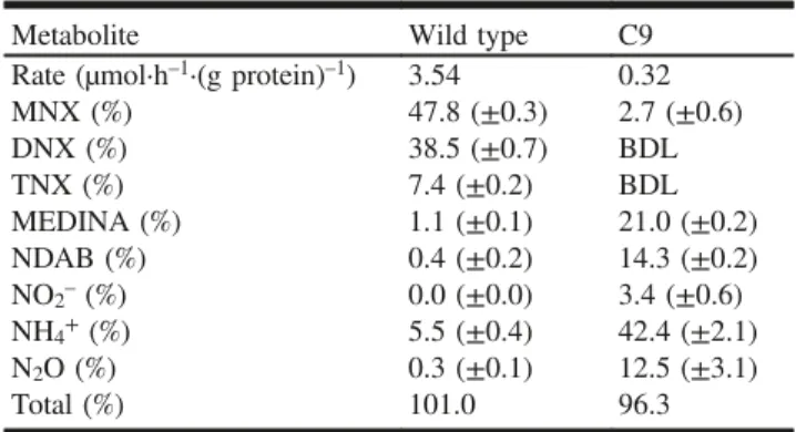
Growth of MR-1 in MMR2 with TMAO was accompanied by the degradation of RDX at a rate of 4.3 µmol·L–1·h–1. After 12 h of incubation, ~50% of RDX was degraded, pro-ducing mostly the three nitroso derivatives MNX (72%), DNX (4%), and TNX (traces). We were unable to analyze for other degradation products such as MEDINA and NDAB because of interference from the medium. Resting cells of MR-1 (2 mg cell protein·mL–1) degraded RDX at a rate of 3.54 µmol·h–1·(g protein)–1 (Table 1). The disappearance of RDX was accompanied by the sequential formation of the three nitroso derivatives MNX (47.8%), DNX (38.5%), TNX (7.4%), and the degradation products MEDINA (1.1%), NDAB (0.4%), HCHO (2.2%), N2O (0.3%), and ammonia Published by NRC Research Press
Can. J. Microbiol. Downloaded from nrcresearchpress.com by National Research Council of Canada on 02/27/12
(5.5%) (Fig. 2a; Table 1). The C- and N-mass balances were calculated at 97% and 101%, respectively.
Anaerobic metabolism of RDX in MR-1 mutants
Mutant C9 grew in MMR2 with TMAO as the sole elec-tron acceptor. C9 cells started to remove RDX at a rate of 0.75 µmol·L–1·h–1 after the cells reached the stationary phase at an OD600 of ~0.35. C9 grew poorly on fumarate, Fe3+ (FeCl3), or nitrate (NaNO3) as the sole electron acceptor (data not shown). Therefore, TMAO was used as the terminal electron acceptor in the MMR2 medium for the metabolic studies. The RDX transformation products were identified in resting cells. RDX removal by C9 occurred at a rate of 0.32 µmol·h–1·(g protein)–1 (Table 1). RDX was degraded mostly via initial denitration leading to ring cleavage and de-composition to MEDINA (21.0%), NDAB (14.3%), NO2– (3.4%), N2O (12.5%), and ammonia (42.4%) (Fig. 2b; Ta-ble 1). Traces of MNX were also detected. MEDINA declined over time as it decomposed to HCHO (12% of the C) and N2O. The N- and C-mass balances were 96% and 36%, re-spectively. The carbon mass balance was finalized by the de-tection of 14CO
2 from incubations with uniformly labelled 14C-[RDX]: 63% of 14CO
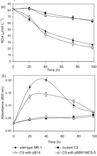
2 was recovered for a C-mass bal-ance of 99%. Growth with fumarate as the sole electron ac-ceptor was partially restored and the ability to transform RDX was completely restored in C9:pB14 (Fig. 3). The mu-tant strain containing pBBR1MCS-5 without insert was used as a control; pBBR1MCS-5 did not restore growth or RDX transformation.
Discussion
Shewanella oneidensis MR-1, a freshwater bacterium iso-lated from a noncontaminated site (Myers and Nealson 1988), degraded RDX efficiently and through the same meta-bolic pathways used by the marine Shewanella strains iso-lated from RDX-contaminated sediments. Similarly, MR-1 did not use RDX as a source of energy, N, or C for growth; RDX transformation proceeded via co-metabolism. MR-1 de-graded RDX via two initial routes as illustrated in Fig. 1: (1) a major route (route A) represented by the sequential
re-duction of the N-NO2 groups to the corresponding N-NO groups to produce MNX, DNX, and TNX and (2) a minor route (route B) involving initial denitration followed by ring cleavage as demonstrated by the detection of key products such as MEDINA, NDAB, N2O, and HCHO. Previously, we identified denitrohydrogenation as the initial step leading to RDX decomposition to MEDINA (Bhushan et al. 2002). In the present study, RDX could have underwent denitrohydro-genation, leading to the formation of the unstable 3,5-dini-tro-1,3,5-triazacyclohexane whose decomposition would also lead to the formation of MEDINA (Fig. 1, route C) (McHugh et al. 2002). The present finding was in line with what we observed during degradation of RDX with the marine strain S. halifaxensis (Zhao et al. 2008); also for both strains, RDX was used as a weak electron acceptor.
To understand the genes responsible for initiating RDX degradation, we constructed a transposon library of MR-1
Table 1. Stoichiometry of nitrogen after degradation of ~45 µmol·L–1RDX by the wild-type (MR-1) and mutant (C9)
strains.
Metabolite Wild type C9 Rate (µmol·h–1·(g protein)–1) 3.54 0.32
MNX (%) 47.8 (±0.3) 2.7 (±0.6) DNX (%) 38.5 (±0.7) BDL TNX (%) 7.4 (±0.2) BDL MEDINA (%) 1.1 (±0.1) 21.0 (±0.2) NDAB (%) 0.4 (±0.2) 14.3 (±0.2) NO2–(%) 0.0 (±0.0) 3.4 (±0.6) NH4+(%) 5.5 (±0.4) 42.4 (±2.1) N2O (%) 0.3 (±0.1) 12.5 (±3.1) Total (%) 101.0 96.3
Note: Values were calculated based on the percentage of reacted RDX using total theoretical numbers of N atoms. BDL, below detection level. RDX, hexahydro-1,3,5-trinitro-1,3,5-triazine; MNX, hexahydro-1-nitroso-3,5-dinitro-1,3,5-triazine; DNX, hexahydro-1,3-dinitroso-5-nitro-1,3,5-triazine; TNX, hexahydro-1,3,5-trinitroso-hexahydro-1,3-dinitroso-5-nitro-1,3,5-triazine;
MEDINA, methylenedinitramine; NDAB, 4-nitro-2,4-diazabutanal.
Fig. 2. Time course of anaerobic transformation of RDX
(~90 µmol·L–1) at 25 °C in 25 mmol·L–1sodium phosphate buffer
(pH 7.0) for wild-type strain MR-1 (a) and for mutant strain C9 (b). The cell protein concentration was 2 mg·mL–1. RDX,
hexahydro-1,3,5-trinitro-1,3,5-triazine; MNX, hexahydro-1-nitroso-3,5-dinitro-1,3,5-triazine; DNX, hexahydro-1,3-dinitroso-5-nitro-hexahydro-1-nitroso-3,5-dinitro-1,3,5-triazine; TNX, hexahydro-1,3,5-trinitroso-1,3,5-triazine; MEDINA, methyl-enedinitramine; NDAB, 4-nitro-2,4-diazabutanal; HCHO, formalde-hyde.
Can. J. Microbiol. Downloaded from nrcresearchpress.com by National Research Council of Canada on 02/27/12
and identified mutant C9 with a reduced ability to degrade RDX in comparison with the wild type. Specifically, the re-duction of the N-NO2 groups of RDX to the corresponding N-NO derivatives (Fig. 1, route A) was affected by disruption of cymA. Other studies have demonstrated that the absence of cymA significantly impedes the ability of MR-1 (i.e., 80%– 100%) to use a range of substrates as terminal electron ac-ceptors, including Fe(III)/Mn(IV) oxides, fumarate, nitrate, nitrite, and DMSO (Myers and Myers 1997; Schwalb et al. 2003; Lies et al. 2005). CymA and menaquinone participate in vanadium reduction (V(V) to V(IV)) (Myers et al. 2004) and arsenate reduction (As(V) to As(III)) in Shewanella spp. (Murphy and Saltikov 2007). CymA, a cytoplasmic mem-brane-bound c-type cytochrome, transfers electrons between the menaquinone pool of the inner membrane and periplas-mic terminal reductases or periplasperiplas-mic c-type cytochrome proteins, such as MtrA (Schuetz et al. 2009). Our results sug-gest that a system of electron transfer involving CymA and possibly other cytochrome c’s or electron transport proteins can reduce RDX in strain MR-1. Although RDX transforma-tion was greatly repressed in mutant C9 (reductransforma-tion of 90% compared with the wild type), it was not completely inhib-ited. Since Shewanella species have approximately 40 c-type
cytochromes (Fredrickson et al. 2008; Zhao et al. 2010), this versatility may be exploited by MR-1 in mutant C9 permit-ting a slower transformation of RDX mainly by denitration. For example, Cordova et al. (2011) have reported that SirCD can functionally replace CymA in several respiratory path-ways of MR-1. CymA does appear to have an important role but the specific nature is still unknown due to the robust electron transport system and cytochrome pool in Shewa-nella. The downstream components involved in RDX trans-formation by MR-1 are also unknown, since CymA can couple to multiple terminal reductases and periplasmic elec-tron transfer proteins (Cordova et al. 2011).
The screening of 2500 transposon mutants certainly did not identify all of the genes involved in the RDX metabolism by MR-1. However, the identification of the cymA gene pro-vided evidence that electron transport components have a role in the anaerobic metabolism of RDX by MR-1. RDX and its N-nitroso derivatives are toxic (Zhang et al. 2006; Pan et al. 2007). On the other hand, the ring-cleavage product MEDINA is unstable and decomposes to HCHO or CO2and N2O (Halasz et al. 2002), while NDAB can be degraded and mineralized by microorganisms such as Methylobacterium and Phanerochaete chrysosporium (Fournier et al. 2004, 2005). As already discussed, CymA is involved in the reduc-tion of several natural substrates. This study demonstrated that cymA was also involved in the transformation of the xenobiotic compound RDX. Zhao et al. (2008) showed that the mechanism of electron transfer had an impact on the RDX transformation route in S. halifaxensis: cells grown with nitrate transformed RDX mainly by denitration while cells grown with TMAO transformed RDX mainly by N-nitroso formation. Similarly, the rate and type of RDX metab-olites formed depended on whether electrons were transferred from biologically reduced Fe(III) or electron shuttles (Bhushan et al. 2006; Kwon and Finneran 2008, 2010). Therefore, supplementary data on the specific electron trans-fer mechanisms occurring in Shewanella (and other anae-robes) that increase the rate and extent of RDX ring-cleavage products is suitable to develop better remediation strategy for RDX contaminated soils and groundwater.
Acknowledgements
This work was supported by the Strategic Environmental Research and Development Program (SERDP), USA (ER1609). We thank J.-S. Zhao and also A. Halasz, D. Manno, L. Paquet, A. Corriveau, and S. Deschamps for the analytic support and D. Labbé for help with the Southern blot. We thank Dr. Maria Brandl for kindly providing the pBBR1MCS-5 vector.
References
Barrett, E.L., and Kwan, H.S. 1985. Bacterial reduction of trimethylamine oxide. Annu. Rev. Microbiol. 39(1): 131–149. doi:10.1146/annurev.mi.39.100185.001023. PMID:3904597. Bhushan, B., Halasz, A., Spain, J.C., and Hawari, J. 2002. Diaphorase
catalyzed biotransformation of RDX via N-denitration mechanism. Biochem. Biophys. Res. Commun. 296(4): 779–784. doi:10.1016/ S0006-291X(02)00874-4. PMID:12200115.
Bhushan, B., Halasz, A., and Hawari, J. 2006. Effect of iron(III), humic acids and anthraquinone-2,6-disulfonate on biodegradation of cyclic nitramines by Clostridium sp. EDB2. J. Appl. Microbiol. Fig. 3. Complementation of mutant strain C9 with plasmid pB14
(i.e., cymA from MR-1 cloned in pBBR1MCS-5). (a) RDX (hexahydro-1,3,5-trinitro-1,3,5-triazine) removal, and (b) growth of the strains in MMR2 mineral medium with fumarate as the terminal electron acceptor.
Published by NRC Research Press
Can. J. Microbiol. Downloaded from nrcresearchpress.com by National Research Council of Canada on 02/27/12
100(3): 555–563. doi:10.1111/j.1365-2672.2005.02819.x. PMID: 16478495.
Boopathy, R., Gurgas, M., Ullian, J., and Manning, J.F. 1998. Metabolism of explosive compounds by sulfate-reducing bacteria. Curr. Microbiol. 37(2): 127–131. doi:10.1007/s002849900350. PMID:9662613.
Burns, J.L., and DiChristina, T.J. 2009. Anaerobic respiration of elemental sulfur and thiosulfate by Shewanella oneidensis MR-1 requires psrA, a homolog of the phsA gene of Salmonella enterica serovar typhimurium LT2. Appl. Environ. Microbiol. 75(16): 5209–5217. doi:10.1128/AEM.00888-09. PMID:19542325. Cordova, C.D., Schicklberger, M.F.R., Yu, Y., and Spormann, A.M.
2011. Partial functional replacement of CymA by SirCD in Shewanella oneidensis MR-1. J. Bacteriol. 193(9): 2312–2321. doi:10.1128/JB.01355-10. PMID:21378180.
Fournier, D., Halasz, A., Spain, J., Fiurasek, P., and Hawari, J. 2002. Determination of key metabolites during biodegradation of hexahydro-1,3,5-trinitro-1,3,5-triazine with Rhodococcus sp. strain DN22. Appl. Environ. Microbiol. 68(1): 166–172. doi:10.1128/ AEM.68.1.166-172.2002.
Fournier, D., Halasz, A., Spain, J., Spanggord, R.J., Bottaro, J.C., and Hawari, J. 2004. Biodegradation of the hexahydro-1,3,5-trinitro-1,3,5-triazine ring cleavage product 4-nitro-2,4-diazabutanal by Phanerochaete chrysosporium. Appl. Environ. Microbiol. 70(2): 1123–1128. doi:10.1128/AEM.70.2.1123-1128.2004. PMID: 14766596.
Fournier, D., Trott, S., Hawari, J., and Spain, J. 2005. Metabolism of the aliphatic nitramine 4-nitro-2,4-diazabutanal by Methylobacter-iumsp. strain JS178. Appl. Environ. Microbiol. 71(8): 4199–4202. doi:10.1128/AEM.71.8.4199-4202.2005. PMID:16085803. Fredrickson, J.K., Romine, M.F., Beliaev, A.S., Auchtung, J.M.,
Driscoll, M., Gardner, T.S., et al. 2008. Towards environmental systems biology of Shewanella. Nat. Rev. Microbiol. 6(8): 592– 603. doi:10.1038/nrmicro1947. PMID:18604222.
Fuller, M.E., McClay, K., Higham, M., Hatzinger, P.B., and Steffan, R.J. 2010. Hexahydro-1,3,5-trinitro-1,3,5-triazine (RDX) bioreme-diation in groundwater: Are known RDX-degrading bacteria the dominant players? Bioremediat. J. 14(3): 121–134. doi:10.1080/ 10889868.2010.495367.
Halasz, A., Spain, J., Paquet, L., Beaulieu, C., and Hawari, J. 2002. Insights into the formation and degradation mechanisms of methylenedinitramine during the incubation of RDX with anaerobic sludge. Environ. Sci. Technol. 36(4): 633–638. doi:10. 1021/es011071g. PMID:11878377.
Heidelberg, J.F., Paulsen, I.T., Nelson, K.E., Gaidos, E.J., Nelson, W. C., Read, T.D., et al. 2002. Genome sequence of the dissimilatory metal ion-reducing bacterium Shewanella oneidensis. Nat. Bio-technol. 20(11): 1118–1123. doi:10.1038/nbt749. PMID: 12368813.
Kovach, M.E., Elzer, P.H., Hill, D.S., Robertson, G.T., Farris, M.A., Roop, R.M., II, and Peterson, K.M. 1995. Four new derivatives of the broad-host-range cloning vector pBBR1MCS, carrying different antibiotic-resistance cassettes. Gene, 166(1): 175–176. doi:10.1016/0378-1119(95)00584-1. PMID:8529885.
Kwon, M.J., and Finneran, K.T. 2006. Microbially mediated biodegradation of hexahydro-1,3,5-trinitro-1,3,5-triazine by extra-cellular eletron shuttling compounds. Appl. Environ. Microbiol. 72(9): 5933–5941. doi:10.1128/AEM.00660-06. PMID:16957213. Kwon, M.J., and Finneran, K.T. 2008. Hexahydro-1,3,5-trinitro-1,3,5-triazine (RDX) and octahydro-1,3,5,7-tetranitro-1,3,5,7-tet-razocine (HMX) biodegradation kinetics amongst several Fe(III)-reducing genera. Soil Sediment Contam. 17(2): 189–203. doi:10. 1080/15320380701873132.
Kwon, M.J., and Finneran, K.T. 2010. Electron shuttle-stimulated
RDX mineralization and biological production of 4-nitro-2,4-diazabutanal (NDAB) in RDX-contaminated aquifer material. Biodegradation, 21(6): 923–937. doi:10.1007/s10532-010-9352-1. PMID:20424887.
Lies, D.P., Hernandez, M.E., Kappler, A., Mielke, R.E., Gralnick, J.A., and Newman, D.K. 2005. Shewanella oneidensis MR-1 uses overlapping pathways for iron reduction at a distance and by direct contact under conditions relevant for biofilms. Appl. Environ. Microbiol. 71(8): 4414–4426. doi:10.1128/AEM.71.8.4414-4426. 2005. PMID:16085832.
McHugh, C.J., Smith, W.E., Lacey, R., and Graham, D. 2002. The first controlled reduction of the high explosive RDX. Chem. Commun. (Camb.), (21): 2514–2515. doi:10.1039/b207885f. Murphy, J.N., and Saltikov, C.W. 2007. The cymA gene, encoding a
tetraheme c-type cytochrome, is required for arsenate respiration in Shewanellaspecies. J. Bacteriol. 189(6): 2283–2290. doi:10.1128/ JB.01698-06. PMID:17209025.
Myers, C.R., and Myers, J.M. 1997. Cloning and sequence of cymA, a gene encoding a tetraheme cytochrome c required for reduction of iron(III), fumarate, and nitrate by Shewanella putrefaciens MR-1. J. Bacteriol. 179(4): 1143–1152. PMID:9023196.
Myers, C.R., and Nealson, K.H. 1988. Bacterial manganese reduction and growth with manganese oxide as the sole electron acceptor. Science, 240(4857): 1319–1321. doi:10.1126/science.240.4857. 1319. PMID:17815852.
Myers, J.M., Antholine, W.E., and Myers, C.R. 2004. Vanadium(V) reduction by Shewanella oneidensis MR-1 requires menaquinone and cytochromes from the cytoplasmic and outer membranes. Appl. Environ. Microbiol. 70(3): 1405–1412. doi:10.1128/AEM. 70.3.1405-1412.2004. PMID:15006760.
Nealson, K.H., and Saffarini, D. 1994. Iron and manganese in anaerobic respiration: environmental significance, physiology, and regulation. Annu. Rev. Microbiol. 48(1): 311–343. doi:10.1146/ annurev.mi.48.100194.001523. PMID:7826009.
Pan, X., Zhang, B., Smith, J.N., Francisco, M.S., Anderson, T.A., and Cobb, G.P. 2007. N-Nitroso compounds produced in deer mouse (Peromyscus maniculatus) GI tracts following hexahydro-1,3,5-trinitro-1,3,5-triazine (RDX) exposure. Chemosphere, 67(6): 1164–1170. doi:10.1016/j.chemosphere.2006.10.077. PMID: 17223165.
Schuetz, B., Schicklberger, M., Kuermann, J., Spormann, A.M., and Gescher, J. 2009. Periplasmic electron transfer via the c-type cytochromes MtrA and FccA of Shewanella oneidensis MR-1. Appl. Environ. Microbiol. 75(24): 7789–7796. doi:10.1128/AEM. 01834-09. PMID:19837833.
Schwalb, C., Chapman, S.K., and Reid, G.A. 2003. The tetraheme cytochrome CymA is required for anaerobic respiration with dimethyl sulfoxide and nitrite in Shewanella oneidensis. Biochem-istry, 42(31): 9491–9497. doi:10.1021/bi034456f. PMID:12899636. Seth-Smith, H.M.B., Rosser, S.J., Basran, A., Travis, E.R., Dabbs, E.R., Nicklin, S., and Bruce, N.C. 2002. Cloning, sequencing, and characterization of the hexahydro-1,3,5-trinitro-1,3,5-triazine de-gradation gene cluster from Rhodococcus rhodochrous. Appl. Environ. Microbiol. 68(10): 4764–4771. doi:10.1128/AEM.68.10. 4764-4771.2002. PMID:12324318.
Tang, Y.J., Meadows, A.L., and Keasling, J.D. 2007. A kinetic model describing Shewanella oneidensis MR-1 growth, substrate con-sumption, and product secretion. Biotechnol. Bioeng. 96(1): 125– 133. doi:10.1002/bit.21101. PMID:16865732.
Thompson, K.T., Crocker, F.H., and Fredrickson, H.L. 2005. Mineralization of the cyclic nitramine explosive hexahydro-1,3,5-trinitro-1,3,5-triazine by Gordonia and Williamsia spp. Appl. Environ. Microbiol. 71(12): 8265–8272. doi:10.1128/ AEM.71.12.8265-8272.2005. PMID:16332812.
Can. J. Microbiol. Downloaded from nrcresearchpress.com by National Research Council of Canada on 02/27/12
Widdel, F., Kohring, G.W., and Mayer, F. 1983. Studies on dissimilatory sulfate-reducing bacteria that decompose fatty acids. III. Characterization of the filamentous gliding Desulfonema limicola gen. nov. sp. nov., and Desulfonema magnum sp. nov. Arch. Microbiol. 134(4): 286–294. doi:10.1007/BF00407804. Zhang, B., Kendall, R.J., and Anderson, T.A. 2006. Toxicity of the
explosive metabolites hexahydro-1,3,5-trinitroso-1,3,5-triazine (TNX) and hexahydro-1-nitroso-3,5-dinitro-1,3,5-triazine (MNX) to the earthworm Eisenia fetida. Chemosphere, 64(1): 86–95. doi:10.1016/j.chemosphere.2005.11.037. PMID:16403555. Zhao, J.S., Halasz, A., Paquet, L., Beaulieu, C., and Hawari, J. 2002.
Biodegradation of hexahydro-1,3,5-trinitro-1,3,5-triazine and its mononitroso derivative hexahydro-1-nitroso-3,5-dinitro-1,3,5-triazine by Klebsiella pneumoniae strain SCZ-1 isolated from an anaerobic sludge. Appl. Environ. Microbiol. 68(11): 5336–5341. doi:10.1128/AEM.68.11.5336-5341.2002. PMID:12406722. Zhao, J.S., Manno, D., Beaulieu, C., Paquet, L., and Hawari, J. 2005.
Shewanella sediminis sp. nov., a novel Na+-requiring and
hexahydro-1,3,5-trinitro-1,3,5-triazine-degrading bacterium from marine sediment. Int. J. Syst. Evol. Microbiol. 55(4): 1511–1520. doi:10.1099/ijs.0.63604-0. PMID:16014474.
Zhao, J.S., Manno, D., Leggiadro, C., O’Neil, D., and Hawari, J. 2006. Shewanella halifaxensissp. nov., a novel obligately respiratory and denitrifying psychrophile. Int. J. Syst. Evol. Microbiol. 56(1): 205– 212. doi:10.1099/ijs.0.63829-0. PMID:16403888.
Zhao, J.S., Manno, D., and Hawari, J. 2008. Regulation of hexahydro-1,3,5-trinitro-1,3,5-triazine (RDX) metabolism in Shewanella halifaxensis HAW-EB4 by terminal electron acceptor and involvement of c-type cytochrome. Microbiology, 154(4): 1026– 1037. doi:10.1099/mic.0.2007/013409-0. PMID:18375796. Zhao, J.S., Deng, Y., Manno, D., and Hawari, J. 2010. Shewanella
spp. genomic evolution for a cold marine lifestyle and in-situ explosive biodegradation. PLoS ONE, 5(2): e9109. doi:10.1371/ journal.pone.0009109. PMID:20174598.
Published by NRC Research Press
Can. J. Microbiol. Downloaded from nrcresearchpress.com by National Research Council of Canada on 02/27/12


