HAL Id: tel-02806368
https://hal.inrae.fr/tel-02806368
Submitted on 6 Jun 2020HAL is a multi-disciplinary open access
archive for the deposit and dissemination of sci-entific research documents, whether they are pub-lished or not. The documents may come from teaching and research institutions in France or abroad, or from public or private research centers.
L’archive ouverte pluridisciplinaire HAL, est destinée au dépôt et à la diffusion de documents scientifiques de niveau recherche, publiés ou non, émanant des établissements d’enseignement et de recherche français ou étrangers, des laboratoires publics ou privés.
Typhimurium. A functional genomics approach
Rodrigo Prado Martins
To cite this version:
Rodrigo Prado Martins. Swine immune response to Salmonella enterica serovar Typhimurium. A functional genomics approach. Immunology. Universidad de Córdoba (Cordoba), 2013. English. �tel-02806368�
AUTOR: RODRIGO PRADO MARTINS
© Edita: Servicio de Publicaciones de la Universidad de Córdoba. 2013
Campus de Rabanales
Ctra. Nacional IV, Km. 396 A
14071 Córdoba
www.uco.es/publicaciones [email protected]
Swine immune response to Salmonella
enterica serovar Typhimurium. A functional
genomics approach
Tesis doctoral
Presentada por:
Rodrigo Prado Martins
Dirigida por:
Prof. Dr. Juan José Garrido Pavón
Portada: Joaquín Sorolla "Una Investigación" 1897
Museo Sorolla. Madrid. (Inv. 417) ©Fundación Museo Sorolla
Dr. Juan José Garrido Pavón, Profesor Titular de Universidad del Departamento
de Genética de la Universidad de Córdoba
CERTIFICA QUE:
El trabajo de investigación presentado por D. Rodrigo Prado Martins titulado “Swine immune response to Salmonella enterica serovar Typhimurium.
A functional genomics approach” ha sido realizado bajo su supervisión y
dirección y reúne los requisitos de originalidad y calidad científica necesarios para constituir una Tesis Doctoral y optar al Grado de Doctor por la Universidad de Córdoba.
Y para que así conste y a efectos oportunos, firma el presente informe.
VoBo del director del trabajo
Prof. Dr. Juan José Garrido Pavón
TÍTULO DE LA TESIS: Swine immune response to Salmonella enterica serovar
Typhimurium. A functional genomics approach.
DOCTORANDO: Rodrigo Prado Martins
INFORME RAZONADO DEL DIRECTOR DE LA TESIS
(se hará mención a la evolución y desarrollo de la tesis, así como a trabajos y publicaciones derivados de la misma).
La idea directriz del trabajo de tesis de D. Rodrigo Prado Martins ha sido la caracterización a nivel molecular de la respuesta del hospedador porcino a la infección por Salmonella typhimurium, utilizando para ello una aproximación integrada de técnicas inmunogenómicas y proteómicas. Los resultados obtenidos se ajustan a los objetivos inicialmente previstos y deberán ser de gran interés para un mejor conocimiento de los mecanismos inmunológicos de defensa contra la infección en la especie porcina. La producción científica derivada de esta tesis es la siguiente:
Martins RP, Collado-Romero M, Martínez-Gomáriz M, Carvajal A, Gil C, Lucena C, Moreno A, Garrido JJ. Proteomic analysis of porcine mesenteric lymph-nodes after Salmonella typhimurium infection. Journal of Proteomics. 2012; 75:4457-4470.
Martins RP, Collado-Romero M, Arce C, Lucena C, Carvajal A, Garrido JJ. Exploring the immune response of porcine mesenteric lymph nodes to Salmonella enterica serovar Typhimurium: an analysis of transcriptional changes, morphological alterations and pathogen burden. Comparative Immunology, Microbiology and Infectious Diseases. 2013; 36:149-160.
Martins RP, Lorenzi V, Arce C, Lucena C, Carvajal A, Garrido JJ. Innate and adaptive immune mechanisms are effectively induced in ileal Peyer’s patches of Salmonella typhimurium infected pigs. Developmental and Comparative Immunology. 2013; 41:100-104.
Por todo ello, se autoriza la presentación de la tesis doctoral. Córdoba, 24 de junio de 2013
Firma del director
Este trabajo ha sido realizado en el Departamento de Genética de la Universidad de Córdoba y financiado por fondos de la Unión Europea (proyectos EADGENE y SABRE), Ministerio de Ciencia e Innovación (AGL2008-00400 y AGL2011-28904) y Junta de Andalucía (P07-AGR-02672). Rodrigo Prado Martins ha sido beneficiario de una beca del Programa de Formación de Profesorado Universitario (FPU) del Ministerio de Educación, Cultura y Deporte.
A todos mis profesores, en especial dos de ellos: mis padres. A todos meus professores, em especial dois deles: meus pais.
No entiendo. Esto es tan vasto que supera cualquier entender. Entender es siempre limitado. Pero no entender puede no tener fronteras. Siento que soy mucho más completa cuando no entiendo. No entender, del modo en que lo digo, es un don. No entender, pero no como un simple estado de ánimo. Lo bueno es ser inteligente y no entender. Es una bendición extraña, como tener locura sin ser loco. Es un manso desinterés, es una dulzura de estupidez. Sólo que de vez en cuando viene la inquietud: quiero entender un poco. No demasiado: pero por lo menos entender que no entiendo.
Não entendo. Isso é tão vasto que ultrapassa qualquer entender. Entender é sempre limitado. Mas não entender pode não ter fronteiras. Sinto que sou muito mais completa quando não entendo. Não entender, do modo como falo, é um dom. Não entender, mas não como um simples estado de espírito. O bom é ser inteligente e não entender. É uma benção estranha, como ter loucura sem ser doida. É um desinteresse manso, é uma doçura de burrice. Só que de vez em quando vem a inquietação: quero entender um pouco. Não demais: mas pelo menos entender que não entendo.
Abstract
Genetic improvement of the resistance to infectious diseases represents an essential step for the development of sustainable and economically viable animal production systems. Control of salmonellosis in swine herds generates increased production costs and public health issues due to the risk of dispersal of antibiotic resistant strains. Therefore, the breeding of resistant animals, in combination with good hygiene practices offers a promising strategy to fight against Salmonella in pigs. Recently, functional genomics approaches combining the power of gene mapping technologies, gene expression studies and modern bioinformatics tools have begun to contribute to a better understanding of the host response to microbial diseases. In light of this, this thesis aimed to identify and describe the molecular pathways and interactions involved in the porcine intestinal immune response to Salmonella enterica serovar Typhimurium (S. Typhimurium).
The first, second and third studies that constitute this thesis aimed to explore the molecular mechanisms occurring in porcine mesenteric lymph-nodes (MLN) at 1, 2 and 6 days post-infection (dpi) with S. Typhimurium. Firstly, the differential expression of immune-related genes was analysed in correlation with changes in tissue morphology and pathogen burden. Results revealed that infection resulted in a substantial infiltration of phagocytes and up-regulation of pro-inflammatory genes. Of note, host defence mechanisms led to a relevant reduction of S. Typhimurium load in tissue, but pathogen was found to maintain itself in MLN at 6 dpi. Subsequently, DIGE-based proteomics was carried out, uncovering that infection caused changes in abundance of proteins involved in diverse host cellular functions, leading to the induction of processes such as phagocyte infiltration, cytoskeleton remodelling, pyroptosis and antigen presentation in infected MLN. Finally, host response to infection was accessed by microarrays analysis and then complemented with gene expression data from pathogen found in tissue. The conjunctive analysis of both parties involved in infection revealed that although S. Typhimurium was able to express virulence factors in porcine MLN, host succeeded in counteracting pathogen strategies by modulating infected cell death and inducing an early cytotoxic response.
The forth and fifth studies reported in this thesis were focused on the role of porcine Peyer’s Patches (PP) upon S. Typhimurium infection. Initially, laser microdissection coupled to qPCR technology uncovered that both innate and adaptive immunity mechanisms are effectively triggered in PP follicles during infection. Afterwards, microarray analysis was carried out to better-explain results from this preliminary approach. It could be confirmed that the bacterial challenge provoked a remarkable inflammatory response and the establishment of multiple
besides the induction of humoral responses.
As conclusions, this thesis highlights that in spite of the sophisticated strategies evolved by pathogen to cause disease, swine appear to induce pyroptosis and inhibit apoptosis of infected cells in MLN to promote clearance of bacteria in the extracellular milieu during S. Typhimurium infections. This mechanism might enable MLN to act as a firewall to prevent pathogen spread beyond intestinal tract. Simultaneously, host might mediate an early cytotoxic response against Salmonella by cross-presentation of bacterial antigens in these organs, coordinating both arms of immunity in order to control infection. Additionally, it could be observed that besides eliciting B-cell-mediated immune responses, PP follicles mediate the generation of effector and memory CD8 T cells during infections by S. Typhimurium, which could represent a novel function for this PP area during Salmonella infections.
Resumen
La mejora de la resistencia genética a las enfermedades infecciosas representa una etapa fundamental para el desarrollo de sistemas de producción animal económicamente viables y sostenibles. El control de la salmonelosis porcina genera elevados costes de producción y supone un riesgo para la salud pública por la posibilidad de desarrollo y dispersión de cepas resistentes a antibióticos. Por lo tanto, la cría de animales resistentes, asociada a buenas prácticas de higiene durante la producción, representa una estrategia prometedora para la lucha contra infecciones por Salmonella en cerdos. Recientemente, la genómica funcional, al combinar la potencia de las tecnologías de mapeo genético, los estudios de expresión génica y las modernas herramientas bioinformáticas empieza a contribuir a una mejor comprensión de la respuesta del hospedador a las infecciones microbianas. Debido a esto, en esta tesis doctoral se planteó como objetivo identificar y describir las rutas e interacciones moleculares involucradas en la respuesta a la infección con Salmonella enterica serovar Typhimurium (S. Typhimurium) en la especie porcina.
Los tres primeros estudios que componen esta tesis doctoral tuvieron como objetivos explorar los mecanismos moleculares que ocurren en los nódulos linfáticos mesentéricos (NLM) porcinos tras 1, 2 y 6 días post-infección (dpi) con S. Typhimurium. Inicialmente, la expresión diferencial de genes relacionados con la respuesta inmunitaria fue analizada y correlacionada con cambios en la morfología tisular y carga de patógeno. Los resultados revelaron que la infección resultó en una abundante infiltración de fagocitos y sobre-expresión de genes pro-inflamatorios. Notablemente, una significativa reducción de la presencia de la bacteria en el tejido fue observada, en asociación con la activación de los mecanismos de defensa del hospedador. A pesar de ello, el patógeno logró mantenerse en los NLM, siendo detectado a los 6 dpi. A continuación, un análisis proteómico mediante DIGE fue llevado a cabo, poniendo de manifiesto que la infección resultó en cambios en la abundancia de proteínas involucradas en distintas funciones celulares del hospedador, promoviendo en los NLM infectados la inducción de procesos como la infiltración de fagocitos, remodelación del citoesqueleto, piroptosis y presentación antigénica. Finalmente, la respuesta del hospedador a la infección fue evaluada mediante análisis de micromatrices y los resultados obtenidos fueron complementados con datos de expresión génica procedentes del patógeno presente en tejido. En conjunto, el estudio reveló que, aunque S. Typhimurium expresa factores de virulencia en NLM porcinos, el hospedador contrarresta de forma efectiva las estrategias de virulencia del patógeno, mediante la modulación de la muerte celular de células infectadas y la inducción de una respuesta citotóxica temprana.
Los estudios cuarto y quinto reportados en esta tesis doctoral estuvieron centrados en el estudio de la función de las placas de Peyer (PP) porcinas durante la infección con S. Typhimurium. Inicialmente, la técnica de microdisección laser, asociada a ensayos de PCR cuantitativa, reveló que mecanismos de respuesta inmune innata y adaptativa son efectivamente inducidos en los folículos de las PP durante la infección. Dichos resultados fueron confirmados y ampliados mediante un análisis de micromatrices que permitió observar que la infección con S. Typhimurium provocó una notable respuesta inflamatoria en los folículos de las PP, resultando al mismo tiempo estimulada la respuesta adaptativa a diferentes niveles. Interesantemente, evidencias de la inducción de cross-presentation y desarrollo de respuesta humoral fueron también observadas.
Como conclusiones, esta tesis doctoral pone de manifiesto que, pese a las sofisticadas estrategias desarrolladas por S. Typhimurium para causar enfermedad, en los NLM porcinos se inducen de manera coordinada mecanismos innatos y específicos de defensa que tienen como objetivo el control de la infección. Entre estos mecanismos, la inducción de piroptosis y la inhibición de la apoptosis de las células infectadas podrían jugar un papel crítico en la función de los NLM porcinos como una barrera para prevenir la dispersión del patógeno más allá del tracto intestinal. A la vez, la infección determinó la activación de una respuesta citotóxica temprana contra Salmonella, mediante la cross-presentation de los antígenos bacterianos. Por otro lado, además de la estimulación de la respuesta mediada por células B, los folículos de las PP promueven la producción de células T CD8 efectoras y de memoria, lo que representa una función no observada hasta el momento en este tejido en su papel como órgano de respuesta frente a la infección con Salmonella.
Contents
1. General introduction ... 1
1.1 The biology of Salmonella ... 3 1.2 Human infections by foodborne Salmonella... 3 1.3 Porcine salmonellosis and public health... 4 1.4 Epidemiology, control and economic impact of S. Typhimurium infections in swine ... 6
1.5 Structural and functional aspects of mesenteric lymph-nodes and Peyer’s Patches... 9
1.5.1 Mesenteric lymph-nodes ... 10 1.5.2 Peyer’s Patches... 11 1.6 The biology of Salmonella Typhimurium infections... 13 1.6.1 The murine model... 13 1.6.2 Differences between infections in mouse and other hosts ... 18
1.7 Swine as a model for the study of Salmonella Typhimurium infections .. 21 1.8 Use of functional genomics to identify target molecules and mechanisms for the improvement of resistance to infectious diseases in animals ... 22 References... 25 2. Objectives... 35 3. Experimental studies... 35
3.1 Exploring the immune response of porcine mesenteric lymph nodes to Salmonella enterica serovar Typhimurium: an analysis of transcriptional changes, morphological alterations and pathogen burden ... 37
3.1.1 Introduction... 39 3.1.2 Materials and methods... 41 3.1.2.1. Experimental infection... 41 3.1.2.2. Histopathology and immunohistochemistry ... 41 3.1.2.3. Nucleic acids purification ... 42 3.1.2.4. Quantitative real-time PCR ... 43
3.1.2.5. Salmonella quantification assay ... 45 3.1.2.6. Isolation of S. typhimurium from MLN samples ... 45 3.1.2.7. Data analysis... 46 3.1.3. Results ... 47 3.1.3.1. Experimental infection... 47
3.1.3.2. Histopathology and immunohistochemistry ... 47 3.1.3.3. Expression of immune-related genes during S. typhimurium infection ... 49 3.1.3.4. Isolation and quantification of S. typhimurium in MLN of
infected pigs ... 53 3.1.3.5. Conjunctive analysis of S. typhimurium burden, phagocyte count and immune-related genes expression... 54 3.1.4. Discussion... 54 Acknowledgements... 61 Appendix A. Supplementary data... 61 References... 61
3.2 Proteomic analysis of porcine mesenteric lymph-nodes after Salmonella typhimurium infection... 69
Abstract ... 70 3.2.1 Introduction... 71 3.2.2 Material and methods ... 72 3.2.2.1. In vivo Salmonella infection and tissue sampling... 72 3.2.2.2. Protein extraction and labeling... 73 3.2.2.3. DIGE ... 73
3.2.2.4. Image acquisition and DIGE analysis ... 74 3.2.2.5. Mass spectrometry and protein identification... 75 3.2.2.6. Systems biology analysis... 76 3.2.2.7. Western blot analysis... 77 3.2.2.8. Histological analysis ... 78
3.3.2.9. Real-time quantitative PCR ... 78 3.2.3 Results ... 79 3.2.3.1. Experimental infection and histological analysis... 79 3.2.3.2. DIGE analysis and identification of differently abundant proteins
... 79
3.2.3.3. Validation of selected proteins by Western blot and
immunohistochemistry... 87 3.2.3.4. Quantification of immune-related genes by qPCR ... 87 3.2.3.5. Biological data interpretation ... 87 3.2.4. Discussion ... 94 Acknowledgments... 100 References... 101
3.3 Pig infections by Salmonella enterica serovar Typhimurium: an insight into the molecular mechanisms carried out in mesenteric lymph-nodes .... 107
Abstract... 108 3.3.1 Introduction... 109
3.3.2 Materials and methods... 110 3.3.2.1 Experimental infection and tissue sampling... 110 3.3.2.2 RNA purification ... 111 3.3.2.3 Microarray analysis... 111 3.3.2.4 Systems biology analysis... 112 3.3.2.5 Relative gene expression analysis by qPCR ... 113
3.3.2.6 Western blot analysis... 113 3.3.2.7 Histopathology, immunohistochemistry and confocal microscopy analysis... 114 3.3.2.8 Cell death analysis ... 115 3.3.2.9 Selective capture of transcribed sequences (SCOTS)... 116 3.3.3. Results ... 117
3.3.3.1 Overview of gene expression in porcine MLN upon Salmonella Typhimurium infection ... 117 3.3.3.2 Validation of microarray data by qPCR ... 117 3.3.3.3 Biological interpretation of microarray data... 117 3.3.3.4 Modulation of immune response mechanisms ... 121 3.3.3.5 Tissue morphology and cell death... 123
3.3.3.6 Salmonella Typhimurium localization and gene expression in vivo
3.3.4. Discussion ... 128 Competing interests ... 134 Authors' contributions... 135 Acknowledgements ... 135 References... 135 3.4 Innate and adaptive immune mechanisms are effectively induced in ileal Peyer’s patches of Salmonella typhimurium infected pigs ... 143
Abstract ... 144 3.4.1 Introduction... 145 3.4.2. Material and methods ... 146
3.4.2.1. Experimental infection... 146 3.4.2.2. Laser-capture microdissection and RNA preparations ... 146 3.4.2.3. Real-time quantitative PCR (qPCR)... 147 3.4.2.4. Bioinformatic data analysis... 147 3.4.2.5. Immunohistochemistry ... 148 3.4.3 Results and discussion ... 148 Acknowledgements ... 153
Appendix A. Supplementary data... 153 References... 154 3.5 Host activates both B-cell and CD8+ T cell-mediated mechanisms in Peyer’s patches follicles to engender immunological memory during
infections by non-typhoid Salmonella ... 157
3.5.1 Introduction... 159 3.5.2 Materials and methods... 160 3.5.2.1 Experimental infection and tissue sampling... 160 3.5.2.2 Laser microdissection and RNA preparations... 161 3.5.2.3 Microarray analysis... 161 3.5.2.4 Systems biology analysis... 162
3.5.2.5 Real-time quantitative PCR (qPCR) ... 163 3.5.2.6 Western blot analysis... 164 3.5.2.7 Histopathology, immunohistochemistry and confocal microscopy
... 164
3.5.3 Results ... 165 3.5.3.1 Genes are mostly up-regulated in Peyer patches follicles after S. Typhimurium infection ... 165 3.5.3.2 Processes related to cell movement and activation are primarily induced in Salmonella infected follicles... 167 3.5.3.3 Adaptive immune responses are properly induced in Peyer’s patches follicles during infections by NTS... 167
3.5.3.4 IFNγ is the main upstream regulator of the transcriptional response carried out in Peyer’s patches during non-typhoidal
salmonellosis ... 171 3.5.3.5 NLRC5 activates different molecules of the antigen presentation via MHCI pathway in Peyer patches follicles of S. Typhimurium infected swine ... 171 3.5.4 Discussion... 176
Supplementary files... 181 References... 181 4. Conclusions ... 187 Conclusions ... 189 5. Appendix ... 193 5.1 Journal information ... 195 5.1.1 Journal of Proteomics ... 195 5.1.2 Comparative Immunology, Microbiology and Infectious Diseases.. 196 5.1.3 Developmental and Comparative Immunology ... 197
1.1 The biology of Salmonella
Salmonella are Gram-negative bacilli belonging to the Enterobacteriaceae family, considered a major cause of disease in cold-blooded and warm-blooded animals (Jacobsen et al., 2011). According to the contemporary nomenclature, the genus Salmonella contains only two species: Salmonella enterica and Salmonella bongori. S. enterica subdivides into the subspecies enterica, salamae, arizonae, diarizonae, houtenae and indica, while S. bongori has no subspecies (Sánchez-Vargas et al., 2011). To date more than 2,500 different Salmonella serovars/serotypes have been characterized, being most of them classified as part of the Salmonella subsp. enterica (Andrews-Polymenis et al., 2010). The characterization of Salmonella serovars is based on their surface antigens: the O (somatic) antigens, which consist in a part of the variable long chain lipopolysaccharide, and the two H (flagellar) antigens (Jacobsen et al., 2011). Besides, Salmonella serotypes can be divided into host restricted, host specific, and generalist serotypes. Host restricted serotypes are predominantly associated with one species, but are able to infect other species as well (ex. S. enterica subsp. enterica ser. Dublin). Host specific only cause disease in one host (ex. S. enterica subsp. enterica ser. Typhi) and host generalist serotypes commonly cause disease in a broad range of hosts (ex. S. enterica subsp. enterica ser. Typhimurium) (Hoelzer et al., 2011).
1.2 Human infections by foodborne Salmonella
Disease caused by Salmonella in humans ranges from gastroenteritis to systemic infections (Monack, 2012). Depending on the clinical syndromes caused in this host, strains can be classified into two groups: typhoid Salmonella and non-typhoid Salmonella. The former group comprises the causative agents of
enteric fever (S. enterica subsp. enterica ser. Typhi and S. enterica subsp. enterica ser. Paratyphi), whereas the latter one includes the remaining serovars (Sánchez-Vargas et al., 2011).
Gastroenteritis by Salmonella is a major concern in developed and developing countries. Majowicz et al. (2010) estimates that each year, non-typhoid Salmonella causes 93.8 million illnesses, of which 80.3 million are foodborne, and 155,000 deaths worldwide. Additionally, these authors highlight that Salmonella infections represent approximately 3% of diarrhoeal illnesses occurring at global scale. In the United States, it is reported that Salmonella alone causes approximately 1 million foodborne infections and costs $365 million in direct medical expenditures annually (CDC, 2010). Similarly, the European Food Safety Authority (EFSA) state that Salmonella was the most frequently reported cause of foodborne outbreaks in 2011 (EFSA, 2013).
A screening by the World Health Organization Global Foodborne Infections Network revealed that S. Enterica and S. Typhimurium are respectively the most common and second most common Salmonella serovars isolated from human infections worldwide, except for North America and Oceania, where the highest prevalence is observed for S. Typhimurium (Hendriksen et al., 2011). S. Typhimurium has a broad host range, causing disease in a variety of animals. In humans, infections by this serovar generally results in a self-limiting gastroenteritis. However, multidrug resistant strains of this pathogen have been reported to cause recurrent systemic infections in humans (Monack, 2012).
1.3 Porcine salmonellosis and public health
Salmonella prevalence estimates for pig farms seem to differ considerably by production and management type. In the USA, average between-herd estimates equals 53%, exceeding 80% for some farrow-to-finish production
systems, while within-herd estimates range from 3.5 to 28% (Foley et al., 2008). At EU level, 28.7% of breeding holdings are Salmonella positive but among the Member States a wide variation (0–64%) exists (De Busser et al., 2013).
Salmonella serovars commonly associated with swine are a major public health concern, being S. Typhimurium is the most frequently reported serovar in pigs and pork in European countries (EFSA, 2013). Infections of pigs with S. Typhimurium may result in long-term asymptomatic carriage of these organisms (Boyen et al., 2008a). Thus, carrier animals can act as reservoirs of pathogen and produce the cross-contamination of carcasses during slaughterhouse operations (Methner et al., 2011). In fact, human infections by this pathogen are mostly associated with the consumption of contaminated pig and meat thereof (EFSA, 2013). The risk of Salmonella infection from consumption of contaminated pork depends on factors that include the level of infection in the pig herd, hygiene during carcass processing in the slaughterhouse, meat storage and distribution conditions and finally the handling of undercooked pork by the consumer (Boyen et al., 2008a).
Pork meat is the most widely consumed meat in Europe and its consumption has grown steadily during the last years (Resano et al., 2011). Since pigs are relevant reservoirs of Salmonella, this tendency increases the potential of exposure to this pathogen (Foley et al., 2008). Curiously, data from the EFSA Panel on Biological Hazards indicate that the contribution of pigs to the prevalence of human infection by Salmonella is currently higher than that of laying hens and eggs. Around 56.8% of the human salmonellosis cases could be attributable to pigs, while the contributions of total reservoirs associated with laying hens, broilers and turkeys are estimated to be 17%, 10.6% and 2.6%, respectively (EFSA, 2013).
1.4 Epidemiology, control and economic impact of S. Typhimurium
infections in swine
S. Typhimurium can cause infection without disease and persist in the host with intermittent faecal shedding. These asymptomatic carriers are difficult to be detected by bacteriological or serological methods (Boyle et al., 2008) and represent the most important source of Salmonella introduction onto pig farms (Hoelzer et al., 2011). However, contact with people, contaminated slurry or sharing contaminated equipment was also proven to be risk factors for the transmission of Salmonella between pig herds and from cattle to pig herds. Additionally, Salmonella infected rodents have been identified as a source of bacteria in pig herds (Wilhelm et al., 2012).
Contaminated feed is recognized as a source of Salmonella for both livestock and poultry. Nevertheless, Doyle and Erickson (2012) state that feed should play only a minor role in the swine industry. Indeed, influence of feed in the prevalence of pig salmonellosis is related to the provision of wet or dry feed in farms. It is reported that dry feed enhances the risk of Salmonella shedding compared to wet feed (Farzan et al. 2006).
Salmonella transmission to pigs occurs mostly via the faeco-oral route, but oro-pharyngeal secretions can also be contaminated so allowing nose-to-nose spread of disease (De Busser et al., 2013). Although all age groups are susceptible to Salmonella infection, disease is most commonly observed among weaned pigs more than eight weeks of age (Hoelzer et al., 2011). It is inferred that the increase in Salmonella shedding by piglets at this stage of production is a consequence of feed transition and a decrease in sow colostral antibodies (Fosse et al., 2009).
Studies highlight the implication of infection status of sows on Salmonella infections in fattening pigs. A high level of seropositivity in sows was observed to
be related to a progressive increase of seropositivity in pigs during farrowing, post-weaning and fattening periods (Lurette et al., 2008). Environmental contamination may also play an important role in maintaining endemic infections. A previous work demonstrated that Salmonella free finishing herds can be produced from endemically infected herds if pigs are strategically moved to clean stalls as they move through the farrow-to-finish system (Hoelzer et al., 2011). In line with this, hygiene measures in farrowing rooms were uncovered to exert a great impact on Salmonella occurrence among all factors explaining Salmonella status in pig herds. Moreover, other factors as mixing pig batches along production, antibiotic treatment during the fattening period or a high number of animals per herd seem to increase the risk of transmission of Salmonella (Correia-Gomes et al., 2013).
Effective food safety interventions to reduce or control foodborne pathogens are needed throughout the food continuum, from the farm to the end user (Methner et al., 2011). Thus, the development of on-farm practices aiming to reduce the number of Salmonella-contaminated animals arriving at the processing plants might contribute significantly toward a reduction of Salmonella contamination of pork (Foley et al., 2008). In order to effectively accomplish this aim, attention should be paid to pig feed, cleaning and disinfection procedures at farms and purchase of animals (Wilhelm et al., 2012). Significant reductions of Salmonella in finisher swine can be further achieved by the use of vaccines that, despite of showing disadvantages of limited protection are able to reduce the length of infectious period and consequently reduce the risk of pathogen dispersal (De Ridder et al., 2013; Doyle and Erickson, 2012).
According to De Busser et al. (2013), interventions at slaughterhouse level are, at present, more likely to produce larger reductions of human illness than actions at the level of primary production. Nevertheless, reducing the prevalence of Salmonella in herds is particularly important because prevalence at
slaughter tends to be considerably higher than on farm. Stress during transport may enhance shedding of Salmonella by non-apparent carriers and subsequently, cause the infection of trucks or interinfection of pigs during lairage, increasing the risk of carcass contamination along the slaughter line. Thus, fasting procedures before slaughter, animal handling during loading and transport and lairage conditions should be critically evaluated to protect food supply from Salmonella (Fosse et al., 2009). Besides, carcass decontamination in addition to hygiene, cleaning and disinfection measures at the slaughter stage as well as education and training are essential to achieve this goal (Botteldoorn et al., 2003).
Efficient improvement of food safety also involves economic considerations. Using a model based on data from Danish programs, Baptista et al. (2011) assert that a substantial cost reduction of about € 400,000 per year for the finisher pig sector would be obtained if no herd surveillance activities were carried out to reduce the prevalence of Salmonella in carcass. Besides, these authors observed that interventions at slaughtering are less cost-effective in small and medium-sized abattoirs compared to large ones. Another study also carried out in Denmark uncovered that costs of pig carcass decontamination ranged from €4 to €5.4 million per year, depending on the decontamination method employed (Lawson et al., 2009). Although measures aiming to control Salmonella represent relevant economic efforts to porcine production system, their importance is unquestionable. Failures at controlling Salmonella in the primary production could lead to the spread of bacteria to other species and the environment, resulting in an increased public health risk via direct transmission and contamination of vegetables (Baptista et al., 2011). Additionally, initiatives during slaughterhouse operations are essential to mitigate the risk of Salmonella in pork (Goldbach et al., 2006).
1.5 Structural and functional aspects of mesenteric lymph-nodes
and Peyer’s Patches
In vertebrates, the immune system is subdivided into the innate and adaptive arms of immunity. The innate immune system is composed of anatomic, physiologic, phagocytic and inflammatory barriers that consist in the first line of defence against infectious disease. At the same time, innate immune components also interact extensively with adaptive components to help them to generate specific humoral response and immunologic memory (Burkey et al. 2009). The integration of the complex cellular interactions that trigger immune responses takes place most efficiently within the organized architecture of secondary lymphoid organs, which include the spleen, lymph nodes, Peyer Patches (PP), tonsils and adenoids (Matsuno et al., 2010). These organs are similarly organized, although differences in their vasculature, mode of antigen entrance, local environment and the stimuli they are subjected may differ. All of them exhibit a compartmentalized T and B areas, antigen presenting cells (APC), lymphoid chemokines, high endothelial venules (HEV), lymphatic vessels and in some cases M cells (Ruddle et al., 2009).
Thus, in addition to the physical barrier provided by epithelia, the intestinal mucosa immune system also uses the gut-associated lymphoid tissues (GALT) to protect the organism and to mediate subsequent innate and adaptive immune responses (Burkey et al., 2009). Traditionally, GALT comprises four distinct lymphoid compartments: the Peyer’s patches (PP) and other lymphoid follicles associated with the follicle associated epithelium (FAE); the lamina propria; intraepithelial lymphocytes (IELs); and mesenteric lymph nodes (MLN) (Acheson and Luccioli, 2004).
1.5.1 Mesenteric lymph-nodes
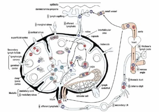
Mesenteric lymph-nodes (MLN), as other lymph-nodes consist of the outer/superficial cortex, inner/deep cortex, medullary cord and marginal, cortical and medullary lymphatic sinuses (Figure 1). The cortical region is composed of primary follicles of densely packed naïve B cells and follicular dendritic cells (FDC), surrounded by the interfollicular area (Matsuno et al., 2010). Upon antigenic stimulus, antigen-activated B cells proliferate giving rise to secondary follicles and germinal centers (Elmore et al., 2006). The inner cortex is the T cell area with DC and HEV. The medullary cord is the plasma cell area with some B cells, while the lymphatic sinuses are populated by macrophages (Newberry and Lorenz, 2005). The collected lymph and cell contents enter the lymph-node via afferent lymphatic vessels and filter the node through the lymphatic sinuses in the medulla or move via the subcapsular sinus to leave through efferent lymphatic vessels. Cells and antigens also enter the lymph-node via an arteriole, which branches into a capillary bed (Ruddle et al., 2009).
In swine, lymph-nodes show some peculiarities when compared to other animals. Both peripheral and mucosa-associated lymph-nodes have a specific structure that is called inverted and are mostly composed of cortex and paracortex, lacking a larger medullary area (Scharek et al., 2007). Besides, lymphocyte trafficking in porcine also differs from that in other animals. As usual, the immigration of lymphocytes into the lymph-node takes place either by afferent lymph vessels or HEV (Rothkotter, 2009). However, in pigs very few lymphocytes leave lymph-nodes in the lymph, since they emigrate via paracortical post-capilary venules and not via efferent lymph vassels (Scharek et al., 2007).
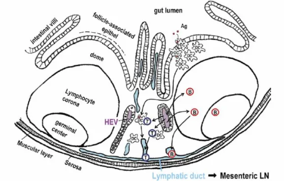
1.5.2 Peyer’s Patches
PP are islands of organized lymphoid tissue located in the small intestine. In human and swine, PP are predominantly found in the ileum show a prenatal development (Makala et al., 2002). As other secondary lymphoid organs, PP contain areas populated by B and T lymphocytes (Figure 2). Follicles are pear shaped zones composed of B-cells and FDC, which are separated by the interfollicular area (T-cell area) (Matsuno et al., 2010). These structures are overlaid with the FAE, which harbours specialized antigen-sampling M cells
Figure 1 – Schematic drawing of the structure of the lymph-node and trafficking routes for T
cells (T) and B cells (B). Numbers in the square indicate the direction of the lymph flow from the initial lymphatic capillary in peripheral organs to the draining lymph-node and then to the blood circulation via the central thoracic duct. A: artery, DC: interdigitating dendritic cell, HEV: high endothelial venule, Mf: sinus macrophage, MM: marginal macrophage, P: plasma cell, V: vein (Matsuno et al., 2000).
interdigitated within the epithelium (Burkey et al., 2009). The PP region between the follicles and the FAE is called the dome and contains mainly T cells, plasma cells, interdigitating DC and macrophages (Makala et al., 2002).
PP are strategically integrated to intestinal surface as a forward defensive system, acting as sites of antigen sampling and induction of mucosal immune responses (Newberry and Lorenz, 2005). Gut antigens enter this organ via uptake by M cells located in the specialized FAE (Rothkotter, 2009). Subsequently, antigens encounter numerous professional APC in the dome that prime naïve T and B cells. These lymphocytes become memory or effector cells and migrate from PP to MLN via efferent lymph and then via the thoracic duct to peripheral blood for subsequent extravasation at mucosal effector sites (Brandtzaeg, 2009).
Figure 2 – Schematic drawing of the structure of Peyer’s patches and trafficking route of T cells
1.6 The biology of Salmonella Typhimurium infections
1.6.1 The murine model
Murine infections by S. Typhimurium have been largely exploited as a model for the study of typhoid and general salmonellosis. S. Typhimurium causes in mice a systemic disease with a pathogenesis resembling typhoid fever in humans (Wick, 2010). S. Typhimurium is a food- and water-borne pathogen. Thus, following ingestion, a proportion of inoculum that succeeds in tolerating the low pH environment of the stomach enters the small intestine to establish infection (Álvarez-Ordóñez et al., 2011). At this point, S. Typhimurium must adhere itself to epithelial cells in the gut by several adhesins and fimbriae and subsequently cross the intestinal epithelium (Broz et al., 2012). As depicted in the Figure 3, multiple traversal routes are involved in Salmonella penetration of host mucosa (Tam et al., 2008).
The host-Salmonella interaction is dominated by the broad array of sophisticated weaponry used by bacteria to overcome host defences (Andrews-Polymenis et al., 2010). S. Typhimurium preferentially targets the M cells located in the PP follicle-associated epithelium, manipulates their function and moves through them, accessing lymphoid cells of GALT (Martinoli et al., 2007). However, this pathogen can also induce its internalization in enterocytes through its virulence-associated type III secretion system (TTSS) encoded by Salmonella pathogenicity Island 1 (SPI-1) (Ly and Casanova, 2007). TTSS encode needle-like complexes that inject bacterial effector proteins that are able to hijack host cell functions, including those associated with cytoskeleton remodelling and immunomodulatory activity (Garai et al. 2012). Invasion also has been proposed to occur by paracellular pathways following disruption of epithelia tight junctions and via DC intercalated between epithelial cells. However, the importance of
these invasion-independent alternative pathways remains to be determined (Broz et al., 2012).
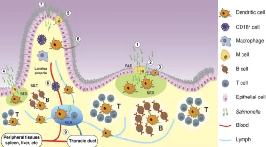
Figure 3 – Entry and capture of orally acquired Salmonella. Salmonella can cross the intestinal
epithelium and enter the host at different sites, mostly at the distal ileum. Two of these sites are organized lymphoid structures, Peyer’s patches (PP) and solitary intestinal lymphoid tissue (SILT). The third site is the intestinal villus. Salmonella can also invade using different mechanisms, which are indicated with numbers. Within PP, Salmonella can traverse the intestinal barrier through: (1) M cells in the follicle-associated epithelium (FAE); (2) epithelial cells forming the FAE, in particular after bacteria compromise M cells and the intestinal barrier; (3) DCs located in the FAE, could capture Salmonella. Once bacteria cross the FAE they can be captured by DCs located in the subepithelial dome (SED). DCs containing Salmonella can then initiate an adaptive immune response by stimulating T cells in the PP (marked T in the figure) or migrate to the mesenteric lymph node (MLN) to initiate adaptive immunity. In addition,
Salmonella can cross the intestinal barrier through SILT, probably in a similar manner as in PP, as
represented in (4). At villi, Salmonella can enter in different ways: (5) through M cells; (6) captured by DCs extending dendrites; or (7) passing through or between compromised epithelial cells. After the bacteria reach the lamina propria, they can access the MLN within migrating DCs, which are abundant in the lamina propria. Finally, the bacteria may be able to reach the blood stream, presumably transported by CD18+ phagocytes, as shown in (8). In addition to being transported to the MLN via lymph within DCs, it remains possible that free bacteria could be transported in lymph. (9) Bacteria can exit the MLN as free bacteria or possibly associated with cells and seed other tissues in the body (Tam et al., 2008).
After crossing the epithelium, Salmonella is taken by phagocytes located in the PP dome that work to remove invading microbes by phagocytosis and alert other immune cells of the infection, either directly or by releasing pro-inflammatory cytokines (Tam et al., 2008). Indeed, a rapid recruitment of neutrophils to infected tissue is the pathological hallmark of gastroenteritis caused by non-typhoidal Salmonella serovars (Andrews-Polymenis et al., 2010). For initiation of responses against microbes, macrophages and dendritic cells express multiple pathogen recognition receptors, including the cytosolic nucleotide-binding and oligomerization domain (NOD)-like receptors (NLR) and the Toll-like receptors (TLR) located on the cell surface or within a vacuolar compartment (Wick, 2011). Furthermore, a subset of NLR induces the assembly of a large multiprotein signaling complex called the inflammasome, which in cooperation with the adaptor protein ASC and activated caspase-1 (CASP1), leads to production and secretion of mature interleukin-1β and IL18 (Figure 4). CASP1 activation also initiates a proinflammatory cell-death called pyroptosis (Miao and Rajan, 2011).
Although pyroptosis and apoptosis are both programmed forms of cell death, pyroptosis is not considered immunulogically silent, since it causes the release of cellular contents and proinflamatory cytokines (Duprez et al., 2009). Of note, induction of pyroptosis was proved to be a mechanism that benefits host during Salmonella infections by eliminating the intracellular niche of the pathogen and re-exposing it to extracellular immune defenses, mainly clearance by neutrophils (Miao et al., 2010). Indeed, this evidence justifies the crucial role of neutrophils to prevent the dissemination of bacteria from the gut during salmonellosis, despite the fact that this S. Typhimurium is mainly considered an intracellular pathogen (Broz et al., 2012).
Figure 4 - The intestinal innate immune response to Salmonella. (A) Following the invasion of the mucosa, the presence of Salmonella is detected
by pattern recognition receptors. Extracellular Salmonella are detected by Toll-like receptors inducing a transcriptional response leading to the expression of proinflammatory cytokines such as IL-23. Intracellular Salmonella activate NOD-like receptors that can induce IL-23 expression, as well as the assembly of NLRC4/NLRP3 inflammasomes that activate Caspase-1, promoting the secretion of mature 1β and 18. (B) 18 and 23 amplify the inflammatory response by paracrine signaling. 18 induces the release of IFNγ from T cells, while 23 induces the release of IL-22 and IL-17. These cytokines induce the increased production of mucins and antimicrobial peptides, and promote the release of CXC chemokines leading to an influx of neutrophils into the mucosa. (C) Infiltrating neutrophils are crucial for the killing of extracellular Salmonella. Although considered an intracellular pathogen, Salmonella can be found extracellularly following transcytosis through M cells or after pyroptosis induced host cell lysis. Besides clearing the pathogen, neutrophil influx can also lead to damage to intestinal tissue, resulting in the loss of epithelial cell barrier function and promoting diarrhea (Broz et al., 2012).
Despite innate mechanisms are triggered by host to fight against infection, Salmonella is able to maintain itself in an intracellular compartment named the Salmonella containing vacuole (SCV) following phagocytosis. To establish this niche, pathogen expresses its SPI-2-efectors that act stabilizing the vacuolar membrane, modulating endocytic trafficking and inducing delayed host cell death (Garai et al., 2012). The SPI-2 TTSS plays an essential role in preventing rapid pathogen clearance by promoting its survival within mononuclear phagocytes (Srikanth et al., 2011).
From intestine, Salmonella can reach the MLN via the draining lymph as free extracellular bacteria or be transported by cells, presumably DC, from the lamina propria and/or Peyer’s patches. At this stage of an orally acquired infection, pathogen is still confined to intestine and associated lymphoid tissue. However, in mice infections virulent Salmonella disseminate and reach other tissues such as the liver, spleen and bone marrow. To cause this systemic dissemination, Salmonella reach bloodstream via lymph that empties into the blood in the thoracic duct or possibly via CD18+ phagocytes infected in the gut (Tam et al., 2008).
Continuous spread of the bacteria from infected cells to new infection foci is one of the key features of murine systemic Salmonella infections. This spread avoids high intracellular bacterial densities within the phagosomal compartment, a situation that would render the bacteria either nutritionally or spatially constricted. Moreover, bacterial spread from established foci to new infection foci at immunologically unprimed sites is also likely to be a mechanism that allows Salmonella to stay one step ahead of the progressive local activation of the inflammatory response (Haimovich and Venkatesan, 2006). S. Typhimurium can be recovered from systemic sites up to one year after infection from asymptomatic mice. Although persisting bacteria sequestered within macrophages appear to enter a dormant-like state, S. Typhimurium survival and
replication in hemophagocytic macrophages may play a key role in the establishment of persistent infections (Monack, 2012).
Effective control and eventual eradication of bacteria during the late phases of a primary infection and the generation of protective immunity against subsequent infections require the development and active recruitment of adaptive immunity mediators (Dougan et al., 2011). However, accumulation evidences have shown that Salmonella is able to inhibit T-cell activation by interfering with the presentation of antigens on the DC surface (Bueno et al., 2012). Since DC are important APC linking innate and adaptive immunity, interfering with their capacity to stimulate naïve T cells may allow pathogens evading adaptive immunity. Such interference would promote pathogen survival and dissemination, both crucial events in Salmonella pathogenesis (Swart and Hensel, 2012).
1.6.2 Differences between infections in mouse and other hosts
When considering host response to salmonellosis, it is important to stress that most of available knowledge was accessed by the study of mouse model, as this has been worked on most extensively. Nevertheless, observations from this approach do not always correlate with human (Tsolis, 2011) and farm animals infections (Bearson and Bearson, 2011). In different hosts, the outcome of infection differs in terms of local versus systemic spread, influence of infecting isolate properties and specific breed responses to pathogen (Dougan et al., 2011). While septicemic episodes have been reported for Salmonella in mice, colonization by S. Typhimurium in food-producing animals is usually limited to the gastrointestinal tract (Hoelzer et al., 2011).
Natural or experimental infection of calves with serotype Typhimurium results in an enteric disease with clinical and pathological features that parallel
the disease in man. The intestinal pathology and the predominant influx of PMN leucocytes observed in infected calves are strikingly similar to that of S. Typhimurium-induced enteritis in humans. In both species, PMN leukocytes are proposed to play a decisive role in the pathogenesis of serotype Typhimurium-induced diarrhoea (Santos et al., 2001). Pig infections with S. Typhimurium are also clinically similar to those observed in human. However, besides some evidences indicating the influx of PMN cells to the porcine gut during S. Typhimurium infections, information on processes behind the induction of diarrhoea is still scarce (Boyen et al., 2008a).
Existing data from the mouse model support that the Peyer’s patch route predominates in traversal of Salmonella. Nevertheless, little is known about how Salmonella penetrates the mucosa in man and other species (Dougan et al., 2011). According to Costa et al. (2012), S. Typhimurium induces ruffling of the plasma membrane at the apical side of intestinal epithelial cells during bovine infections, invading M cells and absorptive enterocytes. Similarly, it has been shown that Salmonella can invade porcine absorptive enterocytes, M-cells and even goblet cells (Boyen et al., 2008a). In a study by scanning and transmission electron microscopy, Meyerholz et al. (2002) demonstrate a preferential adherence of S. Typhimurium to M cells within 5 minutes post infection of swine ileum. In addition, these authors demonstrated that this pathogen may use sites of cell extrusion as an additional mechanism for early invasion. Moreover, in accordance to evidences from murine infections, Boyen et al. (2006) asserted that SPI-1 is necessary to promote the invasion of porcine intestinal epithelial cells by S. Typhimurium, thereby contributing to the efficient short-term colonization of the porcine gut and to the induction of influx of neutrophils into the intestinal lumen.
S. Typhimurium triggers the up-regulation of pro-inflammatory genes and
2009). Moreover, swine jejunum, ileum and colon respond differently to in vivo infections with S. typhimurium. In spite of the up-regulation of genes coding for innate immunity mediators, ileum showed a down-regulation of IL12 and CASP1, both relevant molecules in the response of host to Salmonella. This result could represent an advantage to pathogen during pig infections and was inferred to be related to the preference of S. Typhimurium for invading intestinal mucosa through ileum (Collado-Romero et al., 2010).
In pigs, S. Typhimurium is sporadically present in liver and spleen shortly after experimental inoculation but does not seem to replicate and is cleared from these organs a few days after inoculation. However, bacteria are able to persist in the gut and gut-associated lymph nodes (Boyen et al., 2008a). Gut-associated lymph-nodes are considered important niches for S. Typhimurium during swine infections. In fact, Salmonella prevalence in lymph-nodes of slaughter pigs from EU countries ranges from 0% to 29% (Baptista et al., 2011). Basing on data from mouse infections, it could be inferred that S. Typhimurium reach MLN via intestinal lymph, being shuttled by infected phagocytes or as extracellular bacteria. Nevertheless, this issue has never been addressed to date by any available report. Uthe et al. (2007) observed that S. Typhimurium could be isolated from pig MLN at 8 hours post oral infection and verified that pathogen was able to maintain itself in these organs at least 21 days post infection. Macrophages located in mesenteric lymph nodes are considered to be important players in the induction of long-term persistence of S. Typhimurium in infected pigs (Boyen et al. 2008a). Epidemiological analysis has established that Salmonella persistent carriers are crucial targets for disease control, because they shed the pathogen in high enough numbers to transmit disease (Gopinath et al., 2012). Indeed, asymptomatic carriers represent the most important source of Salmonella introduction onto pig farms (Hoelzer et al., 2011). For this reason, a comprehensive view of the processes involved in the maintenance of pathogen in
host lymphoid organs and the conversion of infected pigs to carriers consist in an essential step for controlling salmonellosis.
As previously discussed, SPI-2 effector are indispensable for systemic infection of mice, since they support the intracellular survival of Salmonella inside host cell, especially macrophages (Garai et al. 2012). Curiously, S. Typhimurium strains mutants for SPI-2 were found to be fully capable of colonizing the intestines of pigs and to establish a long-term intestinal persistent infection after oral inoculation (Boyen et al., 2008b), suggesting that S. Typhimurium might employ different strategies to promote the carrier state during mouse and pig infections. Therefore, this divergence highlights the need of models other than murine infections to elucidate the mechanisms involved in the induction of persistent infections by Salmonella and reinforces the notion that the extrapolation of data from murine model to other species should be done with prudence.
1.7 Swine as a model for the study of Salmonella Typhimurium
infections
Animal models are valuable tools for a better understanding of pathogenesis mechanisms behind human diseases. To date, mouse has been widely used to provide novel knowledge in distinct fields of biology and medicine. However, mouse disease models often do not faithfully mimic the relevant human conditions, bringing to light the need of better animal models (Luney, 2007).
Swine are increasingly appreciated for their potential to model human disease and this tendency was further reinforced by the recent advances in pig genomics and proteomics (Prather, 2013; Bendixen et al., 2010). Besides, pigs and humans are remarkably similar in terms of physiology. Interestingly, the
porcine immune system shows a higher level of similarity to human one than murine. Genomic comparisons between human, murine and porcine sequences also demonstrate more resemblances between human and porcine than human and murine sequences (Luney, 2007).
The swine model has been used to study as a model for many human infectious diseases and represent a valuable intermediate species to validate the applicability of knowledge obtained using rodent models to human (Meurens et al., 2012). However, although former reports of experimental infections of pig with Salmonella can be found in the literature (Williams et al. 1978, Baskerville et al. 1972), it was the 1990s when the use of this species for the study of salmonellosis became more current.
Advances in mammalian models of Salmonella infection are expected to result in new understanding of salmonellosis pathogenesis, contributing to the control and cure of human cases (Gopinath et al., 2012). Upon infection with S. Typhimurium, swine undergo a self-limiting enterocolitis which parallels the clinical manifestation observed in man (Sanchez-Vargas et al. 2011; Boyen et al., 2008a). In light of this and the potential of swine as models of human disease, pigs can be stressed as an ideal model for investigating the pathogenesis of human non-typhoidal salmonellosis.
1.8 Use of functional genomics to identify target molecules and
mechanisms for the improvement of resistance to infectious
diseases in animals
Resistance and tolerance to infectious pathogens are important characteristics of livestock to counteract the potential detrimental impact of pathogens on animal health and production (Doeschl-Wilson et al., 2012a).
Strong evidence has accumulated that livestock species from birds to mammals, harbour genes that control protective responses to the various classes of pathogen from viruses to nematodes. However, there remains a gap for traits linked to disease resistance and tolerance because the main polymorphisms that underpin these traits have not been identified. This is partly because host-pathogen interactions are highly complex, involving many different molecules and cell types which interact together over time (Glass, 2012).
Disease traits have been difficult to target by traditional selection, but recent developments in high throughput genomics provide opportunities to dissect host responses to infectious pathogens and to increase the accuracy of selection (Doeschl-Wilson et al., 2012b). Knowledge obtained from the complete genome sequencing of many species has enabled discovery of new aspects of the host–pathogen interaction. Moreover, the development of genomics, transcriptomics, proteomics and computational biology has resulted in the systematic study of the host–pathogen interaction. Thus, functional genomics approaches have enabled the integration of experimental data from the diverse high-throughput methods to create a global picture of host–pathogen interplay in space and time (Hartlova et al., 2011).
In order to gain access to the host environment, a pathogen must cross the mucosal surfaces of the gut, respiratory tract, mammary gland and genital tract. After invading, these organisms should be able to tolerate and overcome host immunity. Thus, genetic variance in molecules taking part in the distinct partners of host defence against invaders might play an important role in the development of resistance or tolerance to infections. Strikingly, some examples have been described in veterinary. Recently, genetic variants in bovine CD46 have been shown to influence cell permissiveness for bovine viral diarrhoea virus and thus carriers of CD46 alleles might vary in resistance to this pathogen. Besides, the gut receptor for E. coli F18 encoded by the FUT1 gene in pigs was found to
confer complete resistance to E. coli (Glass, 2012). Another report, exploring the genetic resistance of bovine to the tick-borne protozoan Theileria annulata, demonstrated that an up-regulation of SIRPβ and TGFβ2, both mediators of inflammatory response, is associated to greater virulence and also higher propensity for invasion observed Holstein cows compared to the Bos indicus cattle breed Sahiwal (Chauessepied et al., 2010).
In the case of S. Typhimurium infections, genetic resistance to systemic disease in mice has been linked to many factors including the major histocompatibility complex (MHC), Toll-like receptor 4 and the natural resistance associated macrophage protein (NRAMP1). NRAMP1 is the most studied of all these factors and exerts effects on macrophage activation including MHC expression, release of pro-inflammatory cytokines and the production of antimicrobial effectors such as nitric oxide and oxidative burst. Mutations in this gene result in mice susceptible to S. Typhimurium and other intracellular pathogens such as Leishmania and Mycobacterium (Wigley, 2004). In fowl, comparisons of different models have successfully identified the genomic regions carrying the genes TLR4, NRAMP1 and the QTL SAL1 as having roles in phenotypic variations related to Salmonella resistance (Calenge et al., 2010).
Control of salmonellosis in swine herds generates increased production costs and public health issues in the use of prophylactic antibiotic potentially leading to development of antibiotic resistant strains. Therefore, the breeding of resistant animals, in combination with good hygiene practices offers a promising low risk strategy to fight against Salmonella in this species. In pigs, NRAMP1 is strongly expressed on macrophages and neutrophils following stimulation with LPS, but any role for this gene in Salmonella infection is yet to be described (Wigley, 2004). On the other hand, other authors have demonstrated the influence of genetic variance in the outcome of S. Typhimurium infections in porcine. Uthe et al. (2009) asserted a positive association between genetic
polymorphisms in the CCT7 gene with Salmonella shedding in swine. Besides, another report by Tuggle et al (2010) detected transcriptional changes in blood of persistent Salmonella shedder pigs which resulted in increased levels of intracellular-oriented responses compared to low shedder animals.
In spite of the association between genetic variation and resistance to salmonellosis in swine, selective breeding for resistance traits aiming to control the disease or the carriage of Salmonella in this species is still not feasible. In fact, targeting of the key host molecules and mechanisms involved in bacterial pathogenesis is indispensable to address this issue and a robust analysis of S. Typhimurium–pig interactions by integrative approaches represent a necessary step towards this aim.
References
1. Acheson DW, Luccioli S. Microbial-gut interactions in health and disease. Mucosal immune responses. Best Pract Res Clin Gastroenterol. 2004; 18:387-404. 2. Andrews-Polymenis HL, Bäumler AJ, McCormick BA, Fang FC. Taming the elephant: Salmonella biology, pathogenesis, and prevention. Infect Immun. 2010; 78:2356-69.
3. Baptista FM, Halasa T, Alban L, Nielsen LR. Modelling food safety and economic consequences of surveillance and control strategies for Salmonella in pigs and pork. Epidemiol Infect. 2011; 139:754-64.
4. Baskerville A, Dow C, Curran WL, Hanna J. Ultrastructure of phagocytosis of Salmonella cholerae-suis by pulmonary macrophages in vivo. Br J Exp Pathol. 1972; 53:641-7.
5. Bearson BL, Bearson SM. Host specific differences alter the requirement for certain Salmonella genes during swine colonization. Vet Microbiol. 2011;150:215-9.
6. Bendixen E, Danielsen M, Larsen K, Bendixen C. Advances in porcine genomics and proteomics--a toolbox for developing the pig as a model organism for molecular biomedical research. Brief Funct Genomics. 2010; 9:208-19.
7. Botteldoorn N, Heyndrickx M, Rijpens N, Grijspeerdt K, Herman L. Salmonella on pig carcasses: positive pigs and cross contamination in the slaughterhouse. J Appl Microbiol. 2003; 95:891-903.
8. Boyen F, Haesebrouck F, Maes D, Van Immerseel F, Ducatelle R, Pasmans F. Non-typhoidal Salmonella infections in pigs: a closer look at epidemiology, pathogenesis and control. Vet Microbiol. 2008a; 130:1-19.
9. Boyen F, Pasmans F, Van Immerseel F, Morgan E, Botteldoorn N, Heyndrickx M, Volf J, Favoreel H, Hernalsteens JP, Ducatelle R, Haesebrouck F. A limited role for SsrA/B in persistent Salmonella Typhimurium infections in pigs. Vet Microbiol. 2008b;128:364-73.
10. Boyen F, Pasmans F, Van Immerseel F, Morgan E, Adriaensen C, Hernalsteens JP, Decostere A, Ducatelle R, Haesebrouck F. Salmonella Typhimurium SPI-1 genes promote intestinal but not tonsillar colonization in pigs. Microbes Infect. 2006; 8:2899-907.
11. Brandtzaeg P. Mucosal immunity: induction, dissemination, and effector functions. Scand J Immunol. 2009; 70:505-15.
12. Broz P, Ohlson MB, Monack DM. Innate immune response to Salmonella typhimurium, a model enteric pathogen. Gut Microbes. 2012; 3:62-70.