Limited evolutionary conservation of imprinting
in the human placenta
D. Monk*†, P. Arnaud‡, S. Apostolidou*, F. A. Hills§, G. Kelsey¶, P. Stanier*, R. Feil‡, and G. E. Moore*
*Institute of Reproductive and Developmental Biology, Imperial College London, London W12 0NN, United Kingdom;‡Institute of Molecular Genetics,
Centre National de la Recherche Scientifique, Unite´ Mixte de Recherche 5535 and University of Montpellier II, 1919 Route de Mende, 34090 Montpellier, France;§School of Health and Social Sciences, Middlesex University, Enfield EN3 4SA, United Kingdom; and¶Laboratory for Developmental Genetics and
Imprinting, The Babraham Institute, Cambridge CB2 4AT, United Kingdom
Edited by R. Michael Roberts, University of Missouri, Columbia, MO, and approved March 7, 2006 (received for review December 21, 2005)
The epigenetic phenomenon of genomic imprinting provides an additional level of gene regulation that is confined to a limited number of genes, frequently, but not exclusively, important for embryonic development. The evolution and maintenance of im-printing has been linked to the balance between the allocation of maternal resources to the developing fetus and the mother’s well being. Genes that are imprinted in both the embryo and extraem-bryonic tissues show extensive conservation between a mouse and a human. Here we examine the human orthologues of mouse genes imprinted only in the placenta, assaying allele-specific ex-pression and epigenetic modifications. The genes from the KCNQ1 domain and the isolated human orthologues of the imprinted genes Gatm and Dcn all are expressed biallelically in the human, from first-trimester trophoblast through to term. This lack of imprinting is independent of promoter CpG methylation and cor-relates with the absence of the allelic histone modifications dim-ethylation of lysine-9 residue of H3 (H3K9me2) and trimdim-ethylation of lysine-27 residue of H3 (H3K27me3). These specific histone modifications are thought to contribute toward regulation of imprinting in the mouse. Genes from the IGF2R domain show polymorphic concordant expression in the placenta, with imprint-ing demonstrated in only a minority of samples. Together these findings have important implications for understanding the evo-lution of mammalian genomic imprinting. Because most human pregnancies are singletons, this absence of competition might explain the comparatively relaxed need in the human for placental-specific imprinting.
histones兩 methylation 兩 epigenetics
G
enomic imprinting is an epigenetic phenomenon that results in monoallelic expression of certain genes in a parent-of-origin-dependent manner (1). Although found in angiosperm plants and marsupials, it has been suggested to be one of the most important regulatory pathways involved in the development and function of the placenta in eutherian mammals.Imprinted genes are often found in close proximity to each other, indicating coordinate regulation by imprinting control regions (ICRs) (reviewed in ref. 1). Distal mouse chromosome 7 (mChr7) harbors the largest known cluster of imprinted genes and has conserved synteny with human chromosome 11p15.5 (hChr11). This cluster has been implicated in disorders such as Beckwith-Wiedemann and Silver-Russell syndrome, malignancies, and aber-rant fetal growth (2, 3). Two ICRs controlling different sets of genes have been identified within this⬎1-Mb cluster, dividing it into two functionally independent groups. The centromeric domain is con-trolled by the paternally methylated H19 ICR (1), whereas the telomeric potassium voltage-gated channel, subfamily Q, member 1 (KCNQ1)兾Kcnq1 domain is controlled by potassium voltage-gated channel differentially methylated region 1 (KvDMR1) (4), a CpG island within intron 10 of the Kcnq1兾KCNQ1 gene that is maternally methylated (5). In the mouse, 14 imprinted transcripts flank this ICR. The majority are expressed predominantly from the maternal allele, with imprinting of the most distal genes restricted to the placenta (6, 7). Interestingly, the placental imprinting of
genes within this domain does not depend on DNA methylation at their promoters (5, 7, 8). The KvDMR1 is also the promoter for the
KCNQ1 overlapping transcript 1 (Kcnq1ot1)兾KCNQ1OT1 (LIT1)
noncoding RNA (ncRNA) (4, 9) that, because of the DNA meth-ylation and abundance of repressive chromatin modifications on the maternal allele, is paternally expressed (6, 7). Polycomb group proteins are thought to be recruited to the paternal allele, poten-tially by the Kcnq1ot1兾KCNQ1OT1 ncRNA, contributing to the paternal repression of the imprinted genes in cis (6).
A second cluster in the mouse that contains placental-specific imprinted genes maps to mChr17. This cluster contains the maternally expressed insulin-like growth factor receptor 2 (Igf2r) transcript and two maternally expressed placental-specific genes,
Slc22a2 and Slc22a3 (10). This cluster shares several similar
features with the KCNQ1 domain. These features include an intronic ICR (region 2 ICE) within the Igf2r gene, which is the functional promoter for the paternally expressed, unspliced ncRNA Air (11, 12). Also, the paternal silencing observed within the Igf2r domain is associated with repressive chromatin (13, 14). In this report, we have assessed the imprinting status for the human orthologues of all reported mouse placental-specific imprinted genes. In contrast to the maternal expression observed in the mouse, the human expression is largely biallelic. We demonstrate that the lack of imprinting correlates with a lack of allelic chromatin modifications.
Results
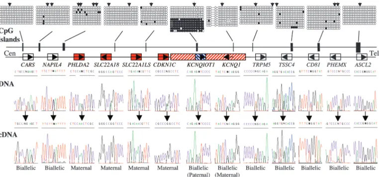
Allelic Expression and Methylation Within theKCNQ1 Domain. To perform an investigation of imprinting across the human KCNQ1 domain, we have analyzed allelic expression, promoter DNA methylation, and histone modifications at specific loci. All genes whose orthologues exhibit placental-specific maternal expres-sion in the mouse, and under the control of the KvDMR1 (NAPIL4, CD81, PHEMEX, and ASCL2) (4, 6, 15, 16) were found to be expressed biallelically in human fetal tissues and in first-trimester and term placental samples (Fig. 1; see also Table 1, which is published as supporting information on the PNAS web site). In contrast, we observed conserved imprinted expres-sion for pleckstrin homology-like domain, family A, member 2 (PHLDA2), SLC22A18, SLC22AILS, and cyclin-dependent ki-nase inhibitor 1C (CDKN1C), with monoallelic expression being detected in all tissues at all gestational ages tested (Fig. 1 and Table 1). These genes are located immediately centromeric to the KvDMR1. The orthologues of these genes are imprinted in both the fetus and placenta in the mouse (6). This data suggests that they are most likely imprinted by a mechanism that is different to that regulating placental-specific imprinting.
Im-Conflict of interest statement: No conflicts declared. This paper was submitted directly (Track II) to the PNAS office.
Abbreviations: ChIP, chromatin immunoprecipitation; ICR, imprinting control region; DMR, differentially methylated region; ncRNA, noncoding RNA.
†To whom correspondence should be addressed. E-mail: d.monk@imperial.ac.uk. © 2006 by The National Academy of Sciences of the USA
printing also was observed for the KCNQ1 and KCNQ1OT1 transcripts in early fetal tissues and first-trimester placentae, but imprinting was relaxed in term placentae (Fig. 1 and Table 1). In the mouse, ubiquitous imprinting of Cdkn1c has been linked to somatic differential DNA methylation in the promoter regions of this gene, which is maintained by Dnmt1 and Lsh (7, 17). We therefore used bisulphite sequencing and Southern blotting to determine the DNA methylation pattern of all genes in the human cluster. Only one differentially methylated region (DMR) was found, which mapped to the previously described KvDMR1 (18). The promoter CpG islands of all of the other genes were hypomethylated in the placenta, liver, muscle, and lymphocyte DNA (Fig. 1 and data not shown). This result suggests that somatic allelic DNA methylation, such as at
Cdkn1c, may be dispensable for conserved imprinting.
Modified Histones and Paternal Repression Within theKCNQ1 Domain.
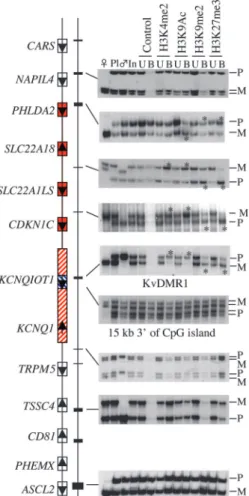
Because of the general lack of DMRs associated with the genes showing maintained ubiquitous imprinted expression, we per-formed qualitative chromatin studies on both sides of the KvDMR1 to determine differences between the parental chromosomes. Our analysis focused on the trimethylation of Lys-27 on histone H3 (H3K27me3) and dimethylation at Lys-9 on histone H3 (H3K9me2) as markers of repressive chromatin and on acetylation at Lys-9 on histone H3 (H3K9ac) and dimethylation of Lys-4 on histone H3 (H3K4me2) as markers of active chromatin. Qualitative chromatin immunoprecipitation (ChIP) was performed on unfixed chromatin fragments prepared from term placenta samples. In all informative placenta samples, enrichment of H3K4me2 and H3K9ac on the maternal chromosome and H3K9me2 and H3K27me3 on the paternal chromosome was limited to those genes
centromeric to the KvDMR1, which showed conserved imprinted expression. None of the other genes showed allele-specific enrich-ment for these chromatin markers (Fig. 2; see also Table 2, which is published as supporting information on the PNAS web site).
At the KvDMR1 region, we detected an opposite allelic pattern, with enrichment of maternal H3K9me2 and H3K27me3 and paternal H3K4me2 and H3K9ac. These data are consistent with transcription from the paternal allele at this promoter. This pattern also was present within the DMR, but not at a distance of 15 kb or 25 kb within the transcribed region of this ncRNA in a first-trimester placenta (10 and 17 weeks; data not shown) or at term (Fig. 2 and Table 2). This finding suggests that the
KCNQ1OT1 ncRNA does not cause changes to the chromatin
over its entire transcribed region in placenta, at least for the modifications analyzed here. Nevertheless, in vivo studies suggest that this ncRNA is essential for epigenetic silencing (19).
Polymorphic Imprinting Within the HumanIGF2R Cluster.To deter-mine whether histone modifications account for placental-specific imprinting in other imprinted clusters, the imprinting status of genes within the IGF2R domain on hChr6q25.3 was analyzed. In the mouse, this cluster contains two genes that are paternally repressed in the placenta without the involvement of promoter DNA methylation (10, 12). Interestingly, this cluster is polymorphically imprinted in humans, with the majority of individuals showing biallelic expression (20, 21). Here we also found IGF2R and SLC22A2 to be polymorphically imprinted, with monoallelic expression detected only in a minority of term placenta samples (Fig. 3 and Table 1). Four of these samples were heterozygous for both genes. Expression was concordant in each placenta, two showing maternal expression and two with
Fig. 1. Schematic representation of the KCNQ1 domain on human chromosome 11p15.5 showing the relative organization of genes and CpG islands. The methylation status of all promoter CpG islands was examined in liver-, muscle-, lymphocyte-, and placenta-derived DNA. Methylation patterns were assessed first by restriction digestion of bisulphite PCR products. The positions of the restriction sites used are shown (2). Example of patterns obtained by sequencing bisulphite PCR product obtained from placenta-derived DNA. Similar patterns were obtained in all tissues analyzed. Each circle represents a single CpG dinucleotide on the strand, a methylated cytosine (F) or an unmethylated cytosine (E). For clarity, only the first 20 CpG dinucleotides from each CpG island are shown. In all tissues analyzed, the only evidence for differential methylation was at the human KvDMR1兾KCNQIOT1 promoter; all other promoter CpG islands were hypomethylated. The imprinted expression for all genes in the cluster was analyzed in first-trimester fetal tissues, term placenta, and cytokeratin 7-positive trophoblast cells. DNA sequence traces for heterozygous term placentae samples are shown for all genes and the resulting RT-PCR sequences. Open boxes depict genes biallelic in all tissues throughout gestation, whereas red boxes represents ubiquitously imprinted, maternally expressed genes. The hatched red box presents the maternal-specific expression of KCNQ1 limited to first-trimester fetal tissues only. The blue hatched box symbolizes the paternal-specific expression of the KCNQ1OT1 transcript observed in first-trimester material only. Both of these genes are biallelically expressed in term placenta.
biallelic expression for both genes. We also analyzed the im-printing of SLC22A1 and SLC22A3 genes in the same region. Similar to the mouse, SLC22A1 was biallelically expressed in all tissues and at all gestational ages tested (Fig. 3 and Table 1);
SLC22A3 exhibited monoallelic expression in first-trimester
placentae (maternal DNA sample was heterozygous) but bial-lelic expression in fetal tissues (of the only heterozygous first-trimester sample set) and term placentae (data not shown). The temporal pattern of imprinting of SLC22A2 and SLC22A3 is similar to that reported for mice (10).
Next we compared the DNA methylation and chromatin configuration for the four samples that showed concordant imprinting or biallelic expression for IGF2R and SLC22A2. The intronic ICR (called region 2 ICE) was the only DMR identified, and this region showed an indistinguishable DNA methylation profile between samples with imprinted or biallelic expression (21) (Fig. 3 and data not shown). We observed no allelic enrichment for active or repressive chromatin markers at gene
promoters throughout the region for all samples (Table 2; see also Fig. 5, which is published as supporting information on the PNAS web site). This lack of allelic histone modification has been reported in a human cell line, although imprinted expres-sion was not analyzed (22). The mouse Igf2r gene contains two DMRs: the germ-line methylated intronic region 2 ICE and the somatic DMR at the promoter CpG island of the Igf2r gene (23). Both these regions are associated with modified histones (13, 14); however, in the Eed null mice, the imprinting of Igf2r is unaffected (24), suggesting that H3K27 methylation is not involved in the imprinting of this gene. This contrast with our human data suggests that a mechanism other than the analyzed histone modifications regulates this domain. The paternally expressed Air ncRNA might fulfill this role; however, as was reported by others (25), we were unable to determine whether the placenta samples that exhibited imprinted expression were associated with polymorphic human AIR expression.
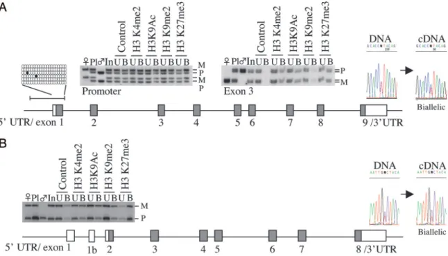
The Biallelic Expression ofDCN and GATM.We have analyzed the human imprinted expression of genes on other chromosomes that exhibit placental-specific imprinting in the mouse, glycine amidinotransferase (Gatm) (mChr2) and decorin (Dcn) (mChr10) (26, 27). In the mouse, both are isolated imprinted genes without associated DMRs. Both human orthologues are biallelically expressed in first-trimester fetal and placenta sam-ples, term placentae, and immunoselected trophoblast cells. The promoters of both genes are not associated with enriched chromatin modifications, suggesting that the lack of allele-specific repressive chromatin in the human placenta also ac-counts for the lack of imprinting (Fig. 4 and Tables 1 and 2).
Discussion
We have analyzed the expression and epigenetic regulation of four imprinted regions in the human. This study has revealed consistent differences in imprinting patterns between mice and humans. In all regions assessed, the mouse has placenta-specific imprinted expres-sion, regulated not by DNA methylation, but most likely by histone modifications (6, 7). Our data indicate that the discrepancy in imprinting between mouse and human correlates perfectly with the lack of allele-specific histone modifications.
Recent studies in the mouse have revealed developmental and mechanistic parallels between imprinted X inactivation and autosomal imprinting of placenta-specific genes (28–30). Both mechanisms are hypothesized to have coevolved with the devel-opment of the placenta and are thought to depend on the presence of ncRNAs and the recruitment of histone-modifying enzymes. The establishment of X inactivation requires the ncRNA, Xist, to be expressed from the paternal X chromosome, whereas the autosomal ncRNAs Kcnq1ot1 and Air may function in a similar manner (30). However, although mouse placental
Xist expression is imprinted, it is biallelic in the human (31, 32).
Therefore, human extraembryonic random X inactivation cor-relates with the lack of autosomal placental imprinting, suggest-ing the loss of a common mechanism.
The lack of autosomal placental-specific imprinting is clearly demonstrated for the genes mapping to the KCNQ1 domain. In the mouse orthologous region, there are 14 imprinted tran-scripts, of which 8 are expressed from the maternal allele in the placenta. In contrast, there are only six imprinted genes in the human, representing those that are ubiquitously imprinted. The evolutionary loss of imprinting in this cluster, therefore, is restricted to those that are placenta-specifically imprinted. It is possible that in the mouse, the placental-specific imprinting may be due to a bystander effect. To date, there is no direct evidence that the placenta-specific genes within this cluster need to be imprinted, only expressed (as in the case of Ascl2; ref. 16). Imprinting of these genes, therefore, may be a consequence of
Fig. 2. Alelle-specific analysis of histone modifications in term placentae by using antibodies against H3K4me2, H3K9ac, H3K9me2, and H3K27me3. For each ChIP, representative data for the placental (pl) and parental (么 and 乆) genotypes, ChIP input (In), unbound fraction (U), and antibody-bound (B) are shown. SNP accession no. and locations are given in Table 3, which is published as supporting information on the PNAS web site. The relative enrichment (*, ⬎2-fold) of the maternal allele in precipitated H3K4me2 (CDKN1C 2.1;
SLC22A18 4.1; no enrichment at PHLDA2 exon 2) and H3K9ac (CDKN1C 7.3; SLC22A18 2.1; PHLDA2 2.5), and for the paternal allele in precipitated
H3K9me2 (CDKN1C 2.8; SLC22A18 2.3; PHLDA2 4.4) and H3K27me3 (CDKN1C 10.1; SLC22A18 6.9; PHLDA2 3.6), is limited to genes with ubiquitous maternal-specific expression. An opposite allelic pattern was observed at the KvDMR1 with relative enrichment of paternal H3K4me2 (3.9) and H3K9ac (2.9) and maternal H3K9me2 (2.1) and H3K27me3 (5.0) precipitation. No enrichment was observed within the transcribed KCNQ1OT1 region.
their proximity to the KvDMR1, with paternal silencing depend-ing on the ICR and involvdepend-ing repressive histone modifications. During the 85 million years of evolution between the diver-gence of mice and humans, there has been a clear reduction in the maintenance of imprinting with conservation of only 29 imprinted transcripts (33). Some of these changes may be attributable to a gradual limiting of chromatin silencing in cis, ultimately maintaining only those that need to be imprinted. Interestingly, the most proximal imprinted genes within the human KCNQ1 cluster are two that show a functional require-ment to be imprinted. Telomerically, the KCNQ1OT1 ncRNA needs to be paternally expressed for the in cis regulation of the entire domain, where as centromeric, PHLDA2 is known to play an important role in human fetal growth, with expression levels correlating with birth weight (ref. 34 and unpublished data).
We have confirmed that imprinting of the IGF2R gene is polymorphic between individual placentae (20, 25) and that this polymorphic trait extends to the SLC22A2 and SLC22A3 genes in a minority of individuals. In its imprinted configuration, the temporal expression patterns for the two placental-specific genes within the cluster mimic that of the mouse. This domain, therefore, may represent a region undergoing a transition to become biallelically expressed throughout the whole population. Interestingly, the epigenetic status at the chromatin and DNA methylation level within this domain is indistinguishable be-tween imprinted and nonimprinted individuals. However, im-printing has been reported for IGF2R (opossum) and MEST (wallaby) genes in marsupials (35, 36), but there is no evidence for differential DNA methylation within the ICRs of both genes. These findings suggest that some imprinted genes either have developed alternative imprinting mechanisms between species or they share mechanisms that have yet to be identified.
Evolutionary Implications for the Lack of Placental-Specific Imprinting in the Human. There is a close relationship between genomic imprinting and the acquisition of a placenta during mammalian
evolution, and many explanations for the evolution of genomic imprinting implicate the placenta as a key tissue. A substantial proportion of imprinted genes are involved in the control of fetal growth and, in general, paternally expressed imprinted genes enhance fetal growth, whereas maternally expressed genes suppress it (37, 38). These parent-of-origin effects on fetal growth and development have resulted in the ‘‘parental conflict hypothesis,’’ based on kinship theory, as the dominant theory of the origin of genomic imprinting (39). This hypothesis predicts that imprinting has evolved because of conflict of resource provisions by the mother to her offspring through the placenta and that paternally expressed genes are ‘‘selfish’’ and are selected to extract more resources from the mother, whereas maternally expressed genes have to balance the nutrition provision to the current fetus with that of future fetuses of the mother (but potentially different fathers). Maternally derived genes, therefore, are more conservative with regards to provision to the fetus. It is possible that mice have acquired an expansion of imprinting to enable the placenta to become more efficient over a much shorter gestational period. This expansion may have led to an accelerated requirement for resource provi-sioning genes and their regulators such that many more genes have become imprinted in placenta. Nevertheless, most human preg-nancies are singletons in contrast to the mouse, where there is increased chance of intralitter competition. Therefore, this lack of competition relieves the pressure for maintaining placental-specific imprinting. With respect to human growth and nutrient transfer, more emphasis may lie with imprinted genes involved in postnatal adaptation and maternal behavior.
Materials and Methods
Collection of Human Material.A total of 45 fetal tissue sets (8 –18 weeks) with corresponding maternal blood samples were ob-tained from termination of pregnancies at Queen Charlotte’s and Chelsea Hospital, London. Local ethical approval for obtaining fetal tissues was granted by the Research Ethics Committee of Hammersmith, Queen Charlottes’s and Chelsea,
Fig. 3. Schematic map of the IGF2R region on human chromosome 6. The methylation status of the CpG islands within this domain was analyzed by bisulphite sequencing and by Southern blotting (data not shown). The only differentially methylated region found maps to the intronic CpG island within intron 2 of the
IGF2R gene. The allele-specific expression was analyzed in first-trimester and term placenta samples. Both IGF2R and SLC22A2 show polymorphic imprinting in
term placenta (horizontal hatched red boxes), whereas SLC22A3 exhibits temporal imprinting, with monoallelic expression limited to first-trimester placenta (diagonal hatched red box). As in the mouse, SLC22A1 was biallelically expressed. No DNA methylation differences were observed between imprinted and nonimprinted samples (data not shown, an example of the methylation profile from a placenta sample showing imprinted expression is given).
and Acton Hospitals’ Research Ethics Committee (2001兾 6028). A set of 240 term placental trio samples consisting of multiple-site placental samples with corresponding maternal and paternal blood samples were collected from consecutive consenting pregnancies at Queen Charlotte’s and Chelsea Hospital (local ethics approval 2001兾6029). All samples were washed in sterile PBS and snap frozen in liquid nitrogen and stored at⫺80°C.
Enrichment for Uncultured Human Villous Trophoblasts.A detailed protocol for the isolation of villous trophoblasts is available on request. Brief ly, we used a protocol of placental tissue diges-tion and negative immunoselecdiges-tion. Dissected placental tissues from both the first trimester (8 –14 weeks) and term were thoroughly washed in PBS and digested with a mixture of trypsin and DNase to release free cells. Unwanted erythro-cytes were removed from the resulting cell suspension by centrifugation through a 40% Percoll solution. The resulting trophoblasts were subjected to negative immunoselection by using monoclonal anti-HLA class 1 (clone w6兾32). All tro-phoblast cell preparations were subjected to cytokeratin 7 immunocytochemistry (⬎98% cytokeratin 7-positive cells; data not shown) and HLA class 1 and Vimentin to assess cell contamination (⬍1.3%; data not shown).
Analysis of Allelic Expression.We extracted total RNA from tissues by using TRIzol Reagent (Invitrogen). After digestion with RNase-free DNase 1 (Invitrogen), we generated first-strand cDNA with Moloney murine leukemia virus reverse transcrip-tase (Promega) by using random primers. Duplicate sets of samples were processed, with reverse transcriptase omitted to detect genomic DNA contamination of the RNA. The presence of cDNA was confirmed by using a GAPDH primer set.
All polymorphisms were identified by interrogating SNP
databases or genomic sequencing and confirmed by sequencing control DNAs (primer sequences are given in Table 4, which is published as supporting information on the PNAS web site). All PCR products were sequenced in both the forward and reverse orientation by using an ABI Prism 3100 DNA sequencer (Ap-plied Biosystems).
Methylation Analysis.For bisulphite sequencing, DNA was con-verted by using EZ DNA Methylation-Gold Kit (Zymo Re-search, Orange, CA). PCR amplification (primer sequences are given in Table 4), cloning, and sequencing were performed by using standard protocols. Combined bisulphite restriction anal-ysis was used to assess the methylation pattern of the amplified region in the overall PCR product and to ensure that there was no cloning bias before sequencing. Southern blotting by using methylation-sensitive restriction enzymes such as SacII and HpaII was performed according to standard protocols.
ChIP. Twelve individual human term placenta samples were analyzed by ChIP. This method generates qualitative differences in allelic enrichment although no conclusions can be drawn on quantitative levels (40). Specificity and efficiency of the ChIP assay was determined by allele specific SSCP or HOT-STOP RFLP analysis of antibody bound (precipitated) and unbound (nonprecipitated) fractions. Appropriate allelic enrichment in the antibody bound fraction is frequently accompanied by a paralleled depletion in the unbound fraction. Mock (control) precipitations indicate specificity of the antibody, ideally, but not always, resulting in no product in the control-bound fraction. Approximately 300 mg of material was used for each ChIP assay. We carried out ChIP as described in refs. 6 and 41. Briefly, we purified nuclei on a sucrose gradient and incubated them with MNase to obtain chromatin fragments of one to six nucleosomes in length. We incubated 20 g of chromatin with 10 g of
Fig. 4. Absence of placental imprinting at the human GATM and DCN genes. (A) The human GATM gene maps to human chromosome 15q21.1 and its mouse orthologue exhibits maternal-specific expression limited to placenta. The human GATM promoter is hypomethylated in all tissues analyzed (bisulphite sequence data from placenta-derived DNA shown), and the gene is biallelically expressed in all fetal and term samples when analyzing coding SNPs in either exon 3 or the 3⬘ UTR. No allele-specific histone modifications were detected within the promoter region or exon 3. (B) The human DCN gene maps to human chromosome 12q21.33, and the mouse orthologue displays maternal-specific expression limited to the placenta. Using a SNP in the 3⬘ UTR, all isoforms of DCN were biallelically expressed. The figure shows the DNA sequence trace used for genotyping and a subsequent RT-PCR that amplifies all isoforms. No allele-specific histone modifications were detected within the promoter region.
antibody overnight at 4°C and then incubated it for 4 h with Protein-A Sepharose beads. For ChIP, we used the following antisera (Upstate Biotechnology) directed against H3K4me2 (07-030), H3K9me2 (07-212), H3K9ac (06-942), and an affinity-purified antiserum against H3K27me3 (donated by Y. Zhang, University of North Carolina, Chapel Hill). We eluted the chromatin–antibody complexes from the beads and purified the DNA by phenol-chloroform extraction and ethanol precipita-tion. Where possible all polymorphisms mapped within 2 kb of the transcription start site or within the first exon (primer sequences are given in Table 3). Only ChIP sample sets that showed enrichment for control regions were used in the analysis. Relative band intensities of the maternal and paternal bands
were determined by usingIMAGEMASTER VDS(Amersham Phar-macia Biotech).
We thank D. Umlauf and A. Wagschal for technical assistance and critical reading of the manuscript, S. Newman for sequencing analysis, and Y. Zhang for providing antiserum against H3K27 methylation. This work was supported by European Molecular Biology Organization, The Wellcome Trust, Sport Aiding Medical Research for Kids, the Dunhill Medical Trust, Institute of Obstetrics and Gynaecology Trust, WellBe-ing, and the Medical Research Council (MRC). P.A. holds a Marie Curie European Reintegration Grant (MERGT-CT-2004-510972). G.K. is supported by the MRC and Biotechnology and Biological Sciences Research Council. R.F. acknowledges grant funding from the Associa-tion pour la Recherche sur le Cancer and Cance´ropoˆle Grand Sud-Ouest.
1. Verona, R. I, Mann, M. R. & Bartolomei, M. S. (2003) Annu. Rev. Cell Dev.
Biol. 19, 237–259.
2. Weksberg, R., Smith, A. C., Squire, J. & Sadowski, P. (2003) Hum. Mol. Genet. 12,Spec. no. 1, R61–R68.
3. Gicquel, C., Rossignol, S., Cabrol, S., Houang, M., Steunou, V., Barbu, V., Danton, F., Thibaud, N., Le Merrer, M., Burglen, L., et al. (2005) Nat. Genet. 37,1003–1007.
4. Fitzpatrick, G. V., Soloway, P. D. & Higgins, M. J. (2002) Nat. Genet. 32, 426–431.
5. Yatsuki, H., Joh, K., Hiagashimoto, K., Soejima, H., Arai, Y., Wang, Y., Hatada, I., Obata, Y., Morisaki, H., Zhang, Z., et al. (2002) Gen. Res. 12, 1860–1870.
6. Umlauf, D., Goto, Y., Cao, R., Cerqueira, F., Wagschal, A., Zhang, Y. & Feil, R. (2004) Nat. Genet. 36, 1296–1300.
7. Lewis, A., Mitsuya, K., Umlauf, D., Smith, P., Dean, W., Walter, J., Higgins, M., Reil, R. & Reik, W. (2004) Nat. Genet. 36, 1291–1295.
8. Tanaka, M., Puchyr, M., Gertsenstein, M., Harpel, K., Jaenisch, R., Rossant, J. & Nagy, A. (1999) Mech. Dev. 87, 129–142.
9. Mancini-DiNardo, D., Steele, S. J. S., Ingram, R. S. & Tilghman, S. M. (2003)
Hum. Mol. Genet. 12, 283–294.
10. Zwart, R., Sleutels, F., Wutz, A., Schinkel, A. H. & Barlow, D. P. (2001) Genes
Dev. 15, 2361–2366.
11. Wutz, A., Smrzka, O. W., Schweifer, N., Schellander, K., Wagner, E. F. & Barlow, D. P. (1997) Nature 16, 745–749.
12. Sleutels, F., Zwart, R. & Barlow, D. P. (2002) Nature 415, 810–813. 13. Fournier, C., Goto, Y., Ballestar, E., Delaval, K., Hever, A. M., Esteller, M. &
Feil, R. (2002) EMBO J. 21, 6560–6570.
14. Yang, Y., Li, T., Vu, T. H., Ucaner, G. A., Hu, J. F. & Hoffman, A. R. (2003)
Endocrinology 144, 5658–5670.
15. Paulsen, M., Davies, K. R., Bowden, L. M., Villar, A. J., Franck, O., Fuermann, M., Dean, W. L., Moore, T. F., Rodrigues, N., Davies K. E., et al. (1998) Hum.
Mol. Genet. 7, 1149–1159.
16. Guillemot, F., Caspary, T., Tilghman, S. M., Copeland, N. G., Gilbert, D. J., Jenkins, N. A., Anderson, D. J., Joyner, A. L., Rossant, J. & Nagy, A. (1995)
Nat. Genet. 9, 235–242.
17. Fan, T., Hagan, J. P., Kozlov, S. V., Stewart, C. L. & Muegge, K. (2005)
Development (Cambridge, U.K.) 132, 635–644.
18. Dao, D., Walsh, C. P., Yuan, L., Gorlov, D., Feng, L., Hensle, T., Nisen, P., Yamachiro, D. J., Bestor, T. H. & Tycko, B. (1999) Hum. Mol. Genet. 8, 1337–1352.
19. Thakur, N., Tiwari, V. K., Thomassin, H., Pandery, R. R., Kanduri, M., Gondor, A., Grange, T., Ohlsson, R. & Kanduri, C. (2004) Mol. Cell. Biol. 24, 7855–7862. 20. Xu, Y., Goodyer, C. G., Deal, C. & Polychronakos, C. (1993) Biochem. Biophys.
Res. Commun. 197, 747–754.
21. Riesewijk, A. M., Xu, Y. Q., Schepens, M. T., Mariman, E. M., Polychronakos, C., Ropers, H. H. & Kalscheuer, V. M. (1998) Biochem. Biophys. Res. Commun. 245,272–277.
22. Vu, T. H., Li, T. & Hoffman, A. R. (2004) Hum. Mol. Genet. 13, 2233–2345. 23. Sto¨ger, R., Kubicka, P., Liu, C. G., Kafri, T., Razin, A., Cedar, H. & Barlow,
D. P. (1993) Cell 73, 61–71.
24. Mager, J., Montgomery, N. D., de Villena, F. P. & Magnuson, T. (2003) Nat.
Genet. 33, 502–507.
25. Oudejans, C. B., Westerman, B., Wouters, D., Gooyer, S., Leegwater, P. A., van Wijk, I. J. & Sleutels, F. (2001) Genomics 73, 331–337.
26. Sandell, L. L., Guan, X.-J., Ingram, R. & Tilghman, S. M. (2003) Proc. Natl.
Acad. Sci. USA 100, 4622–4627.
27. Mizuno, Y., Sotomaru, Y., Katsuzawa, Y., Kono, T., Mergo, M., Oshimura, M., Kawai, J., Tomaru, Y., Kiyosawa, H., Nikaido, I., et al. (2002) Biochem. Biophys.
Res. Commun. 290, 1499–1505.
28. Zeng, S. & Yankowitz, J. (2003) Placenta 24, 270–275. 29. Lee, J. T. (2003) Curr. Biol. 13, R242–R254.
30. Reik, W. & Lewis, A. (2005) Nat. Rev. Genet. 6, 403–410.
31. Kay, G. F., Penny, G. D., Patel, D., Ashworth, A., Brockdorff, N. & Rastan, S. (1993) Cell 72, 171–182.
32. Daniels, R., Zuccotti, M., Kinis, T., Serhal, P. & Monk, M. (1997) Am. J. Hum.
Genet. 61, 33–39.
33. Morison, I. M., Ramsey, J. P. & Spencer, H. G. (2005) Trends Genet. 21, 457–465. 34. McMinn, J., Wei, M., Schupf, N., Cusmai, J., Johnson, E. B., Smith, A. C.,
Weksberg, R., Thaker, H. M. & Tycko, B. (2006) Placenta 27, 119–126. 35. Killian, J. K., Byrd, J. C., Jirtle, J. V., Munday, B. L., Stoskopf, M. K.,
MacDonald, R. G. & Jirtle, R. L. (2000) Mol. Cell 5, 707–716.
36. Suzuki, S., Renfree, M. B., Pask, A. J., Shaw, G., Kobayashi, S., Kohda, T., Kaneko-Ishino, T. & Ishino, F. (2005) Mech. Dev. 122, 213–222.
37. Reik, W., Constancia, M., Fowden, A., Anderson, N., Dean, W., Ferguson-Smith, A., Tycko, B. & Sibley, C. (2003) J. Physiol. 547, 35–44.
38. Constancia, M., Kelsey, G. & Reik, W. (2004) Nature 432, 53–57. 39. Moore, T. & Haig, D. (1991) Trends Genet. 7, 45–49.
40. Gregory, R. I., Randall, T. E., Johnson, C. A., Khosla, S., Hatada, I., O’Neill L. P., Turner, B. M. & Feil, R. (2001) Mol. Cell. Biol. 21, 5426–5436. 41. Umlauf, D., Goto, Y. & Feil, R. (2004) Methods Mol. Biol. 287, 99–120.