REVIEW
Novel therapeutic concepts
Human atrial fibrillation substrate: towards a
specific fibrotic atrial cardiomyopathy
Hans Kottkamp
*
Department of Electrophysiology, Hirslanden Hospital, Witellikerstrasse 40, Zurich 8032, Switzerland Received 11 March 2013; revised 11 April 2013; accepted 15 May 2013; online publish-ahead-of-print 11 June 2013
The atrial structure/substrate of patients with atrial fibrillation (AF) and clinically similar characteristics can present very differently, and also the ‘phenotype’ (i.e. paroxysmal, persistent, and long standing persistent) of the arrhythmia cannot comprehensively explain these differences. It was unclear why some patients stay in paroxysmal AF for decades, whereas other patients with the same characteristics progress to persistent AF within a few months. In this review, evidence is described that AF patients without apparent structural heart disease have a chronic fibrotic bi-atrial substrate. There is also evidence from intraoperatively obtained specimen analysis, post-mortem autopsy findings, electroanatomic mapping studies, and delayed enhancement-MRI investigations that a higher mean value of fibrosis is detected in patients with persistent vs. par-oxysmal AF but that the variability in the extend of fibrosis is always very high with part of parpar-oxysmal AF patients having massive fibrosis and part of persistent AF patients showing mild fibrosis. In addition, patients undergoing ablation very early after the first AF episodes show already sig-nificant fibrosis. These data do not support a causal relationship that AF (sigsig-nificantly) produces fibrosis in the sense of ‘AF begets AF’ instead of being a consequence of the fibrotic process. In patients with mitral stenosis, evidence for reverse atrial remodelling after commissurotomy was reported, however, in patients with ‘lone’ AF, the atrial substrate progressed after successful AF elimination indicating towards the independent/ progressive disease process of an underlying structural atrial disease called fibrotic atrial cardiomyopathy. Other ‘conventional wisdoms’ also need to be re-considered including the aetiological role of age and arterial hypertension for human structural atrial remodelling.
-Keywords Atrial fibrillation † Fibrosis † Cardiomyopathy † Substrate † Pathophysiology
Different substrates for atrial
fibrillation
The observation of tachycardia-induced experimental electrical re-modelling providing the prerequisites for atrial fibrillation (AF) to
persist led to the landmark concept of ‘AF begets AF’.1However,
the underlying pathophysiological process of human structural atrial remodelling remained less completely understood.
The atria of patients with AF and clinically similar characteristics can present very differently during electroanatomic mapping. Inter-estingly, even patients with the worst (bi-)atrial substrate may present with paroxysmal (instead of persistent) AF. Left atrial size, underlying heart disease, age, and ‘phenotype’ (i.e. paroxysmal, per-sistent, and long-standing persistent) of the arrhythmia alone cannot explain these differences. Some patients with no or limited heart disease stay in paroxysmal AF for decades or ‘forever’, whereas some patients with the same clinical characteristics progress
to persistent AF directly or within weeks or months (Figure1). The
experimental concepts of atrial remodelling induced by the arrhyth-mia itself cannot conclusively explain these differences.
The clinical and pathophysiological differentiation between patients where the PV trigger in paroxysmal ‘focal’ AF is the only driver of the disease in a self-perpetuating process and patients with substrate-based AF as a consequence of, e.g. mitral stenosis with a sig-nificantly enlarged left atrium seems clear. In addition, there is now increasing evidence that even in patients with so-called lone or idio-pathic AF, the AF is an arrhythmic manifestation of a structural atrial disease which has recently been defined and described as fibrotic
atrial cardiomyopathy (FACM).2,3Different expressions can be found
from mild (FACM I), moderate (FACM II) to excessive fibrosis (FACM III), and wide clinical variations from asymptomatic to mul-tiple arrhythmic manifestations (including AF, left and/or right atrial re-entrant tachycardia, sinus, and/or AV node disease). Furthermore, the seemingly clear role of co-existing structural cardiovascular
*Corresponding author. Tel:+41 44 387 2000, Fax: +41 44 387 2001, Email:hans.kottkamp@hirslanden.ch
Published on behalf of the European Society of Cardiology. All rights reserved.&The Author 2013. For permissions please email: journals.permissions@oup.com
European Heart Journal (2013) 34, 2731–2738 doi:10.1093/eurheartj/eht194
diseases (e.g. hypertension) and also the role of age for human struc-tural atrial remodelling need to be re-analysed.
Atrial structural remodelling
and atrial arrhythmias
The presence of interstitial (micro-)fibrosis leading to changes in cel-lular coupling results in spatial ‘non-uniform anisotropic’ impulse propagation and is a potential cause of atrial activation abnormalities that may underlie the initiation and perpetuation of re-entrant
arrhythmias including fibrillation.4,5Importantly, the arrhythmogenic
conduction events were described to occur within very small areas,
e.g. re-entrant circuits were found as small as 0.6× 2.6 mm in human
atrial bundles. Accordingly, increased fibrosis as a structural correlate of AF has been reported in humans using intra-operatively obtained
specimen.6,7Cardiac atrial and/or ventricular fibrosis is characterized
by excessive accumulation of collagenous material in the extracellular space. Electroanatomic bipolar voltage mapping has been described to define the relationship between anatomic and electrophysiological
abnormalities in an experimental model8and is now used in clinical
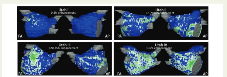
electrophysiological studies for substrate description. Alternatively, the utility of delayed enhancement (DE)—magnetic resonance imaging (MRI) was described in detecting, quantifying and localizing atrial fibrosis including four categories of structural changes (‘Utah
stages I – IV’)9,10(Figure2).
Atrial remodelling in conditions
predisposing to atrial fibrillation
John et al.11described electroanatomic remodelling of the left and
right atria in 24 patients with rheumatic mitral stenosis undergoing mitral commissurotomy. Importantly, patients were excluded if they had any suggestion of atrial arrhythmia/AF or other structural heart disease besides mitral stenosis. In comparison with 24 control patients, the patients with mitral stenosis showed—beyond left atrial enlargement—a significantly reduced biatrial voltage (left atrium 1.8 + 0.6 vs. 3.6 + 0.6 mV, right atrium 1.9 + 0.6 vs. 3.3 + 0.5 mV), reduced conduction velocity, and prolonged effective
re-fractory periods.11These abnormalities may clearly play a role in
the increased propensity to AF in patients with mitral stenosis. Indeed, patients with mitral stenosis were more susceptible to induc-tion of AF with extrastimulus provocainduc-tion. Similar left atrial remod-elling was described in patients with haemodynamically significant atrial septal defects (but no AF) compared with control patients
in-cluding significantly reduced left atrial voltage.12
Hypertension is another common co-existing factor which is
described to be associated with AF. Medi et al.13described results
from electroanatomic mapping and reported right atrial changes in 10 patients with systemic hypertension plus left ventricular hyper-trophy (but no AF) compared with 10 control patients. In patients with chronically treated hypertension, a significant slowing of con-duction velocity was described. Furthermore, sustained AF was induced in 30% of hypertension patients but no controls. Overall,
Figure 1 The progression from paroxysmal to persistent atrial fibrillation may be very different in clinically matched patients. (A) 7-day-ECG in a 69-year-old patient with paroxysmal ‘lone’ atrial fibrillation. The patient had a history of paroxysmal atrial fibrillation of 22 years with frequent epi-sodes per week or even per day (asterisks), however, the individual epiepi-sodes stayed short within minutes or maximally few hours with no evidence for progression. (B) During sinus rhythm, this patient presented with frequent premature atrial complexes with ‘P-on-T’ phenomenon (black asterisks) and also frequent atrial salvoes (white asterisks). (C) 7-day-ECG in another 69-year-old patient with paroxysmal ‘lone’ atrial fibrillation. This patient had a short history of atrial fibrillation of only 6 months, however, he presented with atrial fibrillation episodes lasting already .48 h in the initial 7-day-ECG (arrows). Hardly any premature atrial complexes were found during periods of sinus rhythm (not shown). (D) This patient progressed to persistent atrial fibrillation within only another 3 months.
the structural changes in hypertensive patients observed in this study were less pronounced (compared with the changes in patients with mitral stenosis or atrial septal defect). Importantly, the mean right atrial voltage as a surrogate for the structural (i.e. fibrotic) atrial re-modelling was identical in the hypertension and control groups (2.2 + 0.5 vs. 2.2 + 0.3 mV).
A chronic bi-atrial substrate in
patients with paroxysmal ‘lone’
atrial fibrillation
The evidence for a chronic and substantial atrial substrate in patients with lone AF is substantiated by an investigation of extracellular matrix proteins in left atrial tissue of patients with ‘lone’ AF (n ¼ 56), with AF plus mitral valve disease (n ¼ 46), and with sinus
rhythm (n ¼ 16).7Left atrial tissue was obtained from atriotomy
during cardiac surgery. The association of human AF with fibrosis was shown with collagen concentrations being significantly increased in patients with AF compared with those in sinus rhythm. Importantly, collagen I—which is the major collagenous product of cardiac fibroblasts—was enhanced in ‘lone’ AF patients even to a similar extent compared with AF in patients with severe mitral valve
disease (Figure3).
Stiles et al.14investigated 25 patients (53 + 8 years) with ‘lone’ AF
during an electrophysiological study after at least 7 days in sinus rhythm and found slower conduction velocity, longer effective refrac-tory periods, and—importantly—significantly lower voltages (left atrium 1.7 + 0.7 vs. 3.3 + 0.7 mv, right atrium 1.7 + 0.4 vs. 2.9 + 0.4 mV) compared with control patients without AF. These findings confirm a substantial chronic structural bi-atrial substrate since the
elec-trical remodelling is reversible within a few days.15It might be that not
all patients with paroxysmal ‘lone’ AF have an (underdetected)
chronic substrate, but many more then assumed (Figure4).
The findings of a chronic substrate in patients with paroxysmal ‘lone’ AF were recently extended in a DE-MRI study of the left
atrial substrate.16 In that study, 40 of 333 included patients met
criteria for ‘lone’ AF. The majority of these ‘lone’ AF patients showed a mild or even moderate degree of left atrial fibrosis, thereby compatible with mild/moderate and/or early forms of FACM I/II.
Is mild-to-moderate left atrial fibrosis necessary on top of PV trig-gers for AF to sustain for hours or days? Clinically, the time relation-ship of the trigger initiating AF and the substrate taking over for maintaining AF is largely unknown. Evidence for short trigger activity came from a study investigating a surgical left atrial linear lesion concept targeting at the prevention of anatomic left atrial re-entry without PV isolation in patients with paroxysmal and persistent
AF.17After a mean follow-up of 3.6 years, atrial ectopy, atrial runs,
Figure 2 Quantification of left atrial structural remodelling with delayed enhancement magnetic resonance imaging (‘Utah I – IV’). Posterior-anterior and Posterior-anterior-posterior view of enhancement (green) vs. normal tissue (blue) in patients with ‘lone’ atrial fibrillation (Courtesy of Nassir F. Marrouche, M.D., CARMA Center, Salt Lake City).
Figure 3 Collagen I expression in left atrial tissue of patients in sinus rhythm, with ‘lone’ atrial fibrillation, and with atrial fibrillation plus mitral valve disease. Collagen I was enhanced in ‘lone’ atrial rillation patients to a similar extent when compared with atrial fib-rillation in patients with severe mitral valve disease (modified from
Boldt et al.7)
and re-occurrence of AF episodes were analysed by 7-day-ECGs in 30 patients. Overall, 87% of the patients were free from AF. However, atrial ectopy was present in all patients and atrial runs in 83% of the patients with a median of 9 runs per patient/week (range, 1 – 321). Importantly, the median duration of the atrial runs as a surrogate for the duration of PV trigger activity measured only 1.2 s (range, 0.7 – 25). This may indicate that a chronic atrial substrate is indeed necessary for even relatively short episodes of AF to sustain.
More atrial substrate in patients
with persistent vs. paroxysmal
atrial fibrillation? Fibrotic changes
as cause for or consequence of atrial
fibrillation?
If the clinical AF episodes were the critical stimulus for the develop-ment of structural (fibrotic) remodelling, then there should be a step-wise increase with longer history and/or longer episodes of AF (higher cumulative AF burden), and especially a clear increase in fi-brosis when patients with paroxysmal and persistent AF are com-pared. However, this does not seem to be the case.
Differences in patients with paroxysmal and persistent AF were analysed in the study with left atriotomy specimen obtained during
cardiac surgery.7Importantly, there was a similar degree of enhanced
collagen expression in paroxysmal and persistent AF (Figure5). While
there was a tendency of an increased mean fibrosis level in patients with persistent AF vs. paroxysmal AF, the variation within the two groups was clearly very high and a statistical difference in fibrosis extent, therefore, could not be demonstrated.
Platonov et al.18confirmed and extended the results from Boldt
et al. by investigating structural abnormalities in post-mortem autopsy atrial specimen from 30 patients in three age-matched groups of patients without AF history, with paroxysmal AF, or with permanent AF. Atrial tissue samples were collected from the right atrium, Bachmann’s bundle, and from the posterior left atrial wall.
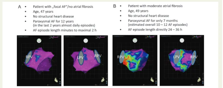
Figure 4 Electroanatomic left atrial voltage mapping in two patients with paroxysmal ‘lone’ atrial fibrillation. (A) The patient had a long history of paroxysmal atrial fibrillation of 12 years, however, the duration of the atrial fibrillation episodes remained very short and the voltage map still shows no evidence at all of fibrosis. These fibrosis-free maps are found in our experience in ,10% of patients with paroxysmal ‘lone’ atrial fibrillation. (B) This patient had a very short history of PAF of only 7 months. Since the patient was extremely symptomatic, an overall cumulative frequency of 10 – 12 atrial fibrillation episodes could be estimated. However, the fibrotic atrial cardiomyopathy process was already substantial with moderate fibrosis at the time of the clinical advent of paroxysmal atrial fibrillation. Projections from anterior and posterior are given. Voltages of mapping points .2.5 mV are annotated purple (normal voltage), voltages ,0.5 mV are indicated red (severely reduced voltage). LPV and RPV, left and right pulmonary veins.
Figure 5 Collagen I expression in human atrial tissue from patients with sinus rhythm, ‘lone’ paroxysmal atrial fibrillation and persistent (CAF) atrial fibrillation, and in patients with mitral valve disease plus sinus rhythm, paroxysmal atrial fibrillation, and CAF, re-spectively. Note, the overlapping wide variation/range of collagen I in patients with paroxysmal atrial fibrillation and CAF, with some paroxysmal atrial fibrillation patients having excessive fibrosis (asterisks) and some CAF patients having mild fibrosis (double
Patients with any history of AF had a three- to five-fold greater extend of fibrosis compared with patients without AF history. A comparison revealed a greater extent of fibrosis in patients with permanent AF compared with patients with paroxysmal AF. However, these differ-ences were statistically significant in the left atrial tissue specimen only at the superior PV level, whereas no significant differences between patients with paroxysmal and permanent AF were found
in the posterior left atrial wall and at the inferior PV level.18
Import-antly, the variation in the data obtained from the posterior level between patients with paroxysmal and permanent AF were very high. In addition, the extent of fibrosis showed no clear correlation with the duration of AF history.
Recently, Teh et al.19described electroanatomic remodelling of the
left atrium in paroxysmal (n ¼ 17) and persistent (n ¼ 14) AF patients without structural heart disease compared with control patients (n ¼ 15). Again, a lower voltage in the AF patients was found compared with the control patients. The mean left atrial voltage difference between patients with paroxysmal and persistent AF even reached a low statis-tical difference, however, in half of the left atrial areas that were inves-tigated, this difference was not statistically significant.
Oakes et al.9reported on the DE-MRI quantification of left atrial
structural remodelling in 81 patients with AF. Mild enhancement was found in 43 patients, 28 of these (65%) had paroxysmal AF. However, 15 of these patients (35%) still had only mild enhancement but were already in persistent AF. Furthermore, from the 30 patients with moderate enhancement, 43% still presented with paroxysmal and 57% with persistent AF, respectively, again indicating towards the high variability of structural remodelling in patients with paroxys-mal and persistent AF.
Atrial fibrillation and the
association to age and other
co-existing cardiovascular diseases
From an epidemiological point of view, human ageing certainly is
asso-ciated with an increased likelihood of developing AF.20However, a
potential causative aetiological relationship of human age and devel-opment of AF is far less well established. Indeed, data on qualitative and quantitative analyses of bi-atrial human tissue from patients with non-valvular AF are scarce. In the human autopsy investigation,
Platonov et al.18 reported on the association between structural
changes in the human atria, age, and history of AF. In contradiction to the ‘conventional wisdom’ about the association between human age and atrial fibrosis, Platonov et al. were not able to detect any correlation between patient age and increase in fibrosis extent. Furthermore, atrial samples from age-matched arrhythmia-free patients contained ‘negligibly’ low amounts of fibrofatty tissue, even in very old patients. These autopsy results match well with data from an electroanatomic mapping study investigating the
impact of age on left and right atrial voltage.21 In that study, the
mean bipolar voltage in young patients≤40 years was somewhat
higher compared with older patients; however, there were hardly any differences in the age group 51 – 60 compared with 61 – 70 and beyond 70 years. Furthermore, age was also equally distributed in the four DE-MRI fibrosis grade groups (mean age 69 years in the total non-lone AF group, 68 years in the Utah I group, 66 years in
the Utah II group, 68 years in the Utah III group, and 70 years in the
Utah IV group, respectively).16
Similar to the assumed aetiological association of human ageing and AF, co-existing cardiovascular diseases (e.g. hypertension) are also thought to play an important aetiological role in the development of
AF. Mahnkopf et al.16compared the left atrial structural changes using
DE-MRI in patients with ‘lone’ AF (n ¼ 40) vs. those with ‘classical’ co-morbidities (n ¼ 293). Importantly, the degree of left atrial structural re-modelling was found to be completely independent of co-morbidities. Furthermore, the distribution of groups Utah I–IV showed no significant differences between patients with lone AF and non-lone AF. For example, fibrosis grade Utah II was found in 65 vs. 64% of lone AF and non-lone AF patients, and fibrosis grade Utah III in 23 vs. 23% of lone
AF and non-lone AF patients, respectively.16In addition, co-existing
car-diovascular diseases were equally distributed in the four fibrosis grade groups, e.g. hypertension in 66% of patients of the total non-lone AF group, in 76% of the Utah I group, in 67% of the Utah II group, in 61% of the Utah III group, and in 68% of the Utah IV group, respectively. These data thereby confirm the autopsy study data, where the
co-morbidity status expressed in the CHA2DS2-VASc score measured
3.8 + 1.8 in patients with AF vs. 4.3 + 1.9 in patients without AF.18
These data also confirm results from electroanatomic mapping, where no atrial voltage differences could be detected in patients with
(arrhythmia free) hypertension patients vs. control patients.13
Evidence for reverse human atrial
structural remodelling?
John et al.22investigated the potential for reverse human atrial
remod-elling in 21 patients undergoing mitral commissurotomy for treat-ment of severe mitral stenosis. Bi-atrial voltage mapping was performed before and after commissurotomy—and in 14 patients,
right atrial mapping was repeated after≥6 months. Importantly, a
further significant increase in atrial voltage was demonstrated late after interventional treatment from 1.8 + 0.7 to 2.8 + 0.6 mV. Therefore, late after reversal of chronic stretch, a progressive im-provement in bipolar voltage could be shown indicating the potential for reverse human atrial remodelling.
If AF itself were the stimulus for the development of this fibrotic sub-strate in patients without structural heart disease, it could be hypothe-sized that this substrate was reversed after successful AF elimination.
Teh et al.23performed right atrial electroanatomic maps in 11 control
patients and 11 ‘lone’ AF patients undergoing catheter ablation at
base-line plus≥6 months following successful elimination of AF. The
sub-strate did not appear to reverse after successful ablation of AF. In contrast, there was a progressive substrate remodelling 6–14 months after successful catheter ablation with further decrease in atrial bipolar voltage and further prolongation of regional refractoriness.
Fibrotic atrial cardiomyopathy:
diagnosis, causality, and
implications for ablation
Electroanatomic mapping and DE-MRI are potential clinical diagnostic tools. Electroanatomic mapping, however, has all limitations of
invasive procedures and does not allow diagnostic follow-up investi-gations. In contrast, the potential of DE-MRI for detecting and quan-tifying atrial fibrosis looks attractive as a non-invasive, repeatable diagnostic tool. Recently, pre-operatively elevated serum markers of collagen synthesis (PICP) have been reported to be associated
with post-surgical AF.24Furthermore, a linear correlation between
PICP and left atrial fibrosis was described. This raises the potential option of identifying subclinical FACM in general before the advent of AF. Overall, the definition and the understanding of the term ‘lone’ AF needs to be modified in the future. FACM to date is difficult to detect by conventional means but constitutes a substantial and chronic structural atrial disease posing the patient to the risk of
devel-oping AF and other manifestations.3
Genetics seem to play a key role in FACM. A familiar aggregation of
lone AF in young persons has been described recently.25Frequently, a
familiar appearance of clinical findings indicating towards FACM can be observed. However, genetic analyses of specific subgroups of AF so far are limited. Besides the key player genetics, additional pathophysiological aspects might include an inflammatory process
as indicated by studies from Frustaci et al.26on biopsy findings in
patients with lone AF, Chung et al.27on C-reactive protein elevation
in patients with atrial arrhythmias, and findings on the role of steroids
to prevent AF recurrences after ablation.28
In patients with ‘pure’ focal AF (i.e. without atrial fibrosis), PV iso-lation can be considered a curative treatment option. In patients with the potentially progressive disease FACM, where AF is a manifest-ation of a structural atrial disease, catheter ablmanifest-ation can also
effective-ly treat AF but ablation cannot be considered curative. Verma et al.29
reported that pre-existent left atrial scarring in patients undergoing ablation was an independent predictor of procedural failure, which
was confirmed in DE-MRI studies.9,10Progressive atrial remodelling
after ablation in FACM cases may explain late recurrences despite durable PV isolation and development of ‘new’ arrhythmias. In add-ition, the association of left atrial fibrosis and the risk of stroke in patients with AF will need to be considered for oral anticoagulation
strategies in the future.3,30
Fibrotic atrial cardiomyopathy:
summary and outlook
There is substantial evidence that the majority of patients without ap-parent structural heart disease (so-called ‘lone’ AF) has a chronic
fi-brotic bi-atrial substrate.7,14,16 A higher mean value of fibrosis is
detected in patients with persistent vs. paroxysmal AF.7,9,18,19
However, the variability in the extend of fibrosis in patients with
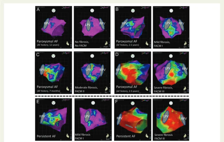
Figure 6 Electroanatomic left atrial voltage mapping in patients with ‘lone’ paroxysmal (A – D) and persistent (E – F) atrial fibrillation. The electro-anatomic maps of the patients with paroxysmal atrial fibrillation show a wide variation of voltage distribution with no correlation to atrial fibrillation history duration: (A) no fibrosis; (B) mild fibrosis; (C) moderate fibrosis; (D) extensive fibrosis. (E and F) Also in patients with persistent atrial fibril-lation, a wide variation of voltage distribution is found. Projection from anterior and posterior is given. Voltages of mapping points .2.5 mV are annotated purple (normal voltage), voltages ,0.5 mV are indicated red (severely reduced voltage). LPV and RPV, left and right pulmonary veins.
paroxysmal and persistent AF is very high with part of paroxysmal AF patients having massive fibrosis as well as part of persistent AF
patients showing mild fibrosis.7,9,18,19 Thus, these data do not
support a causal relationship that AF (significantly) produces fibrosis in the sense of ‘AF begets AF’ instead of being a consequence of the
fi-brotic process in FACM (Figure6). There are obviously patients with
‘lone’ AF, where the substrate is already very advanced with massive
bi-atrial fibrosis before the advent of AF.3Accordingly, no clear
cor-relation could be demonstrated between the extend of fibrosis and
the duration of AF history.16,18The analysed studies with different
methodologies are very consistent, however, the relatively small size of some of these studies necessitates confirmation and extension in future trials.
Other ‘conventional wisdoms’ also need to be re-considered. In the autopsy investigation specifically addressing the role of age, no correlation at all could be detected between patient age and fibrosis
extent.18 Instead, the clinical manifestation of FACM with cardiac
arrhythmias in most patients occurs in advanced ages (≥60 years)
and less frequently in younger ages. In the same line, the degree of left atrial structural remodelling was found to be independent of co-morbidities (e.g. hypertension, diabetes) in DE-MRI and autopsy
studies.16,18Given these results that age and ‘classical’ co-morbidities
have no association with the extent of atrial fibrosis, these data all in-dicate that the chronic bi-atrial substrate of patients with AF is the result of a specific FACM. Further striking evidence for the existence of FACM in patients with ‘lone’ AF comes from electroanatomic mapping studies on the reversibility of human atrial structural
remod-elling.22,23In patients with mitral stenosis, there was clear evidence
for reverse atrial remodelling after commissurotomy. However, in patients with ‘lone’ AF, the atrial substrate progressed despite success-ful AF elimination indicating towards the independent and potentially progressive disease process of FACM.
Different expressions of the FACM disease exist from mild-to-severe fibrosis (FACM I – III), and with wide clinical variations from asymptomatic cases to cases with multiple arrhythmic manifes-tations, atrial mechanical dysfunction as well as thromboembolic
complications.3Clinically, the co-existence of sick sinus node plus
paroxysmal AF (‘bradycardia – tachycardia – syndrome’) is the most
frequent FACM manifestation combination.3,31
Recently, localized electrical ‘rotors’ have been described as prevalent sustaining mechanisms of human AF, and catheter ablation
at patient-specific sources improved clinical outcome.32It might be
speculated that these patient-specific sources correlate with areas of atrial fibrosis where the site-specific micro-architecture of con-nective tissue fibres and the remaining myocardial fibres allows re-entrant/rotor activation to occur and to sustain. The combination of localizing atrial fibrosis plus mapping of specific functional areas allowing re-entrant/rotor activation may hold promise for catheter-based AF substrate modification in the future.
Conflict of interest: H.K. served as a consultant for BiosenseWeb-ster within the past 2 years.
References
1. Wijffels MC, Kirchhof CJ, Dorland R, Allessie MA. Atrial fibrillation begets atrial fib-rillation. A study in awake chronically instrumented goats. Circulation 1995;92: 1954 – 1968.
2. Kottkamp H. Atrial fibrillation substrate: the ‘unknown species’—from lone atrial fibrillation to fibrotic atrial cardiomyopathy. Heart Rhythm 2012;9:481 – 482. 3. Kottkamp H. Fibrotic atrial cardiomyopathy: a specific disease/syndrome supplying
substrates for atrial fibrillation, atrial tachycardia, sinus node disease, AV node disease, and thromboembolic complications. J Cardiovasc Electrophysiol 2012;23: 797 – 799.
4. Spach MS, Dolber PC. Relating extracellular potentials and their derivatives to aniso-tropic propagation at a microscopic level in human cardiac muscle: evidence for elec-trical uncoupling of side to side fiber connections with increasing age. Circ Res 1986; 56:356 – 371.
5. Spach MS, Boineau JP. Micofibrosis produces electrical load variations due to loss of side-to-side cell connections: a major mechanism of structural heart disease arrhyth-mias. Pacing Clin Electrophysiol 1997;20:397 – 413.
6. Kostin S, Klein G, Szalay Z, Hein S, Bauer EP, Schaper J. Structural correlate of atrial fibrillation in human patients. Cardiovasc Res 2002;54:361 – 379.
7. Boldt A, Wetzel U, Lauschke J, Weigl J, Gummert J, Hindricks G, Kottkamp H, Dhein S. Fibrosis in left atrial tissue of patients with atrial fibrillation with and without underlying mitral valve disease. Heart 2004;90:400 – 405.
8. Callans D, Ren JF, Michele J, Marchlinski FE, Dillon SM. Electroanatomic left ventricu-lar mapping in the porcine model of healed anterior myocardial infarction. Correl-ation with intracardiac echocardiography and pathological analysis. CirculCorrel-ation 1999;100:1744 – 1750.
9. Oakes RS, Badger TJ, Kholmovski EG, Akoum N, Burgon NS, Fish EN, Blauer JJE, Rao SN, DiBella EVR, Segerson NM, Daccarett M, Windfelder J, McGann CJ, Parker D, MacLeod RS, Marrouche NF. Detection and quantification of left atrial structural remodeling with delayed-enhancement magnetic resonance imaging in patients with atrial fibrillation. Circulation 2009;119:1758 – 1767.
10. Akoum N, Daccarett M, McGann C, Segerson N, Vergara G, Kuppahally S, Badger T, Burgon N, Haslam T, Kholmovski E, MacLeod R, Marrouche N. Atrial fibrosis helps select the appropriate patient and strategy in catheter ablation of atrial fibrillation: A DE-MRI guided approach. J Cardiovasc Electrophysiol 2011;22: 16 – 22.
11. John B, Stiles MK, Kuklik P, Chandy ST, Young GD, Mackenzie L, Szumowski L, Joseph G, Jose J, Worthley SG, Kalman JM, Sanders P. Electrical remodelling of the left and right atria due to rheumatic mitral stenosis. Eur Heart J 2008;29: 2234 – 2243.
12. Roberts-Thomson KC, John B, Worthley SG, Brooks AG, Stiles MK, Lau DH, Kuklik P, Shipp NJ, Kalman JM, Sanders P. Left atrial remodeling in patients with atrial septal defects. Heart Rhythm 2009;6:1000 – 1006.
13. Medi C, Kalman JM, Spence SJ, Teh AW, Lee G, Bader I, Kaye DM, Kistler PM. Atrial electrical and structural changes associated with longstanding hypertension in humans: Implications for the substrate for atrial fibrillation. J Cardiovasc Electrophysiol 2011;22:1317 – 1324.
14. Stiles MK, John B, Wong CX, Kuklik P, Brooks AG, Lau DH, Dimitri H, Roberts-Thomson KC, Wilson L, De Sciscio P, Young GD, Sanders P. Paroxysmal lone atrial fibrillation is associated with an abnormal atrial substrate: characterizing the ‘second factor’. J Am Coll Cardiol 2009;53:1182 – 1191.
15. Garratt CJ, Duytschaever M, Killian M, Dorland R, Mast F, Allessie MA. Repetitive electrical remodeling by paroxysms of atrial fibrillation in the goat: no cumulative effect on inducibility or stability of atrial fibrillation. J Cardiovasc Electrophysiol 1999; 10:1101 – 1108.
16. Mahnkopf C, Badger TJ, Burgon NS, Daccarett M, Haslam TS, Badger CT, McGann CJ, Akoum N, Kholmovski E, MacLeod RS, Marrouche NF. Evaluation of the left atrial substrate in patients with lone atrial fibrillation using delayed-enhanced MRI: implications for disease progression and response to catheter ablation. Heart Rhythm 2010;7:1475 – 1481.
17. Tanner H, Hindricks G, Kobza R, Dorszewski A, Schirdewahn P, Piorkowski C, Gerds-Li JH, Kottkamp H. Trigger activity more than three years after left atrial linear ablation without pulmonary vein isolation in patients with atrial fibrillation. J Am Coll Cardiol 2005;46:338 – 343.
18. Platonov PG, Mitrofanova LB, Orshanskaya V, Ho SY. Structural abnormalities in atrial walls are associated with presence and persistency of atrial fibrillation but not with age. J Am Coll Cardiol 2011;58:2225 – 2232.
19. Teh AW, Kistler PM, Lee G, Medi C, Heck PM, Spence SJ, Sparks PB, Morton JB, Kalman JM. Electroanatomic remodeling of the left atrium in paroxysmal and persist-ent atrial fibrillation patipersist-ents without structural heart disease. J Cardiovasc Electrophy-siol 2012;23:232 – 238.
20. Miyasaka Y, Barnes ME, Gersh BJ, Cha SS, Bailey KR, Abhayaratna WP, Seward JB, Tsang TS. Secular trends in incidence of atrial fibrillation in Olmsted County, Minne-sota, 1980 to 2000, and implications on the projections for future prevalence. Circu-lation 2006;114:119 – 125.
21. Tuan TC, Chang SL, Tsao HM, Tai CT, Lin YJ, Hu YF, Lo LW, Udyavar AR, Chang CJ, Tsai WC, Tang WH, Suenari K, Huang SY, Lee PC, Chen SA. The impact of age on the electroanatomical characteristics and outcome of catheter
ablation in patients with atrial fibrillation. J Cardiovasc Electrophysiol 2010;21: 966 – 972.
22. John B, Stiles MK, Kuklik P, Brooks AG, Chandy ST, Kalman JM, Sanders P. Reverse remodeling of the atria after treatment of chronic stretch in humans. J Am Coll Cardiol 2010;55:1217 – 1226.
23. Teh AW, Kistler PM, Lee G, Medi C, Heck PM, Spence SJ, Morton JB, Sanders P, Kalman JM. The long-term effects of catheter ablation for lone atrial fibrillation. Pro-gressive atrial electroanatomic substrate remodelling despite successful ablation. Heart Rhythm 2012;9:473 – 480.
24. Swartz MF, Fink GW, Sarwar MF, Hicks GL, Yu Y, Hu R, Lutz CJ, Taffet SM, Jalife J. Elevated pre-operative serum peptides for collagen I and III synthesis result in post-surgical atrial fibrillation. J Am Coll Cardiol 2012;60:1799 – 1806.
25. Oyen N, Ranthe MF, Carstensen L, Boyd HA, Olesen MS, Olesen SP, Wohlfahrt J, Melbye M. Familial aggregation of lone atrial fibrillation in young persons. J Am Coll Cardiol 2012;60:917 – 921.
26. Frustaci A, Chimenti C, Bellocci F, Morgante E, Russo MA, Maseri A. Histological sub-strate of atrial biopsies in patients with lone atrial fibrillation. Circulation 1997;96: 1180 – 1184.
27. Chung MK, Martin DO, Sprecher D, Wazni O, Kanderian A, Carnes CA, Bauer JA, Tchou PJ, Niebauer MJ, Natale A, Van Wagoner DR. C-reactive protein elevation in patients with atrial arrhythmias. Circulation 2001;104:2886 – 2891.
28. Koyama T, Tada H, Sekigucki Y, Arimoto T, Yamasaki H, Kuroki K, Machino T, Tajiri K, Zhu XD, Kanemoto-Igarashi M, Sugiyasu A, Kuga K, Nakata Y, Aonuma K. Prevention of atrial fibrillation recurrence with corticosteroids after radiofrequency catheter ablation. J Am Coll Cardiol 2010;56:1463 – 1472.
29. Verma A, Wazni OM, Marrouche NF, Martin DO, Kilicaslan F, Minor S, Schweikert RA, Saliba W, Cummings J, Burkhardt JD, Bhargava M, Belden WA, Abdul-Karim A, Natale A. Pre-existent left atrial scarring in patients undergoing pul-monary vein antrum isolation. An independent predictor of procedural failure. J Am Coll Cardiol 2005;45:285 – 292.
30. Daccarett M, Badger TJ, Akoum N, Burgon NS, Mahnkopf C, Vergara G, Kholmovski E, McGann CJ, Parker D, Brachmann J, MacLeod RS, Marrouche NF. As-sociation of left atrial fibrosis detected by delayed-enhancement magnetic reson-ance imaging and the risk of stroke in patients with atrial fibrillation. J Am Coll Cardiol 2011;57:831 – 838.
31. Akoum N, McGann C, Vergara G, Badger T, Ranjan R, Mahnkopf C, Kholmovski E, MacLeod R, Marrouche N. Atrial fibrosis quantified using late Gadolinium enhance-ment MRI is associated with sinus node dysfunction requiring pacemaker implant. J Cardiovasc Electrophysiol 2012;23:44 – 50.
32. Narayan SM, Krummen DE, Shivkumar K, Clopton P, Rappel WJ, Miller JM. Treat-ment of atrial fibrillation by the ablation of localized sources. J Am Coll Cardiol 2012;60:628 – 636.
CARDIOVASCULAR FLASHLIGHT
. . . .
doi:10.1093/eurheartj/eht224
Online publish-ahead-of-print 25 July 2013
Epicardial Wolff – Parkinson – White ablation
Christoph Scharf* and Lam Dang
Clinic im Park, Zu¨rich, Switzerland
*Corresponding author. Tel:+41 12558762, Fax: +41 12554401, Email:christoph.scharf@gmail.com
A 45-year-old female patient without structural heart disease was referred for redo Wolff – Parkinson – White (WPW) ablation after an unsuc-cessful endocardial procedure. The refractory period of the accessory pathway had been mea-sured at 230 ms and antiarrhythmic treatment with flecainide was unsuccessful.
(1) The surface electrogram shows the largest negative d-wave in lead III.
(2) During endocardial mapping of the ventricular insertion the earliest activation was found near the coronary sinus ostium (x); however, radiofrequency application was not successful. Note the morphology of the unipolar signal (Abl uni) with an initially positive small spike (red arrow) resulting from epi-endocardial acti-vation followed by the broad negative ventricu-lar activation signal.
(3) Epicardial mapping via subxiphoidal access
showed the earliest signal (XX) opposite to the endocardial site (X). The distance between the two locations was 1.5 cm. Note the morphology with an entirely negative small pathway spike (red arrow) followed by the broad negative ventricular activation signal. Irrigated radiofrequency ablation was successful after 25 s and increase of power from 20 to 30 W.
Epicardial access can be necessary, if the largest negative d-wave is in lead III on the surface ECG. The morphology of the unipolar signal indicates the true epicardial origin of the ventricular pathway insertion. Success can be observed even after delayed response to RF delivery. In Panel 1: ECG, electrocardiogram; LAO, left anterior oblique; Abl, ablation electrode; CS, coronary sinus; CSp, coronary sinus elec-trode proximal; CSd, coronary sinus elecelec-trode distal; RV, right ventricular elecelec-trode; LAT, local activation time measured relative to onset of QRS signals on surface ECG. The local activation time is measured conventionally (middle) and visualized in a coloured Carto-3D map (red earliest).