HAL Id: hal-01542927
https://hal-amu.archives-ouvertes.fr/hal-01542927
Submitted on 23 Jun 2017HAL is a multi-disciplinary open access archive for the deposit and dissemination of sci-entific research documents, whether they are pub-lished or not. The documents may come from teaching and research institutions in France or abroad, or from public or private research centers.
L’archive ouverte pluridisciplinaire HAL, est destinée au dépôt et à la diffusion de documents scientifiques de niveau recherche, publiés ou non, émanant des établissements d’enseignement et de recherche français ou étrangers, des laboratoires publics ou privés.
The iron-sulfur cluster sensor IscR is a negative
regulator of Spi1 Type III Secretion System in
Salmonella enterica Cellular Microbiology
Alexandra Vergnes, Julie P M Viala, Ouadah-tsabet Rabah, Bérengère
Pocachard, Laurent Loiseau, Stephane Meresse, Frédéric Barras, Laurent
Aussel
To cite this version:
Alexandra Vergnes, Julie P M Viala, Ouadah-tsabet Rabah, Bérengère Pocachard, Laurent Loiseau, et al.. The iron-sulfur cluster sensor IscR is a negative regulator of Spi1 Type III Secretion Sys-tem in Salmonella enterica Cellular Microbiology. Cellular Microbiology, Wiley, 2016, 19, pp.e12680. �10.1111/cmi.12680�. �hal-01542927�
1
The iron-sulfur cluster sensor IscR is a negative regulator of Spi1 Type III
1
Secretion System in Salmonella enterica
2 3
Running title: Regulation of Spi1 TTSS by IscR
4 5 6
Alexandra Vergnes1, Julie P.M. Viala1,#, Rabah Ouadah-Tsabet1, Bérengère
7
Pocachard1, Laurent Loiseau1, Stéphane Méresse2, Frédéric Barras1, and Laurent
8 Aussel1* 9 10 11 12
1Aix Marseille Université, CNRS, LCB UMR 7283, IMM, 13402, Marseille,
13
France 2Aix Marseille Université, CNRS, INSERM, CIML, Marseille, France.
14 15 16 17 18
*Corresponding author. Mailing address: Laboratoire de Chimie Bactérienne, 31
19
Chemin Joseph Aiguier, 13402 Marseille Cedex 20, France.
20
Phone: +33 4 91 16 44 31. Fax +33 4 91 71 89 14. E-mail: aussel@imm.cnrs.fr
21 22 23
# Present address: Laboratoire d'Ingénierie des Systèmes Macromoléculaires, 31
24
Chemin Joseph Aiguier, 13402 Marseille Cedex 20, France.
2
Summary
26 27
Iron-sulfur (Fe-S)-containing proteins contribute to various biological processes,
28
including redox reactions or regulation of gene expression. Living organisms have
29
evolved by developing distinct biosynthetic pathways to assemble these clusters,
30
including ISC (Iron Sulfur Cluster) and SUF (Sulfur mobilization). Salmonella enterica
31
serovar Typhimurium is an intracellular pathogen responsible for a wide range of
32
infections, from gastroenteritis to severe systemic diseases. Salmonella possesses all
33
known prokaryotic systems to assemble Fe-S clusters, including ISC and SUF.
34
Because iron starvation and oxidative stress are detrimental for Fe-S enzyme
35
biogenesis and because such environments are often met by Salmonella during its
36
intracellular life, we investigated the role of the ISC and SUF machineries during the
37
course of the infection. The iscU mutant, which is predicted to have no ISC system
38
functioning, was found to be defective for epithelial cell invasion and for mice infection,
39
whereas the sufBC mutant, which is predicted to have no SUF system functioning, did
40
not present any defect. Moreover, the iscU mutant was highly impaired in the
41
expression of Spi1 TTSS that is essential for the first stage of Salmonella infection.
42
The Fe-S cluster sensor IscR, a transcriptional regulator matured by the ISC
43
machinery, was shown to bind the promoter of hilD, which encodes the master
44
regulator of Spi1. IscR was also demonstrated to repress hilD and subsequently Spi1
45
gene expression, consistent with the observation that an iscR mutant is hyperinvasive
46
in epithelial cells. Collectively, our findings indicate that the ISC machinery plays a
47
central role in Salmonella virulence through the ability of IscR to down-regulate Spi1
48
gene expression. At a broader level, this model illustrates an adaptive mechanism used
3
by bacterial pathogens to modulate their infectivity according to iron and oxygen
50
availability.
4
Introduction
52 53
Iron-sulfur (Fe-S) cluster-containing proteins play major roles in a wide variety of
54
process, including redox reactions, regulation of gene expression or metabolic
55
pathways (Kiley and Beinert, 2003). Assembly and delivery of Fe-S clusters are
56
catalysed by multi-protein systems, ISC (Iron Sulfur Cluster) and SUF (Sulfur
57
mobilzation), conserved in most living organisms. Briefly, sulfur is mobilized from
L-58
cysteine by IscS/SufSE (Hidese et al., 2011), and scaffold proteins (IscU/SufBC) bind
59
both sulfur and iron to assemble a cluster before transferring it to the apo-protein via
60
the A-type carriers IscA/SufA (Bonomi et al., 2005; Vinella et al., 2009; Wollers et al.,
61
2010) (Figure S1). In Escherichia coli and most Gram-negative bacteria, the ISC
62
system is encoded by the iscRSUA-hscBA-fdx operon while the SUF system is
63
encoded by the sufABCDSE operon (Fig. S1). Other so-called "non-ISC non-SUF"
64
proteins intervene at the sulfur production step (CyaY), at the transfer steps (ErpA,
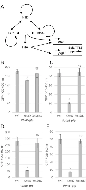
65
NfuA, Mrp), or, possibly, at the repair step (YggX).
66 67
In the last decade, several studies reported the importance of ISC and/or SUF systems
68
in sustaining virulence of various bacteria. Indeed, iron starvation and oxidative stress
69
are detrimental for Fe-S enzyme biogenesis, and such environments are often met by
70
bacterial pathogens upon infection of their host. Thus, the SUF and the ISC systems
71
are required for full virulence of the plant pathogen Dickeya dadantii (Nachin et al.,
72
2001; Rincon-Enriquez et al., 2008). In Shigella flexneri, inactivation of the ISC system
73
prevents plaques formation because the strain is non-invasive (Runyen-Janecky et al.,
74
2008). The SUF system is essential for Mycobacterium tuberculosis survival and
5
therefore required for virulence (Huet et al., 2005). In this context, the case of the iscR
76
gene deserves specific attention.
77 78
In E. coli, IscR is a [2Fe-2S]-containing protein whose maturation is primarily catalyzed
79
by the ISC system. The holo-form of IscR acts as a repressor of a large number of
80
genes, including the isc operon. The apo-form of IscR acts as an activator of a few
81
genes, including the suf operon. Hence, under iron limitation or in the presence of
82
reactive oxygen species (ROS), IscR exists primarily under its apo-form that activates
83
expression of SUF and no longer represses expression of isc, the amount of Fe-S
84
clusters produced being at its highest level. As IscR maturation is catalyzed by the ISC
85
system, a defect in this latter leads to the accumulation of apo-IscR and activation of
86
the SUF system. This regulatory connection between ISC and SUF must be taken into
87
account when interpreting the phenotype of ISC-defective strains. For instance, a D.
88
dadantii iscR mutant is non-invasive on Saintpaulia ionantha plants because the SUF
89
system can't be activated by IscR, but it presents a wild-type phenotype on Arabidopsis
90
thaliana presumably because the demand in Fe-S clusters to be satisfied for the
91
bacterium to grow is met by ISC in this case (Rincon-Enriquez et al., 2008). In fact,
92
IscR appears to be important for full virulence of several pathogens. Mutations in iscR
93
reduce virulence of Pseudomonas aeruginosa and Xanthomonas campestris (Kim et
94
al., 2009; Fuangthong et al., 2015). Moreover, recent studies have shed new light on
95
the role of IscR by expanding its zone of influence beyond the usual Fe-S cluster
96
containing proteins; namely, in enterotoxigenic Escherichia coli, IscR was shown to
97
bind upstream cfaA and to activate CFA/I fimbriae production in response to iron
98
starvation (Haines et al., 2015). In Yersinia pseudotuberculosis, IscR was found to be
99
involved in the regulation of the Type III secretion system (TTSS) master regulator LcrF
6
and was demonstrated to be essential for the virulence of Yersinia in a mouse model
101
of infection (Miller et al., 2014).
102 103
Salmonella enterica serovar Typhimurium is a facultative intracellular pathogen that is
104
associated with a wide range of infections in mammals, ranging from self-limiting
105
gastroenteritis to severe systemic diseases. During the first stage of infection, the
106
bacterium invades intestinal epithelial cells using a TTSS encoded by Salmonella
107
Pathogenicity Island 1 (Spi1) (Galán and Curtiss, 1989). Afterwards, it survives and
108
replicates in macrophages within a membrane-bound compartment called the
109
Salmonella-containing vacuole (SCV) using a TTSS encoded by Spi2 (Figueira and
110
Holden, 2012). Synthesis of the Spi1 TTSS apparatus and most of its effectors is
111
controlled by a complex regulatory scheme. HilA activates directly the expression of
112
the inv and prg operons, which encode the TTSS apparatus (Ellermeier et al., 2005).
113
Expression of hilA is under the control of many environmental conditions and three
114
transcriptional activators: HilC, HilD and RtsA (Ellermeier et al., 2005).
115 116
Among the key points leading to the establishment of infection is the ability of the
117
pathogen to adapt to various environments and to resist different stresses, including
118
iron limitation and ROS production. Recently, CyaY and YggX, both involved in
iron-119
sulfur biogenesis, were demonstrated to be important for Salmonella virulence in mice
120
(Velayudhan et al., 2014). Here, we investigated the involvement of ISC and SUF in
121
Salmonella. Whereas no major role was attributed to the SUF system, the ISC
122
machinery was found to be essential for epithelial cell invasion and we explained this
123
by revealing a molecular link between Fe-S cluster and Spi1 TTSS functioning. In
124
particular, we showed that IscR represses the synthesis of the Spi1-carried regulator
7
HilD, decreasing the expression of genes encoding the TTSS apparatus. Thus, the
126
IscR regulator can be exploited by Salmonella as a sensor to modulate its invasiveness
127
depending on the stage of the infection and the compartment infected.
8
Results
129 130
The ISC machinery is essential for HeLa cell invasion and virulence in mice
131
In order to evaluate the contribution of the ISC and SUF systems to Salmonella
132
intracellular proliferation, we infected activated RAW 264.7 mouse macrophages with
133
Salmonella wild-type strain, iscU and sufBC mutants. Bacterial proliferation was
134
assayed by calculating the proliferation index as a ratio of the intracellular bacteria
135
between 16 and 2 hours post-infection. The proliferation index of the wild-type strain
136
and the iscU mutant were very close (8.8 and 9.4, respectively), whereas it was slightly
137
higher for the sufBC mutant (12.2) (Fig. 1A), reflecting no major role for Fe-S proteins
138
during the intracellular proliferation of macrophages. Next, HeLa cell invasion was
139
carried out. Each strain was incubated for ten minutes with HeLa cells followed by one
140
hour of gentamicin treatment. We found the iscU mutant to invade epithelial cells much
141
less efficiently than the wild-type (Fig. 1B), whereas the sufBC mutant invasion showed
142
no defect compared to the wild-type (Fig. 1B). Next, mixed infections of mice were
143
carried out using wild-type and mutants in order to determine the competitive index
144
(CI) and to compare virulence of the strains (Beuzón and Holden, 2001). The injection
145
of the inoculum was either intraperitoneal (IP), during which bacterial cells can rapidly
146
reach their target organs (Fig. 1C & Table S1), or oral to force the pathogen to cross
147
the intestinal barrier before accessing the blood system (Fig. 1D & Table S2). By
148
performing intraperitoneal infections, we found the iscU mutant to be affected for its
149
virulence (CI = 0.304) (Fig. 1C & Table S1). In contrast, the CI of the sufBC mutant did
150
not show any significant alteration of virulence compared with the wild-type strain (Fig.
151
1C & Table S1). Lastly, mice oral infections were carried out. We observed a severe
9
virulence defect of the iscU mutant (CI = 0.011) (Fig. 1D & Table S2), 30-fold less
153
compared to intraperitoneal infection and 90-fold less compared to the wild-type. The
154
sufBC mutation had no effect (CI = 0.758) (Fig. 1D & Table S2). Taken together, these
155
results indicate that the ISC machinery plays a major role in Salmonella virulence
156
during the invasion of epithelial cells and in the mouse model.
157
158
The ISC system plays a key role in the regulation of Spi1 Type III Secretion
159
System gene expression
160
To test the involvement of Fe-S biosynthesis machineries in Spi1 TTSS regulation, the
161
promoter of hilD - which was shown to be activated to allow Spi1 TTSS biogenesis
162
(Fig. 2A) - was fused to GFP and inserted into the pFPV25 plasmid, yielding the
PhilD-163
gfp transcriptional fusion. Such constructs were previously used in Salmonella to report
164
gene expression within host cells (Valdivia and Falkow, 1997; Aussel et al., 2011).
165
Cells were grown in LB and the fusion expression levels were measured after 16 hours
166
of culture. Expression of the PhilD-gfp fusion was found to be 22% lower in an iscU
167
mutant compared with the wild-type, whereas the sufBC mutation had no effect on the
168
expression of this fusion (Fig. 2B). We next tested whether the effect of the
iscU-169
mediated reduction had an impact on the expression of the hilD-regulated genes such
170
as rtsA, prgH and invF. Transcriptional fusions were constructed for each gene and
171
their expression assayed in iscU and sufBC backgrounds. Compared to the wild-type
172
strain, the iscU mutant exhibited a decrease in the expression of rtsA (4.1-fold), prgH
173
(5.3-fold) and invF (7-fold) (Fig. 2C, 2D and 2E). The sufBC mutation had no effect on
174
the expression level of these genes. Altogether, these results showed that Spi1 TTSS
10
genes are down-regulated in an iscU mutant, conferring a key role to the ISC
176
machinery in the synthesis of this secretion apparatus.
177 178
IscR binds on the promoter of hilD
179
Because the ISC machinery was found to play a role in the regulation of Spi1 TTSS
180
gene expression, we aimed at identifying a protein matured by ISC and involved in the
181
control of the Spi1 TTSS apparatus. Two putative binding sites of IscR - a
[2Fe-2S]-182
containing regulator matured by the ISC system - were found in the hilD promoter
183
region. Only three amino acids differ between E. coli and Salmonella IscR proteins
184
(98% identity) and none of these residues are in the DNA binding domain, supporting
185
the idea that identical motifs can be recognized by both proteins (Rajagopalan et al.,
186
2013). The two putative IscR binding sites upstream of hilD resemble Type 2 motifs
187
which can be bound by both holo- and apo-IscR (Giel et al., 2006). Sites 1 and 2 lie,
188
respectively, from -105 to -81 (ATTACCCAAATTTGGGTTCTTTTGG) and from -65 to
189
-41 (ATTGCCAACACACGCTAATAAAGAG) upstream of the hilD gene transcription
190
start site (Fig. 3A). Comparison of these two sites with the IscR sites from E. coli hyaA
191
and Yersinia pseudotuberculosis lcrF shows that they contain five and four of the nine
192
bases in this binding motif, respectively (Fig. 3B) (Giel et al., 2006). Importantly, Site 1
193
overlaps with a previously identified HilD binding site (Fig. 3A) (Olekhnovich and
194
Kadner, 2002). To test the interaction between IscR and the promoter of hilD (PhilD),
195
electrophoretic mobility shift assays (EMSA) were carried out. When incubated with
196
increasing concentrations of purified His6-IscR, the PhilD probe shifted gradually (Fig.
197
3C). IscR was able to form four different complexes, consistent with the presence of
198
two Type 2 motifs on PhilD and with the ability of IscR to form dimers (Fig. 3C). Next,
11
three nucleotides were mutated at the positions 1, 5 and 6 of the Type 2 motifs on the
200
hilD promoter (ATTACC was changed to CTTAGG on the site 1 and ATTGCC was
201
changed to CTTGGG on the site 2). When incubated with increasing concentrations of
202
IscR, the resulting DNA fragment, called PhilD*, displayed a more modest shift
203
compared to PhilD, highlighting the importance of these conserved nucleotides in the
204
interaction between IscR and the promoter of hilD (Fig. 3D and 3E). Finally, to confirm
205
the direct interaction between IscR and PhilD, DNA-protein interaction experiments
206
were carried out using Bio-Layer Interferometry (BLI), a technique previously used to
207
study protein-protein interactions (Arlet et al., 2014). IscR was also found to interact
208
with PhilD in this assay, whereas no interaction was detected with the promoter of
209
ahpC used as a negative control (Fig. 3F). Interestingly, this experiment allowed us to
210
measure a dissociation constant (Kd) of 390 nM between IscR and PhilD, consistent
211
with previous values of 93 and 112 nM measured between E. coli IscR and the iscR
212
promoter (Giel et al., 2013). Taken together, the results presented in this panel
213
demonstrate a direct interaction between IscR and the promoter of hilD.
214
215
IscR represses the expression of hilD gene and HilD targets
216
Next, to investigate the regulatory role of IscR on hilD, the expression of the hilD fusion
217
was measured and found to be about 1.5-fold up-regulated in an iscR mutant (Fig. 4A),
218
suggesting that IscR acts as a negative regulator. Interestingly, the expression level of
219
hilD was reduced to a similar extent (2.5-fold) in the hilD and in the hilD iscR mutants,
220
consistent with a scenario where IscR could act as an anti-activator on HilD. Because
221
the extent of the regulatory effect was only 1.5-fold, we tested whether the
IscR-222
mediated regulation of hilD had any effect on the expression of downstream
12
regulated genes. The expression of the rtsA, prgH and invF fusions was measured in
224
a wild-type, iscR, hilD and hilD iscR mutant strains. A hilD mutation abrogated the
225
expression of rtsA, prgH and invF (Fig. 4B, 4C and 4D). In contrast, the iscR mutant
226
exhibited a 1.5-fold increase in rtsA expression (Fig. 4B) and a 2-fold increase in prgH
227
and invF expression (Fig. 4C and 4D). In the hilD iscR mutant, the expression level of
228
the rtsA, prgH and invF fusions was found to be lower than 10 units but higher than the
229
hilD mutant values (Fig. 4B, 4C and 4D). The iscR and hilD mutant strains were also
230
complemented by plasmid-borne wild-type copies of iscR and hilD under an
arabinose-231
inducible promoter, respectively (Fig. S2). To investigate further the regulatory effect
232
of IscR, quantitative reverse transcription PCR (qRT-PCR) experiments were carried
233
out with the genes previously analysed. The iscR mutation led to a 5.5-fold increase in
234
hilD mRNA level compared to the wild-type strain, confirming that IscR acts as a
235
repressor of hilD (Fig. 4E). Consistently, rtsA, prgH and invF mRNA levels were found
236
to increase significantly in the iscR mutant (2.5-, 7- and 5.5-fold, respectively) whereas
237
they decreased in the hilD mutant (Fig. 4F, 4G and 4H). Noticeably, IscR was not found
238
to shift the PinvF probe in EMSA, supporting the notion that expression of invF is
239
indirectly regulated by IscR (Fig. S3). In the hilD iscR mutant, the rtsA, prgH and invF
240
mRNA levels were 360-, 60- and 460-fold lower compared to the wild-type strain,
241
showing that the expression of rtsA, prgH and invF is totally abrogated in the absence
242
of both iscR and hilD and suggesting a possible Spi1 gene expression reprogramming
243
process in this background (Fig. 4F, 4G and 4H). Collectively, these results indicate
244
that IscR acts as a repressor of hilD gene expression, leading to the down-regulation
245
of HilD targets.
246
247
Apo- and holo-IscR down-regulate Spi1 TTSS gene expression
13
To investigate whether IscR acts on Spi1 TTSS gene expression under its apo- or
holo-249
form, we built a plasmid encoding an IscR variant (IscR-3CA). This IscR variant has all
250
three cluster-coordinating cysteine residues (Cys92, Cys98 and Cys104) converted
251
into alanine, which prevents binding of the [2Fe-2S] cluster (Rajagopalan et al., 2013).
252
The iscR mutant was transformed with an empty plasmid, with a plasmid encoding
253
IscR or with a plasmid encoding IscR-3CA. In an iscR mutant, expression of the prgH
254
gene was down-regulated 2.4-fold in the presence of piscR while it was down-regulated
255
1.6-fold in the presence of piscR-3CA (Fig. 5A, left). This indicated that both forms
256
have a negative effect on prgH expression. Similarly, the invF gene was
down-257
regulated 1.7-fold in an iscR-/piscR strain and 1.3-fold in an iscR-/piscR-3CA strain,
258
again indicating that apo- and holo-IscR modified invF gene expression (Fig. 5A, left).
259
The experiment was also carried out in a wild-type strain in which the overexpression
260
of iscR reduced the expression of prgH and invF (2.9- and 2.7-fold, respectively) (Fig.
261
5A, right). The same observation was made when the iscR-3CA variant was
262
overexpressed (1.9- and 2.1-fold repression compared to the empty plasmid). Taken
263
together, these results show that both apo- and holo-IscR down-regulate the
264
expression of the Spi1 TTSS gene.
265 266
IscR mediates iron-dependent control of Spi1 TTSS genes
267
It was previously shown that iron limitation down-regulates the expression of Spi1
268
TTSS genes (Ellermeier and Slauch, 2008; Teixidó et al., 2011). Therefore, we tested
269
whether IscR could have a role in mediating this regulation. Expression of prgH and
270
invF genes was monitored in bacteria grown in the presence of 2,2’-dipyridyl (dip), an
271
iron chelator (Fig. S4). In the presence of dip, the level of prgH and invF expression
14
dropped 8.8- and 5.7-fold compared with untreated cells (Fig. 5B). In an iscR mutant,
273
the addition of dip was still inhibiting expression of the two genes but to a much reduced
274
extent, i.e. 3.2-fold for prgH and 1.7-fold for invF fusions (Fig. 5B). These data indicate
275
that IscR contributes significantly to the iron-mediated regulation of Spi1 TTSS gene
276
expression.
277
278
IscR reduces the invasion of HeLa cells by Salmonella
279
Because IscR was shown to decrease the expression of Spi1 TTSS genes, we
280
evaluated its contribution to Salmonella virulence. After ten minutes of incubation with
281
HeLa cells, the iscR mutant presented an efficiency of entry 1.44-fold higher than the
282
wild-type strain (Fig. 6A). Finally, the wild-type and the iscR mutant strains were used
283
in mice co-infection experiments. The inoculum was either injected intraperitoneally or
284
ingested orally. Virulence of the iscR mutant was reduced as compared with the
wild-285
type, using both methods: a CI of 0.144 was obtained using intraperitoneal infections
286
(Fig. 6B left & Table S3), whereas a value of 0.255 was found after oral ingestion (Fig.
287
6B right & Table S3). These results show that during the first step of the infection, IscR
288
can reduce HeLa cell invasion by Salmonella, consistent with its negative role in Spi1
289
TTSS expression.
15
Discussion
291 292
In this study, we showed that the Iron Sulfur Cluster (ISC) machinery is required for
293
the virulence of Salmonella enterica. The reduced invasion of an iscU mutant in HeLa
294
cells combined to its drastic virulence attenuation during oral mouse infections confer
295
a central role to the ISC system in the establishment of the Salmonella infectious
296
process. One reason underlying the reduced virulence of an iscU mutant could be a
297
decrease in Spi1 TTSS gene expression. Indeed, we showed that apo-IscR, which
298
prevails in an iscU mutant, downregulates the expression of the master regulator hilD
299
and thereby of several downstream HilD-regulated genes. Our findings support the
300
idea that the ISC machinery is important for Salmonella virulence through the ability of
301
the Fe-S cluster sensor IscR to regulate Spi1 gene expression (Fig. 7). They also
302
reflect an adaptive mechanism used by Salmonella to favour its infectivity in the gut
303
lumen and tissues, where oxygen is rare and iron available to the bacterium (Kortman
304
et al., 2014; Jennewein et al., 2015).
305 306
In a strain lacking the ISC machinery, the expression of iscR increases significantly
307
(Fig. S5) and the synthesized IscR is mostly under its apo-form. Apo-IscR binds on
308
Type 2 DNA-binding motifs and such motifs were found in the promoter region of hilD
309
(Fig. S6). Therefore, the increase in iscR expression observed in an iscU mutant
310
should lead to a decrease in Spi1 gene expression. This prediction was demonstrated
311
experimentally as Spi1 TTSS genes were found to be down-regulated in an iscU
312
mutant and up-regulated in an iscR mutant. Moreover, our results using an Fe-S
313
cluster-free variant indicated that the apo-form of IscR negatively regulates the
314
expression of the Spi1 TTS genes. Recently, Yersinia IscR was shown to bind
16
upstream of the operon encoding the TTSS master regulator LcrF, which endowed
316
IscR with a positive role in transcription of Yersinia TTSS genes (Miller et al., 2014).
317
Thus, both pathogens appear to have recruited IscR with a primary role in their
318
virulence program. Based on the phenotypes exhibited by iscR mutants, it appears that
319
IscR targets TTSS regulation in opposite ways, as an activator in Yersinia and a
320
repressor in Salmonella. Further studies are required to characterize the physiological
321
significance of the TTSS regulation by IscR in both species.
322 323
In recent years, studies from different groups reported that the presence of iron in the
324
environment induced the expression of hilA (Ellermeier and Slauch, 2008) and hilD
325
(Teixidó et al., 2011). The question arose whether the iron sensing regulator Fur was
326
involved in this iron-mediated activation (Ellermeier and Slauch, 2008; Teixidó et al.,
327
2011). Slauch and colleagues proposed two models to explain Fur activation of Spi1:
328
either by indirect control of the HilD protein or by affecting the threshold value of HilD
329
concentration required to activate the hilD promoter (Ellermeier and Slauch, 2008).
330
More recently, Campoy and colleagues proposed a different model in which Fur would
331
bind upstream of the hilD promoter and directly stimulate the expression of hilD
332
(Teixidó et al., 2011). In the present study, we also found iron to be required for Spi1
333
gene expression. Expression of prgH and invF decreased 9- and 6-fold under iron
334
limited conditions, this regulatory mechanism being largely mediated by Fur. In an iscR
335
mutant, the expression of these two genes was also reduced under iron limited
336
conditions compared to LB, but to a much reduced extent (3- and 2-fold, respectively).
337
Thereby, we wondered if IscR could be involved in the iron regulation of Spi1 genes.
338
Our results show that under iron replete conditions, IscR represses Spi1 expression
339
1.5-fold only (WT vs. ΔiscR in LB), whereas under iron limited conditions, IscR exerts
17
a 5-fold repression effect (WT vs. ΔiscR in LB + dip). So going from iron limited to iron
341
replete conditions will shift the equilibrium from apo- to holo-IscR, thereby repressing
342
iscR and inducing Spi1 gene expression by alleviating IscR repression. Altogether,
343
these results suggest that IscR could act as an additional iron-sensor in the regulation
344
of Spi1 TTSS.
345 346
Expression of Spi1 TTSS is one of the most important factors leading to the success
347
of Salmonella invasion of intestinal epithelial cells. The composition of such an
348
environment is a complex issue because in vivo studies in the gut are extremely
349
challenging and because bacterial infections trigger an inflammatory response which
350
modifies the nature of this environment. Nevertheless, recent studies shed light on
351
oxygen and iron availability. Except for a relatively aerobic zone adjacent to the
352
mucosal surface, the gut lumen contains low levels of oxygen (Marteyn et al., 2010;
353
Jennewein et al., 2015). In contrast, iron is present in sufficient amounts to sustain
354
bacterial replication in the gut, although the production of siderophores by enteric
355
pathogens suggests a limited availability (Kortman et al., 2014). Both limited oxygen
356
tensions and available iron define optimal conditions for Fe-S cluster stability. Hence,
357
IscR is predicted to be mostly under its holo-form and at low cellular concentrations,
358
favoring Spi1 TTSS gene expression (Fig. S5) (Schwartz et al., 2001; Vinella et al.,
359
2013). In previous studies, we have shown that ROS were produced by macrophages
360
and that intracellular Salmonella were capable of sensing these ROS (Aussel et al.,
361
2011). Moreover, an increase in iron efflux was observed in Salmonella-infected
362
phagocytes, reducing both the cytoplasmic labile iron and the ferritin storage within
363
macrophages, thus restricting the acquisition of iron by intracellular Salmonella (Nairz
364
et al., 2007). Under such environmental conditions - iron starvation and oxidative stress
18
-, the apo-IscR form is predicted to dominate, decreasing Spi1 TTSS gene expression
366
and limiting energetic expenses for the pathogen during macrophage infection. This
367
model is supported by transcriptomic and deep-sequencing analysis showing reduced
368
expression for most of the invasion-associated Spi1 genes in macrophages (Eriksson
369
et al., 2003; Srikumar et al., 2015). Indeed, in macrophages, hilD gene expression
370
decreased around 3-fold whereas iscR gene expression increased around 5-fold
371
compared to LB medium (Eriksson et al., 2003). Thus, this scenario describes an
372
adaptive mechanism used by S. enterica to favor its virulence in the gut and to succeed
373
in epithelial cell invasion. From this viewpoint, IscR is predicted to play a key role in
374
sensing oxygen and iron levels since its expression and maturation can be modulated
375
as a function of the host environmental conditions.
376 377
Iron-sulfur clusters are used by a large number of protein species - over 130 in E. coli
378
and Salmonella - which participate in diverse biological processes (Py and Barras,
379
2010). In this study, the defect of the iscU mutant in HeLa cell invasion and oral mice
380
infection can be partially attributed to the down-regulation of Spi1 TTSS gene
381
expression by IscR. Other cellular processes dependent on enzymes carrying Fe-S
382
clusters, like motility, adhesion or respiration, could also be affected as shown in other
383
pathogens (Lim and Choi, 2014). Conversely, the iscR mutant was shown to be more
384
invasive in HeLa cells and less virulent in mice compared to the wild-type, consistent
385
with a possible regulation of other genes involved in Salmonella pathogenesis by IscR.
386
Analysis of iscR and iscU contributions to Salmonella virulence at the genomic level
387
might identify additional actors and will constitute the scope of future studies.
19 Experimental Procedures 389 390 Ethics Statement 391
Animal experimentation was conducted in strict accordance with good animal practice
392
as defined by the French animal welfare bodies (Law 87–848 dated 19 October 1987
393
modified by Decree 2001-464 and Decree 2001-131 relative to European Convention,
394
EEC Directive 86/609). All animal work was approved by the Direction Départementale
395
des Services Vétérinaires des Bouches-du-Rhône (authorization number 13.118 to
396
SM, Application number AR 1A09382857717).
397 398
Bacterial strains and growth conditions
399
The bacterial strains used in this study are described in Table 1. Strains were routinely
400
grown at 37°C in Luria-Bertani (LB) medium. Ampicillin (50 µg/ml), kanamycin (25
401
µg/ml) and chloramphenicol (25 µg/ml) were added when necessary. Deletions of
402
various genes and concomitant insertion of an antibiotic resistance cassette was
403
carried out using Lambda-Red-mediated recombination (Datsenko and Wanner,
404
2000). Mutations were moved to 12023 wild-type strain by P22 transductions.
405 406
Plasmid constructions
407
The cloning vector used to monitor gene expression was pFPV25, carrying
408
promotorless gfpmut3a gene (Valdivia and Falkow, 1997). The inserts carrying 300 bp
409
upstream prgH, invF, rtsA or hilD start codon were PCR-amplified from S. enterica
20
12023 by using the primers listed in Table S4. PCR products were digested using XbaI
411
and KpnI, and cloned into pFPV25 vector, yielding PprgH-gfp, PinvF-gfp, PrtsA-gfp
412
and PhilD-gfp plasmids (Table 1). For iscR and hilD complementation, the coding
413
sequence of each gene was digested using XbaI and NcoI and cloned into pKI*,
414
yielding piscR and philD (Table 1). For IscR purification, the coding sequence of iscR
415
was digested using NdeI and XhoI and cloned into pet22b(+), yielding pHis6-iscR
416
(Table 1). The plasmids expressing IscR variant (piscR-3CA and pHis6-iscR-3CA) were
417
PCR-amplified in two steps using two central oligonucleotides carrying the mutations
418
(C92A/C98A/C104A), the iscR-3CA fw and iscR-3CA-rv oligonucleotides (Table S4).
419
All the inserts were verified by DNA sequencing.
420
421
Bacterial infection of macrophages
422
RAW 264.7 macrophages were seeded at a density of 4105 cells per well in 6-well
423
tissue culture plates containing DMEM with 10% fetal bovine serum (FBS) (HyClone).
424
Macrophages were supplemented with IFN- (10 U/ml, ImmunoTools) 24 hours before
425
use when indicated. Bacteria were cultured over-night at 37°C with shaking, and
426
opsonized in DMEM containing FBS and normal mouse serum (10%, Perbio) for 30
427
minutes. The macrophages were activated with 0.2 µM phorbol 12-myristate
13-428
acetate (PMA) (Sigma Aldrich) before infection when indicated. Bacteria were added
429
to the monolayers at a multiplicity of infection 10:1, centrifuged at 400 g for 5 min at
430
4°C, and incubated for 30 min at 37°C in 5% CO2. The macrophages were washed
431
three times, and incubated with DMEM containing FBS and 100 µg/ml gentamicin for
432
60 minutes, after which the gentamicin concentration was decreased to 10 µg/ml for
433
the remainder of the experiment. For enumeration of intracellular bacteria,
21
macrophages were washed two times with PBS, lysed with 0.1% Triton X-100, and a
435
dilution series was plated on LB agar.
436 437
Invasion assay of HeLa cells
438
HeLa cells were seeded at a density of 5.104 cells/ml in 6-well plates and incubated
439
overnight at 37°C with 5% CO2 in DMEM with 10% FBS. Bacterial strains were cultured
440
aerobically overnight at 37°C in LB. Bacteria were diluted 100-fold and sub-cultured
441
for 3 h at 37°C. HeLa cells were infected for 10 minutes with bacterial strains at MOI
442
100:1. Cells were washed three times with PBS and incubated for 1 h in DMEM
443
supplemented with 100 μg/ml gentamicin. After incubation, HeLa cells were washed
444
two times with PBS and lysed with 0.2% Triton X-100. Colony-forming units (cfu) of
445
intracellular bacteria were counted by plating the appropriate dilution on LB agar
446 plates. 447 448 Competition assays 449
Eight- to 10-week-old C57/B6 mice were inoculated intraperitoneally or orally with
450
equal amounts of two bacterial strains for a total of 105 bacteria per mouse. The
451
spleens were harvested 48 hours after inoculation (i.p. infection) or 5 days after
452
ingestion (oral infection), then homogenized. Bacteria were recovered and enumerated
453
after plating a dilution series onto LB agar and LB agar with the appropriate antibiotics.
454
Competitive indexes (CI) were determined for each mouse (Beuzón and Holden,
455
2001). The CI is defined as the ratio between the mutant and wild-type strains within
456
the output (bacteria recovered from the mouse after infection) divided by their ratios
22
within the input (initial inoculum). A one-sample t test was used to determine whether
458
the CIs were significantly different. All statistical analyses were performed by using
459
Prism (GraphPad, San Diego, CA). The two-tailed P value was calculated.
460 461
Purification of His6-iscR and His6-iscR-3CA
462
E. coli strain BL21(DE3) / piscR-His6 was grown at 37°C in LB medium. At an OD600nm
463
= 0.6, 0.5 mM IPTG was added and growth was carried on for 2 h. Cells were harvested
464
by centrifugation, and the pellet was resuspended in 8 ml of buffer A (25 mM Tris-HCl,
465
pH 7.5, 0.5 M NaCl, 10 mM imidazole). Resuspended cells were broken by two
466
passages through an ice-chilled French pressure cell at 0.5 tons. The resulting crude
467
extract was centrifuged twice at 10000 x g for 30 min at 4°C. The supernatant was
468
applied to a 1-ml His-trap column prepacked with nickel (GE Healthcare life Sciences)
469
and equilibrated with buffer A. Proteins were eluted with a 40-ml gradient running from
470
10 to 500 mM imidazole. Fractions were analysed by a sodium dodecyl sulfate-15%
471
polyacrylamide gel electrophoresis (SDS-PAGE). After elution, the His6
-IscR-472
containing fractions were pooled and the solution was concentrated by ultrafiltration on
473
Amicon ultra 10 K (Millipore) (2500 x g, 4°C). Protein concentration was determined
474
spectrophotometrically at 280 nm (NanoDrop 1000; Thermo Fisher Scientific). Identical
475
protocol was used to purify the His6-IscR-3CA protein.
476 477
Mutagenesis and preparation of DNA substrates
478
Substrates for EMSA were produced using 5′-Cy5 labelled primers (Eurogentec) at one
479
extremity. The resulting DNA fragments include 200 nucleotides upstream and 40
480
nucleotides downstream the start codon of hilD and invF genes, yielding PhilD and
481
PinvF. PhilD* was obtained in two steps using two central oligonucleotides carrying the
23
mutations (See Table S4 for details). Sequence accuracy of the PCR products was
483
checked by sequencing.
484 485
Electrophoretic Mobility Shift Assays (EMSA)
486
Reaction mixes (10 μl) contained Cy5 labelled DNA (100 nM) in Tris-HCl, pH 7.5, 0.5
487
M KCl buffer containing 0.3 mg/ml sonicated calf thymus DNA. Appropriate protein
488
concentrations indicated in figure legends were added prior to incubation at 25 °C for
489
30 min. Samples were loaded on a 6% native PAGE in TBE 1× at 90 V, and ran for 5
490
hours at 135 V. Migration profiles were analysed by scanning the gel using a 635 nm
491
laser and a LPR filter (FLA5100, Fujifilm).
492 493
BioLayer Interferometry (BLI)
494
Protein-DNA interaction experiments were conducted at 25°C with the BLItz instrument
495
from ForteBio (Menlo Park, CA, USA). The BLI consists in a real time optical
496
biosensing technique exploits the interference pattern of white light reflected from two
497
surfaces to measure biomolecular interactions (Concepcion et al., 2009). Purified His6
-498
IscR protein ligand was immobilized onto two different Ni-NTA biosensors (ForteBio)
499
in duplicate at 6 μM. PCR amplified DNA fragment of hilD and ahpC promoters were
500
used as the analytes throughout the study at the 240 nM. The assay was conducted in
501
25 mM TRIS-HCl, pH 7.5, 0.5M NaCl. The binding reactions were performed with an
502
initial baseline for 30 seconds, an association step at 120 seconds and a dissociation
503
step of 120 seconds with lateral shaking at 2200 rpm. A double reference subtraction
504
(sensor reference and His6-IscR) was applied to account for non-specific binding,
505
background, and signal drift to minimize sensor variability.
506 507
24
RNA Preparation and Reverse Transcription
508
RNAs were prepared from 1 ml culture of S. enterica grown in LB until OD600nm = 1.5.
509
The cells were harvested and frost at -80°C. Total RNAs were isolated from the pellet
510
using the SV Total RNA Isolation System (Promega). The RNA quality was assessed
511
by an Experion chip (Bio-Rad) and the absence of DNA contamination was confirmed
512
by PCR. For cDNA synthesis, 650 ng total RNA and 0.5 μg random primers were used
513
with the GoScript™ Reverse transcriptase (Promega).
514 515
Quantitative Real-Time-PCR for Transcriptional Analyses
516
Quantitative real-time PCR (qPCR) analyses were performed on a CFX96 Real-Time
517
System (Bio-Rad). The reaction volume was 15 μl and the final concentration of each
518
primer was 0.5 μM. The cycling parameters of the qRT-PCR were 98°C for 2 min,
519
followed by 45 cycles of 98°C for 5 s, 56°C for 10 s, 72°C for 1s. A final melting curve
520
from 65°C to 95°C is added to determine the specificity of the amplification. The yejA
521
gene was used as a reference for normalization. For each point, a technical duplicate
522
was performed. The amplification efficiencies for each primer pairs were comprised
523
between 75 and 100%. All of the primer pairs used for qRT-PCR are reported in the
524
table S4.
25
Table 1. Bacterial strains and plasmids.
526 527 528
Strain Relevant genotype Source or reference
12023 Wild-type Laboratory stock
ST360 iscU::Kn This study
ST332 sufB-sufC::Cm This study
ST491 iscR::Kn This study
ST532 iscR (KnS) This study
ST520 hilD::Cm This study
ST533 hilD::Cm iscR::Kn This study
Plasmids
pFPV25 GFP reporter fusion vector (Apr) (Valdivia and Falkow,
1997)
PprgH-gfp pFPV25 derivative carrying the prgH-gfp
promoter (Apr) This study
PinvF-gfp pFPV25 derivative carrying the invF-gfp
promoter (Apr) This study
PrtsA-gfp pFPV25 derivative carrying the rtsA-gfp
promoter (Apr) This study
PhilD-gfp pFPV25 derivative carrying the hilD-gfp
promoter (Apr) This study
pET-22b(+) Expression vector carrying a C-terminal
His tag Novagen
pHis6-iscR pET-22b(+) derivative carrying the
His6-iscR (Apr) This study
pHis6 -iscR-3CA
pET-22b(+) derivative carrying the
His6-iscR-3CA (Apr) This study
pKI* pBAD24 derivative carrying a
kanamycin resistance cassette (Knr) This study
piscR pKI* derivative carrying iscR (Knr) This study
26
piscR-3CA pKI* derivative carrying
iscR-C92A/C98A/C104A (Knr) This study
529 530
27
Figure legends
531 532
Figure 1. The ISC machinery is essential for HeLa cell invasion and mice
533
infection.
534
(A) Opsonized bacteria (wild-type, iscU, sufBC) were phagocytosed by RAW 264.7
535
cells activated with IFN- and PMA. Two and 16 hours post-infection, mouse
536
macrophages were lysed for enumeration of intracellular bacteria
(gentamicin-537
protected) determined by colony-forming unit (cfu) counts. The values shown represent
538
the proliferation index calculated as a ratio of the intracellular bacteria between 16 and
539
2 hours post-infection. (B) Wild-type, iscU or sufBC strains were inoculated with
540
HeLa cells for 10 minutes, then treated with gentamicin for one hour. Epithelial cells
541
were lysed for enumeration of intracellular bacteria determined by cfu counts. The
542
values shown represent the invasion of the strains relative to WT, which was set at 1.
543
Results presented in the panels A and B are the means ± standard deviation of at least
544
three independent experiments each in triplicate. Asterisks indicate a statistically
545
significant difference between mutants and the WT. ns, not significant; *P ≤ 0.05
546
(Mann-Whitney U test). (C & D) C57BL/6 mice were inoculated intraperitoneally (C) or
547
orally (D) with a 1:1 mixture of mutant and wild-type Salmonella strains. 48 h (C) or five
548
days (D) post-inoculation, spleens were harvested for bacterial counts. Competitive
549
indexes of wild-type versus mutant strains in mice were determined. Each circle
550
represents one mouse and horizontal bars correspond to the mean. A one-sample t
551
test was used to determine whether the CI was significantly different from 1. ns, not
552
significant; ***P ≤ 0.001.
553
28
Figure 2. The ISC system is required for the expression of Spi1 Type III Secretion
555
System.
556
(A) Simplified model for the regulation of Spi1. The arrows indicate direct activation of
557
gene expression. For clarity, the genes encoding HilD, HilC, RtsA, and HilA are not
558
shown. See reference 24 for a more complete model of Spi1 regulation. (B-E)
Wild-559
type, iscU and sufBC mutant strains were transformed with a plasmid carrying the
560
PhilD-gfp (B), the PrtsA-gfp (C), the PprgH-gfp (D) and the PinvF-gfp (E) fusions.
561
Strains were grown aerobically in LB for 16 hours and GFP synthesis was measured
562
using a fluorimeter. The fluorescence levels shown on the graphics are calculated as
563
the GFP values normalised to the OD600nm. Results are the means ± standard deviation
564
of at least five independent experiments. Asterisks indicate a statistically significant
565
difference between mutants and the WT. ns, not significant; *P ≤ 0.05 (Mann-Whitney
566
U test).
567 568
Figure 3. IscR binds to the hilD promoter in vitro.
569
(A) Key features of the hilD promoter, including HilD binding site (bold bar), 10 and
-570
35 sites, the transcription start site (+1, arrow) and the translation initiation codon of
571
the hilD gene (ATG) (Olekhnovich and Kadner, 2002). The IscR Type 2 binding sites
572
are represented by the two boxes, the arrow indicating the orientation from 5’ to 3’. (B)
573
Sequence alignment of the two putative IscR-binding sites from S. enterica hilD with
574
the Escherichia coli hyaA and Yersinia pseudotuberculosis lcrF domains. (C-E)
575
Electrophoretic mobility shift assays using Cy5-DNA fragments (240 pb) corresponding
576
to the promoters of hilD (PhilD, C) and hilD mutated in the IscR Type 2 binding sites
577
(PhilD*, D). In each experiment, 100 nM of Cy5-DNA fragments were incubated with
29
increasing concentrations of IscR (1, 1.9, 2.8, 3.7, 4.6 and 5.5 µM) or BSA as a control
579
(5.5 µM) and run on a 6% native PAGE. (E) Quantification of the shifted DNA (reflecting
580
the bound DNA) in panels C and D. The values shown on this figure represents the
581
means ± standard deviation of at least three independent experiments. (F). Average
582
binding curves (light colours) and duplicates (dark colours) of immobilized IscR
583
incubated with PhilD (red) and PahpC (blue) at a concentration of 240 nM obtained by
584
biolayer interferometry.
585 586
Figure 4. IscR represses hilD gene expression.
587
(A-D) Wild-type, iscR, hilD and hilD iscR mutant strains were transformed with a
588
plasmid carrying the PhilD-gfp (A), PrtsA-gfp (B), PprgH-gfp (C) and PinvF-gfp (D)
589
fusions. Strains were grown aerobically for 16 hours and the fluorescence was
590
measured afterwards. Levels shown on the graphics are calculated as the GFP values
591
normalised to the OD600nm. (E-H) Expression pattern of hilD (E), rtsA (F), prgH (G) and
592
invF (H) encoding genes in the iscR, hilD and hilD iscR mutants compared to the
wild-593
type strain. RNA was extracted from the wild-type, iscR and hilD mutant strains grown
594
in LB to an OD600nm = 1.5. Quantitative real-time PCR was performed to amplify the
595
hilD, rtsA, prgH and invF genes. ND, Not Determined. Results are the means ±
596
standard deviation of at least three independent experiments. Asterisks indicate a
597
statistically significant difference between mutants and the WT. *P ≤ 0.05
(Mann-598
Whitney U test).
599 600
Figure 5. Apo- and holo-IscR negatively regulates the expression of Spi1 TTSS
601
genes.
30
(A) iscR mutants (left) or wild-type cells (right) carrying the PprgH-gfp or the PinvF-gfp
603
fusion were transformed with an empty plasmid (green bars), with a plasmid encoding
604
IscR (orange bars) or with a plasmid encoding the IscR-3CA variant (grey bars). The
605
fluorescence was measured after 16 hours of aerobic growth in LB. (B) Wild-type or
606
iscR mutant strains carrying the PprgH-gfp fusion (left) or the PinvF-gfp fusion (right)
607
were grown for 6 hours in LB with or without 2,2’-dipyridyl (dip), and the fluorescence
608
was measured afterwards. The values shown on this figure were calculated as the
609
GFP values normalised to the OD600nmand represents the means ± standard deviation
610
of at least five independent experiments. Asterisks indicate a statistically significant
611
difference between two strains. ns, not significant; *P ≤ 0.05; **P ≤ 0.01 (Mann-Whitney
612
U test).
613 614
Figure 6. Salmonella iscR mutant is hyperinvasive in HeLa cells.
615
(A) Wild-type or iscR strains were inoculated with HeLa cells for 10 minutes, then
616
treated with gentamicin for one hour. Epithelial cells were lysed for enumeration of
617
intracellular bacteria determined by cfu counts. The values shown represent the
618
invasion of the strains relative to WT, which was set at 1. These data are the means ±
619
standard deviation of at least three independent experiments each in triplicate.
620
Asterisks indicate a statistically significant difference between the iscR mutant and the
621
WT. ns, not significant; *P ≤ 0.05 (Mann-Whitney U test). (B) C57BL/6 mice were
622
inoculated intraperitoneally (IP, left) or orally (right) with a 1:1 mixture of iscR mutant
623
and wild-type Salmonella strains. 48 h (IP) or 5 days (oral) post-infection, spleens were
624
harvested for bacterial counts. Competitive index of wild-type versus iscR mutant
625
strains in mice were determined. Each circle represents one mouse and horizontal bars
31
correspond to the mean. A one-sample t test was used to determine whether the CI
627
was significantly different from 1. ns, not significant; ***P ≤ 0.001.
628 629
Figure 7. Simplified model for the regulation of Spi1 TTSS.
630
On the promoter region of hilD, two IscR Type 2 binding sites flank and partially overlap
631
the HilD binding site. A competition occurs between HilD (transcriptional activator) and
632
IscR to bind hilD promoter, leading to a modulation in Spi1 TSSS gene expression.
633
Because IscR regulates its own expression depending upon iron and oxygen
634
availability, the hilD repression by IscR can be alleviated or increased in function of the
635
environment encountered by Salmonella during the infection.
636
32
Acknowledgments
638 639
Thanks are due to the members of the FB group for fruitful discussions, in particular to
640
Béatrice Py and Béatrice Roche for extremely helpful advice. We thank Sandrine
641
Ollagnier-de Choudens (LCBM Grenoble) for biochemical expertise. We are grateful
642
to Paul Hoffman (University of Virginia) and Josep Casadesús (University of Seville)
643
for providing plasmid and strain. We thank Yann Denis (Transcriptomics Facility, IMM),
644
Deborah Byrne (Protein Expression Facility, IMM), Rebecca Stevens (GAFL, Avignon)
645
and Kévin Forin for technical assistance. This work was funded by the Agence
646
Nationale de la Recherche (ANR, Programme M.I.E. "Salmo-sensor", Grant
ANR-08-647
MIEN-025-01 and ANR Blanc SPV05511), the Centre National de la Recherche
648
Scientifique (CNRS, PICS07279), and Aix-Marseille Université. BP was funded by the
649
Fondation pour la Recherche Médicale (FRM).
33
References
651 652
Arlet, J.-B., Ribeil, J.-A., Guillem, F., Negre, O., Hazoume, A., Marcion, G., et al.
653
(2014) HSP70 sequestration by free α-globin promotes ineffective erythropoiesis in
654
β-thalassaemia. Nature 514: 242–246.
655
Aussel, L., Zhao, W., Hébrard, M., Guilhon, A.-A., Viala, J.P.M., Henri, S., et al.
656
(2011) Salmonella detoxifying enzymes are sufficient to cope with the host
657
oxidative burst. Mol Microbiol 80: 628–640.
658
Beuzón, C.R., and Holden, D.W. (2001) Use of mixed infections with Salmonella
659
strains to study virulence genes and their interactions in vivo. Microbes Infect Inst
660
Pasteur 3: 1345–1352.
661
Bonomi, F., Iametti, S., Ta, D., and Vickery, L.E. (2005) Multiple turnover transfer of
662
[2Fe2S] clusters by the iron-sulfur cluster assembly scaffold proteins IscU and
663
IscA. J Biol Chem 280: 29513–29518.
664
Concepcion, J., Witte, K., Wartchow, C., Choo, S., Yao, D., Persson, H., et al. (2009)
665
Label-free detection of biomolecular interactions using BioLayer interferometry for
666
kinetic characterization. Comb Chem High Throughput Screen 12: 791–800.
667
Datsenko, K.A., and Wanner, B.L. (2000) One-step inactivation of chromosomal
668
genes in Escherichia coli K-12 using PCR products. Proc Natl Acad Sci U S A 97:
669
6640–6645.
670
Ellermeier, C.D., Ellermeier, J.R., and Slauch, J.M. (2005) HilD, HilC and RtsA
671
constitute a feed forward loop that controls expression of the SPI1 type three
672
secretion system regulator hilA in Salmonella enterica serovar Typhimurium. Mol
673
Microbiol 57: 691–705.
34
Ellermeier, J.R., and Slauch, J.M. (2008) Fur regulates expression of the Salmonella
675
pathogenicity island 1 type III secretion system through HilD. J Bacteriol 190: 476–
676
486.
677
Eriksson, S., Lucchini, S., Thompson, A., Rhen, M., and Hinton, J.C.D. (2003)
678
Unravelling the biology of macrophage infection by gene expression profiling of
679
intracellular Salmonella enterica. Mol Microbiol 47: 103–118.
680
Figueira, R., and Holden, D.W. (2012) Functions of the Salmonella pathogenicity
681
island 2 (SPI-2) type III secretion system effectors. Microbiol Read Engl 158:
682
1147–1161.
683
Fuangthong, M., Jittawuttipoka, T., Wisitkamol, R., Romsang, A., Duang-nkern, J.,
684
Vattanaviboon, P., and Mongkolsuk, S. (2015) IscR plays a role in oxidative stress
685
resistance and pathogenicity of a plant pathogen, Xanthomonas campestris.
686
Microbiol Res 170: 139–146.
687
Galán, J.E., and Curtiss, R. (1989) Cloning and molecular characterization of genes
688
whose products allow Salmonella typhimurium to penetrate tissue culture cells.
689
Proc Natl Acad Sci U S A 86: 6383–6387.
690
Giel, J.L., Nesbit, A.D., Mettert, E.L., Fleischhacker, A.S., Wanta, B.T., and Kiley, P.J.
691
(2013) Regulation of iron–sulphur cluster homeostasis through transcriptional
692
control of the Isc pathway by [2Fe–2S]–IscR in Escherichia coli. Mol Microbiol 87:
693
478–492.
694
Giel, J.L., Rodionov, D., Liu, M., Blattner, F.R., and Kiley, P.J. (2006) IscR-dependent
695
gene expression links iron-sulphur cluster assembly to the control of O2-regulated
696
genes in Escherichia coli. Mol Microbiol 60: 1058–1075.
697
Haines, S., Arnaud-Barbe, N., Poncet, D., Reverchon, S., Wawrzyniak, J., Nasser,
698
W., and Renauld-Mongénie, G. (2015) IscR Regulates Synthesis of Colonization
35
Factor Antigen I Fimbriae in Response to Iron Starvation in Enterotoxigenic
700
Escherichia coli. J Bacteriol 197: 2896–2907.
701
Hidese, R., Mihara, H., and Esaki, N. (2011) Bacterial cysteine desulfurases:
702
versatile key players in biosynthetic pathways of sulfur-containing biofactors. Appl
703
Microbiol Biotechnol 91: 47–61.
704
Huet, G., Daffé, M., and Saves, I. (2005) Identification of the Mycobacterium
705
tuberculosis SUF machinery as the exclusive mycobacterial system of [Fe-S]
706
cluster assembly: evidence for its implication in the pathogen’s survival. J Bacteriol
707
187: 6137–6146.
708
Jennewein, J., Matuszak, J., Walter, S., Felmy, B., Gendera, K., Schatz, V., et al.
709
(2015) Low-oxygen tensions found in Salmonella-infected gut tissue boost
710
Salmonella replication in macrophages by impairing antimicrobial activity and
711
augmenting Salmonella virulence. Cell Microbiol 17: 1833–1847.
712
Kiley, P.J., and Beinert, H. (2003) The role of Fe-S proteins in sensing and regulation
713
in bacteria. Curr Opin Microbiol 6: 181–185.
714
Kim, S.-H., Lee, B.-Y., Lau, G.W., and Cho, Y.-H. (2009) IscR modulates catalase A
715
(KatA) activity, peroxide resistance and full virulence of Pseudomonas aeruginosa
716
PA14. J Microbiol Biotechnol 19: 1520–1526.
717
Kortman, G.A.M., Raffatellu, M., Swinkels, D.W., and Tjalsma, H. (2014) Nutritional
718
iron turned inside out: intestinal stress from a gut microbial perspective. FEMS
719
Microbiol Rev 38: 1202–1234.
720
Lim, J.G., and Choi, S.H. (2014) IscR is a global regulator essential for pathogenesis
721
of Vibrio vulnificus and induced by host cells. Infect Immun 82: 569–578.
36
Marteyn, B., West, N.P., Browning, D.F., Cole, J.A., Shaw, J.G., Palm, F., et al.
723
(2010) Modulation of Shigella virulence in response to available oxygen in vivo.
724
Nature 465: 355–358.
725
Miller, H.K., Kwuan, L., Schwiesow, L., Bernick, D.L., Mettert, E., Ramirez, H.A., et al.
726
(2014) IscR is essential for yersinia pseudotuberculosis type III secretion and
727
virulence. PLoS Pathog 10: e1004194.
728
Nachin, L., El Hassouni, M., Loiseau, L., Expert, D., and Barras, F. (2001)
SoxR-729
dependent response to oxidative stress and virulence of Erwinia chrysanthemi: the
730
key role of SufC, an orphan ABC ATPase. Mol Microbiol 39: 960–972.
731
Nairz, M., Theurl, I., Ludwiczek, S., Theurl, M., Mair, S.M., Fritsche, G., and Weiss,
732
G. (2007) The co-ordinated regulation of iron homeostasis in murine macrophages
733
limits the availability of iron for intracellular Salmonella typhimurium. Cell Microbiol
734
9: 2126–2140.
735
Olekhnovich, I.N., and Kadner, R.J. (2002) DNA-binding activities of the HilC and
736
HilD virulence regulatory proteins of Salmonella enterica serovar Typhimurium. J
737
Bacteriol 184: 4148–4160.
738
Py, B., and Barras, F. (2010) Building Fe-S proteins: bacterial strategies. Nat Rev
739
Microbiol 8: 436–446.
740
Rajagopalan, S., Teter, S.J., Zwart, P.H., Brennan, R.G., Phillips, K.J., and Kiley, P.J.
741
(2013) Studies of IscR reveal a unique mechanism for metal-dependent regulation
742
of DNA binding specificity. Nat Struct Mol Biol 20: 740–747.
743
Rincon-Enriquez, G., Crété, P., Barras, F., and Py, B. (2008) Biogenesis of Fe/S
744
proteins and pathogenicity: IscR plays a key role in allowing Erwinia chrysanthemi
745
to adapt to hostile conditions. Mol Microbiol 67: 1257–1273.