HAL Id: hal-02329364
https://hal.archives-ouvertes.fr/hal-02329364
Submitted on 23 Oct 2019HAL is a multi-disciplinary open access archive for the deposit and dissemination of sci-entific research documents, whether they are pub-lished or not. The documents may come from teaching and research institutions in France or abroad, or from public or private research centers.
L’archive ouverte pluridisciplinaire HAL, est destinée au dépôt et à la diffusion de documents scientifiques de niveau recherche, publiés ou non, émanant des établissements d’enseignement et de recherche français ou étrangers, des laboratoires publics ou privés.
Production and Characterization of Oxyhydroxyapatites
Liene Pluduma, Karlis Agris Gross, Christian Rey, Arnolds Ubelis, Astrida
Berzina
To cite this version:
Liene Pluduma, Karlis Agris Gross, Christian Rey, Arnolds Ubelis, Astrida Berzina. Production and Characterization of Oxyhydroxyapatites. Key Engineering Materials, Trans Tech Publications, 2018, 762, pp.48-53. �10.4028/www.scientific.net/KEM.762.48�. �hal-02329364�
OATAO is an open access repository that collects the work of Toulouse researchers and makes it freely available over the web where possible
Any correspondence concerning this service should be sent
to the repository administrator: [email protected]
This is an author’s version published in: http://oatao.univ-toulouse.fr/24529
To cite this version:
Pluduma, Liene and Gross, Karlis Agris and Rey, Christian and Ubelis, Arnolds and Berzina, Astrida Production and Characterization of Oxyhydroxyapatites. (2018) Key Engineering Materials, 762. 48-53. ISSN 1662-9795
Production and Characterization of Oxyhydroxyapatites
Liene Pluduma
1,a*, Karlis Agris Gross
1,b, Christian Rey
2,c, Arnolds Ubelis
3,dand Astrida Berzina
4,e1Institute of Inorganic Chemistry, Riga Technical University, Latvia 2ENSIACET, University of Toulouse, France
3Institute of Atomic Physics and Spectroscopy, University of Latvia, Latvia 4Institute of Technical Physics, Riga Technical University, Latvia
a[email protected], b[email protected], c[email protected], d[email protected], e[email protected]
Keywords: hydroxyapatite, oxyhydroxyapatite, oxyapatite, hydroxyl ions, fourier transform infrared spectroscopy, deconvulation
Abstract. The amount and alignment of hydroxyl ions influence the bioactivity of hydroxyapatite.
Hydroxyl ions in hydroxyapatite are the most mobile and upon heating are the first to leave the lattice to form oxyhydroxyapatite. This work describes a method for producing hydroxyapatite with different amounts of hydroxyl ions, and reports on the changes in Fourier transform infrared absorption at increasing level of dehydroxylation. Detailed analysis of spectra in the 500-700 cm-1
range showed a peak shift for the hydroxyl ion absorption line at 632 cm-1 to 637 cm-1 and an
increase in the wavenumber for the phosphate line at 575 cm-1.
Introduction
Hydroxyapatite (HA, Ca10(PO4)6(OH)2) is widely used as a ceramic or coating for biomaterial
applications. Considerable work has been conducted on altering the amount of calcium and phosphorous in the material as well as substitutions for calcium, but the detection and alterations of the hydroxyl ions (OH-) has received the least attention. Hydroxyl ions in hydroxyapatite are most mobile and upon exposure to high temperatures are the first to leave the lattice. Since the surface properties are influenced by the OH- content, there is a growing need for the quantification of
hydroxyl ions, methods for altering the OH- concentration and alignment within the lattice.
Preparation of HA ceramics requires heating that will lead to dehydroxylation (loss of hydroxyl ions). During dehydroxylation, a single water molecule is released from two OH- ions.
Dehydroxylation introduces vacancies (V) to create oxyhydroxyapatite (OHA; Ca10(PO4)6(OH) 2-2xOxVx) with a similar crystal structure to HA [1, 2]. Dehydroxylation can occur over a wide range
of temperatures, it depends not only on the composition of the sample, but also on the heating atmosphere. Removal of hydroxyl ions begins at temperatures at about 900 °C in air and 850 °C in a water-free atmosphere [1, 3].
Pure oxyapatite (OAp; Ca10(PO4)6OxVx) does not contain any hydroxyl ions, but has O2- ions and
vacancies. As the temperature range for OAp stability is very narrow (around 800-1050 °C) [4, 5] and it easily absorbs moisture from the atmosphere [6], according to the literature all attempts at making a pure OAp have been unsuccessful as the product always contain a small concentration of hydroxyl ions. Heating at excessively high temperature and/or for unnecessarily long durations lead to decomposition of the apatite [3].
A very important property of highly dehydroxylated HA is that it can be easily rehydroxylated in water vapour at a temperature as low as 400 °C [2, 7].
The amount and alignment of hydroxyl ions in HA ceramics influence the bioactivity of this material. Hydroxyl ions in apatite structure enables thermal stability of stoichiometric HA up to 1400 °C [6], improve the structure and surface properties of HA coatings [8], as well as they might have a positive effect on the biological response of HA biomaterials [9].
In this study a highly dehydroxylated OHA was obtained using a crystalline stoichiometric HA as a starting powder. Samples with different hydroxyl concentration were prepared by heating HA at various temperatures in atmoshere containg water. The amount of hydroxyl ions was evaluated by Fourier transform infrared spectroscopy calculating the area of hydroxyl ion absorption line at 632 cm-1 and ν
4 PO43- aborption lines.
Materials and Methods
HA powder was precipitated by neutralizing a calcium nitrate solution (Ca(NO3)2) with
ammonium hydrogen phosphate solution ((NH4)2HPO4) while adjusting the pH with ammonium
hydroxide solution (NH4OH). The synthesized apatite powder was heated at 1000 °C for 15 h in
water vapor to provide a high concentration of hydroxyl ions.
OHA with a low amount of OH- in the structure was prepared by heating at high temperature
(1000 °C) for 20 to 45 hours in vacuum (1.3 x 10⁻⁴ Pa). A sorption pump (Leybold-Heraeus) was used to provide high vacuum level in a closed quartz system, cylindrical furnace (LabEc) was used for heating. Samples were prepared in custom-made quartz ampules, which were flame-cut and closed by fusing after heating at 1000 °C. This approach protected the sample from influence of moisture in the ambient atmosphere. The ampule was opened after the sample had reached room temperature.
Different amounts of hydroxyl ions in HA were achieved by rehydroxylating OHA produced in vacuum at 1000 °C for 43 h. Thermal gravimetric equipment (Setaram, Setsys Evolution) provided a 90% humidity and 10 ml/min gas flow. 90 mg powder sample was put in a Pt crucible and heated up to 350, 400 or 700 °C at a heating rate of 5 °/min. Dwelling time at maximum temperature was 30 minutes at 350, 400 and 700 °C and 1 h at 400 °C.
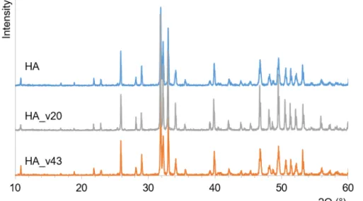
Powder X-ray diffraction (XRD) patterns were recorded on a D8 Advance diffractometer (Brücker) using Cu Kα radiation (λ=1.54 Å) at 40 kV and 40 mA passing through a Kβ Ni-filter (0.020 mm).
The diffracted intensity was recorded over a 10-60o diffraction angle range (2θ) with a scanning step of 0.02o on a position sensitive detector. All samples were ground with a mortar and pestle before
measurement. Crystalline phases were identified using ICDD (International Centre for Diffraction Data) diffraction patterns from pure phases.
A Fourier transform infrared spectrometer (FTIR) Nicolet Is50 (Thermo Fisher Scientific) was used to determine functional groups in the samples. Spectra were taken over a 400-4000 cm-1 range
at a resolution of 4 cm-1. A total of 64 scans were taken and then averaged. Powdered samples were
pressed into KBr tablets: a small amount (about 2 mg) of the sample was ground in a mortar, and combined with 200 mg of KBr (Uvasol®, Merck) by mixing (no grinding) to a homogeneous mixture. Three KBr pellets for each sample were prepared and measured. Deconvolution of the spectral range between 500 and 700 cm-1 was performed using MagicPlotStudent software and area
of the OH- absorption peak at 632 cm-1 and ν
4 PO43- absorption peaks were measured. The OH/PO4
ratio and the percentage of OH- ions were calculated for all samples.
The calcium concentration was measured by atomic absorption spectroscopy with a contra 300 (AnalytikJena) spectrophotometer at 442 nm wavelength. Phosphorous was determined as the phosphovanadomolybdate complex with a UV−1800 (Shimadzu) spectrophotometer using a wavelength of 460 cm-1. 6 replicate samples were used and averaged.
Results and Discussion
The starting HA was highly crystalline, and according to the ICDD database contained all the diffraction peaks corresponding to HA (Fig. 1). The bonding in the thermally modified HA was studied with FTIR spectroscopy. All functional groups represented by absorption peaks in the spectra were characteristic of HA (Fig. 2). Spectra showed a clear hydroxyl vibration peak, at both 631 cm-1 and 3573 cm-1; the second peak representing asymmetrical and symmetrical stretching
most intense absorption bands of HA situated at 1043 cm-1 and 1090 cm-1 corresponded to the
asymmetric stretching modes of PO43-, while the peaks at 962 and 472 cm-1 arose from the
symmetric stretching and bending mode of PO43-, respectively. The two very sharp and separated
peaks at 601 and 573 cm-1 represented the bending mode of the phosphate group.
The measured calcium content was 39.39±0.19%, while phosphorous content was 18.39±0.12%, which gave the Ca/P molar ratio of 1.66. Detailed previous analysis using thermal gravimetric analysis proved that the HA used in this study is fully hydroxylated and contains 100% hydroxyl ions [10].
Fig. 1. XRD pattern of the initial HA, and vacuum treated HA (HA_v20 and HA_v43)
Fig. 2. FTIR spectra of the initial HA, and vacuum treated HA (HA_v20 and HA_v43)
To produce OHA with the lowest amount of hydroxyl ions in the structure, namely, with the largest amount of OAp phase, a vacuum in high temperatures provided conditions to favor the removal of hydroxyl ions. As the pure OAp is believed to be unstable and according to the literature has not been obtained, it is important to choose the right heating conditions, so that only the diffusion of hydroxyl ions occurs while maintaining the apatite structure.
OHA with the lowest amount of OH- in the structure was prepared by heating at high temperature (1000 °C) for 20 or 43 hours in vacuum in a closed quartz system. For 20 h heated HA sample was labeled as HA_v20 and for 43 h heated HA sample was labeled as HA_v43. XRD results (Fig. 1) confirmed that the prepared OHA samples retained the apatite structure. FTIR results showed absorption bands characteristic only to apatite (Fig. 2). Traces of OH- absorption bands at 3570 and 632 cm-1 and PO
bands at 433, 475 and 945 cm-1 were characteristic of OAp [11, 12]. XRD for all rehydroxylated
samples showed no traces of decomposition. FTIR spectra also confirmed HA phase and indicated the presence of OAp.
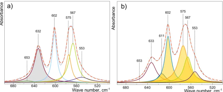
The 632 cm-1 OH- absorption band was chosen for calculating the OH- amount in the products as this band is more sensitive than the absorption band at 3571 cm-1 [13]. The OH- peak was
deconvulated from the phosphate peaks within the 500-700 cm-1 spectral area to enable the
measurement of the OH- and PO
43- peak areas.
Fig. 3. Deconvulated FTIR spectra of (a) HA, and (b) OHA (sample: HA_v43_reh400-1h). For better visualization, OH- peak area used for further calculations is colored in the figure a, and
PO43- peak area is colored in the figure b
The best fit for deconvolution used 7 Lorentzian curves for the OHA samples (Fig. 3). The absorption peak at 653 cm-1 was assumed to arise from the KBr during sample preparation that
could have been introduced from the adsorbed water. The peak at 653 cm-1 was not used for the OH -calculations.
Table 1. Calculated hydroxyl ion content in OHA samples, and information about the absorption peak positions in FTIR spectra range from 500 to 700 cm-1
Sample name Average (OH/ PO4)
[a.u.]
OH- amount
± stdev [%]
Wavenumber from FTIR spectra [cm-1]
OH- PO 4 3-Start HA 0.38 100 632 - 602 575 567 553 HA_v43_reh700 0.37 98 ± 1 632 - 602 575 567 553 HA_v43_reh400-1h 0.26 67 ± 3 633 611 602 575 567 553 HA_v43_reh400 0.22 59 ± 2 633 611 602 577 568 553 HA_v43_reh350 0.14 37 ± 2 634 612 602 580 568 553 HA_v20 0.05 12 ± 1 636 613 603 582 568 553 HA_v43 0.03 8 ± 1 637 614 604 583 568 553
The analysis of the deconvuluted peaks led to the following findings (Table 1):
i. The OH- peak shifts to a higher wavenumber at lower OH- concentrations, i.e., 632 cm-1 for
HA with 100% OH- and 637 cm-1 for the sample with the lowest amount of OH-.
ii. There is an additional PO43- peak at 611-614 cm-1 for OHA samples (this absorption line was
not observed for pure HA samples).
iii. There is a shift for another PO43- peak: 575 cm-1 for 100% HA shifts to 583 cm-1 for sample
A peak shift and broadening occurs with dehydroxylation. A large decrease in wavenumber from 633 cm1 occurs when larger cations take the place of calcium [14], but this work shows that the
absence of OH- ions gives a minor increase in wavenumber. An increase in peak width has been
noted when OH- is absent - this could in part be related to an increase in disorder, in agreement with observed FTIR peak broadening in case of low-crystalline apatites [15]. Since both of these changes are marginal, deconvolution is essential to detect these changes. If previously changes in the OH
-content were approximated from the 633 cm-1 peak intensity, this work shows a slight increase in
wavenumber and an increase in peak width as additional signs to confirm the reduction in OH -content.
Conclusions
Heating well crystalline hydroxyapatite in a closed quartz system under high vacuum at 1000 °C for 43 h produced 90% oxyapatite sample that was stable in air. Oxyhydroxyapatite can be made by rehydroxylating oxyapatite in a humid atmosphere trough varying heating temperature and dwelling time. More detailed analysis of FTIR spectra in the 500-700 cm-1 range showed an increase in the
wavenumber of the hydroxyl ion absorption line at 632 cm-1 and phosphate line at 575 cm-1 with
increasing the level of dehydroxylation.
Acknowledgements
We would like to thank A. Krumina from the Institute of Inorganic Chemistry, Riga Technical University for performing part of the XRD measurements. This work has been partly supported by the European Council Seventh Framework Program M-ERA.NET project “Implants signal to bone for bone growth and attachment” Nr. ESRTD/2017/4, and is a topic of investigation in the IRSES Project Nr. 612691 “Refined Step”.
References
[1] Wang T, Dorner-Reisel A, Müller E. Thermogravimetric and thermokinetic investigation of the dehydroxylation of a hydroxyapatite powder. Journal of the European Ceramic Society 2004;24:693-8.
[2] White AA, Kinloch IA, Windle AH, Best SM. Optimization of the sintering atmosphere for high-density hydroxyapatite-carbon nanotube composites. Journal of the Royal Society Interface 2010;7:S529-S39.
[3] Liao CJ, Lin FH, Chen KS, Sun JS. Thermal decomposition and reconstitution of hydroxyapatite in air atmosphere. Biomaterials 1999;20:1807-13.
[4] Park HC, Baek DJ, Park YM, Yoon SY, Stevens R. Thermal stability of hydroxyapatite whiskers derived from the hydrolysis of α-TCP. Journal of Materials Science 2004;39:2531-4. [5] Cihlář J, Buchal A, Trunec M. Kinetics of thermal decomposition of hydroxyapatite
bioceramics. Journal of Materials Science 1999;34:6121-31.
[6] Gross KA, Berndt CC, Stephens P, Dinnebier R. Oxyapatite in hydroxyapatite coatings. Journal of Materials Science 1998;33:3985-91.
[7] Tonsuaadu K, Gross KA, Pluduma L, Veiderma M. A review on the thermal stability of calcium apatites. Journal of Thermal Analysis and Calorimetry 2012;110:647-59.
[8] Yang C-W, Lui T-S. Kinetics of hydrothermal crystallization under saturated steam pressure and the self-healing effect by nanocrystallite for hydroxyapatite coatings. Acta Biomaterialia 2009;5:2728-37.
[9] Baxter FR, Bowen CR, Turner IG, Dent ACE. Electrically active bioceramics: A review of interfacial responses. Annals of Biomedical Engineering 2010;38:2079-92.
[10] Pluduma L. Hydroxyl ion quantification in hydroxyapatite and the effect on the biological response. PhD Thesis. Riga: Riga Technical University; 2017.
[11] Elliott JC. Structure and chemistry of the apatites and other calcium orthophosphates. Elsevier Inc, Amsterdam 1994:387.
[12] J.C.Trombe. Contribution a l'etude de la decomposition et de la reactivite de certaines apatites hydroxylees et carbonatees. Annales des Chimie 1973;8:251-69.
[13] Rapacz-Kmita A, Paluszkiewicz C, Slosarczyk A, Paszkiewicz Z. FTIR and XRD investigations on the thermal stability of hydroxyapatite during hot pressing and pressureless sintering processes. Journal of Molecular Structure 2005;744:653-6.
[14] Engel G, Klee WE. INFRARED-SPECTRA OF HYDROXYL IONS IN VARIOUS APATITES. Journal of Solid State Chemistry 1972;5:28-&.
[15] Rey C, Combes C, Drouet C, Lebugle A, Sfihi H, Barroug A. Nanocrystalline apatites in biological systems: Characterisation, structure and properties. Materialwissenschaft und Werkstofftechnik 2007;38:996-1002.