HAL Id: inserm-02296607
https://www.hal.inserm.fr/inserm-02296607
Submitted on 25 Sep 2019HAL is a multi-disciplinary open access archive for the deposit and dissemination of sci-entific research documents, whether they are pub-lished or not. The documents may come from teaching and research institutions in France or abroad, or from public or private research centers.
L’archive ouverte pluridisciplinaire HAL, est destinée au dépôt et à la diffusion de documents scientifiques de niveau recherche, publiés ou non, émanant des établissements d’enseignement et de recherche français ou étrangers, des laboratoires publics ou privés.
Impact of the acute local inhibition of soluble epoxide
hydrolase on diabetic skin microcirculatory dysfunction
Yann Savina, Thomas Duflot, Frédéric Bounoure, Sylvain Kotzki, Pierre-Alain
Thiébaut, Pierre-Alex Serreau, Mohamed Skiba, Jean-Michel Picquenot,
Marie Cornic, Christophe Morisseau, et al.
To cite this version:
Yann Savina, Thomas Duflot, Frédéric Bounoure, Sylvain Kotzki, Pierre-Alain Thiébaut, et al.. Im-pact of the acute local inhibition of soluble epoxide hydrolase on diabetic skin microcirculatory dys-function. Diabetes and Vascular Disease Research, SAGE Publications, 2019, Epub ahead of print. �10.1177/1479164119860215�. �inserm-02296607�
https://doi.org/10.1177/1479164119860215
Diabetes & Vascular Disease Research 1 –7
© The Author(s) 2019 Article reuse guidelines: sagepub.com/journals-permissions DOI: 10.1177/1479164119860215 journals.sagepub.com/home/dvr
Background
Diabetic foot ulcers (DFUs) are a common and serious complication of diabetes mellitus and are associated with major morbidity. Indeed, diabetes is the primary cause of non-traumatic lower limb amputation.1 The skin’s
micro-circulation, by maintaining perfusion and delivering
oxygen and nutrients, plays a key role in tissue survival. Skin microvascular dysfunction occurs early in the patho-physiology of diabetes and contributes to poor wound heal-ing and the development of foot complications in diabetic patients.2–4 Endothelial dysfunction, as well as sensory and
Impact of the acute local inhibition
of soluble epoxide hydrolase on diabetic
skin microcirculatory dysfunction
Yann Savina
1,2, Thomas Duflot
3,4,5, Frederic Bounoure
6,7,
Sylvain Kotzki
1,2, Pierre-Alain Thiebaut
8, Pierre-Alex Serreau
3,6,
Mohamed Skiba
6,7, Jean-Michel Picquenot
8, Marie Cornic
8,
Christophe Morisseau
9, Bruce Hammock
9, Laurent Imbert
3,4,
Jean-Luc Cracowski
1,2, Vincent Richard
3,5, Matthieu Roustit
1,2and Jeremy Bellien
3,5Abstract
The impact of the local inhibition of soluble epoxide hydrolase, which metabolizes vasodilator and anti-inflammatory epoxyeicosanoids, on diabetic skin microvascular dysfunction was assessed. In diabetic db/db mice, basal skin blood flow assessed using laser Doppler imaging was similar to that of control mice, but thermal hyperemia was markedly reduced. At 2 h after the topical administration of an aqueous gel containing the soluble epoxide hydrolase inhibitor trans-4-[4-(3-adamantan-1-yl-ureido)-cyclohexyloxy]-benzoic acid (t-AUCB: 400 mg/L), the peak concentration of t-AUCB was detected in the skin of diabetic mice, which quickly decreased thereafter. In parallel, 2 h after application of t-AUCB treatment, thermal hyperemia was increased compared to the control gel. Quantification of t-AUCB in plasma of treated animals showed no or low systemic diffusion. Furthermore, haematoxylin and eosin histological staining of skin biopsies showed that skin integrity was preserved in t-AUCB-treated mice. Finally, for pig ear skin, a surrogate for human skin, using Franz diffusion cells, we observed a continuous diffusion of t-AUCB from 2 h after application to beyond 24 h. A single topical administration of a soluble epoxide hydrolase inhibitor improves microcirculatory function in the skin of db/db mice and might represent a new therapeutic approach for preventing the development of skin complications in diabetic patients.
Keywords
Diabetes, skin microvascular dysfunction, soluble epoxide hydrolase, topical form
1Université Grenoble Alpes, HP2 UMR INSERM 1042, Grenoble, France 2 CHU Grenoble Alpes, Pôle Recherche, INSERM CIC1406, Grenoble,
France
3 Department of Pharmacology, Rouen University Hospital, Rouen, France 4 Laboratory of Pharmacokinetics, Toxicology and Pharmacogenetics,
Rouen University Hospital, Rouen, France
5 Normandie University, UNIROUEN, INSERM U1096, FHU
REMOD-VHF, Rouen, France
6 Department of Galenic, Normandy University, UNIROUEN, Rouen,
France
Original Article
7 INSERM U1239 Normandy University, UNIROUEN, Rouen,
France
8Department of Pathology, Henri Becquerel Center, Rouen, France 9 Department of Entomology and Cancer Center, University of
California, Davis, CA, USA
Corresponding author:
Jeremy Bellien, Department of Pharmacology, Rouen University Hospital, 76031 Rouen, France.
2 Diabetes & Vascular Disease Research 00(0) autonomic neuropathies, is thought to contribute to a
reduc-tion in the funcreduc-tional capacity of the microvasculature,2–4
and topical treatments targeting this microvascular dys-function may help to improve wound healing while mini-mizing potential systemic side effects.5
Increasing evidence suggests that alterations in the endothelium-derived epoxyeicosatrienoic acids (EETs) pathway are involved in the pathophysiology of the endothelial dysfunction associated with type 2 diabetes.6,7
One interesting approach would thus be to increase the bioavailability of EETs, which are formed by the action of cytochrome P450 and display powerful vasodilating, anti-inflammatory and angiogenic properties.6,7 In vivo, EETs
are rapidly converted to the less active dihydroxyeicosa-trienoic acids by soluble epoxide hydrolase (sEH), which is the target of a new class of pharmacological inhibitors.8,9
Interestingly, exogenous EET administration as well as genetic or pharmacological inhibition of sEH has been shown to accelerate wound epithelialization and neovascu-larization in ob/ob mice and in the hairless mouse ear wound model.8–10 However, to date, no study has
evalu-ated their impact on skin microcirculatory function. Since in animals the systemic administration of sEH inhibitors has been successfully used to improve endothelial function of aorta and coronary arteries,11,12 we hypothesized that
local cutaneous sEH inhibition may improve endothelium-dependent microvascular reactivity.
In this context, the aim of this study was to assess the effect of a topical formulation containing a sEH inhibitor on skin microcirculation in diabetic mice.
Methods
Animals and treatments
The protocol was approved by the local institutional review committee (decision number: C 38 516 10 006, n°2017011312598602-V5#8531) and conducted in accord-ance with the National Institutes of Health (NIH) Guide for the Care and Use of Laboratory Animals. Nine-week-old male wild-type C57BL/6J and db/db [BKS(D)-Leprdb/ JOrlRj] mice, a genetic model of type 2 diabetes, were acquired from Janvier Labs (Le Genest-Saint-Isle, France). These mice were allowed to acclimate to the photoperiod (12 h of light/12 h of darkness) and temperature conditions (22 ± 1°C) for 1 week prior to the start of the study. A 2-h topical administration (20 µL) of a newly developed gel-like, aqueous pharmaceutical preparation containing the sEH inhibitor trans-4-[4-(3-adamantan-1-yl-ureido)-cyclohexyloxy]-benzoic acid (t-AUCB: 400 mg/L) dis-solved in dimethyl sulfoxide (DMSO) or a vehicle control gel was applied to the dorsal skin of db/db mice, depilated 2 days before the experiments. Assessment of microvascu-lar function, skin biopsies (50 mm2) and intra-cardiac
blood sampling was performed at 2 and 24 h after gel
application. Animals were anaesthetized with isoflurane (induction at 3% during 3 min and then maintained at 2%) and placed on a heated pad to maintain stable core body temperature (37.5 ± 0.5°C).
Local and systemic quantification of t-AUCB
Plasma and skin levels of t-AUCB were quantified by liq-uid chromatography coupled to tandem mass spectrome-try (LC-MS/MS).13 Briefly, skin tissues were mixed with
1 mL of methanol–water (50:50 v/v) and ultrasonicated for 10 min, or 100 µL of plasma were mixed with 300 µL of methanol, allowing protein precipitation. Then, skin and plasma samples were thoroughly vortexed for 10 s and centrifuged at 16,100g for 5 min. The resulting super-natants were collected and analysed by LC-MS/MS. Chromatographic separation was performed on a Kinetex C18 column (2.6-μm particle size, 50-mm length × 3-mm inner diameter). The autosampler temperature was set at 8°C, the column oven at 30°C, the injected volume was 20 μL and the flow rate was 400 μL/min. The mobile phase was 0.2% formic acid in methanol (solvent A) and 2-mM ammonium formate with 0.2% formic acid in water (solvent B). The elution started with 95% B (0–2 min), 95–5% B (2–5 min), 5% B (5–10 min), 5–95% B (10– 11 min) and 95% B (11–12 min). The following multiple reaction monitoring (MRM) transitions, m/z 412.9 to m/z 135.1 and m/z 412.9 to m/z 93.0 in positive ion mode, were used to detect t-AUCB (quantification and confir-mation transitions, respectively). Skin levels were nor-malized to tissue weight.
Assessment of skin microvascular function
Skin microvascular reactivity to local heating was used as an index of endothelium-dependent function.14 Dorsal skin
blood flow was measured by laser Doppler imaging (LDI; PeriScan PIM, Perimed, Järfälla, Sweden) over 10 min before heating (baseline flow). The skin was then heated at 41°C for 20 min using a 0.5-cm2 heating probe regulated
with an internal thermometer. Skin blood flow was then recorded during the following 15 min.
Data were digitized, stored on a computer and analysed offline with signal processing software (PimSoft v1.5.4.8078, Perimed). Baseline and peak hyperemia were expressed as arbitrary perfusion units (APU), averaged over 3 min immediately before and 1 min immediately after heating, respectively. Thermal hyperemia was subse-quently calculated as the difference between peak hypere-mia and baseline skin blood flow.
Skin integrity
Skin biopsies were carefully sampled and immediately fixed in a 4% formalin solution for 24 h. After proper
fixation, tissue samples were embedded in paraffin and stored at room temperature until analysis, when 4-µm sec-tions were deparaffinized and stained with standard haematoxylin–eosin (H&E) stains. Slides were analysed by an experienced pathologist (J.-M.P.).
Transdermal passage of t-AUCB across
pig ear skin
The percutaneous absorption of t-AUCB was studied using Franz diffusion cells.15 The skin from pig’s ears was
cho-sen for the experiments as it is very similar to that of human skin and closer than mouse skin.16,17 The Franz’s
cells had a contact area of 2 cm2, and the experiments were
conducted at 32°C. The donor compartment was filled with 20 µL of the t-AUCB-containing gel (400 mg/L). The receptor compartment contained 4.5 mL of phosphate-buffered saline (PBS) and was under magnetic stirring. Samples from the receptor compartment were collected at different time points over 24 h to determine the percutane-ous flow of t-AUCB. The t-AUCB quantification was per-formed by LC-MS/MS.
Statistical analysis
All values are expressed as mean ± standard error of mean (SEM). The Shapiro–Wilk test was used to assess normality. Analyses of the differences between diabetic and control mice for basal skin blood flow and thermal hyperemia were performed using an unpaired t-test or the nonparametric Mann–Whitney rank-sum test. Analyses of the variation in basal skin blood flow and thermal hyperemia induced by the t-AUCB-containing gel were performed using mixed effects models with time as fixed effect and the mouse as a random effect followed, in case of signi ficance, by Bonferroni post hoc
tests to compare the baseline value to other time points after application. Analyses of the differences in the effect of the t-AUCB-containing gel and the vehicle gel on basal skin blood flow and thermal hyperemia were performed by repeated measures analysis of variance (ANOVA), and we assessed the influence of the group, of time and the time × group inter action. Statistical anal-yses were performed with NCSS software (version 07.1.14). A two-sided p < 0.05 was considered as statis-tically significant.
Results
At untreated skin sites, cutaneous blood flow was slightly, but significantly, lower in db/db mice compared to control mice (Figure 1(a)). In contrast, there was a more marked reduction in thermal hyperemia in db/db mice compared to controls (Figure 1(b)), showing the presence of diabetic skin microcirculatory dysfunction.
We carefully compared the effect of the t-AUCB- containing gel to that of a vehicle control gel in db/db mice. Both gels were applied on the same animal, with a minimal distance of 1 cm between the two application sites. Both gels increased basal skin blood flow after a 2-h long application, but with no significant difference between groups (Figure 2(a)). However, the t-AUCB- containing gel significantly increased thermal hyperemia compared to the vehicle control gel (Figure 2(b)).
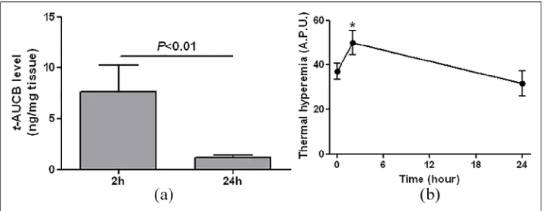
After the 2-h gel application, t-AUCB was detectable in skin biopsies, and these levels had drastically decreased 24 h after application, demonstrating transdermal permea-tion of t-AUCB across db/db mouse skin (Figure 3(a)). Consistently, thermal hyperemia returned to baseline val-ues 24 h after gel application (Figure 3(b)).
Analysis of plasma samples showed no systemic diffu-sion of t-AUCB, assessed at 2 and 24 h after application of the t-AUCB-containing gel, except for one animal (Table 1).
Figure 1. (a) Basal skin blood flow and (b) thermal hyperemia measured by laser Doppler imaging in control (n = 7) and db/db mice
(n = 31).
4 Diabetes & Vascular Disease Research 00(0)
In addition, no significant inflammatory infiltration was observed in mouse skin at either 2 or 24 h after gel applica-tion (Figure 4).
Finally, at the same dosage, a continuous diffusion of t-AUCB was observed across pig ear skin from 2 h up to 24 h after application (Figure 5).
Figure 2. (a) Basal skin blood flow and (b) thermal hyperemia measured by laser Doppler imaging before and after a 2-hour topical
application of the t-AUCB-containing gel (20 µL at 400 mg/L) and the vehicle control gel on the dorsal skin of db/db mice (n = 13).
APU: arbitrary perfusion unit.
*p < 0.05 versus before topical application; †p < 0.05 versus vehicle control gel.
Figure 3. (a) Skin levels of t-AUCB, quantified by liquid chromatography coupled to tandem mass spectrometry, 2 (n = 5) and
24 hours (n = 6) after topical application of the t-AUCB-containing gel (20 µL at 400 mg/L) on the dorsal skin of db/db mice. (b) Thermal hyperemia measured by laser Doppler imaging before (n = 19), 2 (n = 13) and 24 hours (n = 6) after the topical application of the t-AUCB-containing gel on the dorsal skin of db/db mice.
APU: arbitrary perfusion unit.
*p < 0.05 versus before topical application.
Figure 4. Representative images of haematoxylin and eosin staining of mouse skin 8 and 24 hours after topical application of
Discussion
The major finding of this preliminary study is that sEH inhibition by t-AUCB using a topical formulation increases thermal hyperemia in the skin, an index of endothelium-dependent microvascular reactivity, in a murine model of diabetes. As microvascular endothelial dysfunction is a hallmark of diabetes, and considering the role of impaired cutaneous microcirculation in poor wound healing in dia-betics, such a strategy may be an interesting therapeutic approach for DFUs.
EETs are endothelium-derived vasodilating factors with powerful anti-inflammatory and pro-angiogenic properties that could be useful in the treatment of the car-diovascular complications of type 2 diabetes.6,7 Despite
increasing evidence suggesting a possible role for EETs in diabetes-related endothelial dysfunction, no study had pre-viously focused on diabetic skin microvascular dysfunc-tion. The use of thermal hyperemia as a reactivity test was motivated by the involvement of EETs, together with NO, in the response to local heating in humans.18
We observed a reduction in basal skin blood flow in diabetic db/db mice compared to wild-type mice, which is probably mainly related to lower vascular density.19 In
addition, although no data were available in animal models of diabetes when we designed the study, we demonstrated altered microvascular reactivity to thermal hyperemia in diabetic mice. Thus, as shown in humans,20 measuring
blood flow response to a standardized local heat stimulus provides a suitable model for the study of the skin micro-vascular dysfunction associated with diabetes in mice.
In this context, we tested the impact of a topical for-mulation containing t-AUCB, an inhibitor of EET degra-dation by sEH,11,13 on microvascular dysfunction in the
skin of db/db mice. Quantification of t-AUCB in skin biopsies revealed significant transdermal permeation of the molecule 2 h after gel application, associated with increased basal skin blood flow, compared to baseline, and thermal hyperemia. However, the vehicle control gel increased basal skin blood flow in a similar manner. This result supports previous data showing a direct vasodilat-ing effect of the vehicle DMSO.21 In fact, the topical
administration of DMSO has even been proposed for use in humans to treat the skin complications of systemic scleroderma, which is also characterized by microvascu-lar dysfunction and a risk of ulcers, but the results of ran-domized controlled trials were disappointing.22,23 While
the DMSO vehicle had no effect on reactivity, in contrast, the t-AUCB-containing gel improved thermal hyperemia compared to the vehicle control gel, demonstrating an improvement in skin microvascular reactivity. This result shows that, as previously demonstrated in coronary and peripheral arteries,11,12 sEH plays a major role in the
vas-cular dysfunction of the skin associated with type 2 dia-betes. Although the objective of this preliminary study was not to assess the effect of sEH inhibition on wound healing, it provides a first proof of principle in an animal model with prolonged wound healing.24
Importantly for potential human use, histological ana-lysis revealed no signs of skin toxicity with the t-AUCB-containing gel. In addition, quantification of t-AUCB in plasma from exposed animals showed low systemic diffu-sion of the drug, except in only one animal out of five. This may be important because, although the first results obtained in the initial phases of clinical development sug-gest that sEH inhibitors were safe,25,26 some data show that
increasing EET bioavailability might be associated with adverse effects and in particular might potentiate tumour development.6,7,27,28 Moreover, because mouse skin is thin
and the animals were shaved for the experiments, which could have led to an underestimation of the time needed for the transdermal passage of t-AUCB compared to that in humans, the pharmacokinetic study was performed on
Table 1. Quantification of t-AUCB in plasma.
2 hours 24 hours
Below LOQ (2.4 nM) 4 (80%) 4 (100%)
Above LOQ (2.4 nM) 1 (20%) 0 (0%)
t-AUCB: trans-4-[4-(3-adamantan-1-yl-ureido)-cyclohexyloxy]-benzoic acid; LOQ: limit of quantification. Data are represented as n (%).
Figure 5. Evolution of t-AUCB level, quantified by liquid
chromatography coupled to tandem mass spectrometry, in the receptor compartment of Franz cells from 0 to 24 hours after gel application (20 µL at 400 mg/L) to pig ear skin (n = 3 per time point).
6 Diabetes & Vascular Disease Research 00(0) isolated, more human-like, pig ear skin. A progressive and
continuous diffusion of t-AUCB was observed, suggesting that topical sEH inhibitors could be particularly useful in the prevention and/or treatment of skin complications in patients with type 2 diabetes.
Conclusion
These results show that the acute topical administration of a sEH inhibitor improves skin microvascular reactivity in a model of type 2 diabetes. The absence of skin toxicity, the limited systemic diffusion and the demonstration of the progressive passage of the sEH inhibitor across a more human-like animal source of skin support the use of this therapeutic strategy in patients with type 2 diabetes, with potential for the prevention of skin complications and in particular DFUs development. The next steps will be to show, using repeated dosing experiments, that this improvement in skin microcirculatory function translates into effective prevention of diabetes-associated dermato-logical complications and to confirm the limited systemic diffusion of the molecule so as to avoid potential long-term side effects.
Acknowledgements
The authors thank Mr Tony Pereira (Laboratory of Pharma-cokinetics, Toxicology and Pharmacogenetics, Rouen University Hospital, France) for his technical assistance in the quantification of t-AUCB and Dr Alison Foote (Grenoble Alpes University Hospital, France) for editing the manuscript.
Declaration of conflicting interests
The author(s) declared no potential conflicts of interest with respect to the research, authorship and/or publication of this article.
Funding
The author(s) disclosed receipt of the following financial support for the research, authorship and/or publication of this article: The study was co-supported by grants from the Fondation de France (2011-20459), the French National Research Agency (ANR-16-CE17-0012) and the National Institute of Health (NIEHS/R01 ES002710).
ORCID iDs
Mohamed Skiba https://orcid.org/0000-0002-0360-7743 Jeremy Bellien https://orcid.org/0000-0002-0383-2342
References
1. Moxey PW, Hofman D, Hinchliffe RJ, et al. Epidemiological study of lower limb amputation in England between 2003 and 2008. Br J Surg 2010; 97: 1348–1353.
2. Dinh T, Tecilazich F, Kafanas A, et al. Mechanisms involved in the development and healing of diabetic foot ulceration. Diabetes 2012; 61: 2937–2947.
3. Chao CY and Cheing GL. Microvascular dysfunction in dia-betic foot disease and ulceration. Diabetes Metab Res Rev 2009; 25: 604–614.
4. Jhamb S, Vangaveti VN and Malabu UH. Genetic and molecular basis of diabetic foot ulcers: clinical review. J
Tissue Viability 2016; 25: 229–236.
5. Valacchi G, Zanardi I, Sticozzi C, et al. Emerging topics in cutaneous wound repair. Ann N Y Acad Sci 2012; 1259: 136–144.
6. Lorthioir A, Guerrot D, Joannides R, et al. Diabetic car-diovascular disease – soluble epoxide hydrolase as a tar-get. Cardiovasc Hematol Agents Med Chem 2012; 10: 212–222.
7. Bellien J, Joannides R, Richard V, et al. Modulation of cytochrome-derived epoxyeicosatrienoic acids pathway: a promising pharmacological approach to prevent endothelial dysfunction in cardiovascular diseases? Pharmacol Ther 2011; 131: 1–17.
8. Zhao H, Chen J, Chai J, et al. Cytochrome P450 (CYP) epoxygenases as potential targets in the management of impaired diabetic wound healing. Lab Invest 2017; 97: 782– 791.
9. Sander AL, Jakob H, Sommer K, et al. Cytochrome P450-derived epoxyeicosatrienoic acids accelerate wound epithe-lialization and neovascularization in the hairless mouse ear wound model. Langenbecks Arch Surg 2011; 396: 1245– 1253.
10. Sander AL, Sommer K, Neumayer T, et al. Soluble epoxide hydrolase disruption as therapeutic target for wound heal-ing. J Surg Res 2013; 182: 362–367.
11. Roche C, Besnier M, Cassel R, et al. Soluble epoxide hydro-lase inhibition improves coronary endothelial function and prevents the development of cardiac alterations in obese insulin-resistant mice. Am J Physiol Heart Circ Physiol 2015; 308: H1020–H1029.
12. Zhang LN, Vincelette J, Chen D, et al. Inhibition of solu-ble epoxide hydrolase attenuates endothelial dysfunction in animal models of diabetes, obesity and hypertension. Eur J
Pharmacol 2011; 654: 68–74.
13. Liu JY, Tsai HJ, Hwang SH, et al. Pharmacokinetic optimi-zation of four soluble epoxide hydrolase inhibitors for use in a murine model of inflammation. Br J Pharmacol 2009; 156: 284–296.
14. Roustit M and Cracowski JL. Assessment of endothelial and neurovascular function in human skin microcirculation.
Trends Pharmacol Sci 2013; 34: 373–384.
15. Godin B and Touitou E. Transdermal skin delivery: predic-tions for humans from in vivo, ex vivo and animal models.
Adv Drug Deliv Rev 2007; 59: 1152–1161.
16. Herkenne C, Naik A, Kalia YN, et al. Pig ear skin ex vivo as a model for in vivo dermatopharmacokinetic studies in man.
Pharm Res 2006; 23: 1850–1856.
17. Abd E, Yousef SA, Pastore MN, et al. Skin models for the testing of transdermal drugs. Clin Pharmacol 2016; 8: 163– 176.
18. Brunt VE and Minson CT. KCa channels and epoxyeicosa-trienoic acids: major contributors to thermal hyperaemia in human skin. J Physiol 2012; 590: 3523–3534.
19. Schaefer C, Biermann T, Schroeder M, et al. Early micro-vascular complications of prediabetes in mice with impaired
glucose tolerance and dyslipidemia. Acta Diabetol 2010; 47: 19–27.
20. Fuchs D, Dupon PP, Schaap LA, et al. The association between diabetes and dermal microvascular dysfunction non-invasively assessed by laser Doppler with local ther-mal hyperemia: a systematic review with meta-analysis.
Cardiovasc Diabetol 2017; 16: 11.
21. Kaneda T, Sasaki N, Urakawa N, et al. Endothelium-dependent and -inEndothelium-dependent vasodilator effects of dimethyl sulfoxide in rat aorta. Pharmacology 2016; 97: 171–176. 22. Scherbel AL. The effect of percutaneous dimethyl sulfoxide
on cutaneous manifestations of systemic sclerosis. Ann N Y
Acad Sci 1983; 411: 120–130.
23. Williams HJ, Furst DE, Dahl SL, et al. Double-blind, mul-ticenter controlled trial comparing topical dimethyl sul-foxide and normal saline for treatment of hand ulcers in patients with systemic sclerosis. Arthritis Rheum 1985; 28: 308–314.
24. Sullivan SR, Underwood RA, Gibran NS, et al. Validation of a model for the study of multiple wounds in the diabetic mouse (db/db). Plast Reconstr Surg 2004; 113: 953–960. 25. Chen D, Whitcomb R, MacIntyre E, et al. Pharmacokinetics
and pharmacodynamics of AR9281, an inhibitor of soluble epoxide hydrolase, in single- and multiple-dose studies in healthy human subjects. J Clin Pharmacol 2012; 52: 319–328. 26. Lazaar AL, Yang L, Boardley RL, et al. Pharmaco-kinetics, pharmacodynamics and adverse event profile of GSK2256294, a novel soluble epoxide hydrolase inhibitor.
Br J Clin Pharmacol 2016; 81: 971–979.
27. Panigrahy D, Edin ML, Lee CR, et al. Epoxyeicosanoids stimulate multiorgan metastasis and tumor dormancy escape in mice. J Clin Invest 2012; 122: 178–191.
28. Sausville LN, Gangadhariah M, Chiusa M, et al. The cytochrome P450 slow metabolizers CYP2C9*2 and CYP2C9*3 directly regulate tumorigenesis via reduced epoxyeicosatrienoic acid production. Cancer Res 2018; 78: 4865–4877.
![La maladie des taches brunes du niébé [vigna unguiculata (l) walp.] au Burkina Faso: connaissance des agents pathogènes impliques et développement de méthodes de lutte](data:image/gif;base64,R0lGODlhAQABAIAAAP///wAAACH5BAEAAAAALAAAAAABAAEAAAICRAEAOw==)