HAL Id: inserm-02440386
https://www.hal.inserm.fr/inserm-02440386
Submitted on 15 Jan 2020
HAL is a multi-disciplinary open access
archive for the deposit and dissemination of
sci-entific research documents, whether they are
pub-lished or not. The documents may come from
teaching and research institutions in France or
abroad, or from public or private research centers.
L’archive ouverte pluridisciplinaire HAL, est
destinée au dépôt et à la diffusion de documents
scientifiques de niveau recherche, publiés ou non,
émanant des établissements d’enseignement et de
recherche français ou étrangers, des laboratoires
publics ou privés.
Characterization of a Toxoplasma effector uncovers an
alternative GSK3/β-catenin-regulatory pathway of
inflammation
Huan He, Marie-Pierre Brenier-Pinchart, Laurence Braun, Alexandra Kraut,
Bastien Touquet, Yohann Coute, Isabelle Tardieux, Mohamed-Ali Hakimi,
Alexandre Bougdour
To cite this version:
Huan He, Marie-Pierre Brenier-Pinchart, Laurence Braun, Alexandra Kraut, Bastien Touquet, et
al.. Characterization of a Toxoplasma effector uncovers an alternative GSK3/β-catenin-regulatory
pathway of inflammation. eLife, eLife Sciences Publication, 2018, 7, pp.e39887. �10.7554/eLife.39887�.
�inserm-02440386�
*For correspondence: mohamed-ali.hakimi@inserm.fr (M-AH);
alexandre.bougdour@univ-grenoble-alpes.fr (AB) Competing interests: The authors declare that no competing interests exist. Funding:See page 23 Received: 06 July 2018 Accepted: 14 October 2018 Published: 15 October 2018 Reviewing editor: Dominique Soldati-Favre, University of Geneva, Switzerland
Copyright He et al. This article is distributed under the terms of theCreative Commons Attribution License,which permits unrestricted use and redistribution provided that the original author and source are credited.
Characterization of a Toxoplasma effector
uncovers an alternative
GSK3/b-catenin-regulatory pathway of inflammation
Huan He
1, Marie-Pierre Brenier-Pinchart
1, Laurence Braun
1, Alexandra Kraut
2,
Bastien Touquet
3, Yohann Coute´
2, Isabelle Tardieux
3, Mohamed-Ali Hakimi
1*,
Alexandre Bougdour
1*
1
Team Host-pathogen interactions & immunity to infection, University of Grenoble
Alpes, Inserm, CNRS, IAB, Grenoble, France;
2University of Grenoble Alpes, CEA,
Inserm, BIG-BGE, Grenoble, France;
3Team Membrane and Cell Dynamics of Host
Parasite Interactions, University of Grenoble Alpes, Inserm, CNRS, IAB, Grenoble,
France
Abstract
The intracellular parasite Toxoplasma gondii, hijacks evolutionarily conserved hostprocesses by delivering effector proteins into the host cell that shift gene expression in a timely fashion. We identified a parasite dense granule protein as GRA18 that once released in the host cell cytoplasm forms versatile complexes with regulatory elements of the b-catenin destruction complex. By interacting with GSK3/PP2A-B56, GRA18 drives b-catenin up-regulation and the downstream effects on host cell gene expression. In the context of macrophages infection, GRA18 induces the expression of a specific set of genes commonly associated with an anti-inflammatory response that includes those encoding chemokines CCL17 and CCL22. Overall, this study adds another original strategy by which T. gondii tachyzoites reshuffle the host cell interactome through a GSK3/b-catenin axis to selectively reprogram immune gene expression.
DOI: https://doi.org/10.7554/eLife.39887.001
Introduction
Toxoplasma gondii is the causative agent of toxoplasmosis, a widespread parasitic disease in humans which has been recognized as leading cause of deaths attributed to foodborne illness in the
United States (Scallan et al., 2015). Severe to life-threatening Toxoplasmosis mainly occur in
immu-nocompromised people, with acquired immunodeficiency syndrome or under chemo- and graft
rejection therapies (Montoya and Liesenfeld, 2004). In addition, outcomes of congenital
toxoplas-mosis significantly vary with the timing of infection from recurrent eye diseases to adverse motor or
neurologic impairments that can cause stillbirth (Halonen and Weiss, 2013). T. gondii belongs to
the protozoan phylum Apicomplexa and as most Apicomplexa species, develops and proliferates inside a surrogate host cell. Remarkably, T. gondii’s host range is exceptionally broad since it can
infect virtually all nucleated cells of mammals, marsupials and birds (Dubey, 2009).
To achieve intracellular lifestyle, the invasive tachyzoite stage of T. gondii triggers the formation of a unique membrane-bound compartment called the Parasitophorous Vacuole (PV). The PV is shaped as a niche kept hidden from harmful endocytic processing thereby enabling tachyzoite
growth and multiplication (Jones et al., 1972; Mordue et al., 1999). In the last decade, several
studies have highlighted the contribution of parasite effectors delivered in the host cell at the very
onset of-or post-invasion to promote folding and maturation of a functional PV (
Delorme-Walker et al., 2012;Hakimi et al., 2017). Effectors are released from two specialized sets of
and the spherical Dense Granules (DG) in which GRA15, GRA16, GRA24, TgIST, GRA6, and GRA25
proteins are stored (Hakimi et al., 2017). Once delivered in the host cell, these effectors either
remain exposed at the cytoplasmic side of the PV Membrane (PVM) or cross the PVM and travel in the cytoplasm. Interestingly, members of the second class (i.e. GRA16, GRA24, and TgIST) have all been assigned the host nucleus as final destination where they target distinct host regulators to modulate the expression of specific sets of genes.
The comprehensive analysis of how the cohort of effectors interplay during infection at cellular and host levels remains a major challenge which implies identifying and functionally characterizing the T. gondii effector repertoire in given cellular and host contexts. With this concern, we searched for new effectors and characterized GRA18 as the first DG protein that strictly remains in the host cell cytoplasm once delivered from the PV-enclosed tachyzoite. We provide evidence that the exported GRA18 is part of multi-partner - that is, versatile - complexes, specifically formed with com-ponents of the b-catenin destruction complex, which includes b-catenin, GSK3a/b, and the PR56/B’-containing PP2A holoenzyme and as such prevents the continual elimination of b-catenin. Accord-ingly, in presence of GRA18, cytoplasmic b-catenin travels to and accumulates in the host cell nucleus where it activates otherwise repressed target genes. Nuclear b-catenin is known as the main effector of the canonical Wnt signaling pathway, acting as a coactivator of the Lymphoid Enhancer-binding Factor (LEF) or T Cell Factor (TCF) proteins to drive Wnt-specific transcriptional programs
depending on cell lineages (Cadigan and Waterman, 2012;Schuijers et al., 2014). In murine
mac-rophages, we showed that GRA18 induces in a b-catenin-dependent fashion the expression of a par-ticular set of chemokines, that is, Ccl17, Ccl22, and Ccl24, notoriously known associated with
anti-inflammatory effects (Biswas and Mantovani, 2010; Mantovani et al., 2004), but which have yet
not been identified as b-catenin targets. In addition to discovering T. gondii GRA18, its partners and the down signaling pathway in the course of infection, this work also uncovers an unexpected involvement of b-catenin during the resolution of inflammatory processes.
Results
GRA18 is secreted and exported to the cytoplasm of infected host cells
The gene TGGT1_288840, hereafter referred as GRA18, was originally found along with the previ-ously characterized genes GRA16, GRA24, and TgIST in an in silico search for candidate genes
encoding proteins delivered by tachyzoites into host cells (Bougdour et al., 2013; Braun et al.,
2013;Gay et al., 2016). GRA18 protein accommodates both a signal peptide for targeting to the
secretory pathway and a canonical TEXEL motif found on PV residing proteins (i.e. GRAs proteins),
some of which traffic across the PVM to reach the host cell cytoplasm (Coffey et al., 2015;
Hakimi et al., 2017; Hammoudi et al., 2015;Hsiao et al., 2013) (Figure 1A). To determine the
localization of GRA18, a three hemagglutinin (HA3) epitope tag or HAFlag (HF)-epitope tags were
inserted either at the carboxyl-terminus of the endogenous GRA18 locus in the type I strain (RH ku80) or as an extra copy in type II strain (Pru ku80), respectively. In extracellular tachyzoites, GRA18-HF partially co-localized with the DG resident protein GRA1 but not with the micronemal
protein MIC2 or the toxofilin rhoptry protein (Figure 1B). When expressed in intracellular replicating
tachyzoites, the GRA18-HA3 protein accumulated in the cytoplasm of infected cells over time
(Figure 1C, upper panel), but was not detected in host cell nucleus in contrast to the yet identified
effectors (i.e. PP2C-hn, ROP16, ROP47, GRA16, GRA24, GRA28, and TgIST; for review, see
Hakimi et al., 2017). In cells infected by type II tachyzoites (Pru ku80) expressing the chimeric
GRA18-HF under the control of the promoter of GRA1, a similar GRA18-HF cytoplasmic distribution
was observed (Figure 1C, lower panel) however the chimeric construct significantly accumulated in
the parasite cytoplasm and the PV space, suggesting that secretion and export are limiting steps for GRA18 trafficking. Moreover, in the absence of the parasite MYR1 protein, the export of GRA18
through the PVM no longer occurred (Figure 1D), indicating that GRA18 uses an export pathway
shared with the other GRA effectors (Franco et al., 2016). GRA18 is a 70 kDa protein predicted to
be partially disordered (Figure 1E) with no apparent homolog counterpart outside of the Coccidia.
While GRA18 shows little polymorphism among the three major strain types of T. gondii (99%
iden-tity, Figure 1A), the protein is highly divergent in Neospora caninum (39% identity). Collectively,
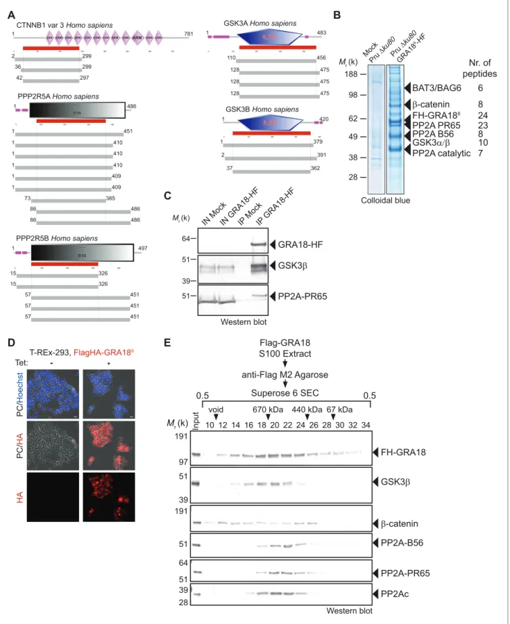
Figure 1. GRA18 is secreted and exported to the host cell cytoplasm. (A) Alignment of GRA18 alleles from T. gondii strains of types I (TGGT1_288840), II (TGME49_288840), and III (TGVEG_288840). The signal peptide sequence (highlighted in green), the B56 SLiM motifs (LxxLx; boxed in red), and the T. gondii Export Element (TEXEL; RRL motif, boxed in blue) are shown. Single amino acid polymorphisms are indicated by red letters. The alignment was done using ClustalW method. (B) GRA18II-HAFlag in Pru ku80 extracellular parasites is contained in cytoplasmic organelles distinct from the apical micronemes (MIC2) and rhoptries (Toxofilin), and partially co-localizing with the dense granule protein GRA1. (C) GRA18 secretion and export to the Figure 1 continued on next page
are exported in the host cell during tachyzoite life cycle (Hakimi et al., 2017;Nadipuram et al.,
2016).
GRA18 forms complexes with host components of the b-catenin
destruction complex
Since GRA18 does not carry any recognizable structural domain that can be used to infer its func-tion, we sought for host molecular partners that could identify the host cell pathway GRA18 could interfere with. Initially, we used a genome-wide yeast-two hybrid (Y2H) screen and screened a human placenta complementary DNA (cDNA) library (referred as prey) using N-terminally fused
GRA18 (aa 27 to 648) from type II strain as a bait (LexA-GRA18II). Of a total of 65.8 million cDNA
fragments screened, 65 positive hits covering 11 different proteins were found. The binding proteins were given Global Predicted Biological Score (Global PBS) ranging from A to D (‘A’ having the
high-est confidence of binding) (Supplementary file 2), if their coding sequences are in-frame and have
no in-frame stop codons. Interestingly, multiple components of the b-catenin destruction complex were found among the interactants: b-catenin itself, the glycogen synthase kinase-3 (GSK3a/b), and
the protein phosphatase 2A (PP2A) regulatory subunit B56a/b/g/d (Figure 2AandFigure 2—figure
supplement 1). These interactions were subsequently validated by chromatography and mass
spec-trometry–based proteomics analysis on proteins extracted from human cells infected by the Pru
PGRA1-GRA18II-HF T. gondii strain. Following Flag affinity chromatography, GRA18 co-eluted with
b-catenin, GSK3a/b, and the PP2A-B56d/e within a quite stable complex that resisted to stringent salt
and detergent conditions (0.5 M KCl and 0.1% NP-40) (Figure 2B). Interestingly, the identification of
the scaffolding subunit PP2A65 RA and the catalytic subunit PP2Ac along with the PP2A-B56, argues for a functional PP2A holoenzyme as part of the complex. The B56 regulatory subunit presumably mediates the recruitment of the PP2A holoenzyme as it was the only PP2A subunit found by Y2H to
interact directly with GRA18 (Figure 2AandSupplementary file 2). Importantly, when purified from
HFFs infected by RH parasites in which GRA18 endogenous locus was fused to the HA-Flag tags (GRA18-HF), GRA18 was found in a similar complex, with the exception of b-catenin that was
pre-sumably below the detection limit, (Figure 2C). Taken together, these data confirm GRA18 as a
partner of GSK3 and the PP2A-B56 subunit and validate the relevance of these interactions in the context of infection by wild-type parasites.
We then detailed the interactions between GRA18 and the associated proteins using an inducible
Flag-tagged GRA18–expressing HEK293 human cell line (T-Rex-293,Figure 2D). GRA18 Flag affinity
coupled to Size-Exclusion Chromatography (SEC) and immunoblot analysis revealed that GRA18 forms distinct complexe(s) with b-catenin, GSK3b, and the PP2A-B56 holoenzyme ranging from 400
to over 700 kDa globular sizes (Figure 2E). While GSK3b and the PP2A-B56 holoenzyme eluted as
discrete and overlapping complexes, in contrast, GRA18 and b-catenin both showed broader elution profiles, possibly reflecting the presence of multiple sub-complexes containing GRA18. Overall, these data confirm the multivalent partnership of GRA18 with the host proteins b-catenin, GSK3a/b, or the PP2A-B56 subunit.
b-Catenin, GSK3a/b, and PP2A-B56 bind different protein domains of
GRA18
To further detail the mode of interaction between GRA18 and the aforementioned partners, we per-formed a domain mapping analysis using the Y2H system and challenged the binding of GRA18
frag-ments to b-catenin, GSK3a/b, or the PP2A-B56 regulatory subunit in yeast (Figure 3A). Interestingly,
Figure 1 continued
host cytoplasm. HFFs were infected with type I RH parasites expressing endogenously tagged GRA18 with hemagglutinin (HA) (upper panel, RHDku80 GRA18-HA3, in red, Triton X-100 permeabilization) or type II Pru strain ectopically expressing a HAFlag (HF)-tagged copy of GRA18IIunder the control of the strong promoter of GRA1 (Pru Dku80 PGRA1-GRA18II-HF, ethanol permeabilization). Cells were fixed 18 hr post-infection (hpi) and stained with
anti-HA antibodies and Hoechst DNA-specific dye (in blue). The white asterisks indicate uninfected HFF cells. (D) MYR1 is required for GRA18 export in the host cell. HFFs were infected with RH WT or RH Dmyr1 parasites transiently transfected with a vector expressing an HF-tagged GRA18 (PGRA1
-GRA18II-HF), and at 18 hpi, the cultures were fixed and stained with antibodies to the HA tag. (E) Schematic representation of GRA18 probability of
disorder. Segments with values < 0 are predicted to be disordered (in red), and segments with values > 0 correspond to folded regions (in green). DOI: https://doi.org/10.7554/eLife.39887.002
1 781 CTNNB1 var 3 Homo sapiens
2 299 36 299 42 297 1 483 110 456 128 475 128 475 128 475
GSK3A Homo sapiens
GSK3B Homo sapiens
1 420
1 379
2 391
37 362
PPP2R5A Homo sapiens
1 451 1 410 73 385 86 486 1 486 1 410 1 410 1 409 1 409 86 486 PPP2R5B Homo sapiens 1 497 15 326 15 326 57 451 57 451 57 451
A
B
T-REx-293, FlagHA-GRA18II Tet: - + PC / H o e ch st PC / HA HAD
10 12 14 16 18 20 22 24 26 28 30 32 34 void 670 kDa 440 kDa 67 kDaFlag-GRA18 S100 Extract anti-Flag M2 Agarose Superose 6 SEC 0.5 0.5 Mr (k) Input 97 191 FH-GRA18 51 39 GSK3! 51 PP2A-B56 191 !-catenin Western blot 51 64 PP2A-PR65 39 28 PP2Ac
E
64 Mr (k) 51 39 51 IN Mo ck GSK3! GRA18-HF PP2A-PR65 IN GRA18 -HF IP Mo ck IP G RA1 8-H FC
Western blot Colloidal blue Mr (k) 98 188 62 49 38 28 Pru "#ku 80 GR A18 II-H F FH-GRA18II 24 PP2A PR65 23 !-catenin 8 BAT3/BAG6 6 PP2A B56 8 PP2A catalytic 7 GSK3$%! 10 Nr. of peptides Mock Pru "#ku 80Figure 2. GRA18 binds directly to the host cell proteins b-catenin, GSK3a/b, and PP2A-B56. (A) Yeast-two hybrid screening of a human placental cDNA library to identify potential host partners for GRA18. Schematic representation of the identified partners having the highest Global PBS scores are shown. Summary of the prey clones that interacted with the GRA18 bait are represented as grey bars. Multiple independent interacting prey clones allowed Selected Interaction Domain (SID, in red) analysis that delineates the shortest fragment that is shared with all the interacting clones, and thus Figure 2 continued on next page
the N-terminal region of GRA18 sheltered the binding site for b-catenin, whereas the C-terminal
fragments accommodated the interaction with the PP2A-B56 subunit (Figure 3B and D). None of
the GRA18 fragments tested other than the full-length protein could support the interactions with GSK3 in this assay, suggesting that binding to GSK3 necessitates a larger contact surface on GRA18 than the other partners.
In order to test the GRA18 binding features in a cellular context, we generated inducible T-Rex cell lines expressing each different flag tagged sub-domains of GRA18, performed immuno-tag
affin-ity purification for each chimeric proteins and analyzed the eluates by Western Blot (Figure 3B–C).
Figure 3C shows that (i) the full-length GRA18 (FH-GRA18FL) pulled-down b-catenin, GSK3b, and
PP2A-B56, (ii) the GRA18Cttruncated version lost the ability to bind to PP2A-B56 in agreement with
the Y2H experiments, while retaining its ability to interact with GSK3b. Conversely, GRA18Ntinduced
in T-Rex cells did not interact with PP2A-B56 and its binding property to GSK3b was significantly impaired when compared to the full-length or the C-terminal region of GRA18. Collectively, these results suggest that the GRA18 C-terminal region mediates the interaction with both the phospha-tase and the kinase. It is noteworthy that both the N- and C-ter truncated fragments were unable to pull down b-catenin, which was unexpected based on the Y2H results. We cannot exclude however the possibility that the harsh washing conditions (0.5 M KCl and 0.1% NP-40) shattered the weak
interactions between b-catenin and the GRA18 N-terminal fragment (Figure 3A). Therefore, we
pro-pose that the N-terminal region of GRA18 mediates labile interaction with b-catenin that can be cap-tured by Y2H, whereas the C-terminal region contributes to a rather strong binding to GSK3b and
the PP2A-B56 holoenzyme (Figure 3D).
GRA18 functions as a positive regulator of b-catenin
As demonstrated above, GRA18 interacts with well-known components of the b-catenin destruction complex, a multiprotein complex with a pivotal role in the Wnt signaling. Indeed, b-catenin regulates the transcription of the Wnt target genes. Central to the Wnt pathway is the regulation of b-catenin levels by a cytoplasmic destruction complex in which b-catenin is embedded. This complex is com-posed of a core scaffold protein, named the axis inhibition protein (Axin) that interacts with factors such as the adenomatous polyposis coli protein (APC), the Ser/Thr kinases GSK3, the casein kinase 1 (CK1), and the PP2A-B56 phosphatase (Reviewed by Stamos and Weis, 2013). In the absence of Wnt signaling, the destruction complex efficiently captures cytoplasmic b-catenin, leading to its phos-phorylation by GSK3 and recognition by the b-TrCP ubiquitin ligase for degradation by the 26S pro-teasome. Wnt ligands trigger functional inactivation of the destruction complex, with ensuing escape of b-catenin from degradation, resulting in b-catenin accumulation and nuclear internalization
(Clevers, 2006;Li et al., 2012;Taelman et al., 2010). Given the interactions between GRA18 and
b-catenin, GSK3b, and PP2A-B56, we hypothesized that GRA18 could interfere with b-catenin regu-lation in the course of infection. To test this hypothesis, we generated parasites knockout for GRA18
(Figure 4A and B) in different strain types (Pru ku80 Dgra18 and 76K Dgra18) which did not show
any obvious growth phenotype in cell culture (Figure 4C and data not shown), and a somewhat
Figure 2 continued
represents a potential region mediating the interaction with GRA18. (B) GRA18 associates with b-catenin, GSK3a/b, and PP2A-B56 in infected host cells. GRA18-associated proteins were purified by Flag affinity chromatography from protein extracts of HFF cells infected with parasites expressing HF-tagged GRA18 (Pru k80, PGRA1-GRA18II-HF). HFFs infected with Pru ku80 parasites were used as a mock. Immunopurified proteins were resolved by
SDS-PAGE, followed by colloidal blue staining and mass spectrometry analysis. The identity of the proteins and their respective number of peptides are indicated on the right of the figure. (C) GRA18 associates with GSK3 and PP2A-B56 when delivered to HFFs by type I RH parasites expressing
endogenously tagged GRA18 with HA-Flag (RH GRA18-HF). IN, input; IP, immunoprecipitation. (D) Immunofluorescence assay (IFA) of FH-GRA18 ectopically and stably expressed in T-Rex-293 cell line. Cells were either left untreated (-) or treated with 1 mg/mL tetracycline for 12 hr before fixation and staining with anti-HA antibodies (in red) and Hoechst DNA-specific dye (in blue). Scale bar, 10 mm. (E) Size-Exclusion Chromatography (SEC) analysis of the GRA18-associated proteins. FH-GRA18 was immunopurified from tetracycline-induced T-Rex cells (T-Rex-GRA18FL). SEC fractions were analyzed by immunoblot using the indicated antibodies.
DOI: https://doi.org/10.7554/eLife.39887.003
The following figure supplement is available for figure 2:
Figure supplement 1. Summary of the hits in the Y2H screen against GRA18. DOI: https://doi.org/10.7554/eLife.39887.004
reduced virulence phenotype in mice when challenged by intraperitoneal injection (Figure 4D). While assessing b-catenin amounts and subcellular localization we showed that b-catenin signals remained unaffected upon T. gondii infection of both human (HFF) and murine (L929) cells
regard-less of the GRA18 status (Figure 4E and F, compare Pru ku80 with Dgra18). Intriguingly, when
para-sites expressed high levels of GRA18, referred to as GRA18+++, b-catenin signals drastically
A
C
Mr (k) 51 39 97 64 97 39 64 51 1 26 648 26 648 FH FH-GRA18FL 233 648 FH-GRA18Ct 26 440 FH-GRA18Nt SP CTNNB1 GSK3A PPP2R5A 26 648 + + + 26 233 + - -233 440 +/- - -420 648 - - + 152 350 - - -350 585 - - +Fragments of GRA18 used for Y2HS
B
1 26 648 SP !-catenin GRA18 GSK3 PP2A B56 CD
GSK3! !-catenin Mock GR A18 FL GR A18 Nt GR A18 Ct IP : Flag-GRA18 FH-GRA18Nt FH-GRA18Ct FH-GRA18FL IP : Flag 64 51 Western blots PP2A-B56 Mock GR A18 FL GR A18 Nt GR A18 Ct InputsFigure 3. b-Catenin, GSK3, and PP2A-B56 recognize distinct domains of GRA18. (A) Interaction domain mapping by Y2H assay using the indicated fragments of GRA18 to delineate the interacting domains of GRA18 with CTNBB1 (b-catenin), GSK3A (GSK3a) and PPP2R5A (PP2A-B56a). CTNNB1 interacts with the N-terminus of GRA18, whereas PPP2R5A interacts with the C-terminal fragments. GSK3A interacted with full-length GRA18 (amino acids 26 – 648) but not with any of the GRA18 fragments tested. (B) Schematic representation of full-length (amino acids 26 – 648) and truncated versions of GRA18 (FH-GRA18Nt(aa 26-440)and FH-GRA18Ct(aa 233-648)) proteins stably
expressed in T-Rex cells. (C) Cytoplasmic fractions from T-Rex cells presented in (B) were immunoprecipitated with anti-Flag antibodies and analyzed by immunoblotting. Untransfected T-Rex cells were used as a mock. (D) Schematic diagram summarizing the interaction domain mapping of GRA18 obtained from Y2H and biochemical approaches.
7 0 5 10 15 Pru ku80 Pru ku80 #gra 18 N u mb e r o f p a ra st ie s/ va cu o le Pru #ku 80 Pru ku80 #gra 18 GRA18II DHFR cassette 5’ region of GRA18 3’ region of GRA18
attB1 attB4 attB3 attB2
P1 P2 P4 P3 P5 1,650 2,000 3,000 4,000 1,000 1,650 2,000 3,000 4,000 6,000 5,000 7.3 kbp 6.0 kbp 1,650 2,000 3,000 4,000 1,000 Pru ku 80 Pru ku 80 # gra 18 Pru ku80 Pru ku80 #gra 18 Primers: P1, P4 P2, P3 1.6 kbp P1, P5 2.3 kbp Mr (k) 97 64 β-catenin 97 51 QRS 39 TBP ui Pru ku80 #gra18GR A18 +++ 64 51 GRA18-HF 97 Western blot ui Pru ku80 #gra18 GRA18+++
Ph a se / H o e ch st Ph a se / ! -ca te n in ! -ca te n in G R A1 8 +++
Phase/Hoechst Phase/GRA18II-HF GRA18II-HF !-catenin
A
B
C
E
F
FH-GRA18I β-catenin GAPDH 0 8 Time: (hrs) -0 -0.5 1 2 6 8 + + + + + + + Wnt3A TetracyclineG
Western blotDays after infection
0 10 20 30 40 50 0 50 100 Pru ku80 (n=6) #gra18 (n=6) W psp *** Pe rce n t su rvi va l
Days after infection
0 5 10 15 20 0 50 100 76K (n=7) #gra18 (n=6) W p * Pe rce n t su rv iv a l
Days after infection
D
Pru ku80 wt Pru ku80 gra18
0.0 0.2 0.4 0.6 0.8
Pru ku80 Pru ku80
#gra18
Pru ku80 Pru ku80
#gra18 Pl a u q e a re a (mm ²)
Figure 4. GRA18 is a positive regulator b-catenin. (A) Generation and confirmation of insertion/deletion of GRA18 in T. gondii type II Pru strain. Schematic representation of the GRA18 locus with the double homologous recombination event between the knockout construct (pDEST14 KO GRA18) and genomic DNA replacing the GRA18 coding sequence with the DHFR cassette used for positive selection. (B) PCR reactions with the indicated primers confirming the deletion/insertion of GRA18 in the mutant parasites. (C) Parasites lacking GRA18 exhibit no growth defect in vitro as determined by fluorescence imaging assays in HFFs and plaque assays. Data are mean value ± s.d. of triplicates from two independent experiments. (D) GRA18 Figure 4 continued on next page
increased in cells infected by the GRA18+++strain compared to uninfected cells (
Figure 4E and F).
b-Catenin upregulation is associated with a strong accumulation of b-catenin in the host nuclei
sug-gesting that a transcriptionally active bulk of b-catenin was produced (Figure 4F). To determine
whether additional parasite factors may contribute to the GRA18-mediated b-catenin induction, we used ectopic expression of GRA18 in HEK293 human cell line. As a positive control, cells were treated with exogenous Wnt3A ligand, a natural inducer of b-catenin, thus confirming that the T-Rex
cells carry a functional and regulatable b-catenin-destruction complex (Figure 4G) (Azzolin et al.,
2014). Induction of GRA18 expression with tetracycline promoted the accumulation of b-catenin,
indicating that GRA18 alone was sufficient to drive b-catenin upregulation to levels comparable to
those obtained with Wnt3A (Figure 4G). Therefore, these data indicate that GRA18, very likely
through the interactions with GSK3 and PP2A-B56, functions as a positive regulator of b-catenin.
GRA18 alters the expression of a specific set of genes in infected cells
The ability of GRA18 to promote nuclear accumulation of the transcriptional regulator b-catenin prompted us to investigate whether GRA18-GSK3/b-catenin partnerships could contribute to the typical changes in gene expression observed in cells infected with tachyzoites. To test this hypothe-sis, we performed a comparative transcriptomic analysis by RNA-sequencing of mouse Bone Mar-row-Derived Macrophages (BMDMs) loaded with parental or Dgra18 parasites of the type II Pru strain. Macrophages were chosen because they are infected in mice and play an essential role in the
early immune response against T. gondii (Dunay et al., 2008;Jensen et al., 2011). Since GSK3 and
b-catenin were involved in the regulation of the inflammatory gene expression in response to
bacte-rial lipopolysaccharides (LPS) (Chattopadhyay et al., 2015;Jang et al., 2017;Martin et al., 2005;
Yang et al., 2010), transcriptomic analysis was also performed on infected macrophages treated
with LPS. We focused our analysis on genes that were modulated with more than 3-fold change and had a signal threshold above 5 Reads Per Kilobase of transcript per Million mapped reads (RPKM) in at least one sample when comparing the wild-type and Dgra18 mutant strains. Filtered data are
pre-sented in Supplementary file 3 (data are accessible through NCBI GEO, accession number
GSE103113). Thirty-eight genes were significantly and differentially regulated with most correspond-ing to genes up-regulated upon macrophage infection by wild-type parasites, but not with the
Dgra18 strain as revealed by hierarchical clustering (Figure 5A). Complementation of Dgra18
muta-tion with GRA18-HF under the control of the GRA1 promoter (Dgra18, GRA18+++) restored the
expression pattern to levels observed with wild-type parasites or even higher, in strong support of the GRA18-dependent induction of those genes. Parasite transcriptome analysis indicated that nei-ther Dgra18 mutation nor LPS treatment had any significant impact on T. gondii gene expression
and that GRA18 expression in the Pru ku80 Dgra18, GRA18+++ complemented strain was restored
with a ~ 35 fold more transcript reads than in the wild-type strain (Figure 5D,Supplementary file 3,
and Figure 5—figure supplement 1A), in line with GRA18 overexpression driven by the GRA1
strong promoter. It is noteworthy that the expression pattern of the aforementioned host genes reg-ulated by GRA18 remained similarly regreg-ulated in the infected macrophages subjected to LPS
stimu-lation (Figure 5A and B and Supplementary file 3), indicating that LPS did not affect GRA18
function. Gene set enrichment analysis (GSEA) highlighted pathways significantly affected in a GRA18-dependent fashion (p values < 0.05) with functions related to the inflammatory response (e.
Figure 4 continued
mutants exhibit decreased virulence in mice. Virulence of the type II Pru ku80Dgra18 and 76K Dgra18 strains were compared to the parental strains Pru ku80 and 76K, respectively, in BALB/c mice. Mice were inoculated with 105tachyzoites of each strain by intraperitoneal injection and survival was monitored. Cumulative results of two independent experiments with at least three mice in each group (n ! 6) are shown. Significance was tested using Log-rank (Mantel-Cox) test; *p=0.0162 and ***p=0.0005 when compared to the respective wild-type strain. (E) Effect of GRA18 on b-catenin levels. Murine L929 cells were left uninfected (ui) or infected with wild-type (Pru ku80), Dgra18, or the Dgra18 GRA18+++complemented (GRA18+++) strains. At
18 hr post-infection, cells were harvested and analyzed by immunoblot using the indicated antibodies. TgQRS was used to control parasite loading. (F) IFA of b-catenin in confluent HFFs left uninfected (ui) or infected with the indicated strains for 18 hr. In the lower panel, IFA was performed using an anti-HA antibody to monitor the HF-tagged version of GRA18 in the GRA18+++complemented strain. Data are representative of at least three
independent experiments. (G) Immunoblot analysis of nuclear fraction of the T-Rex FH-GRA18 cell line left untreated or induced with tetracycline for the indicated periods of time. The Wnt3A ligand was used as a positive control.
A
C
D
0.0 -2.0 2.0 Pru ku80 #gra1 8 GR A18 +++ Pru ku80 #gra1 8 GR A18 +++ - + LPS:E
F
CCL22 (p g /m L ) i 0 6 + 0 10000 20000 30000 40000 ui Pru ku80 #gr a18 GRA 18 +++ CCL17 (p g /m L ) ui 0 6 ++ 0 20000 40000 60000 ui Pru ku80 #gr a18 GRA 18 +++ I T 0 5000 10000 15000 20000 I T 0 50 100 150 200 Ccl22 I T 0 1000 2000 3000 Ccl24 Ccl22 Ccl17 ui Pru ku80 #gr a18 GRA 18 +++ RQ i K 6 0 1000 2000 3000 i K 6 0 5 10 15 Ccl24 RQ ui K 6 0 200 400 600 800 ui 76K #gr a18 ui 76K #gr a18 RQ ui Pru ku80 #gr a18 GRA 18 +++ ui Pru ku80 #gr a18 GRA1 8+++ ui 76K #gr a18B
Ccl17 Ccl22 Pru psp6 6ttt PS PS PS PS [1] ui Pru psp6 6ttt PS PS PS PS Pru sp6 6ttt PS LPS PS PS Ccl22 cl22 ui Pru sp6 6ttt PS PS PS LPS 0 Pru 0 0 sp6 0 0 P6ttt 0 0 PS 3 0 PS 3 0 PS 3 0 LPS 3 0 [1] Ccl24 [2] [3] [1] [2] ui Pru sp6 P6ttt PS PS PS LPS Ccl24 ui Pru ku80 #gra18 GRA18+++ U n s ti m u la te d L PS s ti m u la ti o n ui Pru ku80 #gra18 GRA18+++ [0-2 500 ] [0-2 500 ] [0-2 000 0 ] [0-4 100 0 ] [0 -30 0 ] [0 -14 0 0] 0 5 10 15 20 25 30 35 40 45 LEUKOCYTE MIGRATION IMMUNE SYSTEM PROCESS MYELOID LEUKOCYTE MIGRATION RESPONSE TO EXTERNAL STIMULUS LYMPHOCYTE MIGRATION LEUKOCYTE CHEMOTAXIS CHEMOKINE MEDIATED SIGNALING PATHWAY CELL MOTILITY LOCALIZATION OF CELL GRANULOCYTE MIGRATION-log(FDR, q-value)
0 5 10 15 20 25 30 35
Genes defining inflammatory response. Genes up-regulated by STAT5 in response
to IL2 stimulation. Genes regulated by NF-kB in response to
TNF. -log(FDR, q-value) Gpr183, Olr1 Tnfrsf9, Ccr7 Rgs16, Penk ui Pru ku80 #gra18 GR A18 +++ ui Pru ku80 #gr a18 GR A18 +++ Unstimulated LPS stimulation -2.0 0.0 2.0 8/200 3/200 3/200 11/259 17/1984 7/99 15/1821 6/49 7/117 6/72 11/835 11/835 6/75 Ccl24, Ccl22 Olr1, Cd34 Ccl17, Ccl1 Xcl1, Ccr7 Ccl24, Ccl22 Ccl17, Ccl1 Itga9, Ccr7 Itgax, Cd34 Ccl24, Ccl22 Ccl17, Ccl1 **** ****
log2 expression scale
log2 expression scale
****
****
****
**** **** ****
Figure 5. GRA18 alters the host cell transcriptome. (A) RNA-Seq analysis of BALB/c BMDMs that were left uninfected (ui) or infected with the indicated strains at an MOI of 1:5. At 18 hr post-infection, cells were left unstimulated or stimulated with LPS (100 ng/mL) for 6 hr. Heat map representation of the differentially expressed mouse genes (!3 fold, RPKM !5 in at least one sample) between parental and Dgra18 infected cells in the absence of LPS. RPKM values were log2 transformed, Gene/Row normalized, and mean centered using MeV. (B) Tracks of the BMDMs RNA-Seq reads for Ccl17, Ccl22, Figure 5 continued on next page
g., Csf1, Olr1, and Tnfrsf9) and chemotaxis (e.g., Ccr7, Ccl1, Ccl17, Ccl22, and Ccl24) (Figure 5C). Surprisingly, none of the typical Wnt/b-catenin target genes were found differentially regulated by
GRA18 (Figure 5—figure supplement 1BandSupplementary file 3), suggesting either that murine
macrophages have a peculiar repertoire of b-catenin target genes or that GRA18 fosters a singular b-catenin transcriptional activity or that its action is independent of b-catenin.
To confirm the transcriptomic profile, we focused on the chemokines Ccl17, Ccl22, and Ccl24
pre-viously identified as selectively up-regulated genes upon T. gondii infection (Hammoudi et al.,
2015;Melo et al., 2013). Quantitative RT-PCR analysis reproducibly demonstrated a pronounced
decrease in Ccl17, Ccl22, and Ccl24 expression from cells infected with Dgra18 mutant parasites
when compared to wild-type Pru or Pru Dgra18, GRA18+++parasites (Figure 5E). Similar data were
obtained with the 76K strain, another type II cystogenic strain, highlighting that T. gondii reproduc-ibly regulated those chemokines in a GRA18-dependent manner. GRA18 clearly plays a major role in regulating CCL17 and CCL22 synthesis and secretion since its loss significantly lowered protein
lev-els in the supernatant of mouse macrophages (Figure 5F). Overall, these results support the
regula-tory function of GRA18 for specific chemokines amongst others.
GRA18 alters host gene expression in a b-catenin-dependent fashion
To evaluate whether b-catenin is required for the GRA18-induced chemokine expression, we gener-ated macrophage RAW264.7-derived cell line mutgener-ated for Ctnnb1 using CRISPR/CAS9-medigener-ated gene editing. Cells were transfected with a vector expressing the CAS9 endonuclease and a guide
RNA targeting the third coding exon of the murine Ctnnb1 gene (Figure 6A). Cells harboring
inser-tion/deletion (indel) mutations at the Ctnnb1 locus lack b-catenin expression when compared to the
wild-type parental cell line (Figure 6A and B). As expected, treatment of the Ctnnb1-/-mutant cells
with the GSK3 inhibitors did not lead to b-catenin expression, otherwise induced in the wild-type parental cell line. These results demonstrate that the RAW264.7 murine cells carry a regulatable b-catenin destruction complex and that the two alleles coding for b-b-catenin were successfully
dis-rupted in the Ctnnb1-/-mutant cells.
The ectopic expression of GRA18 in RAW264.7 macrophages caused a significant increase in
endogenous b-catenin (Figure 6C and D, compare the effect of mCherry with GRA18 expression),
similarly to the effect of GRA18 when delivered by the parasites in HFF or L929 cells (Figure 4E and
F). As expected for RAW264.7 macrophages whose Ctnnb1 alleles are disrupted, no b-catenin was
detected upon ectopic expression of GRA18 (Figure 6C and D) in contrast to the significant
amounts of nuclear b-catenin found in wild-type RAW264.7 macrophages expressing GRA18. In the same line Ccl17, Ccl22, and Ccl24 were selectively induced in GRA18-expressing wild-type
RAW264.7 macrophages when compared to mock - that is, mCherry - transfected cells (Figure 6D
and E) whereas Ccl17 and Ccl24 were no longer induced in the b-catenin lacking ones. The slight
reduction of Ccl22 expression in the GRA18-expressing mutant RAW264.7 macrophages (Figure 6E)
Figure 5 continued
and Ccl24 in the indicated samples. (C) Enrichment analysis in functional annotation and GO analysis of the differentially expressed genes defined in
Figure 5Ausing GSEA. Top-scoring pathways regulated in a GRA18-dependent manner, number of genes per pathway, and names of representative genes are shown. (D) Heat map of expression values obtained by RNA-Seq analysis of T. gondii genes from the samples in (A). A selected set of T. gondii ROPs and GRAs are shown. (E) Quantitative chemokine expression was determined by qRT-PCR on BMDMs infected with the wild-type and Dgra18 mutant strains in the type II strains Pru and 76K. Values were normalized to the amount of TATA box binding protein (Tbp) in each sample. Data are mean value ± s.d. of three replicates. The P-values were calculated using two-tailed unpaired Student’s t-test; ****p<0.001. Data are representative of two independent experiments. (F) CCL17 and CCL22 levels were measured by ELISA from supernatants collected 24 hr after BMDM infection with the indicated strains at an MOI of 1:5. Supernatant from uninfected cells was used as a control (ui). Means of four independent experiments are shown. ****, p<0.0001 following one-way ANOVA analysis and multiple-comparison post hoc tests.
DOI: https://doi.org/10.7554/eLife.39887.007
The following figure supplements are available for figure 5: Figure supplement 1. RNA-Seq analysis of mouse and T. DOI: https://doi.org/10.7554/eLife.39887.008
Figure supplement 2. Deletion of the aspartyl protease ASP5 compromises GRA18 export and phenocopies the deletion of GRA18 in promoting host cell gene expression.
39 TBP Western blot β-catenin 97 64 wt Ctnnb 1 -/-Ctnnb1 !-catenin Western blot P-GSK3α (Ser21) GSK3β P-GSK3β (Ser9) FH-GRA18 TBP mCherry-HF Mr (k) 39 51 39 51 mC herry GR A18 II mC herry GR A18 II WT Ctnnb1 -/-97 64 51 64 39 28 19 39 51 Western blot Mr (k) 39 TBP 97 !-catenin MockCHIRBIO WT MockCHIRBIO Ctnnb1 -/-Wild-type locus Mutated loci 1 bp deletion 2 bp deletion
A
B
FH-GRA18II mCherry-HF WT HA / H o e ch st ! -ca te n in FH-GRA18II mCherry-HF Ctnnb1-/-C
D
E
Mr (k) y 6 0 2 4 6 8 Ccl17 RQ RAW wt RAW Ctnnb1 -/-* y 6 0 2 4 6 Ccl22 RQ * y 6 0 20 40 60 80 mCherry FH-GRA18 Ccl24 RQ * RQ rry 8 0 20 40 60 80 100 Ifnb1 mCherry FH-GRA18 RAW wt RAW Ctnnb1 -/-* i 0 1000 2000 3000 Ccl17F
Pru ku80 #gra18 ui RAW wt RAW Ctnnb1 -/-RQ * * i 0 10 20 30 40 + RQ Ccl22 Pru ku80 ui #gra18 GRA18 +++ 0 200 400 600 800 ns * *Figure 6. GRA18 promotes Ccl17, Ccl22 and Ccl24 chemokines expression in a b-catenin-dependent fashion. (A) Schematic diagram of gRNAs targeting the Ctnnb1 locus. The Protospacer Adjacent Motif (PAM) sequence is lined and highlighted in red; the targeting sequences is shown in green. Bi-directional arrow indicates the Cas9 cleavage site. DNA sequences from the wild-type and mutated RAW264.7-derived cell lines were analyzed by DNA sequencing; the identified deletions are indicated with (-). No mutations in control samples were observed. (B) Immunoblot analysis of b-catenin in Figure 6 continued on next page
suggests a different mode of regulation for the three chemokines. Consistent with b-catenin
upregu-lation, similar data were obtained with Ifnb1 (Figure 6E), a known b-catenin target gene in
macro-phages (Ma et al., 2014; Rathinam et al., 2010; Yang et al., 2010). To ascertain the b-catenin
requirement for GRA18 activity under more physiological conditions, chemokine expression was monitored upon infection of RAW264.7 cells disrupted or not for Ctnnb1 with wild-type and Dgra18 mutant parasites. While b-catenin was required for full induction of the Ccl17 chemokine, Ccl22
expression remained unaffected in Ctnnb1-/-mutant host cells (Figure 6F), again consistent with a
different mode of regulation orchestrating Ccl17 and Ccl22 expression. Importantly, the moderate role played by GRA18 in the regulation of Ccl22 in RAW264.7 cells when compared to BMDMs, sug-gests that a GRA18-independent pathway controls Ccl22 expression, which is reminiscent of the aforementioned b-catenin independent mechanism (see discussion). Indeed, Ccl22 was much less
induced in RAW264.7 than in BMDM cells (compareFigures 5E and6F), indicating differences in
responsiveness to T. gondii infection. In addition, these data also argue that using the transformed RAW264.7 cell line to monitor GRA18 activity on Ccl22 induction may be less relevant than relying on the primary BMDM cells. Altogether, these results indicate that basal levels of b-catenin are required for full activity of GRA18 towards Ccl17.
GRA18 acts as an inhibitor of GSK3 to trigger b-catenin transcriptional
activity
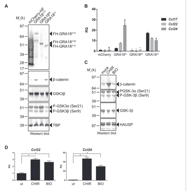
In order to better map the GRA18 active domains we assayed the different sub-domains of GRA18
(GRA18Nt and GRA18Ct) described above for their ability to upregulate b-catenin and to induce
expression of the downstream target genes in macrophages. While expression of GRA18FL in
RAW264.7 macrophages caused a significant increase in endogenous b-catenin protein levels,
lead-ing to the expression of the downstream chemokines (Figure 7A and B), expression of GRA18Nt,
which no longer interacts with GSK3 and PP2A-B56 (Figure 3), failed to induce b-catenin and
accordingly chemokine transcripts. Given that the N-terminal region of GRA18 has retained the
abil-ity to interact with b-catenin (Figure 3A), these data suggest that the interactions with b-catenin
alone are not sufficient to promote its accumulation and transcriptional activity. In contrast,
GRA18Ct, shown previously to interact with GSK3 and PP2A-B56 (Figure 3) has retained full activity
on catenin and downstream transcriptional consequences, further confirming the pivotal role of b-catenin in driving the expression of the aforementioned chemokines. While the effect of GRA18 on b-catenin protein levels required interactions with GSK3 and/or PP2A-B56, direct interaction between GRA18 and b-catenin seems to be dispensable. Altogether these results support a scenario in which GRA18, through its interactions with GSK3 and PP2A-B56 inhibits the b-catenin-destruction complex, hence stabilizing b-catenin thereby acting as a positive regulator of b-catenin level to pro-mote gene expression. Additional evidence for this model is the expression of Ccl22 and Ccl24
induced by GSK3 inhibitors in a manner that resembles the effect of GRA18 (Figure 7C and D).
Figure 6 continued
parental RAW264.7 and the Ctnnb1-/-mutant confirmed the absence of b-catenin expression. As positive controls, cells were treated with 3 mM of BIO
or 2 mM of CHIR GSK3 inhibitors for 12 hr. TBP was used as loading control. (C–E) RAW264.7 (WT) and b-catenin-deficient (Ctnnb1-/-) RAW264.7-derived cell lines were transfected with mCherry vector control (pcDNA-mCherry-HF) or the FH-GRA18IIexpression vector (pcDNA-FH-GRA18FL). At 18
hr after transfection, cells were either (C) fixed for IFA using anti-HA (red) and anti-b-catenin (green) antibodies or (D) cells were harvested and analyzed by immunoblot using the indicated antibodies. Anti-HA was used to detect FH-GRA18 and mCherry-HF. In (E) and (F) transcripts for Ccl17, Ccl22, Ccl24, and Ifnb1 were quantified by qPCR and normalized using Tbp. Data are mean value ± s.d. of three replicates. The P-values were calculated using two-tailed unpaired Student’s t-test or one-way ANOVA with Bonferroni posttests analysis of variance; *p<0.05 and P values greater than 0.05 were considered not significant (ns). In (F) RAW264.7 (WT) and b-catenin-deficient (Ctnnb1-/-) RAW264.7-derived cell lines were either left uninfected or
infected with the indicated wild-type and Dgra18 mutant strains at a MOI of 1:6 for 18 hr. Data are representative of two independent experiments. DOI: https://doi.org/10.7554/eLife.39887.010
Discussion
GRA18 export mechanism
In search of new T. gondii effectors from the GRA family that would be not only secreted into the PV space but would also traffic beyond - that is, across the PVM - and be delivered in the infected host cell, we identified a novel T. gondii GRA protein that we termed GRA18. However, contrasting with the recently described GRA16, GRA24, GRA28 or TgIST, whose final destination is the nucleus of the host cell, GRA18 remained strictly cytoplasmic throughout the intracellular life cycle of the tachyzoite. rry L Nt Ct 0 10 20 30 40 Ccl17 Ccl22 Ccl24 nd Mr (k) 97 β-catenin mC he rry-HF GR A18 II FL GR A18 Nt GR A18 Ct 64 51 39 28 17 FH-GRA18II FL FH-GRA18II Nt FH-GRA18II Ct 97 64 GSK3β 39 51 39 51 P-GSK3α (Ser21) P-GSK3β (Ser9) 39 TBP Western blot A B !-catenin Western blot Mr (k) PGSK-3α (Ser21) GSK-3! 39 51 97 64 39 51 P-GSK-3β (Ser9) mock 97 HAUSP CHIRBIO C D 40 30 20 10 0 RQ
mCherry GRA18FL GRA18Nt GRA18Ct
RQ R O 0 1 2 3 4 RQ R O 0 20 40 60 Ccl22 Ccl24
ut CHIR BIO ut CHIR BIO
* * * *
* * *
Figure 7. GRA18 activity is dependent on its interactions with GSK3 and PP2A-B56. (A–B) RAW264.7 cells were transfected with mCherry vector control or the FH-GRA18 expression vectors pcDNA-FH-GRA18FL(GRA18FL), pcDNA-FH-GRA18Nt(GRA18Nt), and pcDNA-FH-GRA18Ct(GRA18Ct). At 18 hr after transfection, cells were harvested and (A) whole cell extracts were analyzed by immunoblot using the indicated antibodies, or (B) quantitative
chemokine expression was determined by qRT-PCR as inFigure 6. Asterisks indicate P-values (p<0.05) obtained when comparing GRA18Ntwith GRA18FL. (C) Treatment of RAW264.7 cells with the GSK3 inhibitors CHIR and BIO led to b-catenin protein accumulation and Ccl22 and Ccl24
upregulation as determined by qRT-PCR in (D). ut, untreated; nd, not detected. Data are mean value ± s.d. of three replicates. The P-values were calculated using two-tailed unpaired Student’s t-test; *p<0.05. Each data set is representative of two independent experiments.
Analyzing GRA18 protein sequence, we recognized in addition to the N-terminal signal peptide, an N-terminal putative TEXEL motif, both embedded in highly intrinsically disordered regions
(Figure 1E). The presence of the TEXEL motif suggests that GRA18 might be processed by the
aspartic protease TgASP5, an enzyme we and others have recently shown to mediate the secretion
or export of a number of GRA proteins (Coffey et al., 2015; Curt-Varesano et al., 2016;
Hammoudi et al., 2015). As expected for a TgASP5-dependent export, GRA18 was no longer
detected in the cytoplasm of cells invaded by TgASP5 mutant parasites (Figure 5—figure
supple-ment 2A) and accordingly, TgASP5 and GRA18 mutant parasites showed similar inability to induce
target genes in macrophages ((Hammoudi et al., 2015),Figure 5—figure supplement 2B). Recent
findings showed that the export pathway to the host cell of most of the dense granule effectors depends on MYR1, another DG protein localized at the PVM. In agreement with a common GRA export mechanism, trafficking of GRA18 through the PVM was also dependent on MYR1
(Figure 1D), (Coffey et al., 2016). The detection of GRA18 in the host cell cytoplasm together with
GRA18 sequence features prompted us to search for host cell cytoplasmic partners.
GRA18, a potential inhibitor of the b-catenin destruction complex
Using a genetic screen combined with affinity- and size-exclusion chromatography under stringent conditions, and mass spectrometry, we have characterized GRA18 as a strong interactor of host GSK3 and the PP2A-B56 holoenzyme, and to a lesser extent with b-catenin. The complex(es) pro-mote b-catenin nuclear accumulation, raising the possibility that GRA18 could modulate b-catenin
stability (see proposed model inFigure 8). However, while GRA18 clearly regulated host cell gene
expression, we did not detect significant changes in b-catenin levels upon infection with wild-type
parasites (Figure 4E and D). As only a small fraction of the total b-catenin pool is destined to
tran-scriptional regulation, possibly GRA18 induces subtle effects on the b-catenin levels that cannot be captured by IFA or immunoblot. Next, to figure out how GRA18 could interplay with b-catenin in cells, we engineered b-catenin mutant in RAW264.7 macrophages and provided definitive evidence for b-catenin acting epistatically to GRA18’s activity on host cell transcription for a restricted set of
target genes (i.e. Ccl17, Ccl24, and Ifnb1) (Figure 6).
b-Catenin is a dual function co-activator protein with distinct pools serving cell-cell adhesion and gene transcription functions, respectively. b-Catenin was originally identified as an element of the adherent junction complex together with cadherin and a-catenin, the latter binding to actin fila-ments in a process that promotes cell–cell contacts. Further, a nonjunctional pool of cytoplasmic b-catenin was recognized as a transcriptional coactivator of Wnt canonical signaling pathway. The Wnt signaling represents a complex and essential pathway starting with a repertoire of Wnt ligands and acting at the heart of embryogenesis, but also throughout life by directing stem cell renewal and senescence. Centerpiece of Wnt signaling is the canonical Wnt/b-catenin pathway genetically dis-sected in both human and Drosophila. Briefly, in the absence of Wnt ligands, cytosolic b-catenin is maintained at low levels by the multiprotein complex composed of APC, Axin, GSK3 and PP2A-B56. Within this complex, also referred to as the b-catenin destruction complex, b-catenin is constantly
phosphorylated for ubiquitylation that directs its degradation by the 26S proteasome (Liu et al.,
2002). Mutation in any of these components (APC, Axin, or b-catenin) leads to inappropriate
stabili-zation of b-catenin, which results in cancer, most notably of the colon (Stamos and Weis, 2013).
Axin, a central scaffold protein of the complex, is typified by intrinsically disordered and flexible regions that mediate direct interactions with all other core components of the destruction complex
(b-catenin, APC, and GSK3) (Stamos and Weis, 2013). Remarkably, the T. gondii tachyzoite GRA18
also carries significant disordered regions (Figure 1E) which could confer the potential of competing
with Axin/APC for binding to b-catenin, GSK3, and PP2A-B56. Accordingly, by disrupting the b-cate-nin destruction complex GRA18 would likely promote the stabilization and nuclear translocation of
b-catenin and ultimately b-catenin-dependent gene expression (Figure 8). RNA-Seq performed on
BMDMs indicated that b-catenin transcripts remained unaffected by GRA18 (Supplementary file 3),
which is consistent with a post-transcriptional regulation of b-catenin. Collectively our data argue for GRA18 acting as a direct regulator of b-catenin and although the exact modus operandi of GRA18 awaits clarification, some scenarios can be proposed. Interestingly, the Y2H interaction assay allowed
defining the region of b-catenin that promotes the interaction with GRA18 (Figure 2A). Indeed, the
SID analysis for interaction between GRA18 and b-catenin is mediated by a region encompassing
residues initially phosphorylated by CK1 (Ser45), and subsequently by GSK3 (Thr41, Ser33 and Ser37). Once phosphorylated, Ser33 and Ser37 mediate the interaction with the b-TrCP adaptor pro-tein for b-catenin degradation. Therefore, the GRA18 binding features to the N-terminal domain of b-catenin appear quite peculiar and differ from the binding sites for Axin and APC mapped at cen-tral ARM domain. As such, the binding interface of GRA18 to the b-catenin N-terminal region would confer to GRA18 a unique ability to interfere with both the phosphorylation cascade catalyzed by CK1 and GSK3, but also at a later step with the binding to b-TrCP. Interestingly, phosphorylation of
GSK3b Ser9 or GSK3a Ser21 is reported to decrease GSK3a/b enzymatic activity (Cross et al.,
1995; Peyrollier et al., 2000). Because GRA18 did not alter the phospho-Ser21/Ser9 levels
(Figures 6D and 7A), we conclude that the parasite protein, which we found to bind GSK3 and
inhibit its pro-degradative activity on cytoplasmic b-catenin, acts independently of the phosphoryla-tion status of Ser21/Ser9.
In the context of the Wnt signaling, the function of the PP2A-B56 when associated with the
b-cat-enin destruction complex remains uncertain (Hsu et al., 1999; Seeling et al., 1999;
Yamamoto et al., 2001). A recent study reported that the PP2A regulatory B56 subunit binds to a
LxxIxE Short Linear Motif (SLiM) on partner proteins as illustrated with the B56 binding to Axin
(Hertz et al., 2016). Interestingly, GRA18 carries two LxxIxE motifs within an intrinsically disordered
region of the C-terminal domain, which is in agreement with the B56 binding assays (Figure 3A and
Figure 8. Model for GRA18 mechanism of action. A model depicting how GRA18 interacts and interferes with the b-catenin destruction complex leading to host cell gene regulation. Possible effect of GRA18 on gene expression through GSK3 but b-catenin-independent or still unidentified T. gondii factor are represented by dashed lines. Question mark indicates the putative direct interaction with b-catenin observed by the Y2H or GRA18 overexpression. See also discussion.
C). As we previously showed (Hakimi et al., 2017), SLiMs is a prominent characteristic of the GRA effector family of proteins, probably reflecting a favorable evolutionary strategy to expand and diversify the binding interfaces between host and parasite proteins.
Our data suggested a simultaneous binding of GRA18 to GSK-3 and PP2A-B56 (Figure 2E), but
whether the binding of PP2A-B56 is essential for GRA18 activity is yet to determine. GSK3 activity can be regulated by phosphorylation of tyrosine residues (Tyr279/Tyr216 in a/b isoforms,
respec-tively) exposed in the activation loop, which enhances its kinase activity (Dajani et al., 2003). An
attractive hypothesis is that PP2A-B56 is recruited to the GRA18 complex in order to dephosphory-late GSK3, hence contributing to the inhibitory activity towards the b-catenin destruction complex. Alternatively, PP2A-B56 could be recruited to GRA18-GSK3 complex to dephosphorylate b-catenin,
leading to the inhibition of b-catenin degradation (Su et al., 2008).
The GRA18-GSK3-b-catenin axis induces the expression of
anti-inflammatory chemokines
Our transcriptomic data have revealed the magnitude by which GRA18 alters the expression of genes of cells infected by T. gondii tachyzoites, particularly the chemokines Ccl17, Ccl22, and Ccl24. Those genes have been found repeatedly induced by T. gondii tachyzoites regardless of the strain
type in murine macrophages (Figure 5—figure supplement 1C) (Hammoudi et al., 2015;
Melo et al., 2013; Morgado et al., 2011), as expected from the overall conservation of GRA18
across the Toxoplasma lineages. Whether this GRA18 property is also expressed in other intermedi-ate hosts or in different cell types is not known, but given the conservation of the Wnt signaling in vertebrates, GRA18 activity in other hosts can be assumed. Interestingly, the human placental cells, which have the unique ability to resist to T. gondii infection, were shown to specifically respond to
the invader by producing the CCL22 chemokine (Ander et al., 2018), whereas other cell types such
as human fibroblasts did not ((Ander et al., 2018) and data not shown). Whether the mechanism
underlying CCL22 induction in human placental cells is driven by GRA18 or differ from murine mac-rophages should be addressed in the future. It is noteworthy that a quite substantial residual
activa-tion of Ccl17, and Ccl22 was observed in the absence of GRA18 (Figures 5E–F andand6F), which
suggests that an alternative mechanism of activation by the parasites exists.
We provide here genetic evidence for GRA18 acting in a b-catenin-dependent manner on chemo-kine expression in macrophages. Although Wnt b-catenin signaling has originally been studied in the
context of thymocyte development and stem cell biology (Reya et al., 2003; Staal et al., 2008;
Verbeek et al., 1995), there is increasing evidence for its contribution in the regulation of innate
immunity. For instance, activation of b-catenin promotes differentiation and activation of dendritic cells to stimulate regulatory T cells (Tregs) and suppresses the inflammatory response
(Manicassamy et al., 2010; Zhou et al., 2009), while b-catenin accumulation triggered by GSK3
inhibitors enables Treg cell survival (Ding et al., 2008). Interestingly, CCL17, CCL22 and CCL24 are
expressed by alternatively activated M2-polarized macrophages or tolerant macrophages and the release of these chemokines results in the recruitment of Treg cells and amplification of a Th2
response (Biswas and Mantovani, 2010). Thereby, an attractive hypothesis could be that once
released in the cell cytoplasm, GRA18 contributes to the characteristic M2 macrophage polarization
associated with the T. gondii parasitic process (Jensen et al., 2011) through the GSK3-b-catenin
axis. It is well acknowledged that cytokines and chemokines act as a frontline defense against T. gon-dii since they promote rapid Th1 cell–mediated pro-inflammatory response typified by the recruit-ment of immune killer cells at play to control the bulk tachyzoite population. However, counterbalancing the Th1-induced inflammatory effects, Th2 chemokines are efficient at dampening the inflammatory response and are therefore crucial to avoid immunopathology and preserve host and parasite survival. Accordingly these Th2 chemokines likely favour parasite dissemination and col-onization of deep organs such as the brain allowing T. gondii bradyzoite to subsequently establish a persistent parasitism within intracellular cysts. Consequently, we propose the following hypothesis in which the infection using Dgra18 strains, which may provide a weaker Th2 chemokine profile (Ccl17, Ccl22, and Ccl24) led to early control and lower chance for long-term persistence in mice than GRA18 wild type parasites.
In conclusion, the discovery and characterization of GRA18 extends our view of how T. gondii has evolved a variety of strategies to interfere with host cells by unveiling the hijacking of the evolution-arily conserved b-catenin destruction complex.
Materials and methods
Parasite and cell culture
T. gondii tachyzoites were maintained by serial passage on human foreskin fibroblast (HFF) mono-layers. The strains used in this study were RH ku80, Pru ku80, and 76K-GFP-LUC (gift of M. Grigg,
National Institutes of Health, Bethesda, MD). HFF primary cells, T-Rex-293 (RRID:CVCL_D585), L929
(Sigma-Aldrich Cat# 85011425), J774 (J774A.1, Sigma-Aldrich Cat# 91051511), and RAW264.7
(ATCC Cat# TIB-71, RRID:CVCL_0493) cell lines were cultured in Dulbecco’s modified Eagle’s
medium (DMEM, Invitrogen) supplemented with 10% heat-inactivated FBS (Invitrogen), 10 mM Hepes buffer, pH 7.2, 2 mM L-glutamine, and 50 mg/ml penicillin and streptomycin (Thermo Fisher
Scientific). Cells were incubated at 37
˚
C in 5% CO2. Stable transgenic parasites or recombinantswere selected with 25 mg/ml mycophenolic acid and 50 mg/ml xanthine or 1 mM pyrimethamine. The cultures were free of mycoplasma, as determined by qualitative PCR and/or IFA.
Reagents
Antibodies raised against Hemagglutinin (Roche Cat# 3F10, RRID:AB_2314622 or Cell Signaling
Technology Cat# 3724, RRID:AB_1549585), GRA1 (provided by J.-F- Dubremetz, UMR 5235 Centre
National de la Recherche Scientifique, Montpellier, France), Toxofilin (provided by I. Tardieux, INSERM 1209, Grenoble, France), MIC2 (provided by D.Sibley, Washington University School of
Medicine, St. Louis, MO), b-catenin (BD Biosciences Cat# 610153, RRID:AB_397554), GSK3b (Cell
Signaling Technology Cat# 12456, RRID:AB_2636978), PP2A-B56 (#MABS270, Millipore), PP2A65
RA (Cell Signaling Technology Cat# 2039S, RRID:AB_10695607), PP2Ac (#2038, Cell signaling),
Phos-pho-GSK3a/b (Cell Signaling Technology Cat# 8566S, RRID:AB_10860069), TBP (Abcam Cat#
ab62126, RRID:AB_2287049), and TgQRS (van Rooyen et al., 2014) were used in the
immunofluo-rescence assay and/or Western blotting. Immunofluoimmunofluo-rescence secondary antibodies were conjugated to Alexa Fluor 488 or Alexa Fluor 594 (Invitrogen). Western blotting secondary antibodies conju-gated to alkaline phosphatase was purchased from Promega. The inhibitors CHIR 99021(#252917-06-9) and BIO (#66746362–9) were purchased from R and D systems. Recombinant bacterial lipo-polysaccharide (LPS) from Escherichia coli O26:B6 (Sigma-Aldrich) was used to stimulate the BMDMs.
Plasmid constructs
The plasmids and primers used in this work are listed inSupplementary file 1. To construct the
vec-tors pLIC-GRA18-HA3-DHFR and pLIC-GRA18-HF-DHFR, the coding sequence of GRA18 was
ampli-fied using primers LICF-288840_F and LICF-288840_R and RH ku80 genomic DNA as the template.
The PCR product was cloned to pLIC-HA3-DHFR and pLIC-HF-DHFR vectors, respectively using the
LIC cloning method as described inHuynh and Carruthers, 2009.
The vector pDEST14 KO GRA18II was generated to construct the deletion/insertion mutation of
GRA18 in Pru (type II) T. gondii strain. The Multisite Gateway Pro3-fragment Recombination system was used to clone the DHFR cassette flanked by the 5’ and 3’ surrounding regions of GRA18 coding
sequence of type II genomic DNA as described inBougdour et al. (2013). Briefly, the 5’ flanking
region of GRA18 of Pru strain was amplified using primer attB1-288840_F and attB4-288840_R, and was cloned into the plasmid pDONR221 P1-P4 (Invitrogen). The 3’ flanking region of GRA18 was amplified using primers attB3-288840_F and attB2-288840_R and was cloned into the plasmid pDONR221 P3-P2. The resulting vectors, pDONR221/5’GRA18 and pDONR221/3’GRA18, respec-tively, were then recombined with pDONR221/DHFR into the destination vector pDEST14 KO,
yield-ing the pDEST14 KO GRA18II.
The plasmid pTOXO_Cas9-CRISPR::sgGRA18 vector was generated as previously described
(Curt-Varesano et al., 2016) to construct the gra18 deletion in the 76K strain. Briefly, the sense and
anti-sense oligos GRA18-CRISPR-FWD and GRA18-CRISPR-REV containing the sgRNA targeting the GRA18 genomic sequence were phosphorylated, annealed and ligated in the pTOXO_Cas9-CRISPR plasmid linearized with BsaI, yielding pTOXO_Cas9-CRISPR::sgGRA18.
To construct the pPGRA1-GRA18II-HF vector, the promoter sequence of GRA1 was amplified by
PCR using the primers LICF-PGRA1_F2 and PGRA1_R. The GRA18 coding sequence of type II T.
resulting PCR products were assembled using the Gibson assembly kit (NEB) and the resulting
PGRA1-GRA18 DNA fragment was cloned into the plasmid pLIC-HF-DHFR, yielding the pPGRA1
-GRA18II-HF vector.
The pX330_hSpCas9::sgCTNNB1 vector was generated using the sense and anti-sense oligos, CTNNB1-CRISPR-FWD and CTNNB1-CRISP-REV, respectively. Annealed oligos were ligated into the
pX330_hSpCas9 plasmid linearized with BbsI (Cong et al., 2013). The expressed guide RNA targets
the second armadillo repeat of genomic Ctnnb1 sequence.
Toxoplasma transfection and generation of GRA18 mutant strains
T. gondii strains were transfected using electroporation parameters established previously
(Bougdour et al., 2013). Stable integrants or recombinants were selected with 25 mg/ml
mycophe-nolic acid and 50 mg/ml xanthine or 1 mM pyrimethamine, and cloned by limiting dilution.
To construct the deletion/insertion mutation of GRA18 the type II Pru ku80 strain, the pDEST14
KO GRA18IIplasmid was amplified by PCR using primers attB1-288840_F and attB2-288840_R. After
sodium acetate/ethanol precipitation, DNA was re-suspended in TlowE buffer and ~20 mg of PCR product was used for transfection. Recombinants were selected with 1 mM pyrimethamine and
sin-gle-cell cloned by limiting dilution and verified by PCR analysis as described inFigure 4.
To generate the GRA18 insertional mutant in the 76K strain, the parasites were cotransfected with a mixture of the pTOXO_Cas9CRISPR::sgGRA18 vector with purified amplicons containing the DHFR cassette flanked by sequences homologous to the sequence targeted by sgGRA18 (5:1 mass ratio). These amplicons were generated by PCR amplification of the DHFR cassette using the primers GRA18-DHFR - F and GRA18-DHFR – R, and a vector carrying the DHFR cassette as template
(Donald and Roos, 1993). Stable recombinants were selected with 1 mM pyrimethamine, single-cell
cloned by limiting dilution and verified by PCR analysis as described inFigure 4.
Immunofluorescence microscopy
Immunofluorescence assays were performed previously (Bougdour et al., 2013). Briefly, cells were
fixed in PBS-3% (vol/vol) formaldehyde and permeabilized with PBS-0.1% Triton X-100 (vol/vol) or
ethanol ("20
˚
C) for 3 min. After blocking in PBS-3% BSA, samples were incubated in PBS-3% BSAcontaining the primary antibodies indicated in the figures, followed by the secondary antibodies coupled with Alexa Fluor 488 or Alexa Fluor 568 (Invitrogen) at a 1:1000 dilution. Nuclei of both host-cells and parasites were stained for 10 min at RT with Hoechst 33258 at 2 mg/ mL in PBS. After four washes in PBS, coverslips were mounted on a glass slide with Mowiol mounting medium. Images were acquired with a fluorescence microscope AxioImager M2 equipped with Apotome module (Carl Zeiss, Inc.).
Mice and experimental infection
Six-week-old female BALB/cJRj mice were obtained from Janvier Labs and were maintained in spe-cific pathogen-free conditions in accordance with institutional and national regulations. Freshly egressed tachyzoites were washed and diluted in Hank’s Balanced Salt Solution (HBSS) supple-mented with 10 mM HEPES at pH7.2. Plaque assays were performed on each inoculum to quantify the number of viable tachyzoites injected, and only experiments where comparable numbers were
obtained were included in our analyses. Mice were infected by intraperitoneal injection of 105
tachy-zoites in 200 mL volume. The health of the mice was monitored daily until they presented severe symptoms of acute toxoplasmosis (bristled hair and complete prostration associated with reduced mobility). All animal experiments were conducted with the approval and oversight of the Institutional Animal Care and Use Committee at the University Grenoble Alpes (agreement # B38 516 10 006).
Yeast two-hybrid screen
Full-length GRA18II (aa 27 to 648) cloned in pB27 (N-LexA-bait-C fusion) was used in a ULTImate
Y2H screen against a human Human Placenta_RP5 complementary DNA Gal4-activating domain-fusion library (Hybrigenics, Paris, France). The construct was checked by sequencing. Prey fragments
of positive clones were amplified by PCR and sequenced at their 50and 30junctions and the resulting