HAL Id: tel-03200561
https://hal.archives-ouvertes.fr/tel-03200561
Submitted on 16 Apr 2021HAL is a multi-disciplinary open access archive for the deposit and dissemination of sci-entific research documents, whether they are pub-lished or not. The documents may come from teaching and research institutions in France or abroad, or from public or private research centers.
L’archive ouverte pluridisciplinaire HAL, est destinée au dépôt et à la diffusion de documents scientifiques de niveau recherche, publiés ou non, émanant des établissements d’enseignement et de recherche français ou étrangers, des laboratoires publics ou privés.
Stories from the wall: Envelope-associated functions in
bacteria. Focus on cell wall synthesis, protein secretion
& signal transduction machineries.
Antoine P. Maillard
To cite this version:
Antoine P. Maillard. Stories from the wall: Envelope-associated functions in bacteria. Focus on cell wall synthesis, protein secretion & signal transduction machineries.. Biochemistry, Molecular Biology. Communauté Université Grenoble Alpes, 2019. �tel-03200561�
Stories from the wall:
Envelope-associated functions in bacteria
Focus on cell wall synthesis, protein secretion & signal transduction machineries.
Mémoire en vue d’obtenir l’Habilitation à Diriger des Recherches
UFR de Chimie-Biologie – Université Grenoble Alpes
Antoine Maillard
Bacterial Pathogenesis & Cell Responses, dir. Ina Attrée Biology of Cancer & Infection, dir. Jean-Jacques Feige Interdisciplinary Research Institute in Grenoble, dir. Jérôme Garin
Soutenue publiquement le mercredi 22 mai 2019 devant le jury composé de : Mme le Prof. Sophie BLEVES, rapportrice
Mme le Dr. Françoise JACOB-DUBUISSON, rapportrice M. le Dr. Jean-Michel JAULT, rapporteur
Mme le Prof. Christelle BRETON, présidente Mme le Dr. Cécile BREYTON, examinatrice Mme le Dr. Ina ATTREE-DELIC, examinatrice
La inspiración existe, pero tiene que encontrarte trabajando.
Acknowledgements - Remerciements
Mes premiers remerciements vont aux membres de mon jury d’habilitation, tout particulièrement aux deux rapportrices et au rapporteur qui ont accepté de consacrer leur expertise et une part appréciable de leur temps à l’évaluation de mon travail. Votre implication à tous m’honore et m’oblige.
Mme le Pr. Christelle Breton a été le guide et le censeur de mes démarches en tant que directrice de l’Ecole doctorale de chimie et des sciences du vivant de l'Université de Grenoble. Christelle Breton a présidé le jury de thèse de Widade Ziani dont j’étais co-encadrant. Dans toutes ces circonstances, j’ai bénéficié d’une écoute à la fois rigoureuse, opérationnelle et, m’a-t-il semblé, bienveillante : je suis très reconnaissant à Christelle Breton d’avoir accepté la présidence de ce jury d’habilitation.
Mme le Pr. Sophie Bleves a une expérience riche et diversifiée des systèmes de sécrétion et de la virulence bactérienne. Par ses travaux en effet, Sophie Bleves a contribué / contribue / contribuera certainement encore à caractériser les voies de sécrétion de type 2, 3, 5, 6… Rencontre récente, je suis heureux que Sophie Bleves ait accepté de juger mon travail en tant que rapportrice et l’en remercie chaleureusement.
Mme le Dr. Françoise Jacob-Dubuisson a baptisé les systèmes TPS, dont j’étudie désormais un exemple. Un travail intense, des collaborations fructueuses et des approches diversifiées ont, malgré les spécificités de FHA du bacille de la coqueluche, rendu ce modèle incontournable pour qui tente de comprendre la fonction d’export de ces machines. Un autre volet majeur de ses recherches, que je connais moins, porte sur un système à deux composants, régulateur majeur de la virulence de Bordetella pertussis. Je suis honoré que Françoise Jacob-Dubuisson ait accepté de juger mon travail en tant que rapportrice et l’en remercie infiniment.
M. le Dr. Jean-Michel Jault fait partie de ceux qui m’ont accueilli à l’Institut de Biologie Structurale. Pendant les premières années, Jean-Michel Jault a suivi les progrès du projet Cnr à travers réunions et discussions au sein du Laboratoire des Protéines Membranaires. Sa grande culture, sa disponibilité et sa bienveillance s’exercent maintenant à Lyon et je le remercie vivement d’avoir accepté d’évaluer mon travail en tant que rapporteur. Mme le Dr. Cécile Breyton est elle-aussi une collègue de l’IBS. Il m’avait été donné de citer son travail sur le système Sec avant même de la rencontrer. Cécile Breyton a suivi les avant-derniers progrès du projet Cnr en tant que membre du Comité de suivi de thèse de Widade Ziani. Je lui suis très reconnaissant d’avoir accepté de participer à mon jury d’habilitation.
Mme le Dr. Ina Attrée-Delic dirige le groupe de recherches Pathogenèse Bactérienne et Réponses Cellulaires dont je fais partie depuis août 2016. Son accueil chaleureux, sa confiance et ses encouragements sont autant de raisons de lui exprimer ma profonde reconnaissance, qui va donc bien au-delà de sa participation à ce jury.
J’ai eu la chance de croiser ou d’accompagner quelques personnes que je suis heureux de pouvoir remercier ici. Certaines ont contribué de près ou de loin à façonner le chercheur que je suis, faisant un métier de ce qui était une vocation. D’autres ont joué un rôle plus ponctuel. Je fais confiance aux personnes concernées pour restituer la reconnaissance, l’affection ou l’admiration que je leur porte.
Merci à ceux qui m’ont fait grandir au début de ma carrière : Yves Gaudin, Michel Arthur et Franck Duong. Merci à Jean-Jacques Feige, Jérôme Garin et Eva Pebay-Peyroula qui m’ont fait confiance et m’ont accueilli dans leurs laboratoires / instituts de recherches.
Merci à Thierry Vernet et Franck Fieschi pour leur accueil et leur générosité quand j’ai manifesté mon intérêt pour travailler à l’Institut de Biologie Structurale.
Merci à Michel Morange, Michel Goldberg, Dominique Anxolabéhère, Claude Nespoulous, Anne Wisner, Gilles Rivière, Michael Domanski, Eric Guittet, Anne Flamand, Félix Rey, Ghada Ajlani, Bruno Robert, Stéphane Mesnage, Jean-Emmanuel Hugonnet, Claudine Mayer, Régis Villet, Anne-Charlotte Sentilhes, Meriem Alami, Dominique Belin, Nhâm Nguyen, Florence Baudin et Rob Ruigrok. (les vies d’avant)
Merci à Richard Kahn, Eric Girard, Juliette Trepreau, Widade Ziani, Isabelle Petit-Härtlein, Eve de Rosny, Stephan Lacour, Annie Kolb, Jacques Covès, Kouloud Mahroug et Loïc Grandvuillemin. Merci aussi à Eric Forest. (cnr) Merci à Juan-Carlos Fontecilla-Camps, Yvain Nicolet, Anne Volbeda, Patricia Amara, Claudine Darnault, Lydie Martin, Monika Spano, Franck Borel, David Cobessi, Xavier Vernède et Jean-Luc Ferrer. (go metallo !)
Merci à Caroline Mas, Michel Vivaudou (XL !!!), Christophe Moreau, Jean Revilloud, Isabel Ayala, Lionel Imbert, Michel Thépaut, Céline Julian-Binard, Corinne Vivès, André Zapun, Claire Durmort, Cécile Morlot, Jérôme Dupuy, Nicole Thielens, Christine Ebel, Hugues Lortat-Jacob et Jacques Neyton.
Merci à mes désormais-'collaborateurs de l’IBS Daphna Fenel, Guy Schoehn, Jean-Marie Teulon, Jean-Luc Pellequer, Quentin Bertrand et Andrea Dessen, pour nos échanges : si ExlA a une forme, nous la découvrirons ! Merci à mes collègues du laboratoire de Biologie du Cancer et de l’Infection pour leur bon accueil. Je n’en cite que quelques-uns : Nadia Alfaidy, Sabine Bailly, Nadia Cherradi, Claude Cochet, Odile Filhol-Cochet, Laurent Guyon et en particulier Isabelle Vilgrain pour ses conseils de biochimie (!) et ses dons de matériel. Nos réunions hebdomadaires sont une véritable invitation à l’introspection biologique pour le microbiologiste que je suis ! Merci à Belinda Champagne, Amandine Morand et Anaëlle Dumazer dont le sérieux et l’implication ont été ou sont bien utiles pour étudier ExlA.
Bravo à Sylvie Elsen, Philippe Huber, Stéphanie Bouillot, Eric Faudry, Mylène Robert-Genthon, Viviana Job, Michel Ragno, François Cretin, Petar Panchev, Claire Bama et Marie-Pierre Mendez, ainsi qu’à Pauline Basso, Emeline Reboud, Alice Berry, Vincent Déruelle, Tuan-Dung Ngo, Stéphane Pont, Julian Trouillon et Manon Janet-Maître qui, avec Ina, font ou ont fait de l’équipe Pathogenèse bactérienne et réponses cellulaires un labo si riche et si vivant !
Il n’y a pas que la science qui avance !
Je pense très fort à mes parents, mes sœurs, mon frère, leurs conjoints et leurs enfants.
Je pense aussi aux amis que l’on fait et à ceux que l’on retrouve, autour du bac à sable ou quelques centaines de kilomètres plus loin.
A tous, je dis : c’est miraculeux de vous (re)trouver !
Ce manuscrit est dédié à… Anne Mathilde Charles
Summary
ACKNOWLEDGEMENTS - REMERCIEMENTS 5 SUMMARY 11 ABBREVIATIONS 13 LIST OF FIGURES 14 CV 15 EDUCATION 15 RESEARCH EXPERIENCE 16 SKILLS 17 FINANCIAL SUPPORT 17 HONORS 17 PUBLICATIONS 18TEACHING & MENTORING STUDENTS 21
SERVICE TO THE COMMUNITY 22
ABSTRACT 23
INTRODUCTION 25
1. BUILDING UP THE WALL 26
1.1. LAYERED STRUCTURE OF THE BACTERIAL CELL ENVELOPE 26
1.1.1. THE INNER MEMBRANE 27
1.1.2. THE PERIPLASM 28
1.1.3. THE PEPTIDOGLYCAN 29
1.1.4. THE OUTER MEMBRANE 29
1.1.5. SURFACE POLYSACCHARIDES 30
1.2. MAKING THE PEPTIDOGLYCAN MESH 32
1.3. FOCUS ON FEMABX ENZYMES, THAT MAKE PG BRANCHING PEPTIDE IN FIRMICUTES 33 1.3.1. PRELIMINARY CHARACTERIZATION OF W. VIRIDESCENS FEMX CATALYTIC MECHANISM 34 1.3.2. USE OF TRUNCATED SUBSTRATES ALLOWED TO RAISE A FIRST IMPRINT OF FEMX CATALYTIC SITE 35 1.3.3. FEMX CRYSTAL STRUCTURES IN THE APO AND PG PRECURSOR-BOUND FORMS 35 1.3.4. PARTIAL CHARACTERIZATION OF FEMX ACTIVE SITE BY SITE-DIRECTED MUTAGENESIS. 36
1.3.5. CONCLUSION 38
2.1. INTRODUCING THE ESSENTIAL SEC TRANSLOCASE 39 2.1.1. THE SECYEG TRANSLOCON AT THE ATOMIC SCALE 39 2.1.2. SEC SUBSTRATE AT THE GATE - ROLE OF THE SIGNAL PEPTIDE 40 2.1.3. POWERING PROTEIN TRANSLOCATION THROUGH THE SECYEG CHANNEL 42 2.1.4. ADDITIONAL SUBUNITS MAKE THE TRANSLOCASE HOLO-ENZYME 43 2.2. FOCUS ON SECY : DYNAMICS OF THE MAIN PROTEIN CHANNEL IN THE INNER MEMBRANE 44 2.2.1. DEMONSTRATION OF A PLUG-OPEN INTERMEDIATE OF THE CHANNEL DURING TRANSLOCATION 44 2.2.2. ROLE OF THE PLUG IN SECY-MEDIATED TRANSLOCATION 48
2.2.3. CONCLUSION 50
3. MONITORING THE ENVELOPE 52
3.1. PREVALENT SIGNALING PATHWAYS IN BACTERIA 52
3.1.1. ONE-COMPONENT AND TWO-COMPONENT SYSTEMS PREVAIL 53
3.1.2. ECF-TYPE SIGMA FACTORS 53
3.2. FOCUS ON CNRYXH-MEDIATED SIGNALING ACROSS THE INNER MEMBRANE 54 3.2.1. MOLECULAR BASIS OF CNRH INHIBITION BY CNRY 56 3.2.2. MOLECULAR BASIS OF METAL SENSING BY CNRX 64
3.2.3. CONCLUSION 71
4. TAMING THE OUTSIDE WORLD 74
4.1. WAYS & MEANS OF PROTEIN SECRETION IN PROTEOBACTERIA 74
4.1.1. THE GENERAL SECRETION PATHWAY OF PROTEINS 74
4.1.2. SECRETING FOLDED PROTEINS 75
4.1.3. AUTOTRANSPORTERS AND TWO-PARTNER SECRETION SYSTEMS 76
4.2. FOCUS ON P. AERUGINOSA PA7 EXOLYSIN 79
4.2.1. LOOKING FOR THE MOLECULAR DETERMINANTS OF EXLA PORE-FORMING ACTIVITY 82 4.2.2. INVESTIGATING EXLA INTERACTION WITH TARGET CELLS 87
4.2.3. BIOGENESIS OF P. AERUGINOSA EXLA 91
4.2.4. CHARACTERIZING THE STRUCTURE OF P. AERUGINOSA EXLA 95
CONCLUSION & OUTLOOK 99
Abbreviations
ASD anti-sigma domain AT autotransporter ATP adenosine triphosphate CD circular dichroism CDF cation diffusion facilitator CL cardiolipin
cryoEM cryo-electromicroscopy
DDM β-D-dodecylmaltoside
DTSSP 3,3'-dithiobis(sulfosuccinimidyl propionate)
ECF extra-cytoplasmic function EM electromicroscopy
EPR electron paramagnetic resonance FHA filamentous hemagglutinin GFP green fluorescent protein
GNAT Gcn5-related N-acetyl transferase IM inner membrane LPS lipopolysaccharide MS mass spectrometry OM outer membrane PA phosphatidic acid PBP penicillin-binding protein PC phosphatidylcholine PE phosphatidylethanolamine PFT pore-forming toxin PG peptidoglycan PI phosphatidylinositol PL phospholipid PMF proton-motive force
POTRA polypeptide transport-associated PS phosphatidylserine
PVDF polyvinylidene fluoride RBC red blood cell
RND resistance nodulation and cell division RP RNA polymerase
SDS sodium dodecylsulfate SPase signal peptidase
SRP signal recognition particle TnS type n secretion
TnSS type n secretion system TA teichoic acid
TAT twin-arginine translocase TF trigger factor
TG transglycosylase
TIC transcription intiation complex TM transmembrane
TP transpeptidase TPS two-partner secretion TPSS two-partner secretion system UM5K UDP-MurNAc-pentapeptide
List of figures
Figure 1. Depiction of Gram-positive and Gram-negative cell envelopes. ... 26
Figure 2. Major membrane lipids of E. coli K-12. ... 27
Figure 3. OM biogenesis machinery. ... 30
Figure 4. Peptidoglycan synthesis in W. viridescens and S. aureus ... 31
Figure 5. Complexes responsible for PG synthesis in E. coli during lateral cell-wall growth and division. ... 32
Figure 6. Coupled enzymatic assay set-up to detect and measure FemX activity. ... 34
Figure 7. Ribbon representation of FemX in complex with the UDP-MurNAc-pentapeptide. ... 36
Figure 8. Structure of the UDP-MurNAc-pentapeptide-binding cavity of FemXWv. ... 37
Figure 9. Crystal structure of the idle SecY channel from Methanocaldococcus jannaschii (PDB code 1RH5). 40 Figure 10. Schematic overview of bacterial inner-membrane protein biogenesis. ... 41
Figure 11. Components and hypothetical organization of the holotranslocon. ... 43
Figure 12. Crosslinking between SecY plug and SecE. ... 45
Figure 13. SecY-plug movement during preprotein translocation. ... 46
Figure 14. Stabilization of the plug in the open state increases the translocase activity. ... 47
Figure 15. In vitro activity of the plug-less translocation channel. ... 48
Figure 16. Stability of the plug-less translocation channel. ... 49
Figure 17. The movement of the plug is essential for protein translocation. ... 50
Figure 18. Schematic presentation of the major types of transmembrane signalling systems in bacteria. ... 52
Figure 19. Promoter recognition by sigma factors. ... 54
Figure 20. CnrY embraces the closed conformation of CnrH. ... 57
Figure 21. Molecular basis of CnrH inhibition by CnrYc. ... 58
Figure 22. Sequence conservation analysis of CnrY-like proteins. ... 59
Figure 23. Testing CnrY-dependent activity of CnrH in E. coli. ... 59
Figure 24. In vivo investigation of CnrY function pinpoints a hotspot in CnrH inhibitory binding. ... 60
Figure 25. CnrY and NepR define a new structural class of anti-s. ... 61
Figure 26. Comparison of CnrH:CnrYC to relevant :anti- complexes. (on previous page) ... 63
Figure 27. CnrXs dimer and close-up views of the metal-binding site with different bound metal ions. ... 65
Figure 28. Comparison of metal-bound CnrXs conformations. ... 67
Figure 29. Metal-induced conformational changes outside the metal-binding site. ... 68
Figure 30. Ni(II) binding to M123A-CnrXs fails to promote a productive conformational change. ... 69
Figure 31. Structural superimposition of CnrXs establish three conformations. ... 70
Figure 32. CnrXs coprecipitation with MBP fusion proteins containing the periplasmic domains of CnrY. ... 72
Figure 33. Effect of dodecylphosphocholine on the dimer interface of NccX. ... 73
Figure 34. Bacterial protein export systems In bacteria. ... 75
Figure 35. Schematic representation of proteins in type 5 secretion pathways. ... 77
Figure 36. Pfam signatures in ExlA sequence and expected structural features. ... 80
Figure 37. Modalities of ExlA-mediated virulence and cytotoxicity. ... 81
Figure 38. Both classes of PFTs damage membranes by a similar multistep mechanism. ... 82
Figure 39. Expression of exlBA in E. coli yields hemolysis around colonies on blood agar plates. ... 83
Figure 40. Kinetic assessment of ExlA-mediated hemolysis. ... 84
Figure 41. Kinetic assessment of ExlA-mediated cytotoxicity on cultivated lung carcinoma (A549) cells. ... 86
Figure 42. Lipid binding properties of ExlA in vitro. ... 88
Figure 43. C-terminal sequence of ExlA. ... 89
Figure 44. Schematic representation of the ExlA secretion pathway, as pictured from ShlA data. ... 92
Figure 45. Schematic representation of E. coli CdiA, B. pertussis FhaB and P. aeruginosa ExlA. ... 94
Figure 46. Taxonomic distribution of ExlA-like sequences with respect to families. ... 95 Figure 47. Aminoacid conservation along ExlA sequence and predicted features of the C-terminal domain . 96 Figure 48. Negative stain electron microscopy imaging of RBC ghosts challenged with urea extracted ExlA . 99
CV
Antoine MAILLARD
Born in Paris on may, 15th 1973 – 45 years old.
Father of two – 9 and 7 years old.
Biochemist with a strong interest for structural and genetic approaches in microbiology. Researcher at the CEA research center in Grenoble since october 2007.
antoine.maillard@cea.fr
Lab Biologie du Cancer et de l’Infection
Team Pathogenèse Bactérienne & Réponses Cellulaires Tel +33 4 38 78 35 74
CEA / DRF / IRIG / DS / BCI / PBRC 17, avenue des Martyrs
38054 Grenoble cedex 9
Education
12/01 Doctorat de Biochimie from the Université Pierre et Marie Curie – Paris 6 grade TH Jury: MM. MORANGE (president), LE MAIRE, DUBUISSON & GAUDIN (supervisor)
NB: written congratulations from the jury have been forbidden at Paris 6 starting september 2001
12/00 - 09/01 Military duty – scientist at the CEA research center in Saclay – DBJC – SBFM.
09/97 DEA de Structure, Fonction & Ingénierie des Protéines – Paris 6 grade AB Dir. A. CHAFFOTTE, M. DESMADRIL, M. GOLDBERG, J. JANIN, M. MORANGE
06/96 Maîtrise de Biochimie – Paris 6 grade AB
Major in biochemistry & biophysics (3rd/210) (dir. C. MARION)
Optional modules: Molecular biology of Eukaryotes (dir. M. MORANGE) In vitro genetics (dir. B DUJON)
Interactions between plants and microorganisms (dir. G BOMPEIX)
06/95 Licence de Biochimie – Paris 6 grade AB
Major in biochemistry & molecular biology (11th/188) (dir. P. SARTHOU)
Mandatory modules of Organic chemistry (dir. A. MARQUET) and Classical genetics.
Research experience
08/16 - present Institut de Recherche Interdisciplinaire de Grenoble (IRIG) team Bacterial pathogenesis & cellular responses – Ina ATTREE Pseudomonas aeruginosa virulence.
10/07 - 07/16 Institut de Biologie Structurale – Grenoble team Heavy Metal & Signaling – Jacques COVES
Transmembrane signaling in response to an extracellular stress agent in proteobacteria.
10/06 - 09/07 Institut de Virologie Moléculaire et Structurale – Grenoble dir. Florence BAUDIN
Structure and function of the NS1 protein from type A influenza virus.
06/04 - 09/06 Duong lab – University of British Columbia – Vancouver dir. Franck DUONG
Molecular dynamics of Escherichia coli translocon SecYEG during protein translocation.
04/02 - 05/04 Laboratoire de Recherche Moléculaire sur les Antibiotiques - INSERM – Paris dir. Michel ARTHUR
Structure & function of UDP-MurNAc-pentapeptide:aminoacyl-transferases of Firmicutes.
10/97 - 12/01 Laboratoire de Virologie Moléculaire et Structurale - CNRS - Gif-sur-Yvette dir. Yves GAUDIN
Identification of elements contributing to the folding of rabies virus envelope protein.
08/96 - 09/97 Unité des Venins - Institut Pasteur – Paris dir. Cassian BON & Anne WISNER
Cloning, expression, molecular modeling of 3 related proteins from Crotalinae snake venom.
04/96 - 05/96 Laboratoire de Dynamique du Génome - Institut Jacques Monod – Paris dir. Martine SIMONELIG
Screening of a human genomic bank with a gene from Drosophila melanogaster.
06/95 - 08/95 Unité de Biochimie et Structure des Protéines - INRA - Jouy-en-Josas dir. Claude NESPOULOUS
Spectroscopic study of the effect of lipids and detergents on the structure of cryptogein, a fungus toxin.
Skills
Biochemistry: chromatographic & electrophoretic separation, immunoprecipitation, enzymology Biophysics: basics in UV /CD / FTIR spectroscopies, equilibrium & sedimentation ultracentrifuge Molecular Biology: PCR cloning, directed mutagenesis, recombinant expression
Cell Biology: cell culture, cell transfection, immunofluorescence
Bioinformatics: databases and their dedicated tools (KEGG, PATRIC, NCBI, Uniprot, MiST, PDB…) Softwares: Excel (VBA programming), UCSF Chimera, Swiss PDB viewer, BioEdit, Jalview… Languages: fluent english ; german spoken, read & written.
Financial support
Personal applications in bold type:
01/11 - 12/13 Région Rhône-Alpes – CIBLE funding (project #1910) 10/07 - present Researcher position at the CEA
10/06 - 09/08 ANR via the CNRS
08/05 - 09/06 University of British Columbia
08/04 - 07/05 INSERM via the INSERM-CIHR agreement 06/04 - 07/04 University of British Columbia
10/03 - 05/04 Association Biologie Infection
04/02 - 09/03 A.C.I. « Nouvelles Cibles Thérapeutiques » from the Ministry of Research 12/00 - 09/01 Military duty
11/00 CNRS
11/97 - 10/00 Ph.D. funding from the Ministry of Research 04/97 DEA fellowship from the Ministry of Research
Honors
01/06 Cecil Green award
Back then, the Cecil Green award for Academic research and Scientific communication was awarded once a year by the Principal of Green College (GC) to selected resident members for their contribution to the academic life of the College. A residential unit, GC is also an academic unit from the Faculty of interdisciplinary studies at the University of British Columbia. The mission prescribed to GC by its founder, Cecil Green, is to bring together academics from various backgrounds such as to promote knowledge and foster ideas & friendship.
Publications
19 articles, 1 book chapter Please note the code below: ● joint first coauthors * corr. author
names of the students I co-supervised & my own.
Citations per year from 2004 to 2017: 2007 = Cnr project starts
2015 = Covès team down 2016 = joined the Attrée team
Research articles
[17] Maillard AP● & Künnemann S●, Grosse C, Volbeda A, Schleuder G, Pe t-Härtlein I, de Rosny E, Nies D, Covès J. (2015) Response of CnrX from Cupriavidus metallidurans CH34 to nickel binding. Metallomics 7 622-31
cited 6 times as of march 20th 2019 (Google Scholar)
[16] Ziani W● & Maillard AP●, Pe t-Härtlein I, Garnier N, Crouzy S, Girard E, Covès J. (2014) The X-ray structure of NccX from Cupriavidus metallidurans 31A illustrates potential dangers of detergent solubilization when generating and interpreting crystal structures of membrane proteins. J. Biol. Chem. 289 31160-72 cited 6 times [15] Maillard AP*, Girard E., Ziani W, Petit-Härtlein I, Kahn R., Covès J. (2014) The crystal structure of the anti-σ factor CnrY in complex with the σ factor CnrH shows a new structural class of anti-σ factors targeting
extracytoplasmic function σ factors. J. Mol. Biol. 426 2313-27 cited 26 times
[14] Trepreau J, Grosse C., Mouesca J.-M., Sarret G., Girard E., Petit-Haertlein I, Kuennemann S., Desbourdes C., de Rosny E, Maillard AP, Nies D., Covès J. (2014) Metal sensing and signal transduction by CnrX from
Cupriavidus metallidurans CH34: role of the only methionine assessed by a functional, spectroscopic, and theoretical study. Metallomics 6 263-73 cited 12 times
[13] Trepreau J, de Rosny E, Duboc C, Sarret G, Petit-Haertlein I, Maillard AP, Imberty A, Proux O, Covès J. (2011) Spectroscopic characterization of the metal-binding sites in the periplasmic metal-sensor domain of CnrX from C. metallidurans CH34. Biochemistry 50 9036-45 cited 9 times
[12] Trepreau J, Girard E, Maillard AP, de Rosny E, Petit-Haertlein I, Kahn R, Covès J. (2011) Structural basis for metal sensing by CnrX. J Mol Biol. 408 766-79 cited 27 times
[11] Pompidor G, Girard E, Maillard A, Ramella-Pairin S, Bersch B, Kahn R & Covès J (2009) Biostructural analysis of the metal-sensor domain of CnrX from Cupriavidus metallidurans CH34 Antonie van Leeuwenhoek 96 141-8 cited 8 times
[10] Pompidor G, Maillard AP, Girard E, Gambarelli S, Kahn R & Covès J (2008) X-ray structure of the metal-sensor CnrX in both the apo- and copper-bound forms FEBS Lett. 582 3954-58 cited 17 times
[9] Villet R, Fonvielle M, Busca P, Chemama M, Maillard AP, Hugonnet J-E, Dubost L, Marie A, Josseaume N, Mesnage S, Mayer C, Valéry J-M, Ethève-Quelquejeu M, Arthur M (2007) Idiosyncratic features in tRNAs participating in bacterial cell wall synthesis. Nucleic Acids Res. 35 6870-83 cited 35 times
[8] Maillard AP, Lalani S, Silva F, Belin D, Duong F (2007) Deregulation of the SecYEG translocation channel upon removal of the plug domain. J. Biol. Chem. 282 1281-7 cited 64 times
[7] Tam PC● & Maillard AP●, Chan KK, Duong F. (2005) Investigating the SecY plug movement at the SecYEG translocation channel. EMBO J. 24 3380-8. cited 121 times.
[6] Maillard AP● & Biarro e-Sorin S●,Villet R, Mesnage S, Bouhss A, Sougakoff W, Mayer C, Arthur M. (2005) Structure-based site-directed mutagenesis of the UDP-MurNAc- pentapeptide-binding cavity of the FemX alanyl transferase from Weissella viridescens. J Bacteriol. 187 3833-8 cited 31 times.
[5] Biarrotte-Sorin S● & Maillard AP●, Dele ré J, Sougakoff W, Arthur M, Mayer C. (2004) Crystal structures of Weissella viridescens FemX transferase and its complex with UDP-MurNAc-pentapeptide: insights into FemABX family substrate recognition. Structure (Camb.) 12 257–267 cited 66 times.
[4] Biarrotte-Sorin S● & Maillard AP●, Dele ré J, Sougakoff W, Blanot D, Blondeau K, Hugonnet J-E, Mayer C, Arthur M. (2003) Crystallization and preliminary X-ray analysis of Weissella viridescens FemX UDP-MurNAc-pentapeptide:L-alanine ligase. Acta Crystallogr. D Biol. Crystallogr. 59 1055-7 cited 2 times
[3] Maillard A, Domanski M, Brunet P, Chaffotte A, Guitet E, Gaudin Y. (2003) Spectroscopic characterization of two peptides derived from the stem of rabies virus glycoprotein. Virus Res. 93 151-8 cited 11 times
[2] Desmézières E, Maillard AP, Gaudin Y, Tordo N, Perrin P. (2003) Differential stability and fusion activity of Lyssavirus glycoprotein trimers. Virus Res. 91(2)181-7 cited 26 times
[1] Maillard AP, Gaudin Y. (2002) Rabies virus glycoprotein can fold in two alternative, antigenically distinct conformations depending on membrane-anchor type. J. Gen. Virol. 83 1465-76 cited 25 times
Review articles & book chapter
[3] Reboud E●, Basso P●, Maillard AP●, Huber P, Attrée I. (2017) Exolysin Shapes the Virulence of Pseudomonas aeruginosa Clonal Outliers. Toxins 9 364
[2] Maillard AP, Chan K, Duong F. (2005) Preprotein Translocation through the Sec Translocon in Bacteria. Landes Bioscience - Springer / edited by Jerry Eichler
[1] Maillard AP, Gaudin Y. (2001) Les changements de conformation des glycoprotéines fusogènes virales. Cahier des Ecoles Physique et Chimie du Vivant n°3 Membranes biologiques : 71-78
Scientific communications
Invited seminars at international conferences:
VAAM annual meeting, Marburg, mar. 1st 2015 – chair: Pr. Dr. T. MASCHER
Metal sensing and transmembrane signaling by the CnrYXH complex from Cupriavidus metallidurans.
5th International IMBG meeting, Autrans, sep. 20th 2012 – chair: Dr. J. COVES
Metal sensing and transmembrane signal transduction via the CnrYXH pathway in Cupriavidus metallidurans CH34.
Invited seminars:
Institut de Biologie Structurale, sep. 27th 2007 – host: Dr. J. COVES
Dynamique du transporteur SecYEG au cours de la translocation des préprotéines à travers la membrane cytoplasmique.
Institut Pasteur de Lille, nov. 23rd 2006 – host: Dr. C. LOCHT
Dynamique du transporteur SecYEG au cours de la translocation des préprotéines à travers la membrane cytoplasmique.
Migenix Inc., Vancouver, aug. 2nd 2006 – host: Dr. D. DUGOURD The envelope of viruses and bacteria: a biochemist grip.
Laboratoire de Recherche Moléculaire sur les Antibiotiques, oct. 10th 2005 – Dr. M. ARTHUR Canal ouvert, canal fermé: dynamique du translocon SecYEG d’E. coli
Posters at international conferences:
Ziani W, Girard E, Petit-Härtlein I, Kahn R, Covès J, Maillard AP. (2013) Transmembrane signaling via ECF-type σ factors: CnrY-mediated inhibition of CnrH in Cupriavidus metallidurans CH34 typifies an emerging class of minimal size antisigmas. FEMS 5th Congress of microbiologists. SIGNALLING/282. Leipzig
Ziani W, Maillard A, Girard E, De Rosny E, Petit-Haertlein I, Coves J. (2014) Metal-induced
transmembrane signal transduction by CnrYXH from Cupriavidus metallidurans CH34: interaction between CnrX and CnrY in the periplasm. International Congress on Bio-Inorganic Chemistry. S391. Grenoble
Maillard AP, Gaudin Y. (2002) Rabies virus glycoprotein can fold in two alternative, antigenically distinct conformations depending on membrane-proximity. XIIth IUMS International Congress of Virology. V1128. Paris
Maillard A, Dominguez Del Angel V, Lee W-H, Arocas V, Bon C, Wisner A (2001) Cloning, expression and molecular modeling of botrocetin, the von Willebrand factor coagglutinin component from Bothrops jararaca venom. XVIIIth Congress of the International Society of Thrombosis and Haemostasis. P234. Paris
Teaching & mentoring students
Stereochemistry & organic chemistry
02/07 – 05/14 UFR Chimie-Biologie at Univ Grenoble-Alpes / CHI120 module
Every year: 20h to 30h teaching classes & practicals to 1st year students.
Technician trainees
07/11 – 10/11 Kouloud Mahroug (Lycée Professionnel Louise Michel, Grenoble) 10/08 – 06/10 Mathias Alcoz (SFP, Moirans)
10/07 – 06/09 Stéphanie Ramella-Pairin (SFP, Moirans) – shared supervision
Undergraduate students
01/19 – 01/19 Jennifer Riondet – L3 tainee (Univ Grenoble Alpes) 06/17 – 07/17 Belinda Champagne – L3 trainee (Univ Grenoble-Alpes) 06/14 – 07/14 Loïc Gandvuillemin – L3 trainee (Univ Grenoble-Alpes)
04/08 – 06/08 William Stafford – L3 trainee / ERASMUS (Univ Grenoble-Alpes)
Graduate students
03/19 – 08/19 Anaëlle Dumazer – M2 trainee (ENSCM)
04/18 – 06/18 Amandine Morand – M1 trainee (Univ Grenoble-Alpes)
10/11 – 09/14 Widade Ziani – PhD student (Univ de Grenoble-Alpes) – shared supervision 04/14 – 07/14 Christopher Arthaud – M1 trainee (Univ de Grenoble-Alpes)
10/08 – 09/11 Juliette Trepreau – PhD student (Univ Grenoble-Alpes) – shared supervision 09/03 – 05/04 Régis Villet – PhD student (Univ Paris 6) – shared supervision
01/03 – 07/03 Anne-Charlotte Sentilhes – M2 trainee (Univ Paris 7) – shared supervision
Teaching or mentoring the general public 07/13 Stage de 3ème
One-week observation internship, mentoring Thibaut Fousse. 10/10 Grenoble Science Fair
Presenter of a protein discovery workshop (structure, function & diversity). 10/09 Grenoble Science Fair
Designer and producer of the slideshow used to introduce proteins to the public visiting the Institut de Biologie Structurale. Presenter to highschool students.
12/08 Stage de 3ème
One-week internship, mentoring Aurélien Cassebras.
04/06 Greater Vancouver Regional Science Fair 2006
The Greater Vancouver Regional Science Fair is a festive public event celebrating the scientific method and its learning by children and teenagers. University scientists are offered to volunteer and as a member of the jury, I was assigned five projects in the junior section, i.e. five children / teenagers who presented their own experiment. Reasoning, experimental set-up and analysis, novelty were the appreciation criteria. Our prime mission of course was to make constructive remarks to everyone, and ranking was performed by judges at round tables to mark a project for a prize.
11/05 Green College, University of British Columbia, Green Speaker Series ‘Getting across the border of the bacterium E. coli: the journey of a macromolecule.’
Service to the Community
Conseil d’Unité of the Institut de Biologie Structurale 01/15 – 07/16 Elected substitute personnel representative.
01/12 – 12/14 Elected personnel representative. Deputy to the Conseil de Direction of ex CEA / DSV. 01/08 – 12/10 Elected personnel representative. Deputy to the Conseil de Direction of ex CEA / DSV. Conseil de Pôle of the COMUE Université de Grenoble – Alpes
New body with counseling role to the COMUE members, even though it is still very much focused toward the university. It gathers ~20 elected representatives from 3 colleges. Decision making body for the IRS call for research projects of the COMUE, consulting body for human ressources (sabbatical applications…).
11/15 – 10/20 Elected personnel representative
11/14 – 10/15 Appointed personnel representative at the first instance of the Conseil
1.7.3 SFBBM-SFB joint congress organization
The organization of the SFBBM-SFB joint 2011 congress was a one year-long project, coordinated by Christine Ebel and whose preparation involved ~20 scientists to various extent including Jean-Michel Jault and Cécile Breyton. With occasional help, my personal involvement covered:
- communication: design of the announcement poster with CNRS communication service, mailing preparation, banner and bag design. I also coined the title that was finally attached to the event.
- booklet design & print - outreach for 2000 € subsidies
- abstract selection for the session « Integrative & structural biology »
- administration: design the solution that helped our technical assistant in the main room (not a professional) to be paid by the SFBBM
Abstract
Bacterial cells are enclosed within an envelope that displays several layers: the cytoplasmic membrane, the periplasmic space or equivalent and the peptidoglycan-based cell wall, made complete with teichoic acids or an outer membrane. The bacterial envelope serves as a physical scaffold, a selective diffusion barrier and a functional hub altogether. By electing projects on peptidoglycan synthesis, protein translocation and signal transduction, I have made my research experience relate to all of these properties. My present project aims at understanding how the exolysin that is the main virulence factor of P. aeruginosa clonal outliers and a pore-forming toxin, acquires its functional properties upon secretion and achieves function upon target membrane encounter. This somehow sounds like an echo of my predoctoral research, when I investigated fungal or snake toxins and a viral membrane-fusion protein. I have always been attracted to questions revolving about structure:function relationships. In my experience, it feels even better when there’s a border to cross.
Introduction
- How do toxins trigger dysfunction?
- What does toxin-induced dysfunction unravel about the target?
Having stated my interest for research in this way while talking with my professors, MM. Morange, Goldberg and Chaffotte, I got soon persuaded to contact the head of the Venom unit at the Pasteur Institute in Paris, where I was to spend the next 13 months as an intern for my master’s degree. The project that I took there aimed at understanding how bothrojaracin, a C-type-lectin-like toxin from the Viperidae snake Bothrops jararaca venom inhibits thrombin, a pivotal blood coagulation factor. Homology modeling eventually allowed to define patches of charged residues that would occlude thrombinexosites, yet there was so much more to this project! For example the fact that totally new functions were associated to the C-type lectin fold through heterodimerization via a swapped interface was a thrilling example of how evolution generated a plethora of new target binding specificities through the parsimonious use of a pre-existing fold. Also, in those days of rising genomics, as part of the perspective section of a paper that was never submitted, I had drafted a project of screening for new hemostatic functions after high-throughput cDNA cloning of a library of C-type-lectin-like toxins, taking advantage of the conserved regulatory DNA sequences that flanked toxin genes. Consituting a portfolio of hemostatic proteins was appealing at a time when some thought that plasminogen activator from Trimeresurus stejnegeri venom would replace tissue plasminogen activator as a blockbuster thrombolytic drug. But the Venom unit was about to enter a phase of negociated shut-down that lasted 6 years and I was fortunate to perform my PhD elsewhere on solid ground.
August 2016, nineteen years later: toxin on my path again as I joined Ina Attrée’s group to work on the newly described exolysin! After working on cryptogein, bothrojaracin and rabies virus membrane-fusion protein, for the fourth time in my career I work on a protein that is used by a predator or a pathogen to weaken a prey or break into a host. Considering how invasive these properties are, I find it funny that the rest of my time was mostly dedicated to the biogenesis and maintenance of a wall.
Living organisms all display an envelope which encloses a space under physiological control. Soft or hard, fully organic or mineral in part, with shape defined or not, the primary role of the envelope is to control energy dissipation so it can be used to build or maintain a living entity.
Bacterial cells are enclosed within an envelope that serves as a physical scaffold, a selective diffusion barrier and a functional hub altogether. By electing projects on peptidoglycan synthesis (section 1), protein translocation (sections 2 & 4) and signal transduction (section 3), I have made my research experience relate to all of these properties, which I am happy to bring together in this document.
1. Building up the wall
Bacterial cells are micrometer-large living entities with a prokaryotic organization, i.e. membrane-delimited intracellular compartment or nucleus have not been observed yet. Based on structural and sequence traits, Bacteria have been acknowledged as one of the three domains of life (Woese et al., 1990) yet their already established role in shaping Eukarya, whose mitochondria and plastid organelles are of alphaproteobacterial and cyanobacterial origin, respectively (Archibald, 2015), and their emerging role in physiological, not only pathogenic processes of multicellular organisms (Byrd et al., 2018; Gensollen et al., 2016; Man et al., 2017; Sonnenburg and Bäckhed, 2016) tend to draw more attention than ever on the pervasive influence of Bacteria in life as we know it.
Despite their contribution to Eukarya evolution, bacteria are often studied for the threat that they pose to health and farming activities (Fisher and Mobashery, 2016). This is how a strong interest has been developed towards the biogenesis of the peptidoglycan, which is an essential component of the bacterial cell wall, a distinctive feature of bacterial cells, a cue for innate immunity and a major drug target (Höltje, 1998; Nikolaidis et al., 2014; Ray et al., 2013; Typas et al., 2011; Zhao et al., 2017).
Figure 1. Depiction of Gram-positive and Gram-negative cell envelopes. (Left) Monoderm cell wall contains one lipidic membrane with a thick peptidoglycan layer and teichoic and lipoteichoic acids. (Right) Lipopolysaccharidic diderm bacteria are enveloped by two membranes with a thin peptidoglycan layer in-between.CAP covalently attached protein; IMP, integral membrane protein; LP, lipoprotein; LPS, lipopolysaccharide; LTA, lipoteichoic acid; OMP, outer membrane protein; WTA, wall teichoic acid. (from Silhavy et al., 2010)
1.1. Layered structure of the bacterial cell envelope
Peptidoglycan is not alone: the bacterial envelope is a complex protective structure made of several layers which together achieve physical robustness and controlled permeability (Höltje, 1998; Morris and Jensen, 2008; Silhavy et al., 2010; Strahl and Errington, 2017). Essentially two organizational plans
have been selected and display a common base: the cytoplasmic membrane, the periplasmic space or its equivalent and the peptidoglycan-based cell wall, made complete with teichoic acids, as in monoderms, or an outer membrane as is the case for diderms (Fig. 1).
1.1.1. The inner membrane
In Gram-negative bacteria, the cytoplasmic membrane is called the inner membrane (IM). Like the eukaryotic plasma membrane for which the fluid mosaic model was coined (Nicolson, 2014), the bacterial plasma membrane is made of a phospholipid bilayer (60%) with proteins (40%) embedded, anchored or bound in various ways (Kuhn et al., 2017; Zückert, 2014).
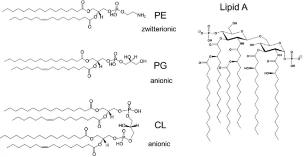
Figure 2. Major membrane lipids of E. coli K-12.
The structures of the most abundant membrane lipids of E. coli K-12 are depicted. (Left) The zwitterionic phospholipid phosphatidylethanolamine (PE; ~75% of total lipid by weight) as well as the anionic phospholipids phosphatidylglycerol (PG; ~20%) and cardiolipin (CL; ~5%) are the major lipids of the IM. They are also found in similar proportions in the inner leaflet of the OM. (Right) The asymmetric OM bilayer consists of LPS whose lipid A moiety makes the outer leaflet of the OM. (modified from May and Silhavy, 2017)
The phospholipids (PL) are produced in the cytosol by a machinery that is very conserved among bacteria (and from which it has actually spread to plants and protozoans by gene transfer, Zhang and Rock, 2008). Lipid composition of membranes (Fig. 2) may be regulated in response to environmental changes, with respect to lipid head-group, acyl chain length and degree of acyl chain saturation (Zhang and Rock, 2008). PL composition not only affects the physicochemical properties of the membrane but
may also drive the localization of proteins (Strahl and Errington, 2017). Finally, specific protein:lipid interactions have been shown to contribute to protein topology or function (Bogdanov et al., 2014; Gold et al., 2010; Hertle et al., 1997).
In bacteria (and in energetic eukaryotic organelles), the cytoplasmic membrane is energized with a proton gradient across the plasma membrane. This proton gradient is maintained by the respiratory chain ATPase and is called the proton-motive force (PMF). This source of energy entails two components, Δψ and ΔpH, which relate to the electric and the chemical gradients, respectively. PMF is used to fuel transport of small molecules and of proteins across the cytoplasmic membrane (see section 2.1.3) and across the outer-membrane as well (see section 1.1.4).
1.1.2. The periplasm
The periplasm was originally defined in diderm bacteria to account for the space between the IM and the outer membrane (OM). Some pictures occasionally showed a peptidoglycan layer detached from the OM. The periplasm is made accessible to small molecules from the extracellular milieu via major porins, whose production is regulated in response to environmental changes, sometimes also upon antibiotic exposure.
The periplasm is the compartment where all proteins from the general secretion pathway fold, unless they need to be maintained in a competent state to translocate across the OM. Soluble proteins SurA, FkpA and Skp are the general chaperones in E. coli periplasm while Spy and other chaperones seem to play more specific roles (Goemans et al., 2014). DegP contributes to periplasm homeostasis by playing a dual role, both as a chaperone and a temperature-dependent protease (Krojer et al., 2008a, 2008b; Meltzer et al., 2008; Spiess et al., 1999). Finally, lipoproteins of the OM are shuttled from the IM by the protein LolA (Fig. 3).
The periplasm is devoid of energy and chaperones cannot promote folding by ATP-regulated interactions (Clare and Saibil, 2013; Mas and Hiller, 2018). Instead, a protein like Spy uses loose electrostatic binding to help client proteins to fold (Clark and Elcock, 2016).
What about monoderm bacteria? In the recent years, evidence has been published that S. aureus, B. subtilis and probably monoderms as a whole display a periplasmic space in-between their cytoplasmic membrane and peptidoglycan (Matias and Beveridge, 2006, 2008). Lipoteichoic acids fill the periplasmic compartment of monoderms which is very different from the periplasm in diderms. Still, in support of the comparison, SurA homolog PrsA is a membrane-tethered chaperone that contributes to secretion (Lin et al., 2018; Wahlström et al., 2003).
1.1.3. The peptidoglycan
The peptidoglycan is bacteria’s very own protective structure. This gigantic molecule called murein is built outside the cell by periplasmic transglycosylases and transpeptidases from units synthesized in the cytoplasm and the inner leaflet of the IM (see below). This exoskeleton forms a so-called sacculus that helps withstanding osmotic pressure around all bacterial cells, with the exception of some intracellular bacteria (Otten et al., 2018).
As a distinct physical barrier, the PG comprises anywhere between a few to dozens of so-called layers, depending on bacterial source and growth state (Höltje, 1998). For example, its thickness has been estimated to 2-to-5 layers in E. coli depending on the growth phase (Leduc et al., 1989) but as little as a single layer was observed in a cryoelectromicroscopy study (Gan et al., 2008). In monoderm bacteria where PG is in contact with the outside, it is functionalized by the covalent attachment of proteins (Jacobitz et al., 2017).
Under selective conditions, it is possible to maintain so-called L-forms of monoderm or diderm bacteria that are devoid of murein: these can also occur naturally, e.g. under the pressure of PG-targeting antibiotics to which they are resistant (Errington et al., 2016). L-forms were used to solve a long-time question: by inactivating the gene murC of the PG precursor synthesis pathway and the restoring it, Kawai et al. (2014) were able to show that no template is necessary and a rod-shape is self-determined in B. subtilis by the balanced activities of the elongasome and divisome PG synthetic machineries (see below).
1.1.4. The outer membrane
In diderm bacteria, the OM bilayer is attached to PG by the so-called Braun’s lipoprotein, Lpp (Braun and Rehn, 1969) on the periplasmic side. As a result, OM proteins may bind PG, e.g. OmpA (Wang et al., 2016). The OM is an asymmetric bilayer whose inner leaflet is made of the same PL as the IM and whose outer leaflet is assembled from lipopolysaccharides (LPS), a distinctive feature (Fig. 2), toward the outside. OM asymmetry is vital to bacteria (Malinverni and Silhavy, 2009; Sutterlin et al., 2016)
LPS is synthesized at the IM, shuttled to the outer leaflet of the OM and assembled in tight arrays by the Lpt pathway. LPS is a major contributor to OM impermeability, sugar moieties keeping hydrophobic molecules away and the lipid cores keeping hydrophilic molecules outside. Indeed, increased antibiotic sentivity is associated with LPS defect in the OM (Sutterlin et al., 2014 for an example). It is also called the endotoxin as the strong immune response it provokes, sometimes causes a septic shock.
The OM is functionalized with proteins that are inserted by the essential β-barrel assembly machine (BAM) (Noinaj et al., 2017) or the translocation and assembly module (TAM) (Stubenrauch and Lithgow, 2019).
Finally, no more energy is stored in the OM than in the periplasm. Still, processes taking place at the OM can be energized using the PMF at the IM. Such is the role of the TonB:ExbB:ExbD complex in the IM, which provides energy through the periplasm to TonB-dependent transporters, a family of β-barrel transporters bearing a semi-conserved 5-residueTonB-box near the N-terminus (Noinaj et al., 2010).
Figure 3. OM biogenesis machinery.
Depicted are the components of three essential cellular machines required for outer membrane (OM) biogenesis. β-barrel proteins and lipoproteins are made initially in the cytoplasm in precursor form with a cleavable signal peptide that directs these precursors to the Sec machinery for translocation across the IM. Chaperones, e.g. SurA deliver β-barrel outer membrane proteins to the β-barrel assembly machine (BAM) for insertion into the OM. For OM lipoproteins, after the signal sequence is removed and lipids are attached to the amino-terminal cysteine residue, the Lol machinery delivers them to the OM. The LipoPolysaccharide Transport (LPT) pathway transports LPS from its site of synthesis in the IM to the cell surface via a hydrophobic conduit formed by oligomerized LptA. (modifed from Konovalova et al., 2017)
1.1.5. Surface polysaccharides
Sugars make a quantitative contribution to the bacterial envelope. They are found in permanent structures like PG, LPS (Bertani and Ruiz, 2018) and TA (van der Es et al., 2017) as well as diversified glycan moieties of proteins that make so-called S-layers (Schäffer and Messner, 2017). All of these are assembled from the same initial cytoplasmic building block that is divergently modified and flipped across the IM upon completion.
Surface-exposed polysaccharides can modulate the immune response of the host like has been found e.g. in P. aeruginosa (McCarthy et al., 2017).
Figure 4. Peptidoglycan synthesis in W. viridescens and S. aureus
The peptidoglycan is the result of the polymerization of a pentapeptide dinucleotide subunit by transpeptidation and transglycosylation reactions. Conserved features of the pentapeptide stem linked to the D-lactyl group of MurNAc include (i) alternate amino acids of the L and D configuration, except for the C-terminal residues (D-alanyl-D-alanine), (ii) presence of a diamino acid residue at the third position (L-lysyl, L-ornithyl, or meso-diaminopimelyl), and (iii) linkage of the latter residue to the side chain carboxyl group of the second residue (γ-D-glutamyl). The linear pentapeptide stem is assembled in the cytoplasm by the stepwise addition of amino acids and of the dipeptide D-Ala-D-Ala onto UDP-MurNAc by the Mur synthetases to form the UDP-MurNAc-pentapeptide precursor. Unlike Fem transferases, Mur synthetases catalyze amide or peptide bond formation by a mechanism involving ATP-dependent activation of carboxyl groups via formation of acyl phosphates. The membrane-associated steps of peptidoglycan synthesis are initiated by the transfer of the phospho-MurNAc-pentapeptide moiety from the nucleotide to the lipid carrier undecaprenyl-phosphate to form lipid intermediate I (undecaprenyl-P-P-MurNAc-pentapeptide). Following addition of GlcNAc to form lipid intermediate II (undecaprenyl-P-P-MurNAc[pentapeptide]-GlcNAc), the complete disaccharide-peptide subunit is translocated to the cell surface and delivered to the peptidoglycan polymerization complexes containing the essential glycosyltransferase and D,D-transpeptidase activities. Insets show side chain synthesis by Fem transferase in W. viridescens and S. aureus. The arrows indicate the direction of the CO → NH pep de bonds. C55 , undecaprenyl lipid carrier.
1.2. Making the peptidoglycan mesh
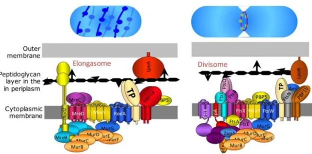
The PG is built from a highly conserved disaccharidic-pentapeptidic brick whose pieces are brought together by the Mur ligases on the inner leaflet of the cytoplasmic membrane (Fig. 4 for details). After the brick has been assembled and flipped to the outer leaflet by flippases such as FtsW or RodA in E. coli, it gets incorporated into the PG mesh by transpeptidase (TP) and transglycosylase (TG) activities that are brought together in multienzyme complexes of two sorts, depending on whether they make PG at the division septum (divisome) or in the lateral cell wall (elongasome). Both machineries perform a similar function and display the same essential set of complementary actors that work to rearrange PG as the bacteria grows: (i) one penicillin binding protein (PBP) that carries both TP and TG activities (class A PBP), (ii) a specific lipoprotein partner of this enzyme that was shown to amplify its efficiency (Caveney et al., 2018; Greene et al., 2018; Typas et al., 2010, 2011), (iii) one PBP carrying a morphogenesis module and TP activity (class B PBP), (iv) hydrolytic enzymes (v) one flippase and (vi) scaffolding proteins (Fig. 5).
Figure 5. Complexes responsible for PG synthesis in E. coli during lateral cell-wall growth and division. Peptidoglycan polymerization involves two complexes, the divisome and the elongasome, responsible for septum formation and lateral cell-wall elongation, respectively. These complexes include all the biosynthetic and hydrolytic enzymes required for the incorporation of new subunits into the expanding peptidoglycan net, as well as cytoskeletal proteins, MreB and FtsZ, acting as trailing guides. (reproduced from Hugonnet et al., 2016)
As a result of the coordinated activities of the PG synthetic machineries, the PG mesh is made of glycan strands running circumferentially (Gan et al., 2008; Tocheva et al., 2013; Turner et al., 2018; Verwer et al., 1978) that are cross-bridged by peptide moieties. Peptide cross-bridges have been extensively
characterized using partial digests and mass spectrometry, unraveling an unexpected flexibility in ways cross-linking is achieved, which sometimes causes antibiotic resistance (Mainardi et al., 2008).
Most often, peptide bridges are forged by PBPs that catalyze the condensation between the carboxyl end of the ‘acyl donor’ peptide and the amino function on the side chain of the ‘receiver’ peptide (Fig. 3). The majority of firmicutes produce PG precursors containing an additional side chain linked to the -amino group of a diamino acid residue at the third position of the pentapeptide stem (Schleifer and Kandler, 1972). The addition of glycine and of L-amino acids is performed by a unique family of nonribosomal peptide bond-forming transferases that use aminoacyl~tRNA as substrate (Hegde and Shrader, 2001; Plapp and Strominger, 1970a). These transferases differ by the type, position, and number of amino acids incorporated into the side chain and by the PG precursor used as substrate (Rohrer and Berger-Bächi, 2003). Most FemABX enzymes are loosely bound to the cytoplasmic side of the inner membrane where they modify the lipid II precursor of PG. In contrast, Weissella viridescens FemX had been shown to use UDP-Mur-N-Ac-pentapeptide as a substrate, on which it adds the first of the two alanyl residues present in the PG (Plapp and Strominger, 1970a, 1970b).
The interest in FemABX enzymes has been boosted when it was observed that the inactivation of non essential enzymes of the FemABX family restored methicillin susceptibility in methicillin-resistant S. aureus (Strandén et al., 1997). Acquired resistance in firmicutes is mostly due to the production of so-called low-affinity PBPs that display a narrow substrate specificity: the addition of a complete side chain to the PG precursors is essential for β-lactam resistance mediated by such low-affinity PBPs in Staphylococcus aureus (Rohrer and Berger-Bächi, 2003) but also Streptococcus pneumoniae (Filipe et al., 2001; Hakenbeck, 2001) and Enterococcus faecalis to a lesser extent (Bouhss et al., 2002). Transferases of the Fem family are therefore considered as potential targets for the development of novel antibiotics which would rehabilitate β-lactam antibiotics against β-lactam-resistant bacteria (Kopp et al., 1996).
1.3. Focus on FemABX enzymes, that make the PG branching peptide in firmicutes
The Weissella viridescens FemX transferase (FemXWv) has been selected as a model enzyme to
elucidate kinetic and structural properties of FemABX enzymes because it catalyzes the transfer of L-Ala onto the cytoplasmic precursor UDP-N-acetyl-muramyl-pentapeptide (UDP-MurNAc-pentapeptide, UM5K) (Hegde and Shrader, 2001; Plapp and Strominger, 1970a), in contrast to other members of the Fem family, which preferentially (Bouhss et al., 2002) or exclusively (Plapp and Strominger, 1970b; Schneider et al., 2004) transfer amino acids to membrane bound peptidoglycan precursors. The research program on FemXWv has been started by Michel Arthur who purified the
enzyme from a batch of W. viridescens and cloned the corresponding gene (Bouhss et al., 2001). Working in collaboration with structural biologists and synthesis chemists, I helped advance the structure:function characterization of FemX (Biarrotte-Sorin et al., 2003, 2004; Maillard et al., 2005; Villet et al., 2007)
1.3.1. Preliminary characterization of W. viridescens FemX catalytic mechanism
W. viridescens FemX is a 336 residue-long soluble protein. Kinetic studies by others have revealed an ordered mechanism involving (i) sequential binding of UDP-Mur-NAc-pentapeptide with a lysyl side chain at position 3 in the pentapeptidic stem (UM5K) and Ala~tRNA, (ii) transfer of L-Ala to the ε-amino group of L-Lys in the pentapeptide stem, and (iii) release of the tRNA and UDP-MurNAc-hexapeptide reaction products (Hegde and Blanchard, 2003; Hegde and Shrader, 2001). Site-directed mutagenesis had yielded identified Asp108 as a candidate for the catalytic base, as the Asp108Asn substitution decreased the catalytic efficiency (V/K) of the enzyme 230-fold, primarily due to a 60-fold decrease in kcat (Hegde and Blanchard, 2003).
We developed our own enzymatic assay by quantifying the transfer of 14C labelled alanine to UM5K:
the UDP-N-acetylmuramic-hexapeptide (UM5K-Ala) product was separated from unreacted alanine by paper chromatography and radioactive spots corresponding to both reacted and unreacted alanine were located by autoradiography, cut out and quantified with a scintillation counter (Maillard et al., 2005). A coupled reaction was set up to ensure a constant supply of the short-lived Ala~tRNA substrate: the tRNA was completely acylated (90%) during the entire 10 minutes reactions, as judged by direct determination of 14C Ala~tRNA. Our results were comparable to those already published (Hegde and
Blanchard, 2003), with for a slightly higher kcat (1,600 versus 660 min-1) and a lower Km for Ala~tRNA
(1.6 versus 15 µM), which we explained by the use of an extensively purified tRNA preparation as opposed to crude in vitro transcription reaction products. Also, the amount of acylated tRNA at steady state was not reported in the first study.
Figure 6. Coupled enzymatic assay set-up to detect and measure FemX activity.
Paper chromatography followed by scintillation counting allowed to quantify the incorporation of 14C Ala into 14C UDP-MurNAc-hexapeptide (UM5K-L-Ala). See text.
1.3.2. Use of truncated substrates allowed to raise a first imprint of FemX catalytic site
FemX first substrate is UM5K. By assessing FemX activity with purified precursors and break-down products of its natural substrate after chemical or enzymatic cleavage, I have identified the chemical groups that are important for FemX:UM5K interaction (Table 1). This seemed to show that the phosphate groups of the UDP nucleotide and the last two aminoacyl residues of the pentapeptide were instrumental in substrate binding (Maillard et al., 2005).
Truncated substrates Code name FemX activity
Ala-Glu-Lys-Ala-Ala pentapeptide not detected P-MurNAc-Ala-Glu-Lys-Ala-Ala P-MurNAc-5K 0.02% P-P-MurNAc-Ala-Glu-Lys-Ala-Ala PP-MurNAc-5K 0.15% UMP-P-MurNAc-Ala-Glu-Lys-Ala-Ala UM5K 100%
UMP-P-MurNAc-Ala-Glu-Lys-Ala UM4K 0.03%
UMP-P-MurNAc-Ala-Glu-Lys UM3K not detected
Table 1. Relative activity of FemX towards truncated substrates
1.3.3. FemX crystal structures in the apo and PG precursor-bound forms
The FemABX family was structurally undefined as we started collaborating with Claudine Mayer (Université Paris 6) and her PhD student Sabrina Biarrotte-Sorin. Protocols allowing quantitative production, selenomethionine incorporation and purification for W. viridescens FemX have been established and our collaborators eventually crystallized FemX apo and UM5K-bound forms (Biarrotte-Sorin et al., 2003).
W. viridescens FemX structure was obtained at 1.7 Å resolution, showing two domains packed together in a globular object. As had been reported for S. aureus FemA (Benson et al., 2002), structural similarity with the acetyltransferases of the Gcn5-related N-acetyl transferase (GNAT) superfamily, which catalyze the transfer of the acetyl group from acetyl coenzyme A to a primary amine (Vetting et al., 2005), was also detected for FemX (Biarrotte-Sorin et al., 2004). Structural deviations between the C-termini of FemX domains and genuine GNAT enzymes coincided with divergence already described for the GNAT fold (Dyda et al., 2000). FemABX enzymes are not the only case of GNAT domain duplication , but the particularity of FemX is that the folding pattern of domain 1 starts with C-terminal residues. A closer look suggested that the FemABX family probably results from a complex evolutionary scenario involving gene duplication, circular permutation and domain insertion. Cirular permutation is considered as a rare event (Vogel and Morea, 2006).
Figure 7. Ribbon representation of FemX in complex with the UDP-MurNAc-pentapeptide.
GNAT-related domains are colored in blue (domain 1) or pink (domain 2). The UM5K is shown as ball-and-stick
New to the field was the structure of a FemABX enzyme in complex with its first substrate, at 1.9 Å resolution. UM5K is bound into a cleft at the interface of the two domains, mostly in contact with domain 1 (Fig. 7) inducing minor structural differences to FemX if any (0.2 Å root mean square deviation). Notable differences were observed for Lys36 through stabilization of its amino group in the complex and for Tyr215 and Tyr256 whose hydroxyl groups are displaced by 0.9 and 2.1 Å, respectively as they shift from a mutual interaction to UM5K binding (Fig. 8). UM5K displays a unique bent structure (Fig. 7), the UDP-MurNAc moiety being clearly stabilized by polar and stacking interactions with the protein (Bmean 22.3 Ų) whereas the pentapeptide showed a higher degree of flexibility (Bmean 50.0 Ų).
1.3.4. Partial characterization of FemX active site by site-directed mutagenesis.
Mutagenesis was performed in three phases, based on the more and more precise informations unraveled. First, conserved positions in multiple sequence alignments within the FemABX family have been mutated: Lys305Met has been the most promising hit identified with this approach as it behaved like the wild type during purification but had a ~4000-fold depressed activity. Yet results obtained with other mutants showed that a 3 log inactivation was very partial (unpublished). Second, when a structural homology with the GNAT superfamily has been recognized, I have tested a hypothetic functional similarity between both catalysts by superimposing individual FemX domains on the model
GNAT Esa1 (Yan et al., 2000) and mutated FemX residues that aligned with Esa1 catalytic residues. Mutating these positions only yielded marginal effects (unpublished).
Finally, when the structure of FemX in complex with its first substrate has been determined, a census of all chemical groups of FemX interacting with UM5K has been made. These residues belonged to both domains and their systematic mutagenesis pointed at Arg211 and Lys36, whose mutation to methionine inactivated the enzyme. Both residues take part into a hydrogen-bonds network that involves many groups of the substrate (Biarrotte-Sorin et al., 2004).
Figure 8. Structure of the UDP-MurNAc-pentapeptide-binding cavity of FemXWv.
(A) Schematic representation of the nine FemXWv residues in contact with the substrate through hydrogen (dashed lines) or stacking (arrows) interactions. The estimates of the transferase activity (v/E) were obtained for the mutant proteins with a single amino acid substitution as described in the text. (B) Close view of the superimposition of the FemXWv UDP-MurNAc-pentapeptide-binding cavity in the structures of the apo wild-type enzyme (4), UDP-MurNAc-pentapeptide:apo wild-type enzyme complex (4), and Lys36Met mutant protein (this work). The bound substrate and secondary structures of FemXWv are colored in magenta and dark blue, respectively. The FemXWv side chains of Trp32, Lys36, Trp39, Arg211, Tyr215, and Tyr256 are colored in yellow for the apo wild-type enzyme, in cyan for the complex, and in orange for the Lys36Met mutant.
Three lines of evidence indicate that a complex hydrogen bond network involving Lys36, Arg211 and, to a lesser extent, Tyr215 is critical for the transferase activity of FemXWv. First, Lys36Met and
Arg211Met substitutions depleted (47,000-fold) activity below detectable levels (Fig. 8). Second, comparison of the CD spectra and crystal structures in the case of the Lys36Met substitution indicated
that conformational changes in the mutant proteins cannot account for the loss of activity. Thus, changes in the chemical nature of the residues at positions 36 and 211 were responsible for the loss of activity. Third, analysis of substrate analogs indicated that a loss of the phosphate end or of the C-terminal D-Ala residue severely depleted FemXWv activity (Table 1). Together, these results indicate
that hydrogen interactions between two residues of FemXWv (Lys36 and Arg211) and two regions of
UM5K (phosphate groups and D-Ala residues) constrain the substrate in a bent conformation which is essential for activity.
In spite of their essential role in activity, Lys36 and Arg211 are not highly conserved in members of the Fem family. Looking for concerted residue-changes in multiple sequence alignments in order to reach out beyond UM5K binding site was unsuccessful (Biarrotte-Sorin et al., 2004).
1.3.5. Conclusion
As no catalytic residue was identified, the burning question remained intact… The mechanism of aminoacyl transfer from Ala-tRNA~Ala to UM5K has been identified when the structure of a complex between FemX and a synthetic peptidyl-RNA conjugate mimicking a bisubstrate reaction intermediate was determined (Fonvielle et al., 2013). Structural comparison with FemX:UM5K complex showed little change in protein structure except for a loop that folded back onto the PG precursor and, significantly, in the first three residues of the pentapeptide stem mimick. This movement suggested a rotation of the acyl acceptor lysyl residue toward the tip of the Lys305 side chain, which was proven to carry the catalytic base.
Taking a step back, my work on FemX completed my PhD experience to make me a biochemist, comforting my keen interest in structure-function relationships. Working with Michel Arthur also pulled me in the field of microbiology, which I enjoy very much up to now.
2. Gating the cytoplasmic membrane
Cells are enclosed in a lipidic cytoplasmic membrane which primarily acts as a diffusion barrier for aqueous solutes. Isolation is no long term option, and the entry or detection of such molecules is achieved by membrane-embedded proteins: transporters and receptors control material and information fluxes.
This section is about the universally conserved Sec protein channel of the general secretion pathway. I joined Franck Duong’s project and helped settling his new laboratory at the University of British Columbia where my work contributed to substantiate functionally relevant intramolecular motions of the channel (Maillard et al., 2007; Tam et al., 2005).
2.1. Introducing the essential Sec translocase
Secretion by the Sec translocase is the fate of proteins with an aminoterminal signal peptide (Blobel and Dobberstein, 1975). The so-called general secretion pathway is universally conserved among living organisms but the definition and functional characterization of its components was spear-headed by S. cerevisiae and E. coli models, owing to the accessibility of biochemical reconstitution (Wickner and Schekman, 2005) and the unsurpassed exploratory power of genetic screens (Bieker et al., 1990). Fifteen years ago, model objects from archaea and other sources have emerged that supported the structural characterization of SecYEG / SecYEβ / Sec61αγβ machineries (Rapoport et al., 2017).
2.1.1. The SecYEG translocon at the atomic scale
Biochemical, biophysical and electrophysiological studies have established that the Sec complex serves as the channel through which preproteins traverse the membrane. The structures of SecYEG (~75 kDa) and SecYEβ complexes, respectively from E. coli and the archaea Methanococcus jannaschii, provided new insights into the translocation mechanism (Breyton et al., 2002; Van den Berg et al., 2004).
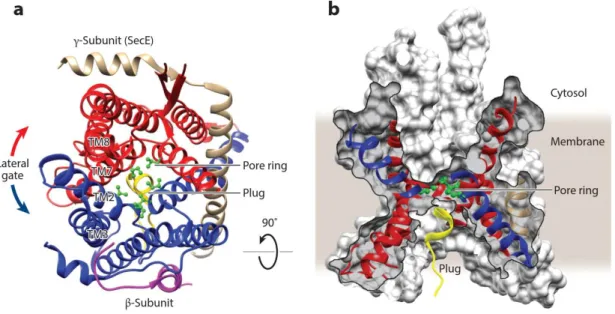
The SecY subunit consists of two subdomains, transmembrane segments TM1–TM5 and TM6–TM10, arranged like a claw and related to each other by a two-fold pseudo-symmetry axis. The essential SecE-subunit docks its TM helix across the interface of the two SecY subdomains, clamping them together.
The proposed translocation channel, in the center of the SecY-subunit, is constricted by a ring of six hydrophobic amino acid residues halfway across the membrane plan, that delineate a 5-8 Å diameter pore. The ring is proposed to seal the channel in dynamic manner during translocation as it should widen enough, probably by shifts in the helices forming the channel, to allow the passage of a


