Effect of Postextubation High-Flow Nasal Oxygen With
Noninvasive Ventilation vs High-Flow Nasal Oxygen Alone
on Reintubation Among Patients at High Risk of Extubation Failure
A Randomized Clinical Trial
Arnaud W. Thille, MD, PhD; Grégoire Muller, MD; Arnaud Gacouin, MD; Rémi Coudroy, MD; Maxens Decavèle, MD; Romain Sonneville, MD, PhD; François Beloncle, MD; Christophe Girault, MD; Laurence Dangers, MD; Alexandre Lautrette, MD, PhD; Séverin Cabasson, MD; Anahita Rouzé, MD; Emmanuel Vivier, MD; Anthony Le Meur, MD; Jean-Damien Ricard, MD, PhD; Keyvan Razazi, MD; Guillaume Barberet, MD; Christine Lebert, MD; Stephan Ehrmann, MD, PhD; Caroline Sabatier, MD; Jeremy Bourenne, MD; Gael Pradel, MD; Pierre Bailly, MD; Nicolas Terzi, MD, PhD; Jean Dellamonica, MD, PhD; Guillaume Lacave, MD; Pierre-Éric Danin, MD; Hodanou Nanadoumgar, MD; Aude Gibelin, MD; Lassane Zanre, MD; Nicolas Deye, MD, PhD; Alexandre Demoule, MD, PhD; Adel Maamar, MD; Mai-Anh Nay, MD; René Robert, MD, PhD; Stéphanie Ragot, PharmD, PhD; Jean-Pierre Frat, MD; for the HIGH-WEAN Study Group and the REVA Research Network
IMPORTANCEHigh-flow nasal oxygen may prevent postextubation respiratory failure in the intensive care unit (ICU). The combination of high-flow nasal oxygen with noninvasive ventilation (NIV) may be an optimal strategy of ventilation to avoid reintubation. OBJECTIVETo determine whether high-flow nasal oxygen with prophylactic NIV applied immediately after extubation could reduce the rate of reintubation, compared with high-flow nasal oxygen alone, in patients at high risk of extubation failure in the ICU.
DESIGN, SETTING, AND PARTICIPANTSMulticenter randomized clinical trial conducted from April 2017 to January 2018 among 641 patients at high risk of extubation failure (ie, older than 65 years or with an underlying cardiac or respiratory disease) at 30 ICUs in France; follow-up was until April 2018.
INTERVENTIONS Patients were randomly assigned to high-flow nasal oxygen alone (n = 306) or high-flow nasal oxygen alternating with NIV (n = 342) immediately after extubation. MAIN OUTCOMES AND MEASURES The primary outcome was the proportion of patients reintubated at day 7; secondary outcomes included postextubation respiratory failure at day 7, reintubation rates up until ICU discharge, and ICU mortality.
RESULTS Among 648 patients who were randomized (mean [SD] age, 70 [10] years; 219 women [34%]), 641 patients completed the trial. The reintubation rate at day 7 was 11.8% (95% CI, 8.4%-15.2%) (40/339) with high-flow nasal oxygen and NIV and 18.2% (95% CI, 13.9%-22.6%) (55/302) with high-flow nasal oxygen alone (difference, −6.4% [95% CI, −12.0% to −0.9%]; P = .02). Among the 11 prespecified secondary outcomes, 6 showed no significant difference. The proportion of patients with postextubation respiratory failure at day 7 (21% vs 29%; difference, −8.7% [95% CI, −15.2% to −1.8%]; P = .01) and reintubation rates up until ICU discharge (12% vs 20%, difference −7.4% [95% CI, −13.2% to −1.8%];
P = .009) were significantly lower with high-flow nasal oxygen and NIV than with high-flow
nasal oxygen alone. ICU mortality rates were not significantly different: 6% with high-flow nasal oxygen and NIV and 9% with high-flow nasal oxygen alone (difference, −2.4% [95% CI, −6.7% to 1.7%]; P = .25).
CONCLUSIONS AND RELEVANCEIn mechanically ventilated patients at high risk of extubation failure, the use of high-flow nasal oxygen with NIV immediately after extubation significantly decreased the risk of reintubation compared with high-flow nasal oxygen alone.
TRIAL REGISTRATIONClinicalTrials.gov Identifier:NCT03121482
JAMA. 2019;322(15):1465-1475. doi:10.1001/jama.2019.14901 Published online October 2, 2019. Corrected on February 25, 2020.
Visual Abstract Editorialpage 1455
Supplemental content CME Quiz at
jamanetwork.com/learning
Author Affiliations: Author
affiliations are listed at the end of this article.
Group Information: The
HIGH-WEAN Study Group and REVA Research Network members are listed at the end of the article.
Corresponding Author: Arnaud W.
Thille, MD, PhD, Médecine Intensive Réanimation, CHU de Poitiers, 2 rue la Milétrie, 86021 Poitiers Cedex, France (aw.thille@gmail.com).
Section Editor: Derek C. Angus, MD,
MPH, Associate Editor, JAMA (angusdc@upmc.edu).
JAMA |
Original Investigation
|
CARING FOR THE CRITICALLY ILL PATIENT
I
n intensive care units (ICUs), approximately 10% to 15% of patients ready to be separated from a ventilator experi-ence extubation failure leading to reintubation.1In pa-tients considered at high risk, these rates can even exceed 20%.1,2Because reintubation is associated with particularly high mortality,3,4a strategy of oxygenation aimed at avoiding reintubation deserves consideration. Although noninvasive ventilation may prevent postextubation respiratory failure in patients at high risk,5-9only 2 small-scale randomized clini-cal trials (RCTs) have shown decreased reintubation rates com-pared with standard oxygen.5,6The most recent international clinical practice guidelines recommend the use of noninva-sive ventilation to prevent postextubation respiratory failure in patients at high risk of extubation failure.10However, up un-til now, no large-scale RCT has demonstrated a significant re-duction of reintubation rates with noninvasive ventilation com-pared with standard oxygen. Thereby, most patients are treated with standard oxygen in clinical practice and only 10% of them receive noninvasive ventilation after extubation in the ICU.2,11 High-flow nasal oxygen is an alternative strategy that may reduce the risk of reintubation in the ICU compared with stan-dard oxygen.12,13
A large-scale RCT has reported that high-flow nasal oxygen was noninferior to noninvasive ventilation in preventing reintubation in patients at high risk.14Whereas high-flow nasal oxygen could be considered as a reference treat-ment after extubation, using high-flow nasal oxygen with non-invasive ventilation may further improve gas exchange and the work of breathing,15thereby avoiding reintubation.
This multicenter RCT involving patients at high risk of ex-tubation failure in the ICU was conducted to determine whether high-flow nasal oxygen with noninvasive ventilation, com-pared with high-flow nasal oxygen alone, after extubation could reduce the rate of reintubation.
Methods
The study was conducted in 30 ICUs in France. For all the cen-ters, the study protocol (Supplement 1) was approved by the central ethics committee (Ethics Committee Ouest III, Poitiers, France; registration No. 2016-A01078-43). Written informed consent was obtained from all patients or next of kin before inclusion in the study.
Adult patients intubated more than 24 hours in the ICU and ready for extubation, after a successful spontaneous breath-ing trial performed accordbreath-ing to the international conference consensus on weaning,16were enrolled if they were at high risk of extubation failure (ie, older than 65 years or had any un-derlying chronic cardiac or lung disease).10Underlying chronic cardiac diseases included left ventricular dysfunction, what-ever the cause, defined by left ventricular ejection fraction equal to or below 45%; history of cardiogenic pulmonary edema; documented ischemic heart disease; or permanent atrial fibrillation. Underlying chronic lung diseases included chronic obstructive pulmonary disease, obesity-hypoventila-tion syndrome, or restrictive pulmonary disease.
The main exclusion criteria were long-term treatment with noninvasive ventilation or continuous positive airway
pres-sure at home, contraindication to noninvasive ventilation, un-derlying chronic neuromuscular disease (myopathy or myas-thenia gravis), or traumatic brain injury leading to intubation, as well as patients who underwent unplanned extubation (ac-cidental or self-extubation) or with a do-not-reintubate order at time of extubation.
The trial was overseen by a steering committee that pre-sented information regarding the progression and monitor-ing of the study at REVA (Réseau Européen de Recherche en Ventilation Artificielle) Network meetings every 6 months. No safety committee was required because the interventions used in the study were strategies of oxygenation typically used in clinical practice. Research assistants regularly monitored all the centers on site to check adherence to the protocol and the accuracy of the data recorded. An investigator at each center was responsible for daily patient screening, enrolling pa-tients in the study, ensuring adherence to the protocol, and completing the electronic case-report form. Although the in-dividual study assignments of the patients could not be masked, the coordinating center and all the investigators re-mained unaware of the study group outcomes until the data were locked in January 2019.
Randomization
Randomization was computer-generated using a centralized web-based management system in permuted blocks of 4 par-ticipants (unknown to investigators), with stratification ac-cording to the center and arterial partial pressure of carbon di-oxide (PaCO2) level (≤45 or >45 mm Hg) measured at the end of the spontaneous breathing trial (or under mechanical ven-tilation before the trial if this latter was not measured). Strati-fication was performed on PaCO2level to include the same num-ber of hypercapnic patients in the 2 groups because noninvasive ventilation may be more effective in these patients.7,8 Pa-tients were randomly assigned in a 1:1 ratio to receive high-flow nasal oxygen alone (control group) or with noninvasive ventilation (intervention group) immediately after extuba-tion (Figure 1).
Interventions
Patients assigned to the control group were continuously treated by high-flow nasal oxygen alone for at least 48 hours
Key Points
QuestionAmong mechanically ventilated patients at high risk of extubation failure, does the use of high-flow nasal oxygen with noninvasive ventilation after extubation reduce the risk of reintubation compared with high-flow nasal oxygen alone?
FindingsIn this randomized clinical trial that included 641 patients, high-flow nasal oxygen with noninvasive ventilation, compared with high-flow nasal oxygen alone, significantly decreased the rate of reintubation within the first 7 days after extubation (11.8% vs 18.2%).
MeaningIn patients at high risk of extubation failure, the use of high-flow nasal oxygen with noninvasive ventilation after extubation significantly decreased the risk of reintubation compared with high-flow nasal oxygen alone.
with a flow of 50 L/min and fraction of inspired oxygen (FIO2) adjusted to obtain adequate oxygenation, with an oxygen satu-ration as measured by pulse oximetry (SpO2) of at least 92%. To provide sufficient humidification, the temperature of the heated humidifier was set at 37°C as during invasive mechani-cal ventilation.
Patients assigned to the intervention group (referred to here as the noninvasive ventilation group) were treated with high-flow nasal oxygen with noninvasive ventilation. Nonin-vasive ventilation was initiated immediately after extubation with a first session of at least 4 hours and minimal duration of at least 12 hours a day during the 48 hours following extuba-tion. Continuous application of noninvasive ventilation was promoted throughout the entire night period. Noninvasive ven-tilation was carried out with an ICU ventilator with noninva-sive ventilation mode or dedicated bilevel ventilator in pressure-support mode with a minimal pressure-support level of 5 cm H2O targeting a tidal volume around 6 to 8 mL/kg of
predicted body weight, a positive end-expiratory pressure level between 5 and 10 cm H2O, and a FIO2adjusted to obtain ad-equate oxygenation (SpO2≥92%). Between noninvasive ven-tilation sessions, high-flow nasal oxygen was delivered as in the control group. Blood gases were performed 1 hour after treatment initiation under high-flow oxygen in the high-flow nasal oxygen alone group and under noninvasive ventilation in the noninvasive ventilation group. In the 2 groups, pa-tients were treated for a minimum of 48 hours. When there were no signs of respiratory failure 48 hours after extubation, treatment was stopped and switched to standard oxygen. According to patient respiratory status, treatment could be con-tinued until complete respiratory recovery. In case of estab-lished postextubation respiratory failure, the use of noninva-sive ventilation was discouraged in accordance with the most recent international clinical practice guidelines,10
given that it has no proven benefit17
and can even increase the risk of death by delaying reintubation.18
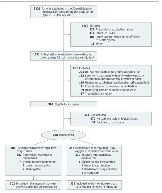
Figure 1. Flow of Patients in the HIGH-Wean Trial of High-Flow Nasal Oxygen With or Without Noninvasive Ventilation
3121 Patients extubated in the 30 participating
intensive care units during the study period (April 2017-January 2018)
1460 Excluded
927 At low risk of extubation failure 414 Intubated <24 h
18 Minor
101 Under law protection or nonaffiliated
to health system
321 Not included
270 No staff available or logistic issues 51 Declined to participate 969 Eligible for inclusion
692 Excluded
274 Do-not-reintubate order at time of extubation 182 Long-term treatment with noninvasive ventilation
or continuous positive airway pressure at home
119 Unplanned extubation (accidental or self-extubation) 41 Contraindication to noninvasive ventilation 39 Underlying chronic neuromuscular disease 37 Traumatic brain injury
648 Randomized
1661 At high risk of reintubation were extubated
after at least 24 h of mechanical ventilationa
302 Included in the intention-to-treat
analysis and in the 90-d follow-up 339 Included in the intention-to-treatanalysis and in the 90-d follow-up
306 Randomized to receive high-flow
oxygen alone
302 Received intervention as
randomized
4 Did not receive intervention 2 Under law protection 2 Missing data
342 Randomized to receive high-flow
oxygen with noninvasive ventilation
339 Received intervention as
randomized
3 Did not receive intervention 1 Under law protection 1 Died before being extubated 1 Missing data
aThose at high risk of reintubation were older than 65 years or had an underlying chronic cardiac or lung disease.
Outcomes
The primary outcome was the proportion of patients who required reintubation within the 7 days following extubation. To ensure the consistency of indications across sites and reduce the risk of delayed intubation, patients were immedi-ately reintubated if 1 of the following criteria was fulfilled: severe respiratory failure, hemodynamic failure with the need for vasopressors, neurological failure (altered con-sciousness with a Glasgow Coma Scale score <12), or cardiac or respiratory arrest. Severe respiratory failure leading to reintubation was defined by the presence of at least 2 criteria among the following: a respiratory rate greater than 35 breaths per minute, clinical signs suggesting respiratory dis-tress with activation of accessory respiratory muscles, respi-ratory acidosis defined as a pH level below 7.25 units and PaCO2greater than 45 mm Hg, hypoxemia defined as a need for FIO2at 80% or more to maintain an SpO2level at 92% or more, or a ratio of the partial pressure of arterial oxygen to the fraction of inspired oxygen (PaO2:FIO2) equal to or below 100 mm Hg.
Secondary outcomes included reintubation at 48 hours, 72 hours, and up until ICU discharge; an episode of postextu-bation respiratory failure within 7 days following extupostextu-bation; the proportion of patients in whom the treatment was con-tinued beyond the first 48 hours following extubation; length of stay in the ICU and in the hospital; and mortality in the ICU, in the hospital, at day 28, and at day 90. An episode of postextubation respiratory failure was defined by the pres-ence of at least 2 criteria among the following: a respiratory rate greater than 25 breaths per minute, clinical signs sug-gesting respiratory distress, respiratory acidosis defined as a pH level less than 7.35 units and PaCO2level greater than 45 mm Hg, hypoxemia defined as a need for FIO2at least 50% to maintain SpO2level of at least 92%, or a PaO2:FIO2ratio equal to or below 150 mm Hg.
Exploratory outcomes included blood gases 1 hour after treatment initiation, time to reintubation, the proportion of pa-tients who met criteria for reintubation, reasons for reintuba-tion, use of noninvasive ventilation as rescue therapy, the pro-portion of patients who were reintubated or died in the ICU, and mortality of reintubated patients.
Statistical Analysis
Enrollment of 590 patients was determined to provide a power of 80% and to show an absolute difference of 8% in the rate of reintubation between the control group using high-flow nasal oxygen alone (rate of reintubation estimated to 18%) compared with the intervention group using high-flow nasal oxygen and noninvasive ventilation (rate of reintu-bation estimated to 10%) at a 2-sided α level of .05 (ie, exactly the same difference as that planned in a previous RCT com-paring high-flow nasal oxygen vs standard oxygen on reintu-bation among patients at low risk of extureintu-bation failure13). To allow for the potential secondary exclusions, the number of patients to be enrolled was then inflated to 650 patients (increased by 10%).
All the analyses were performed by the study statistician according to a predefined statistical analysis plan. The
analy-sis was performed on all randomized patients who were ex-tubated and for whom the primary outcome was completed. Patients were analyzed according to their randomization group regardless of the treatment applied. The proportions of pa-tients having needed reintubation within the 7 days follow-ing planned extubation were compared between the 2 groups by means of the χ2
test.
Kaplan-Meier curves were plotted to assess time from ex-tubation to reinex-tubation and were compared by means of the log-rank test at day 7. Reintubation rates at the various pre-defined times, postextubation respiratory failure rates at day 7, and mortality rates in the ICU and hospital were compared between the 2 groups by means of the χ2
test. Kaplan-Meier curves were plotted to assess the time from extubation to death and were compared by means of the log-rank test at day 90.
A multiple logistic regression analysis was performed for the primary outcome to adjust on the stratification variable (PaCO2level) and on potential baseline unbalanced variables. Lack of balance was defined as P < .05, and the only variable ultimately included in the model was underlying chronic lung disease. The results were presented as odds ratios with 95% CIs. A post hoc random-effects multilevel logistic regres-sion model was used to take into account the effect of the hospital. Treatment group was introduced in the model as a fixed effect and hospital was introduced in the model as a random effect. A subgroup analysis was performed for the primary and secondary outcomes according to the PaCO2 level (≤45 or >45 mm Hg) prior to extubation after an interac-tion test carried out to detect heterogeneity of treatment effect between hypercapnic and nonhypercapnic patients. Because of the potential for type I error due to multiple com-parisons, findings for analyses of secondary end points should be interpreted as exploratory. A 2-tailed P value of less than .05 was considered to indicate statistical signifi-cance. We used SAS software version 9.4 (SAS Institute) for all analyses.
Results
Study Participants
From April 2017 through January 2018, 3121 patients were extubated in the 30 participating units, 969 were eligible for inclusion in the study, and 648 underwent randomization (mean [SD] age, 70 [10] years; 219 women [34%]) (Figure 1). Seven patients were secondarily excluded because they were protected under French law, which was not known at ran-domization (n = 3), died before extubation (n = 1), or were missing data for the primary outcome (n = 3), leaving 641 patients included in the analysis: 302 patients were assigned to high-flow nasal oxygen alone and 339 to high-flow nasal oxygen with non-invasive ventilation.
The characteristics of the patients at inclusion were similar in the 2 groups except for a higher proportion of patients with underlying chronic lung disease in the noninva-sive ventilation group (Table 1). The median duration of mechanical ventilation prior to extubation was 5 days (interquartile range [IQR], 3-10), and weaning was considered
Table 1. Baseline Patient Characteristicsa
Characteristic
No. (%)
High-Flow Nasal Oxygen Alone (n = 302)
High-Flow Nasal Oxygen With NIV (n = 339)
Characteristics of the patients at admission
Age, mean (SD), y 70 (10) 69 (10)
Sex
Men 195 (65) 230 (68)
Women 107 (35) 109 (32)
Body mass index, mean (SD)b 28 (6) 28 (7)
SAPS II score at admission, mean (SD)c 55 (17) 55 (20)
Main reason for intubation
Acute respiratory failure 158 (52) 167 (49)
Coma 55 (18) 57 (17)
Shock 30 (10) 37 (11)
Cardiac arrest 26 (9) 35 (10)
Surgery 28 (9) 35 (10)
Other reason 5 (2) 8 (2)
Risk factors of extubation failure
Age >65 y 223 (74) 237 (70)
Underlying chronic cardiac diseased 145 (48) 161 (47)
Ischemic heart disease 78 (26) 88 (26)
Atrial fibrillation 58 (19) 45 (13)
Left ventricular dysfunction 39 (13) 52 (15)
History of cardiogenic pulmonary edema 21 (7) 25 (7)
Underlying chronic lung diseased 87 (29) 126 (37)
Chronic obstructive pulmonary disease 64 (21) 86 (25)
Obesity-hypoventilation syndrome 16 (5) 20 (6)
Chronic restrictive pulmonary disease 12 (4) 24 (7)
Characteristics of the patients the day of extubation
SOFA score, mean (SD)e 4.2 (2.5) 4.4 (2.7)
Duration of mechanical ventilation, median (IQR), d 5 (3-9) 6 (3-11) Weaning difficultyf Simple 203 (67) 232 (68) Difficult 92 (31) 93 (27) Prolonged 7 (2) 14 (4)
Ventilator settings before the spontaneous breathing trial
Assist-control ventilation 42 (14) 49 (14)
Pressure-support ventilation 260 (86) 290 (86)
Pressure-support level, mean (SD), cm H2O 9.6 (2.8) 9.3 (2.9)
Positive end-expiratory pressure, mean (SD), cm H2O
5.7 (1.6) 5.9 (1.6)
Tidal volume, mean (SD)
mL 468 (125) 479 (144)
mL/kg 7.7 (2.4) 7.9 (2.5)
Respiratory rate, mean (SD), breaths/min 22 (7) 22 (6)
FIO2, mean (SD), % 35 (10) 35 (11) Median (IQR) 30 (30-40) 30 (30-40) PaO2:FIO2, mean (SD), mm Hg 274 (93) 275 (89) pH, mean (SD), units 7.45 (0.06) 7.45 (0.05) PaCO2mean (SD), mm Hg 40 (8) 40 (8) (continued)
difficult or prolonged in 32% of patients (206 of 641 patients). At the time of extubation, 111 patients (17%) had hypercapnia (PaCO2>45 mm Hg).
Initial mean (SD) settings were as follows: in the high-flow nasal oxygen alone group, the gas flow rate was 50 (5) L/min with FIO2of 0.41 (0.13); in the noninvasive ventilation group, the pres-sure-support level was 7.8 (2.5) cm H2O, PEEP was 5.3 (1.1) cm H2O, and FIO2was 0.34 (0.10), resulting in a tidal volume of 8.6 (2.9) mL/kg of predicted body weight.
In the noninvasive ventilation group, noninvasive venti-lation was delivered for a mean (SD) of 22 (9) hours within the first 48 hours following extubation (mean of 13 hours within the first 24 hours) and was delivered for 4 hours or less due to intolerance in 20 patients (6%). In the high-flow nasal oxy-gen alone group, high flow nasal oxyoxy-gen was delivered for a mean (SD) of 42 (11) hours within the first 48 hours.
Primary Outcome
The reintubation rate at day 7 was 11.8% (95% CI, 8.4%-15.2%) with noninvasive ventilation and 18.2% (95% CI, 13.9%-22.6%) with high-flow nasal oxygen alone (difference, −6.4% [95% CI, −12.0 to −0.9]; P = .02) (Figure 2).
Secondary Outcomes
Reintubation rates were also significantly lower with nonin-vasive ventilation than with high-flow nasal oxygen alone at 48 hours, 72 hours, and until ICU discharge (Table 2). The pro-portion of patients with postextubation respiratory failure at day 7 was significantly lower with noninvasive ventilation than with high-flow nasal oxygen alone (21% vs 29%; difference, −8.7% [95% CI, −15.2% to −1.8%]; P = .01). In the noninvasive ventilation group, noninvasive ventilation was continued be-yond the first 48 hours for incomplete recovery of respiratory Table 1. Baseline Patient Characteristicsa(continued)
Characteristic
No. (%)
High-Flow Nasal Oxygen Alone (n = 302)
High-Flow Nasal Oxygen With NIV (n = 339)
Characteristics at the end of the spontaneous breathing trial
T-piece trial 188 (62) 206 (61)
Low level of pressure support 114 (38) 133 (39)
Duration of the trial, median (IQR), min 60 (30-60) 60 (30-60)
Arterial pressure, mean (SD), mm Hg
Systolic 136 (21) 136 (22)
Diastolic 67 (14) 68 (16)
Heart rate, mean (SD), bpm 92 (17) 92 (18)
Respiratory rate, mean (SD), breaths/min 23 (6) 23 (6)
SpO2, mean (SD), % 96 (3) 96 (3) PaO2, mm Hg Mean (SD) [No.] 87 (28) [221] 86 (27) [241] pH, units Mean (SD) [No.] 7.45 (0.06) [221] 7.45 (0.05) [241] PaCO2, mm Hg Mean (SD) [No.] 39 (8) [221] 40 (9) [241]
Administration of steroids before extubation 42 (14) 53 (16)
Ineffective cough, No./No. (%) 65/284 (23) 86/322 (27)
Abundant secretions, No./No. (%) 121/288 (42) 114/326 (35)
Participating centers (n = 30)
No. of centers that participated 30 (100) 30 (100)
No. of patients per center, median (IQR) 10 (9-25) 13 (9-23)
Abbreviations: bpm, beats per minute; FIO2, fraction of inspired oxygen; IQR, interquartile range; NIV, noninvasive ventilation; PaCO2, arterial partial pressure of carbon dioxide; PaO2, partial pressure of arterial oxygen; SAPS, Simplified Acute Physiology Score; SOFA, Sequential (Sepsis-Related) Organ Failure Assessment; SpO2, oxygen saturation as measured by pulse oximetry.
a
The only significant differences in baseline characteristics between the 2 trial groups were the proportion of patients with underlying chronic lung disease (P = .02).
b
Calculated as weight in kilograms divided by height in meters squared. cThe SAPS II score was calculated from 17 variables at admission. Scores range
from 0 to 163, with higher scores indicating more severe disease and higher
mortality risk. Patients with a SAPS II score of 55 at admission have a predicted mean 43% chance of survival.
d
Patients could have more than 1 underlying chronic cardiac or lung disease. eThe SOFA score was calculated from 6 variables the day of extubation. Scores
range from 0 to 24, with higher scores indicating more severe organ failure and higher mortality risk. Patients with a SOFA score between 4 and 5 have a predicted mean chance of survival >90%.
f
Weaning difficulty was defined as follows: simple weaning included patients extubated after success of the first spontaneous breathing trial, difficult weaning included patients who failed the first spontaneous breathing trial and were extubated within the 7 following days, and prolonged weaning included patients extubated more than 7 days after the first spontaneous breathing trial.
status in 86 patients (25%), whereas in the high-flow nasal oxy-gen alone group, high-flow nasal oxyoxy-gen was continued in 106 patients (35%) (difference, −9.7% [95% CI, −16.8% to −2.6%];
P < .01). Mortality in the ICU, in the hospital, and at day 90 were
not significantly different between groups (Table 2; eFigure in Supplement 2).
Exploratory Outcomes
One hour after treatment initiation, PaO2:FiO2was higher with noninvasive ventilation than with high-flow nasal oxygen alone (mean [SD], 291 [97] mm Hg vs 254 [113] mm Hg; difference, 37.0 [95% CI, 19.7 to 54.3]; P < .001), whereas the proportion of patients with hypercapnia did not differ (21% vs 19%, re-spectively; difference, 1.2% [95% CI, −5.4% to 7.7%]; P = .72). The median time to reintubation was not significantly dif-ferent between groups: 33 hours (IQR, 7-81) with noninvasive ventilation and 39 hours (IQR, 12-67) with high-flow nasal oxy-gen alone (difference, −5.0 [95% CI, −42.0 to 32.0]; P = .76).
Among the 100 patients who were reintubated in the ICU, 96% met prespecified criteria for reintubation: 95% (39/41) in the noninvasive ventilation group vs 97% (57/59) in the high-flow nasal oxygen alone group (difference, −1.5% [95% CI, −13.0% to 7.4%]; P = .99). The reason for reintubation was se-vere respiratory failure in 88 patients, neurological failure in 37 patients, hemodynamic failure in 16 patients, and respiratory or cardiac arrest in 10 patients (eTable 1 inSupplement 2).
Among the 88 patients who had postextubation respira-tory failure with high-flow nasal oxygen alone, 28 patients (32%) were treated with noninvasive ventilation as rescue therapy delivered for a mean (SD) of 20 (18) hours, of whom 12 patients (43%) needed reintubation. Results for additional exploratory outcomes are shown in Table 2.
Subgroup Analysis and Additional Analyses
No significant interaction was noted between PaCO2at enroll-ment and treatenroll-ment group with respect to the primary out-come (P for interaction = .25). Among the 111 patients with PaCO2 greater than 45 mm Hg before extubation, the reintubation rate at day 7 was significantly lower with noninvasive ventilation than with high-flow nasal oxygen alone (8% vs 21%; differ-ence, −12.9% [95% CI, −27.1% to −0.1%]; P = .049) (Figure 3). Among the 530 patients with PaCO2of 45 mm Hg or less, rein-tubation rates at day 7 were not significantly different be-tween groups (13% with noninvasive ventilation vs 18% with high-flow nasal oxygen alone; difference, −5.0% [95% CI, −11.2% to 1.1%]; P = .10) (eTables 2, 3, and 4 inSupplement 2).
After adjustment for PaCO2level at enrollment (≤45 or >45 mm Hg, stratification randomization variable) and under-lying chronic lung disease (variable unbalanced between both groups indicated in Table 1), the odds ratio for reintubation at day 7 was significantly lower with noninvasive ventilation than with high-flow nasal oxygen alone (adjusted odds ratio, 0.60 [95% CI, 0.38-0.93]; P = .02). The post hoc analysis showed a lower reintubation rate with noninvasive ventilation than with high-flow nasal oxygen alone after adjustment for the hospi-tal random effect (P = .02 without hospihospi-tal random effect, and
P = .02 after adjustment for the hospital random effect)
(eTable 5 inSupplement 2).
During the study, there were no severe adverse events at-tributable to the randomization group.
Discussion
In this multicenter, randomized, open-label trial, high-flow na-sal oxygen with noninvasive ventilation, compared with high-flow nasal oxygen alone, decreased the rate of reintubation within the first 7 days after extubation in the ICU.
This study was designed to assess noninvasive ventila-tion in a large populaventila-tion of patients who are particularly easy to identify and extubated daily in the different ICUs. Al-though noninvasive ventilation may be beneficial on out-comes of hypercapnic patients,7,8these patients account for only about 20% to 30% of patients at high risk of extubation failure in the ICU.7,19
Patients older than 65 years or with un-derlying chronic cardiac or respiratory disease are also at high risk of reintubation20and could benefit from noninvasive ventilation.21
To our knowledge, the combination of high-flow nasal oxy-gen with noninvasive ventilation had not been previously as-sessed after extubation in the ICU. A preliminary study ob-served a reintubation rate of 15% at day 7 with noninvasive ventilation and standard oxygen in exactly the same population.21
Therefore, the study hypothesized that a new strategy combining high-flow nasal oxygen with noninvasive ventilation could further reduce the rate of reintubation, whereas the estimated rate would exceed 15% in the control group.13,14
For an overall reintubation rate around 15% in the ICU,22
an absolute difference of at least 5% (relative reduc-tion of one-third) would be considered clinically significant and in the range of previous large multicenter RCTs assessing re-intubation as the main outcome.13,23Reintubation rates were Figure 2. Kaplan-Meier Analysis of Time From Extubation
to Reintubation for the Overall Study Population
25 20 15 10 5 0 Patients R equiring R e intubation, %
Time Since Extubation, d High-flow nasal oxygen alone
0 302 339 1 276 321 2 265 314 3 253 308 4 248 305 6 244 292 5 246 294 7 243 291 No. at risk
High-flow nasal oxygen Alone
With noninvasive ventilation
Log-rank P = .02 High-flow nasal oxygen with noninvasive ventilation
The median observation time was 7 days (interquartile range, 7-7) in both treatment groups.
almost exactly the expected rates in the 2 groups (18.2% and 11.8%), reinforcing the external validity of the study.
To date, only 2 RCTs have observed lower reintubation rates with noninvasive ventilation than with standard oxygen.5,6To our knowledge, the present study is the largest RCT showing a reduced risk of reintubation after extubation in the ICU with noninvasive ventilation. Unlike a previous RCT
that reported similar reintubation rates between noninvasive ventilation and high-flow nasal oxygen applied 24 hours af-ter extubation in nonhypercapnic patients,14
this study com-bined noninvasive ventilation and high-flow nasal oxygen for at least 48 hours and treatment was prolonged if necessary. Al-though the beneficial effects of noninvasive ventilation on oxy-genation, alveolar ventilation, and work of breathing are well Table 2. Primary, Secondary, and Exploratory Outcomes
No. (%) Absolute Difference, % (95% CI) P Value High-Flow Nasal Oxygen Alone (n = 302) High-Flow Nasal Oxygen With NIV (n = 339) Primary Outcome
Reintubation at day 7 55 (18) 40 (12) −6.4 (−12.0 to −0.9) .02
Secondary Outcomes
Postextubation respiratory failure at day 7 88 (29) 70 (21) −8.5 (−15.2 to −1.8) .01 Reintubation
At 48 h 36 (12) 24 (7) −4.8 (−9.6 to −0.3) .04
At 72 h 47 (16) 30 (9) −6.7 (−11.9 to −1.7) .009
Up until ICU discharge 59 (20) 41 (12) −7.4 (−13.2 to −1.8) .009
Length of stay, median (IQR), days
In ICU 11 (7 to 19) 12 (7 to 19) 0.5 (−1.6 to 2.6) .55 In hospital 23 (15 to 39) 25 (15 to 42) 2.3 (−1.4 to 6.1) .31 Mortality In ICU 26 (9) 21 (6) −2.4 (−6.7 to 1.7) .25 In hospital 46 (15) 54 (16) 0.7 (−5.0 to 6.3) .80 At day 28 33 (11) 39 (12) 0.6 (−4.4 to 5.5) .82 At day 90 65 (21) 62 (18) −3.2 (−9.5 to 2.9) .30 Exploratory Outcomes
Patients meeting reintubation criteria during ICU stay
65 (22) 49 (14) −7.1 (−13.1 to −1.1) .02
Mortality or reintubation in ICU 64 (21) 51 (15) −6.2 (−12.2 to −0.2) .04
Mortality of reintubated patients 21/59 (36) 11/41 (27) −8.8 (−25.7 to 9.9) .35
Abbreviations: ICU, intensive care unit, IQR, interquartile range; NIV, noninvasive ventilation.
Figure 3. Kaplan-Meier Analysis of Time From Extubation to Reintubation According to Predefined Strata
25 20 15 10 5 0 Patients R equiring R eintubation, %
Time Since Extubation, d High-flow nasal oxygen alone
High-flow nasal oxygen with noninvasive ventilation
High-flow nasal oxygen with noninvasive ventilation 0 48 63 1 44 63 2 44 61 3 39 59 4 38 58 6 37 58 5 37 58 7 37 58 Hypercapnic patients (PaCO2 >45 mm Hg)
A
No. at risk
High-flow nasal oxygen Alone With noninvasive ventilation Log-rank P = .049 25 20 15 10 5 0 Patients R equiring R eintubation, %
Time Since Extubation, d High-flow nasal oxygen alone
0 254 276 1 232 258 2 221 253 3 214 249 4 210 247 6 207 234 5 209 236 7 206 233 Nonhypercapnic patients (PaCO2 ≤45 mm Hg)
B
No. at risk
High-flow nasal oxygen Alone
With noninvasive ventilation
Log-rank P = .11
Results in hypercapnic patients with arterial partial pressure of carbon dioxide (PaCO2) greater than 45 mm Hg (A) and in nonhypercapnic patients with PaCO2 of 45 mm Hg or less (B) are shown. The median observation time was 7 days (interquartile range, 7-7) in both treatment groups.
demonstrated,24,25continuation of high-flow nasal oxygen be-tween sessions of noninvasive ventilation may further pro-vide clinical improvement by decreasing work of breathing.15,26 Approximately one-third of patients treated with high-flow nasal oxygen alone received noninvasive ventilation as rescue therapy in case of postextubation respiratory failure. Although noninvasive ventilation as rescue therapy may avoid reintuba-tion in a number of cases, it has been shown to possibly be del-eterious and increase mortality in this setting.18Moreover, inter-national clinical practice guidelines suggest that noninvasive ven-tilation should not be used in the treatment of patients with established postextubation respiratory failure.10
Limitations
This study has several limitations. First, high-flow nasal oxy-gen rather than standard oxyoxy-gen was used in the control group. However, it has been shown that reintubation rates with high-flow nasal oxygen were reduced as compared with standard oxygen.12,13According to clinical practice in participating cen-ters, the use of standard oxygen alone would have been consid-ered a suboptimal strategy for patients at high risk. Therefore, high-flow nasal oxygen was used in the control group to pro-mote equipoise and facilitate inclusions in different centers.
Second, attending physicians could not be blinded to the study group and this could have modified the decision of re-intubation by promoting early rere-intubation in patients treated with high-flow nasal oxygen alone. However, almost all rein-tubated patients met prespecified criteria for reintubation, and they had particularly high mortality (exceeding 30%), con-firming high severity of patients who were reintubated.
Third, the weaning protocol and the type of spontaneous breathing trial performed before extubation may have influ-enced the results.27In addition, inclusion criteria identifying patients at high risk were different from previous studies.6,13,14 However, international clinical practice guidelines specify that patients at high risk who may benefit from noninvasive ven-tilation are those older than 65 years or who have any under-lying cardiac or respiratory disease.10
Conclusions
In mechanically ventilated patients at high risk of extubation fail-ure, the use of high-flow nasal oxygen with noninvasive venti-lation immediately after extubation significantly decreased the risk of reintubation compared with high-flow nasal oxygen alone.
ARTICLE INFORMATION
Accepted for Publication: September 9, 2019. Published Online: October 2, 2019.
doi:10.1001/jama.2019.14901
Correction: This article was corrected online on
February 25, 2020, to clarify the intervention description in the Abstract and Methods section; to correct data reported for exploratory outcomes in the Results section; and to fix the x-axis labels in Figure 2 and Figure 3.
Author Affiliations: Centre Hospitalier
Universitaire de Poitiers, Médecine Intensive Réanimation, Poitiers, France (Thille, Coudroy, Robert, Frat); INSERM Centre d’Investigation Clinique 1402 ALIVE, Université de Poitiers, Poitiers, France (Thille, Coudroy, Robert, Ragot, Frat); Groupe Hospitalier Régional d’Orléans, Médecine Intensive Réanimation, Orléans, France (Muller, Nay); Centre Hospitalier Universitaire de Rennes, Hôpital Ponchaillou, Service des Maladies Infectieuses et Réanimation Médicale, Rennes, France (Gacouin, Maamar); Groupe Hospitalier Pitié-Salpêtrière Charles Foix, Service de Pneumologie, Médecine Intensive et Réanimation (Département R3S), AP-HP, INSERM, UMRS1158 Neurophysiologie Respiratoire Expérimentale et Clinique, Sorbonne Université, Paris, France (Decavèle, Demoule); Hôpital Bichat–Claude Bernard, Médecine Intensive Réanimation, AP-HP, Université Paris Diderot, Paris, France (Sonneville); Centre Hospitalier Universitaire d’Angers, Département de Médecine Intensive Réanimation, Université d’Angers, Angers, France (Beloncle); Centre Hospitalier Universitaire de Rouen, Hôpital Charles Nicolle, Département de Réanimation Médicale, Normandie Université, UNIROUEN, EA3830-GRHV, Institute for Research and Innovation in Biomedicine (IRIB), Rouen, France (Girault); Centre Hospitalier Universitaire Félix Guyon, Service de Réanimation Polyvalente, Saint Denis de la Réunion, France (Dangers); Centre Hospitalier Universitaire de Clermont-Ferrand,
Hôpital Gabriel Montpied, Service de Réanimation Médicale, Clermont-Ferrand, France (Lautrette); Centre Hospitalier de La Rochelle, Service de Réanimation, La Rochelle, France (Cabasson); Centre Hospitalier Universitaire de Lille, Centre de Réanimation, Université de Lille, Lille, France (Rouzé); Hôpital Saint-Joseph Saint-Luc, Réanimation Polyvalente, Lyon, France (Vivier); Centre Hospitalier Universitaire de Nantes, Médecine Intensive Réanimation, Nantes, France (Le Meur); Hôpital Louis Mourier, Réanimation Médico-Chirurgicale, AP-HP, INSERM, Université Paris Diderot, UMR IAME 1137, Sorbonne Paris Cité, Colombes, France (Ricard); Hôpitaux universitaires Henri Mondor, Service de Réanimation Médicale DHU A-TVB, AP-HP, Créteil, France (Razazi); Groupe Hospitalier Régional Mulhouse Sud Alsace, site Emile Muller, Service de Réanimation Médicale, Mulhouse, France (Barberet); Centre Hospitalier Départemental de Vendée, Service de Médecine Intensive Réanimation, La Roche Sur Yon, France (Lebert); Centre Hospitalier Régional Universitaire de Tours, Médecine Intensive Réanimation, CIC 1415, Réseau CRICS-Trigger SEP, Centre d'étude des pathologies respiratoires, INSERM U1100, Université de Tours, Tours, France (Ehrmann); Centre Hospitalier de Pau, Service de Réanimation, Pau, France (Sabatier); Centre Hospitalier Universitaire La Timone 2, Médecine Intensive Réanimation, Réanimation des Urgences, Aix-Marseille Université, Marseille, France (Bourenne); Centre Hospitalier Henri Mondor d’Aurillac, Service de Réanimation, Aurillac, France (Pradel); Centre Hospitalier Universitaire de Brest, Médecine Intensive Réanimation, Brest, France (Bailly); Centre Hospitalier Universitaire Grenoble Alpes, Médecine Intensive Réanimation, INSERM, Université Grenoble-Alpes, U1042, HP2, Grenoble, France (Terzi); Centre Hospitalier Universitaire de Nice, Médecine Intensive Réanimation, Archet 1, Université Cote d’Azur, Nice, France (Dellamonica); Centre Hospitalier de Versailles, Service de
Réanimation Médico-Chirurgicale, Le Chesnay, France (Lacave); Centre Hospitalier Universitaire de Nice, Réanimation Médico-Chirurgicale Archet 2, INSERM U 1065, Nice, France (Danin); Centre Hospitalier Universitaire de Poitiers, Réanimation Chirurgicale, Poitiers, France (Nanadoumgar); Hôpital Tenon, Réanimation et USC
médico-chirurgicale, CARMAS, AP-HP, Faculté de médecine Sorbonne Université, Collegium Galilée, Paris, France (Gibelin); Centre Hospitalier Emile Roux, Service de Réanimation, Le Puy en Velay, France (Zanre); Hôpital Lariboisière, Réanimation Médicale et Toxicologique, AP-HP, INSERM UMR-S 942, Paris, France (Deye).
Author Contributions: Dr Thille had full access to
all of the data in the study and takes responsibility for the integrity of the data and the accuracy of the data analysis. All authors give their agreement to be accountable for all aspects of the work, and ensure the accuracy and integrity of any part of the work. Concept and design: Thille, Girault, Dellamonica, Lacave, Zanre, Ragot, Frat.
Acquisition, analysis, or interpretation of data: All authors.
Drafting of the manuscript: Thille, Coudroy, Girault, Cabasson, Nanadoumgar, Zanre, Demoule, Ragot, Frat. Critical revision of the manuscript for important intellectual content: Thille, Muller, Gacouin, Coudroy, Decavèle, Sonneville, Beloncle, Girault, Dangers, Lautrette, Rouzé, Vivier, Le Meur, Ricard, Razazi, Barberet, Lebert, Ehrmann, Sabatier, Bourenne, Pradel, Bailly, Terzi, Dellamonica, Lacave, Danin, Gibelin, Zanre, Deye, Demoule, Maamar, Nay, Robert, Ragot, Frat.
Statistical analysis: Thille, Zanre, Ragot. Obtained funding: Thille, Pradel, Zanre.
Administrative, technical, or material support: Thille, Gacouin, Dangers, Rouzé, Le Meur, Razazi, Lebert, Lacave, Zanre, Demoule, Robert.
Supervision: Thille, Zanre, Robert, Ragot.
Conflict of Interest Disclosures: Dr Thille reported
and personal fees and nonfinancial support from Fisher & Paykel Healthcare during the conduct of the study and personal fees from Maquet-Getinge, GE Healthcare, and Covidien outside the submitted work. Dr Sonneville reported receiving grants from the French Ministry of Health, the European Society of Intensive Care Medicine, and the French Society of Intensive Care Medicine and personal fees from Baxter outside the submitted work. Dr Beloncle reported receiving personal fees from Lowenstein Medical and nonfinancial support from GE Healthcare, Getinge Group, and Covidien outside the submitted work. Dr Girault reported receiving grants, personal fees, and nonfinancial support from Fisher & Paykel Healthcare during the conduct of the study and grants and nonfinancial support from ResMed outside the submitted work. Dr Ricard reported receiving travel and accommodation expenses from Fisher & Paykel Healthcare outside the submitted work. Dr Ehrmann reported receiving grants, nonfinancial support, and other funding from Fisher & Paykel Healthcare during the conduct of the study; grants, personal fees, nonfinancial support, and other funding from Aerogen; grants from Hamilton; personal fees from La Diffusion Technique Française; and personal fees from Baxter outside the submitted work. In addition, Dr Ehrmann had a patent to EP17305015 issued. Dr Terzi reported receiving personal fees from Boehringer Ingelheim and Pfizer outside the submitted work. Dr Danin reported receiving fees for lectures from Fisher and Paykel during the conduct of the study. Dr Deye reported receiving lecture and travel fees from Zoll and Bard outside the submitted work. Dr Demoule reported receiving personal fees from Medtronic, Baxter, Hamilton, and Getinge; grants, personal fees, and nonfinancial support from Philips and Lungpacer; personal fees and nonfinancial support from Fisher & Paykel Healthcare; and grants from the French Ministry of Health and Respinor outside the submitted work. Dr Frat reported receiving personal fees and nonfinancial support from Fisher & Paykel Healthcare during the conduct of the study and personal fees and nonfinancial support from SOS Oxygen outside the submitted work. No other disclosures were reported.
Funding/Support: The study was funded by the
“Programme Hospitalier de Recherche Clinique National 2015” of the French Ministry of Health through the University Hospital of Poitiers, Poitiers, France.
Role of the Funder/Sponsor: The funder had no
role in the design and conduct of the study; collection, management, analysis, and interpretation of the data; preparation, review, or approval of the manuscript; and decision to submit the manuscript for publication. Fisher & Paykel provided to all the participating centers the high-flow nasal oxygen equipment and masks for noninvasive ventilation but had no other involvement in the study.
Group Information: Members of the HIGH-WEAN
Study Group and REVA Research Network include the following: Florence Boissier (Centre Hospitalier Universitaire de Poitiers, Médecine Intensive Réanimation, Poitiers), Delphine Chatellier (Centre Hospitalier Universitaire de Poitiers, Médecine Intensive Réanimation, Poitiers), Céline Deletage (Centre Hospitalier Universitaire de Poitiers, Médecine Intensive Réanimation, Poitiers), Carole Guignon (Centre Hospitalier Universitaire de
Poitiers, Médecine Intensive Réanimation, Poitiers), Florent Joly (Centre Hospitalier Universitaire de Poitiers, Médecine Intensive Réanimation, Poitiers), Morgane Olivry (Centre Hospitalier Universitaire de Poitiers, Médecine Intensive Réanimation, Poitiers), Anne Veinstein (Centre Hospitalier Universitaire de Poitiers, Médecine Intensive Réanimation, Poitiers), Dalila Benzekri-Lefevre (Groupe Hospitalier Régional d’Orléans, Médecine Intensive Réanimation, Orléans), Thierry Boulain (Groupe Hospitalier Régional d’Orléans, Médecine Intensive Réanimation, Orléans), Yves Le Tulzo (Centre Hospitalier Universitaire de Rennes, Hôpital Ponchaillou, Service des Maladies Infectieuses et Réanimation Médicale, Rennes), Jean-Marc Tadié (Centre Hospitalier Universitaire de Rennes, Hôpital Ponchaillou, Service des Maladies Infectieuses et Réanimation Médicale, Rennes), Suela Demiri (Groupe Hospitalier Pitié-Salpêtrière Charles Foix, Service de Pneumologie et Réanimation Médicale, AP-HP, Paris), Julien Mayaux (Groupe Hospitalier Pitié-Salpêtrière Charles Foix, Service de Pneumologie et Réanimation Médicale, Paris), Lila Bouadma (Hôpital Bichat–Claude Bernard, Médecine Intensive Réanimation, Paris), Claire Dupuis (Hôpital Bichat–Claude Bernard, Médecine Intensive Réanimation, Paris), Pierre Asfar (Centre Hospitalier Universitaire d’Angers, Département de Médecine Intensive Réanimation, Angers), Marc Pierrot (Centre Hospitalier Universitaire d’Angers, Département de Médecine Intensive Réanimation, Angers), Gaëtan Béduneau (Centre Hospitalier Universitaire de Rouen, Hôpital Charles Nicolle, Département de Réanimation Médicale, Rouen), Déborah Boyer (Centre Hospitalier Universitaire de Rouen, Hôpital Charles Nicolle, Département de Réanimation Médicale, Rouen), Benjamin Delmas (Centre Hospitalier Universitaire Félix Guyon, Service de Réanimation Polyvalente, Saint Denis de la Réunion), Bérénice Puech (Centre Hospitalier Universitaire Félix Guyon, Service de Réanimation Polyvalente, Saint Denis de la Réunion), Konstantinos Bachoumas (Centre Hospitalier Universitaire de Clermont-Ferrand, Hôpital Gabriel Montpied, Clermont-Ferrand), Edouard Soum (Centre Hospitalier Universitaire de Clermont– Ferrand, Hôpital Gabriel Montpied,
Clermont-Ferrand), Marie-Anne Hoppe (Centre Hospitalier de La Rochelle, Service de Réanimation, La Rochelle), Quentin Levrat (Centre Hospitalier de La Rochelle, Service de Réanimation, La Rochelle), Saad Nseir (Centre Hospitalier Universitaire de Lille, Center de Réanimation, Lille), Olivier Pouly (Centre Hospitalier Universitaire de Lille, Center de Réanimation, Lille), Gaël Bourdin (Hôpital Saint-Joseph Saint-Luc, Réanimation Polyvalente, Lyon), Sylvène Rosselli (Hôpital Saint-Joseph Saint-Luc, Réanimation Polyvalente, Lyon), Charlotte Garret (Centre Hospitalier Universitaire de Nantes, Médecine Intensive Réanimation, Nantes), Maelle Martin (Centre Hospitalier Universitaire de Nantes, Médecine Intensive Réanimation, Nantes), Guillaume Berquier (Hôpital Louis Mourier, Réanimation Médico-Chirurgicale, Colombes) Abirami Thiagarajah (Hôpital Louis Mourier, Réanimation Médico-Chirurgicale, Colombes), Guillaume Carteaux (Hôpitaux Universitaires Henri Mondor, Service de Réanimation Médicale, Créteil), Armand Mekontso-Dessap (Hôpitaux Universitaires Henri Mondor, Service de Réanimation Médicale, Créteil), Antoine Poidevin (Groupe Hospitalier Régional Mulhouse Sud Alsace, site Emile Muller, Service de
Réanimation Médicale, Mulhouse), Anne-Florence Dureau (Groupe Hospitalier Régional Mulhouse Sud Alsace, site Emile Muller, Service de Réanimation Médicale, Mulhouse), Marie-Ange Azais (Centre Hospitalier Départemental de Vendée, Service de Médecine Intensive Réanimation, La Roche Sur Yon), Gwenhaël Colin (Centre Hospitalier Départemental de Vendée, Service de Médecine Intensive Réanimation, La Roche Sur Yon), Emmanuelle Mercier (Centre Hospitalier Régional Universitaire de Tours, Médecine Intensive Réanimation, Tours), Marlène Morisseau (Centre Hospitalier Régional Universitaire de Tours, Médecine Intensive Réanimation, Tours), Alexandre Massri (Centre Hospitalier de Pau, Service de Réanimation, Pau), Walter Picard (Centre Hospitalier de Pau, Service de Réanimation, Pau), Marc Gainnier (CHU La Timone 2, Médecine Intensive Réanimation, Marseille), Thi-My-Hue Nguyen (Centre Hospitalier Henri Mondor d’Aurillac, Service de Réanimation, Aurillac), Gwenaël Prat (Centre Hospitalier Universitaire de Brest, Médecine Intensive Réanimation, Brest), Carole Schwebel (Centre Hospitalier Universitaire Grenoble Alpes, Médecine Intensive Réanimation, Grenoble), and Matthieu Buscot (Centre Hospitalier Universitaire de Nice, Réanimation Médicale Archet 1, Université Cote d’Azur, Nice).
Meeting Presentation: Presented at the annual
congress of the European Society of Intensive Care Medicine, October 2, 2019, Berlin, Germany.
Additional Contributions: We thank Jeffrey
Arsham (a translator employed by CHU de Poitiers, Poitiers, France) for reviewing and editing the original English-language manuscript.
Data Sharing Statement: SeeSupplement 3. REFERENCES
1. Thille AW, Richard J-CM, Brochard L. The
decision to extubate in the intensive care unit. Am J Respir Crit Care Med. 2013;187(12):1294-1302. doi: 10.1164/rccm.201208-1523CI
2. Esteban A, Frutos-Vivar F, Muriel A, et al.
Evolution of mortality over time in patients receiving mechanical ventilation. Am J Respir Crit Care Med. 2013;188(2):220-230. doi:10.1164/rccm. 201212-2169OC
3. Epstein SK, Ciubotaru RL, Wong JB. Effect of
failed extubation on the outcome of mechanical ventilation. Chest. 1997;112(1):186-192. doi:10.1378/ chest.112.1.186
4. Frutos-Vivar F, Esteban A, Apezteguia C, et al.
Outcome of reintubated patients after scheduled extubation. J Crit Care. 2011;26(5):502-509. doi:10. 1016/j.jcrc.2010.12.015
5. Ornico SR, Lobo SM, Sanches HS, et al.
Noninvasive ventilation immediately after extubation improves weaning outcome after acute respiratory failure: a randomized controlled trial. Crit Care. 2013;17(2):R39. doi:10.1186/cc12549
6. Nava S, Gregoretti C, Fanfulla F, et al.
Noninvasive ventilation to prevent respiratory failure after extubation in high-risk patients. Crit Care Med. 2005;33(11):2465-2470. doi:10.1097/01. CCM.0000186416.44752.72
7. Ferrer M, Valencia M, Nicolas JM, Bernadich O,
Badia JR, Torres A. Early noninvasive ventilation averts extubation failure in patients at risk: a randomized trial. Am J Respir Crit Care Med.
2006;173(2):164-170. doi: 10.1164/rccm.200505-718OC
8. Ferrer M, Sellarés J, Valencia M, et al.
Non-invasive ventilation after extubation in hypercapnic patients with chronic respiratory disorders: randomised controlled trial. Lancet. 2009;374(9695):1082-1088. doi: 10.1016/S0140-6736(09)61038-2
9. Vargas F, Clavel M, Sanchez-Verlan P, et al.
Intermittent noninvasive ventilation after extubation in patients with chronic respiratory disorders: a multicenter randomized controlled trial (VHYPER). Intensive Care Med. 2017;43(11):1626-1636. doi:10.1007/s00134-017-4785-1
10. Rochwerg B, Brochard L, Elliott MW, et al.
Official ERS/ATS clinical practice guidelines: noninvasive ventilation for acute respiratory failure. Eur Respir J. 2017;50(2):1602426. doi:10.1183/ 13993003.02426-2016
11. Demoule A, Chevret S, Carlucci A, et al;
oVNI Study Group; REVA Network (Research Network in Mechanical Ventilation). Changing use of noninvasive ventilation in critically ill patients: trends over 15 years in francophone countries. Intensive Care Med. 2016;42(1):82-92. doi:10.1007/ s00134-015-4087-4
12. Maggiore SM, Idone FA, Vaschetto R, et al.
Nasal high-flow versus Venturi mask oxygen therapy after extubation. Effects on oxygenation, comfort, and clinical outcome. Am J Respir Crit Care Med. 2014;190(3):282-288. doi:10.1164/rccm. 201402-0364OC
13. Hernández G, Vaquero C, González P, et al.
Effect of postextubation high-flow nasal cannula vs conventional oxygen therapy on reintubation in low-risk patients: a randomized clinical trial. JAMA. 2016;315(13):1354-1361. doi:10.1001/jama.2016.2711
14. Hernández G, Vaquero C, Colinas L, et al. Effect
of postextubation high-flow nasal cannula vs noninvasive ventilation on reintubation and postextubation respiratory failure in high-risk patients: a randomized clinical trial. JAMA. 2016;316 (15):1565-1574. doi:10.1001/jama.2016.14194
15. Di Mussi R, Spadaro S, Stripoli T, et al. High-flow
nasal cannula oxygen therapy decreases postextubation neuroventilatory drive and work of breathing in patients with chronic obstructive pulmonary disease. Crit Care. 2018;22(1):180. doi: 10.1186/s13054-018-2107-9
16. Boles JM, Bion J, Connors A, et al. Weaning
from mechanical ventilation. Eur Respir J. 2007;29 (5):1033-1056. doi:10.1183/09031936.00010206
17. Keenan SP, Powers C, McCormack DG, Block G.
Noninvasive positive-pressure ventilation for postextubation respiratory distress: a randomized controlled trial. JAMA. 2002;287(24):3238-3244. doi:10.1001/jama.287.24.3238
18. Esteban A, Frutos-Vivar F, Ferguson ND, et al.
Noninvasive positive-pressure ventilation for respiratory failure after extubation. N Engl J Med. 2004;350(24):2452-2460. doi:10.1056/ NEJMoa032736
19. Thille AW, Boissier F, Ben Ghezala H, Razazi K,
Mekontso-Dessap A, Brun-Buisson C. Risk factors for and prediction by caregivers of extubation failure in ICU patients: a prospective study. Crit Care Med. 2015;43(3):613-620. doi:10.1097/CCM. 0000000000000748
20. Thille AW, Harrois A, Schortgen F, Brun-Buisson
C, Brochard L. Outcomes of extubation failure in medical intensive care unit patients. Crit Care Med. 2011;39(12):2612-2618. doi:10.1097/CCM. 0b013e3182282a5a
21. Thille AW, Boissier F, Ben-Ghezala H, et al. Easily
identified at-risk patients for extubation failure may
benefit from noninvasive ventilation: a prospective before-after study. Crit Care. 2016;20(1):48. doi:10. 1186/s13054-016-1228-2
22. Thille AW, Cortés-Puch I, Esteban A. Weaning
from the ventilator and extubation in ICU. Curr Opin Crit Care. 2013;19(1):57-64. doi:10.1097/MCC. 0b013e32835c5095
23. François B, Bellissant E, Gissot V, et al;
Association des Réanimateurs du Centre-Ouest (ARCO). 12-H pretreatment with methylpred-nisolone versus placebo for prevention of postextubation laryngeal oedema: a randomised double-blind trial. Lancet. 2007;369(9567):1083-1089. doi:10.1016/S0140-6736(07)60526-1
24. Brochard L, Mancebo J, Wysocki M, et al.
Noninvasive ventilation for acute exacerbations of chronic obstructive pulmonary disease. N Engl J Med. 1995;333(13):817-822. doi:10.1056/
NEJM199509283331301
25. Vitacca M, Ambrosino N, Clini E, et al.
Physiological response to pressure support ventilation delivered before and after extubation in patients not capable of totally spontaneous autonomous breathing. Am J Respir Crit Care Med. 2001;164(4):638-641. doi:10.1164/ajrccm.164.4. 2010046
26. Pisani L, Fasano L, Corcione N, et al. Change in
pulmonary mechanics and the effect on breathing pattern of high flow oxygen therapy in stable hypercapnic COPD. Thorax. 2017;72(4):373-375. doi:10.1136/thoraxjnl-2016-209673
27. Subirà C, Hernández G, Vázquez A, et al. Effect
of pressure support vs T-piece ventilation strategies during spontaneous breathing trials on successful extubation among patients receiving mechanical ventilation: a randomized clinical trial. JAMA. 2019; 321(22):2175-2182. doi:10.1001/jama.2019.7234