by
ROSEMARY SANCHES
B.S., Instituto de Fisica e Quimica de
S. Carlos - USP
Brazil
(1973)
SUBMITTED IN PARTIAL FULFILLMENT
OF THE REQUIREMENTS FOR THE
DEGREE OF
MASTER OF SCIENCE
at the
MASSACHUSETTS INSTITUTE
OF TECHNOLOGY(AUGUST 1977)
Signature
of Author ...'10
DovaU
ment
of Biology, Aug. 1977
Biology, Aug. 1977...Certified
by ..
...
Thesis Supervisor
Accepted by ...
-- -- _j - -ARHIE
`<, ,'.19·' 1~
A 1~ ·· ,IrjChairman, Department of Biology
Graduate Committee
k
?,'> i) i.7
A;Pf5v t& - ¢-. .
.."t
ib~
-2-APPLICATIONS OF RAMAN SPECTROSCOPY by
ROSEMARY SANCHES
Submitted to the Department of Biology
on August 1977 in partial fulfillment of the requirements for the Degree of Master of Science
ABSTRACT I
The frequency of vibration of the C = 0 group in dimethylformamide, N-methylformamide, N-methylacetamide, acetone and methylacetate has been measured in a number of solvents. A marked contrast in the effect of the solvent on the solutes acetone, dimethylformamide and methylacetate on the one hand, and N-methylformamide and N-methylacetamide on the other hand, has been found. This suggests that the measurement of carbonyl stretching frequency as a function of different solvents can be an
effective way to distinguish between solvent exposed and solvent protected
carbonyl groups. In a plot of vc=o (carbonyl stretching vibration
frequency) versus the dielectric constant would indicate free C = 0
groups, while an increase in v 0with , followed by a decrease in
v
c=0
= would indicate partially protected or hydrogen bonded carbonylgroups. The use of Bellamy plots also distinguishes these two different
cases.
ABSTRACT II
The preliminary Raman spectra of Fragment-1 prothrombin plus Ca2+
in the a-helix and random coil contents, and in the carbonyl stretching vibration frequency. These dissimilarities may represent real conforma-tional differences for Fragment-1 with or without Ca2+.
Thesis Supervisor: Paul R. Schimmel
-4-TABLE OF CONTENTS
Page
Introduction ... 10
CHAPTER I:
The Physics of the Phenomenon of Laser Raman
Spectroscopy ... 12
CHAPTER II:
Solvent Effects oh the Carbonyl Stretch Vibration
Frequency... 23
CHAPTER III:
Application of Raman Spectroscopy to Fragment- of
ABBREVIATIONS
CHAPTER I
£ : electric field associated with a wave
6E : maximum electric field
o
v : frequency of oscillation of the wave (exciting light)
exc
t time
: dipole moment
a polarizability
Qi vibrational normal mode
v.i : vibration frequency of the molecule
v : frequency of the scattered light
I : power radiated by a dipole
n vibrational level h Planck's constant k : Boltzmann constant T : absolute temperature CHAPTER II v =0: c V : V =0: c
carbonyl stretching vibration frequency dielectric constant
CHAPTER III Fragments of prothrombin: I-1: F-i: F-2: F-1.2: Intermediate-1 Fragment-1 Fragment-2 Fragment-1.2
LIST OF FIGURES AND TABLES
CHAPTER I
Figure 1 Energy diagram and transitrons for Rayleigh and Raman
effect.
Intensity for Stokes and anti-Stokes lines.
Raman spectroscopy compared to infrared spectroscopy: energy diagram and spectrum.
Energy diagram showing fluorescence phenomenum and how it can be avoided.
Experimental device for Raman spectroscopy.
CHAPTER II
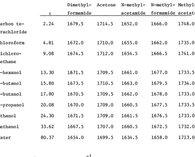
Table I : Lists vcO0 for each solute as a function of solvents.
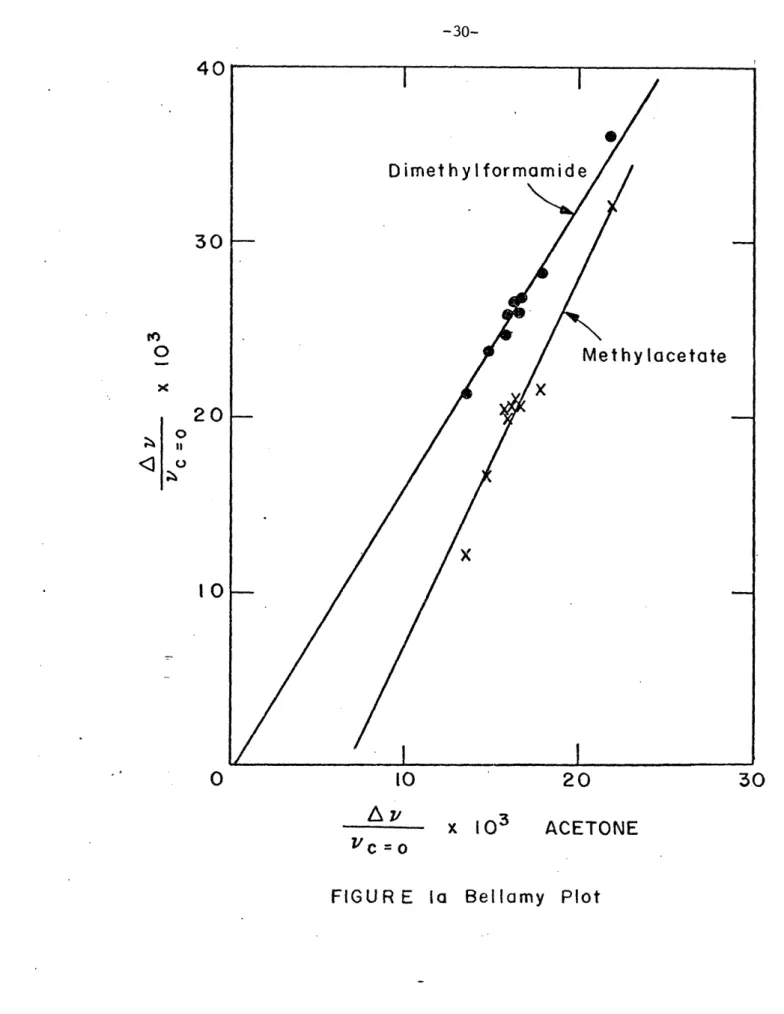
Figure la and Figure lb: Bellamy plot.
Figure 2 to 6: vc=0 versus solvent for all solutes.
CHAPTER III
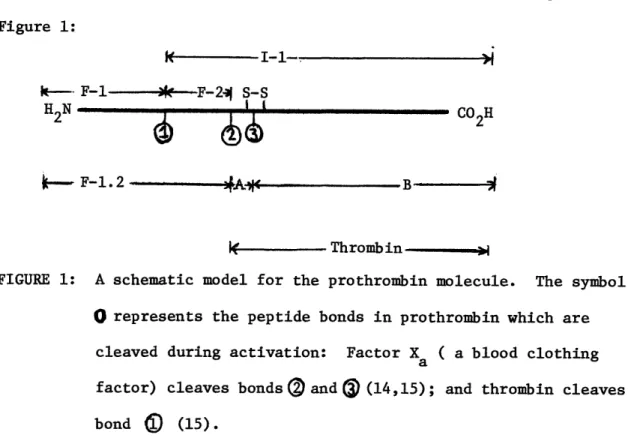
Figure 1 : A schematic model for the prothrombin molecule.
Table 1 : Amino acid composition of prothrombin, Intermediate-i
and Fragment-l.
Spectra of Fragment-i and Fragment 1 + Ca+ +
Spectrum of Buffer Figure Figure 2 3 Figure 4 : Figure 5 :
-8-ACKNOWLEDGEMENTS
My warmest and deepest thanks to Prof. Eugene Stanley, my thesis advisor, whose enthusiasm and encouragement was a vital ingredient in this work.
I am very grateful to Ken Rothschild for his continual help and interest in the preparation of this study.
MY most sincere thanks to Prof. Paul Schimmel and Prof. Richard Lord for suggestions in the manuscript.
I want to recall and acknowledge Prof. Sergio Mascarenhas, who has been my advisor for many years and through whom I discovered and
enjoyed Biophysics.
I am also indebted to all my other friends, from whom I learned immensely.
-10-INTRODUCTION
Raman scattering was discovered nearly 50 years ago, in 1928. During this period, Raman spectroscopy has advanced to take its place among other, older methods of investigating the structure and composition
of matter, and its importance is still increasing.
The kind of information provided by laser Raman spectroscopy consists
essentially of:
1. The frequencies of intra and inter-molecular vibrations in the -l
range 10-4000 cm1.
2. The spectral line or band intensities associated with these
frequencies. The intensity is to a good approximation a linear function of the molar concentration of the sample molecules.
The methods of drawing structural conclusions from this kind of infor-mation are mainly those of reasoning by analogy. Spectra of model
compounds of known conformation and appropriate composition are obtained. Empirical correlations between frequencies (and sometimes intensities)
and conformations can often be worked out and then used for structural studies of new systems. Clearly, this kind of structural study is
vastly inferior to x-ray diffraction when the latter is applicable. Since most biopolymers are hard to obtain in crystalline form, and in any event are not crystalline in vivo, the structural information obtainable from their Raman spectra is potentially very useful.
In chapter I we have a brief description of the Raman effect. A conceptual derivation of the scattering phenomenon is followed by a discussion of the advantages of the technique and then the experimental arrangement is described.
In chapter II we study the dependence of the carbonyl stretch
vibration frequencies upon the environment of the carbonyl groups. The effect of solvent interactions is studied by dissolving a compound
containing the carbonyl group in solvents with different polarity (e.g.,
CC14, CHC13, CH30H,...). Raman spectroscopy may be used to examine the
conformational changes in a molecule as it binds ions or moves into a different chemical environment, and here it is used as a probe of
hydrogen bonding. A further step would be to investigage the ability of compounds to complex selectivity with cations. By studying compounds which bind a wide variety of cations, information about the relative
importance of steric and electrostatic forces in cation-ionophore inter-actions can be obtained.
In chapter III Raman spectroscopy is applied to a biological macro-molecule: a fragment of prothrombin. This fragment is known to be
the relevant part of the protein for the binding of Ca+ + through carbonyl
groups. The Raman spectrum of this fragment in the presence and absence
-12-CHAPTER I: THE PHYSICS OF THE PHENOMENON OF LASER RAMAN SPECTROSCOPY
LASER RAMAN SPECTROSCOPY
The Phenomenon
When light passes through a solution, a fraction of the light is scattered. Most of the scattered radiation has the same frequency as the incident radiation. This scattering, refered to as Rayleigh
scattering, may be viewed as the elastic scattering of a photon by the molecule.
In 1928 Raman (1) discovered experimentally that there was some-times also present in the scattered light weak radiation of
frequencies different than the incident light. The frequency shifts from the incident frequency correspond to the vibrational frequencies of the scattering molecules and hence are a source of information on
molecular structure. Raman scattering may be viewed as inelastic
scattering of a photon by the molecule.
The Interpretation (2)
It is assumed that light is scattered from a free molecule as a result of the induced oscillations of the dipole of the molecule by the irradiating electromagnetic field.
Consider a free molecule subjected to an electromagnetic wave, where
C = o cos(2vex t)
that is, the wave is a monochromatic plane wave. Here, s0 is the
maximum electric field associated with the wave, vexc is the frequency of oscillation of the wave, and t is the time. This wave induces a
-14-dipole moment p in the molecule, where
p = as
a is the polarizability of the molecule, i.e., a measure of the
electron-ic deformation of the electron cloud by the field. The polarizability is a function of the interatomic distances of the molecule and is hence dependent upon the vibrational modes. Then, if the amplitude of the motion is small
a(Qi) o + aQ Qi +
where Qi is a particular vibrational normal mode of the molecule, i.e., Qi is the displacement from equilibrium for some kind of possible
harmonic motion. Qi is assumed periodic in time
Qi = Q cos (27vit)
with vi as the frequency of the vibration. Then
P1
=
+
°a
-
-)Qi cos(2Tvit)
+
..s cos (2vex
c t) =0 cos (27rvexc )
c
exc
Q tcos 2fr(v + vi)t ++cos 2r(v x c - vi)t +
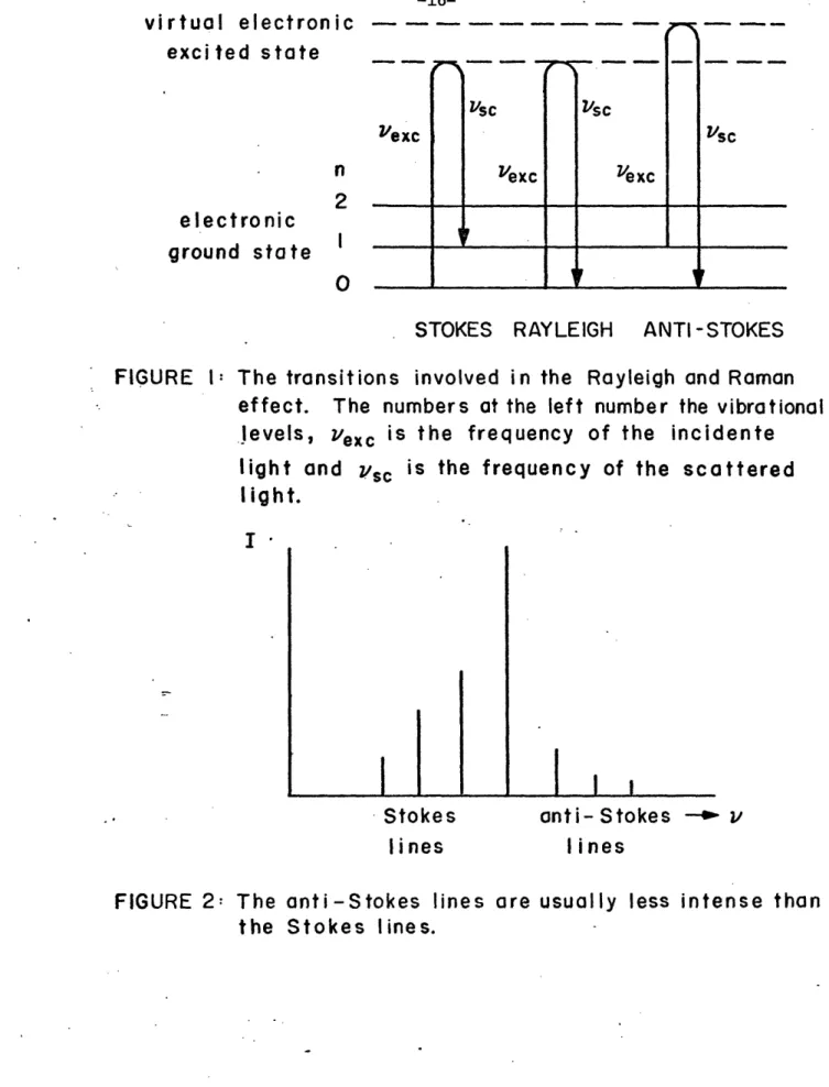
Therefore if the polarizability of the molecule changes periodically in consequence of the vibration, then in the scattered radiation the sum and the difference of the incident frequency and the molecular vibration frequency will appear. These are respectively the anti-Stokes and the S-okes Raman scattering. In other words, if the net transition is
from a lower to a higher energy level, so that v < Vexc (v is the
frequency called of the scattered light), the process Stokesis
scattering; the opposite case, in which v > is termed
anti-sc exc
Stokes scattering (Figure 1).
In addition to the fundamental vibrations in the Raman effect, for which the frequency of a normal vibration has to be added or
subtracted from the frequency of the incident radiation, there are also overtones and combination tones. The reasons for these are mechanical and electrical anharmonicity. This means, there are terms of a
quadratic or higher order in the dependence of thepolarizability on the displacements.
The total power I radiated by an oscillating dipole in classical electromagnetics(3) is
I a Wp|
2Consideration of the energy level diagram (Figure 1) shows that the intensities of the Stokes and anti-Stokes Raman lines are not equal,
as suggested by the preceding classical analysis. According to
quantum theory (4) and in agreement with observation the anti-Stokes lines have a much smaller intensity (Figure 2), since the number of molecules in the initial state n = 1 of the anti-Stokes lines is only exp(-hv /kT) times the number of molecules in the initial state n = 0 of the Stokes lines (ground state). Here n numbers the vibrational
levels, h is Planck's constant, k is the Boltzmann constant, v is the vibration frequency, and T is the absolute temperature. One has to
remember that the selection rule for the vibrational Raman effect is
-16-_ . _
virtual electronic
excited state
Vexcelectronic
ground state
n2
I0
_ _ - --I/vs Vexc'exc
Vs cSTOKES RAYLEIGH ANTI -STOKES
FIGURE 1:
The transitions
involved in the Rayleigh and Raman
effect.
The numbers at the left number the vibrational
levels,
exc
is the frequency of the incidente
light and vsc is the frequency of the scattered
I ight.
I
I I
anti- Stokes
lines
FIGURE 2: The anti-Stokes
lines
are
the Stokes
lines.
usually less intense than
Stokes
lines
!Fr
I
i IF · II · l i-l I I Vsc I ilevels, the other has to be n = 1. The intensity ratio of the anti-Stokes lines to the corresponding anti-Stokes lines is in agreement with the Boltzmann factor.
Comparison with Infrared Spectroscopy
The Raman effect of molecules provides the same kind of information as does infrared absorption, chiefly the characteristic frequencies of molecular vibrations. Only those vibrations that are connected with a
change of dipole moment can give rise to an infrared transition, and only those vibrations that are connected with a linear change of
polarizability can give rise to a Raman transition (4). Since selection rules for the Raman effect differ from those for the infrared absorption (5), the vibrational transitions observed by one method supplement those observed by the other, but sometimes one can be used when the other
cannot. A great impediment to the use of infrared analysis of
biological materials is the very strong absorption by water in the mid and far infrared. Raman spectra are then advantageous for study of biological materials (6-8) in aqueous solution (9).
In infrared spectroscopy one illuminates the sample with a variety of individual wavelengths of infrared light, and measures the trans-mitted intensity at different incident frequencies. In Raman
spectro-scopy one illuminates the sample with a fixed wavelength and measures the scattered intensity as a function of the frequency shift from the incident frequency - Raman spectrum (7, 10). One can repeat Raman
-18-spectrum is identical, so long as one plots ones data as a function of the shift in frequency (Figure 3). Raman spectrum does not change when one changes the exciting frequency, but fluorescence does. In fluor-escence phenomena the laser excites an electronic level in a region
where there are many closely spaced excited levels to which the molecules may decay (Figure 4). Then there is a final decay from some electronic level to the vibrational ground state. Thus the fluorescence signal competes with the Raman signal, and since fluorescence phenomena are generally intense (and since the noise is roughly proportional to the total light emitted) one may have difficulty seeing the Raman
scattered light superposed on the fluorescence signal. One solution to this problem is to vary the frequency of the laser light until one is below the fluorescence band.
Technique
The laser is an ideal source for the excitation of Raman spectra and enables the study of samples as small as 1 mg or less. The Raman spectrum is recorded by directing a laser beam on the sample to be studied (often contained in a capillary) and collecting the scattered light into a monochromator-detector system that can measure the
intensity of the light at different frequencies (Figure 5). The Raman effect is the result of inelastic scattering of the photons in the laser beam by molecules, in which process the energy of each photon may be decreased or increased by transfer of vibrational energy to or from the molecule. If the molecule receives energy (the more likely event) the scattered photons have lost it and thus have lower frequency.
Energy
hZ exc
hvexc
hv
O traonint
INFRAREDabsorbed4
rad.
mif
tr
RAMAN RAMAN _ _ _ _ _ _ ._ _ _ _ _ _ _n w _exciting
rad.
(vexc)
ensity
in
scatt.
rad.
(Zvs )attered
tensity
exciting
rad.
(Yexc )
/
Freq.
1exc Freq.Freq.
3: Infrared spectroscopy measures transmitted intensity
as a function of incident frequency,
while
Raman
spectroscopy measures scattered intensity as
a
function of frequency shift from a fixed incident
light frequency.
scatt.
rad.
(z4c)scattered
intensity
FIGUREI
-I I I B I I -I III I I I I I _ -, -1 F, 'I 9i
!r
-20-excited
electronic
3
2
state
,
2
virtual level
electronic
2
ground
2
state
O
I1
-RAMAN
FIGURE
4: Fluorescenece
-9FLUORESCENCE
phenomena are much stronger than
Raoman scattering.
cases by choosing
Sample
They can be removed in many
a different
laser frequency.
Analyser
Photodetector
5:
The light
scattered
by the sample is collected
large lens and frequency analyzed by a double
grating monochromator.
FIGURE
by a
I _~~~~~~~~~~~~~~~~~~~~~~~~4--~--
11' 1 l= ! = -r I$ T - F ! ! a -,_
When visible radiation is used for excitation, the scattered radiation still lies in or near the visible spectrum because the vibrational energy transferred is a small fraction of the energy of visible photons.
Hence the optical components and instrumentation for Raman spectra are those of visible spectroscopy.
The spectrum is usually recorded with the intensity of the Raman scattering in relative units on the ordinate scale and wavenumber
dis--1
placement in cm from the laser exciting frequency, which is taken
-22-REFERENCES
1. Raman, C.V. (1928) Indian J. Phys. 2, 387
2. Schutte, C.J.H. (1976) The Theory of Molecular Spectroscopy (North Holland, Elsevier)
3. Jackson, J.D. (1962) Classical Electrodynamics (New York, John Wiley)
4. Hibben, J.B. (1939) The Raman Effect and its Applications (New York, Reinhold)
5. Herzberg, G. (1945) Infrared and Raman Spectra (New Jersey, D. Van Nostrand)
6. Koenig, J.L. (1972) J. Polymer; Sci. part D 60, 59
7. Lord, R.C. (1971) XXII Int. Congr. Pure Appl. Chem. 7, 179 8. Whittman, R. (1974) Transport and Diffusion in Red Blood Cells
(London, Edward Arnold)
9. Yu, N.T.; Lui, C.S. and O'Shea, D.C. (1972) J. Mol. Biol. 70, 117 10. Tobin, M.C. (1971) Laser Raman Spectroscopy (New York, John Wiley)
CHAPTER II: SOLVENT EFFECTS ON THE CARBONYL STRETCH VIBRATION FREQUENCY
-24-INTRODUCTION
It has been shown that the stretching frequency of a carbonyl group,vc=O is sensitive to many factors including inductive effects, hydrogen bonding, and ionic interactions (1-5). Hence it is often difficult to draw any conclusions about the local interactions of a
carbonyl group from the absolute value of vc=O . We are interested in
investigating whether the changes of vc=O as a function of different dielectric and polar solvents can be used to probe the local environ-ment of a carbonyl group. For example, it has been found that the
vc 0o of intramolecular hydrogen bonded amide carbonyl groups of
valinomycin increases in frequency with a increase in the dielectric of the solvent, whereas non-hydrogen bonding ester carbonyl groups decrease in frequency. It is not known, however, to what extent these changes are due to direct solvent effects on the carbonyl groups versus induced conformational changes of the molecule (6).
One of the first attempts to relate vc = 0 to changes in properties of the solvent medium was made by Kirkwood, Bauer and Magat (7,8). Their
theory relates the frequency shift Av , where Av c=0 c=0)and vc=O is the
carbonyl stretch frequency of the vapor phase, to the dielectric
constant of the solvent by
Av C- (1)
vc=O = 2 + 1
where the factor C is complex and depends on details of the model used for the vibrating solute dipole. In a test of this theory, Thompson
has shown (9) that if Av/vc=0 is plotted against (-1)/(2e+1), a straight line can be drawn from the origin which passes through points
corresponding to nonpolar solvents he measured, but not for most other solvents. He suggested that the deviation does not seem to arise from hydrogen bonding, since this should lower vc=0 and increase Av(l), whereas the converse is found.
Detailed studies by Josien et al. (10-16) and by Bayliss et al. (17,18) have also shown there is a relationship between frequency shifts and bulk solvent properties, in those cases where such specific inter-actions such as hydrogen bonding are minimal. Bellamy (19,20) has
studied the carbonyl frequencies of a wide range of compounds in many solvents and concluded that all the frequency shifts follow a common pattern and seem to be produced mainly by local association effects, and not by dielectric constant factors. The order of solvent effectiveness
in lowering vc=O is therefore always the same, and a quantitative relationship exists between the effects of one solvent and another. Thus, the relative frequency shifts (Av/Vc=0 ) of any one carbonyl group in a series of solvents can be plotted directly against the values for some other carbonyl group in the same solvents, to give a straight line. The failure of the slopes of these lines to correlate directly with the proton-accepting powers of the carbonyl group, shows that the use of relative carbonyl shifts cannot safely be used as a measure of the hydrogen bonding powers of different solvents.
The effect of the solvent on vc=0 for various carbonyl compounds has been examined in terms of bulk dielectric constant also by
-26-Kagarise (21). He suggested that dispersion forces, bulk dielectric effects and specific interaction effects all make a contribution to
Av . He suggested that the stronger the proton donating tendency of the
solvent, the greater the proportion of Av arising from the formation of stable compounds possessing large interaction energies and well defined structures.
Although these studies indicate that vc=0 cannot be directly inter-preted easily in terms of microscopic interactions, it is possible that
vc=O for a group of solvents can be used to distinguish between two
different classes of carbonyl groups, i.e., those strongly hydrogen bonded and those non-hydrogen bonded. For this purpose, we have studied c=O for three compounds, dimethylformamide
(CH3 0O), acetone(CH3 ), and methylacetate (CH3-0
N-C C=O C=O
CH3 H CH3 CH3
which do not form strong intermolecular bonds with themselves, and
two compounds, N-methylformamide (CH3 0) and N-methylacetamide
N-C
H H
(CH3 O) which can form hydrogen bonds with themselves.
N-C
/ I
MATERIALS AND METHODS
The solvents used were: carbon tetrachloride, chloroform, di-chloromethane, 1-hexanol, 2-butanol, 1-butanol, -propanol, ethanol, methanol, and water. The solvents and solutes were obtained from Baker
and Allied Chemical and had purity for spectroscopic studies.
The solutions were prepared immediately prior to usage and sealed in glass capillaries. They were centrifuged to eliminate bubbles. The molar ratio of solute to solvent was 1/8 for all samples.
Raman spectra were measured using a Spex Ramalog 4 system and a Spectra-Physics Model 164-03 Ar laser (488 nm excitation). The incident
power was 300-400 mw, resolution 3-4 cm and scanning speed 1.2-30cm-1
/
min.
Only the Raman scattered light with polarization parallel to the polarization of the incident light was measured (the laser light used
to illuminate the sample is typically plane polarized perpendicular to the plane determined by the incident light beam and the entrance slit of the spectrometer).
-28-RESULTS AND DISCUSSION
-1
Carbonyl stretch vibrations occur in the 1600-1800 cm region.
Table I lists vc=0 for each solute as a function of solvents. If we plot vc=0 of each solute in a particular solvent as a function of vc=O for acetone in the same solvent (Bellamy plot) (see Fig. la and lb), we find dimethylformamide and methylacetate fall on straight lines whereas N-methylformamide and N-methylacetamide clearly are not on straight lines.
If we also plot the stretching frequency versus the dielectric constant of the solvent (Fig. 2 to 6), dimethylformamide, acetone, and methylacetate showed a similar behavior: a decrease in the stretching
frequency with an increase in the dielectric constant. The points are almost in a straight line with the consistent exception of carbon tetra-chloride and chloroform. However, for N-methylformamide and N-methyl-acetamide there is a jump in c=0 for carbon tetrachloride, chloroform, and dichloromethane to higher dielectric solvents such as hexanol, and
then a decrease in vc=0 with increasing . Here, at least for
N-methyl-formamide, the points fit a straight line. For N-methylacetamide, the point
using water as solvent may be wrong due to interference of the carbonyl
stretching vibration peak with the water peak. If this point is 10 cm 1
higher, the points for N-methylacetamide will also fit roughly a straight line.
From inspection of this data it is evident that two kinds of patterns are found for hydrogen bonding versus non-hydrogen bonding (with itself)
TABLE I - vc=0 for each solute in different solvents (the dielectric constant of the solvents is also given)
Dimethyl-formamide Carbon te-trachloride Chloroform Dichloro-methane 1-hexanol 2-butanol 1-butanol 1-propanol Ethanol Methanol Water 2.24 4.81 9.08 13.30 15.80 17.80 20.08 24.30 33.62 80.37 1679.5 1672.0 1674.5 1671.5 1673.5 1670.5 1670.0 1671.5 1667.5 1654.0 Acetone N-methyl-acetamide 1714.5 1652.0 1710.0 1712.0 1709.5 1710.5 1709.5 1709.0 1709.0 1707.0 1699.5 1655.0 1654.5 1661.0 1663.0 1662.0 1660.5 1661.5 1660.5 1634.5 (vc=O is given in cm ) N-methyl-formamide 1666.0 1662.0 1666.5 1677.0 1679.5 1678.0 1677.5 1676.5 1672.5 1658.0 Methyl-acetate 1748.0 1735.0 1741.0 1733.5 1734.0 1733.0 1733.5 1733.0 1732.0 1713.0 F_
-30-40
30
0
20
0
I< I 00
10
20
30
A
v
x 103
ACETONE
vc
=0
X I N-methylacetamide - X X X
Xx
0N-methylformamide--
0
40 TO10
Azc ;/c = O20
x 103
30
ACETONE
lb
- Bellamy Plot
40
X30
O
x
< °
20
O0 O I I I I IFIGURE
-32-0
0"
0
0
CD CD CD ( W3) O- 40
E
-o
0
E
0
-0
._ I0
-0
o
u U)o
I0
', cO 'IO
0
CO co 0 oO
-Do
O0
0
0
o
.r
0)4-O
o
O
,,
C\ o0
o
rr>
0
;:
D CD Ii L ta.0
04~(
0
0
0
0)
o
N-
,-_
r-
(0
(I W3) O=%//
-34-0
10
20
30
40
50
60
70
80
e
versus esolvent Solute - methylacetate
1I 3U
1740
E 0 11 #)1730
1720
1710
! . . HeFIGURE 4
v
'
10
20
30
40
50
60
70
80
c
versus solvent Solute -
N-1680
1670
E O 01660
1650
I I I II I I I 2 -hufnnnl I I I I I I I I _ _ · methylformmamide _0
FIGURE 5 Vc o-36-Aow
10
20
30
40
50
60
70
80
E
versus (solvent Solute-N 1660 1650 I E o 0 u 1640
1630
0
- methylacetamide
FIGRE 6 "C=0an increase in the dielectric constant is similar to that observed by Rothschild et al. (6) for the non-hydrogen bonded ester carbonyl
groups of valinomycin. They found also an initial jump in vc=O and
then a decrease with increasing for amide carbonyl groups which are
believed to be able to form hydrogen bonds.
The dielectric plot can be explained considering that the solute will form hydrogen bonds with the solvents and will decrease vc=O. The
strong specific interaction apparent in polar solvents is due to more or less stable complexation with the solute molecules. Such complexes are formed by way of a hydrogen bond, e.g., in the case of methanol:
C = O .... H - - CH3
The c=O frequency decrease with an increase in the strength of the
hydrogen bond is a reflection of a lowered C=O bond force constant (22). This explanation may be accepted with confidence, as it is entirely consistent with the independent experimental observation that the C=O distance increases with decreasing 0....0 distance (in the case of methanol) (23). It is well established that for a given type of bond a decrease in force constant goes together with a bond lengthening (24). However, no theoretical explanation was found for the linearity of the frequency of vibration with the dielectric constant.
For those compounds in which a solutesolute hydrogen bond is possible (N-methylformamide and N-methylacetamide (16)), there is an
increase in vc=O with increasing (for low values of £). The
-38-CH o N-C OH CH3 CH
i
N-C/o
H CH3Either weakening or breaking this hydrogen bond by solvent could cause an increase in vc=0. But if the solute-solvent hydrogen bond formed is stronger than the solute-solute one, a decrease in the stretching frequency will occur. This happens for high dielectric constant solvents.
In Fig. 2 to 4 the two points that are consistently out of a
straight line are carbon tetrachloride and chloroform. It is known that chloroform solutions give exceptionally large frequency shift compared with its dipole moment and correlation time (25). This might explain its position below the line compared to dichloromethane. In contrast, carbon tetrachloride is somewhat above the line which may reflect the relative weakness of the
C = O . .. C1 C
-interaction.
These results seem to indicate that the lack of a straight line
for the Bellamy plots and the lack of monotonically decreasing dielectric versus vc=O plot, can be used as an indication that the C = 0 group
in question is not able to freely interact with the solvent. In the case of amide C=O groups of valinomycin, this is caused by intramolecular hydrogen bonds, whereas N-methylformamide and N-methylacetamide it is caused by intermolecular solute-solute hydrogen bonding. Here specific information might be derived from deviations in the normal position of carbon tetrachloride and chloroform as defined by our standard solutes. In this regard, the fact that vc=O of the ester carbonyl groups in valinomycin for carbon tetrachloride does not appear raised could indicate some type of local interaction or protection of the ester carbonyl groups.
Our next step would be to try this method with other materials. Enniatin-B, for example, is an antibiotic related to valinomycin that
cannot form intramolecular hydrogenbonds since it contains N-CH3 instead
of the N-H present in valinomycin . Interestingly its Raman spectrum
shows peaks that could correspond to hydrogen bonded carbonyl groups (26). It may be possible to distinguish between valinomycin hydrogen bonded carbonyl groups and enniatin-B carbonyl groups on the basis of
a Bellamy plot or vc=O versus plot, as discussed in this paper.
Further application of this method might be generally applicable where one wishes to determine if an intramolecular C=O group is solvent exposed or solvent protected.
-40-REFERENCES
1. Bellamy, L.J. (1975) Advances in Infrared Group Frequencies (London, Chapman and Hall Ltd.)
2. Whetsel, K.B. and Kagarise, R.E. (1962) Spectrochim. Acta 18, 315 3. Kagarise, R.E. (1955) J. Amer. Chem. Soc. 77, 1377
4. Kosower, E.M. (1958) J. Amer. Chem. Soc. 80, 3253 5. Schuber, W.M. (1960) J. Amer. Chem. Soc. 82, 1353.
6. Asher, I.M.; Rothschild, K.J. and Stanley, H.E. (1974) J. Mol. Biol.
89, 205
7. West, W. and Edwards, R.T. (1937) J. Chem. Phys. 5, 14 8. Bauer, E. and Magat, M. (1939) J. Phys. Radium 9, 319
9. Heald, C. and Thompson, H.W. (1962) Proc. Roy. Soc. A268, 89 10. Josien, M.L. and Fuson, N. (1954) J. Chem. Phys. 22, 1169
11. Josien, M.L. and Fuson, N. (1954) J. Chem. Phys. 22, 1264 12. Josien, M.L. and Lascombe, J. (1954) Compt. Rend. 239, 51 13. Josien, M.L. and Lascombe, J. (1955) J. Chem. Phys. 52, 162
14. Josien, M.L; Leicknam, J.P. and Fuson, N. (1958) Bull. Soc. Chim. France 188
15. Josien, M.L.; Lascombe, J. and Leicknam, J.P. (1958) Compt. Rend. 246, 1418
16. Rey-Lafon, M.;Lascombe, J. and Joslen, M.L. (1963) Ann. Chim. 8, 493
17. Bayliss, N.S.; Cole, A.R.H. and Little, L.H. (1955) Australian J.
Chem. 8, 26
18. Bayliss, N.S.; Cole, A.R.H. and Little, L.H. (1959) Spectrochim
Acta 15, 12
19. Bellamy, L.J. and Williams, R.L. (1960) Proc. Roy. Soc. (London)
20. Bellamy, L.J. and Williams, R.L. (1959) Trans. Faraday Soc. 55, 14 21. Kagarise, R.E. and Whetsel, K.B. (1962) Spectrochim. Acta 18, 341 22. Davies, M.M. and Sutherland, G.B.B.M. (1938) J. Chem. Phys. 6, 755 23. Nakomoto, K.; Margoshes, M. and Rundle, R.E. (1955) J. Amer. Chem.
Soc. 77, 6480
24. Badger, R.M. (1935) J. Chem. Phys. 3, 710 25. Koga, K. (1973) J. Mol. Spect. 47, 107 26. Rothschild, K.J. , personal communication.
-42-CHAPTER III: APPLICATION OF RAMAN SPECTROSCOPY TO FRAGMENT-1 OF PROTHROMBIN
INTRODUCTION
Bovine prothrombin is a glycoprotein (1-3) of molecular weight 68,000 to 74,000 (4-7) and consists of a single polypeptide chain (6,8, 9). Activation of prothrombin yields a serine protease thrombin (10,11) with a molecular weight of 37,000 (12,13). The amino acid sequence of bovine thrombin has been reported (9). It has been known for a long
time that thrombin accounts for only one-half of the mass of the prothrom-bin molecule (14). What is the function of the nonthromprothrom-bin- forming half of prothrombin? Two other products are formed (designed Fragment-1 (F-l) of molecular weight 22,000 to 24,000 and Fragment-2 (F-2) of molecular weight 12,800) derived from the amino terminal half of prothrombin (14). The structural information is summarized by the schematic diagram in
Figure 1: I - I-1-. I k-- F-1---*--F-2- S-S
H
N t - COH 2 -bu2 i--_ F-1.2 DA" - B i WIII- Thrombin-FIGURE 1: A schematic model for the prothrombin molecule. The symbol
O represents the peptide bonds in prothrombin which are
cleaved during activation: Factor Xa ( a blood clothing
factor) cleaves bonds
®
and D (14,15); and thrombin cleaves
-44-Fragment-1, the NH2-terminal region of the propiece (Fragment 1.2) has been shown to be responsible for the binding of prothrombin
to phospholipids in the presence of Ca2+(16). Other studies have shown
that the Fragment-1 region contains the sites for Ca2+ binding, and
that all Ca2 + binding to prothrombin occurs via the Fragment-i region,
consistent with the Ca2+ requirement for phospholipid binding (17, 18).
Fragment-2, the COOH-terminal region of the propiece, is required for acceleration of prothrombin activation by Factor Va (19).
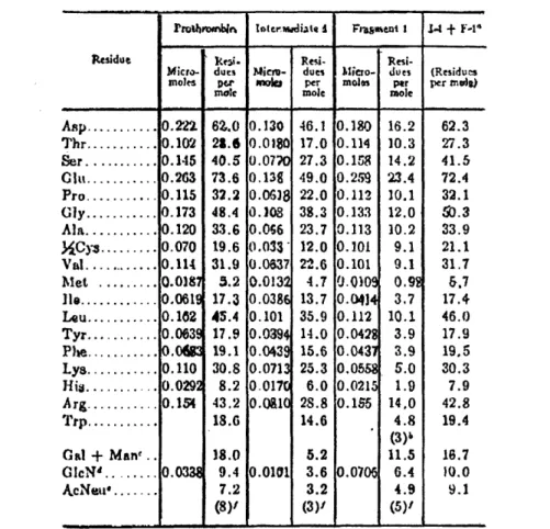
The amino acid composition of prothrombin, Intermediate-1 (I-1), and Fragment-1 are given in Table 1 (20).
The Fragment-1 region
The Fragment-1 region is responsible for the Ca+2 mediated binding
of prothrombin to phospholipid vesicle surfaces (16,18) and contains the modified glutamyl residues (21) which are necessary for normal
Ca binding by prothrombin (21-24). Carboxylation of 10 glutamyl
to y -carboxyglutamyl residues (23, 25) in the Fragment-1 region has been shown to occur as a consequence of the action of the vitamin K (26) and to be carried out postribosomally (26). Sequence analysis have
indicated that these residues are found primarily in pairs (25, 27-29) and it has been proposed that the cluster of 4 carboxyl groups provided
by this structure is responsible for Ca2+ binding by these proteins.
Abnormal prothrombin (induced by the vitamin K antagonist, dicoumarol) lacks these modified glutamic acid residues (the only protein primary structure difference from normal prothrombin) and that is the reason why
mrombl Totiler.madicrte I Fauaent 1 -4 + F-l4
Residut ke.i. Resi-
Resi-Micro- dues icm- dues Mliero- dues (Residues moles per moIa per molas per per mles)
mole mole mole
Asp ... 0.22... 62.0 0.130 46.1 0.180 16.2 62.3 Thr ... 102 23.6 0.0180 17.0 0.114 10.3 27.3 Ser... . 145 40.5 0.077 27.3 0.158 14.2 41.5 Clt ... 0.263 73.6 0.138 49.0 0.2259 23.4 72.4 Pro... 0.115 32.2 0.06G8 22.0 0.112 10.1 32.1 ly ... 0.173 48.4 0.108 38.3 0.133 12.0 0.3 Ala ... 0.120 33.6 0.066 23.7 0.113 10.2 33.9 XCys ... 0.070 19.6 0.033' 12.0 0.101 9.1 21.1 Val .... 0.114 31.9 0.0637 22.6 0.101 9.1 31.7 Met ... 0.0187 5.2 0.0132 4.7 0.0109 0.92 6.7 lie ... 00619 17.3 0.0386 13.7 0.0414 3.7 17.4 Lou ... 0.102 45.4 0.101 35.9 0.112 10.1 46.0 Tyr ... 0.063c 17.9 0.0394 14.0 0.0428 3.9 17.9 Phe ... 0.0 19.1 0.0439 15.6 0.0431 3.9 19.5 Lys ... 0.110 30.8 0.0713 25.3 0.0558 5.0 30.3 His ... 0.029 8.2 0.0170 6.0 0.0215 1.9 7.9 Arg ... 0.154 43.2 0.0610 28.8 0.155 14.0 42.8 Trp... 18.6 14.6 4.8 19.4 (3)' Gal + Man .. 8.0 5.2 11.5 16.7 GlcNd... . 0.038 9.4 0.0101 3.6 0.0706 6.4 10.0 A.cNeu ... 7.2 3.2 4.9 9.1 (8)(3) (5)'
TABLE
Is Amino acid composition of bovine prothrombin and
products of thrombin-catalyzed proteolysis of
-46-abnormal prothrombin does not bind Ca2+ and is non-functioning in
blood coagulation (18, 21, 30).
COOH
I
COOH COOH\ /
1 2 - vitamin K - ) H
CH2 CH2
H2N-CH-COOH H2N-CH-COOH
Some investigators have observed positive cooperativity in the
binding of Ca2+ to prothrombin (23, 31), whereas others claim that
exists two distinct classes of binding sites (17,32). Some authors (24,33) found that more than glutamate residue carboxylation is required
for the normal Ca2 + and phospholipid binding by the vitamin K related
proteins. The results of their experiments have led to the discovery
that positive cooperativity in the Ca2+ binding to Fragment-1 depends
upon having intact disulfide bridges (there are four disulfide bridges in Fragment-l) and, by inference, specific protein tertiary structure. These results do not eliminate the possibility that groups in the
Fragment-1 other than the carboxyl groupd of y -carboxyglutamic acid are involved in tight calcium binding and the detailed calcium-binding structure is still unknown.
Stenflo (34) did a structural comparison of normal and discoumarol-induced bovine prothrombin. He found that the two prothrombins have the same main antigenic determinants and that the sedimentation
co-efficients, the Stokes molecular radius, the tyrosine titration curves, and the fluorescence emission spectra were identical indicating that
there is no gross difference involving the entire molecule. However, the quantitative immunological precipitin curves suggest that there is a conformational difference between normal prothrombin with and without calcium ions.
Investigation of the conformation of the normal and abnormal
(dicoumarol-induced) prothrombins in the presence and absence of Ca2+
was reported using optical rotatory dispersion (ORD) and circular
dichroism (CD) (35). Both techniques showed no conformational differences
between the two proteins, in the absence of Ca2 +
. A small a -helix
content was found. But, normal prothrombin exhibits a marked change in
the aromatic region of the spectrum on binding of Ca2+
ions, whereas no such conformational change was found in the case of the dicoumarol-induced protein. This suggests that the conformational change is local and occurs in the neighborhood of an aromatic amino acid (28).
An interesting comparison between a vitamin K dependent peptide from human prothrombin to the known corresponding bovine segment has shown that a number of residues which could be considered as the most important are the same (36). The 8 glutamic acid residues which have been proved to be modified in the bovine peptide are present in the human material as well as the two cysteine residues which are involved in a disulfide bridge in the bovine molecule. The main differences are the absence of proline in the human polypeptide and conversely the presence of valine and tyrosine which are absent in the bovine material (the amino
-48-acid composition from reference 20 indicates the presence of 32 VAL and 18 TYR residues per mole of bovine prothrombin - see Table 1).
The ORD and CD results (37) indicate the existence of marked alterations in the secondary structure of Profragment-l obtained from human prothrombin as a result of changes in hydrogen ion and calcium ion concentration. These results are consistent with the hypothesis
that ionization of the y -carboxyglutamyl side chain carboxyl groups,
localized in a quite small region of Profragment-l leads to considerable electrostatic repulsion resulting in a conformational change and a
2+
nonfunctional configuration near neutral pH. Addition of Ca diminishes
the electrostatic repulsive forces by charge neutralization and induces shifts to a conformation resembling the low pH form of Profragment-l.
Binding of protein to phospholipid through calcium ions, a function
ascribed to - carboxyglutamic acid, can be fulfilled by other structures
as avidenced by S-100 protein from brain which binds to phospholipid through calcium ion (38) but contains no y-carboxyglutamic acid (39).
Preliminary investigations of Ca2 + binding to Fragment- by
difference ultraviolet absorption spectroscopy indicates a significant environment change for tryptophanyl and possibly tyrosyl chromophores
during Ca2+ binding (Peng and Jackson, unpublished observation).
We decided to study the binding of Ca+2 to Fragment-l, using
Raman spectroscopy. We hoped to see the effect of binding in the carbonyl
-1
stretching frequence (1600-1800 cm region of the spectrum) and
possible conformational changes. Also the pH and ionic strength dependence could be investigated.
MATERIAL AND METHODS
Fragment-1 samples were provided by Dr. Craig Jackson from
Washington University. Purification of prothrombin and isolation of Fragment-1 is described in reference 20. Bovine Fragment-1 was used at a concentration of 60 mg/ml in Tris-buffer, with (10 mM) or without
Ca . The Raman system used was improved with a computer and accumulation
and manipulation of the data made it possible to increase the signal: noise ratio.
-50-RESULTS AND DISCUSSION
The spectra of buffer, Fragment-1 without Ca 2 +
, and Fragment-1
2+
plus Ca is shown. These samples were made in water and the carbonyl
-1
stretching frequency (1600-1800 cm ) is not accessible due to the
water peak. Samples in D20 are necessary to study this region.
There are substancial frequency differences between the Raman
spectrum of Fragment-1 and Fragment-1 plus Ca+2 which may represent
real conformational differences. The disulfide bonds vibration is seen,
-L
in both spectra, around 509 cm , and the presence of these bonds is confirmed for both samples. The a -helix and random coil contents shown
by the peaks around 1275 and 1250 cm 1 respectively seem to be different
for the two spectra. The region around 1400 cm 1
shows also
dis-similarities. In this region is the CO stretching vibration frequency
and the binding of Ca2+
to the y-carboxyglutamyl residues is supposed to cause variation in this frequency. It was not possible to decide if there is or if there is not an environmental change for tryptophanyl
and tyrosyl residues upon Ca2+
binding. The vibrations of the aromatic amino acids appear in various regions of the spectrum. One of these
frequencies is not the same in the two spectra. The C-H rocking vibration
appears at 1172 cm- 1
for Fragment-1, and at 1178 cm- 1 for Fragment-1
plus Ca 2+ , and this may indicate a different environment for tyrosyl or
tryptophanyl residues.
We have to remember that the buffer spectrum is superimposed to the protein spectrum, and it would have to be subtracted from the overall
419 -434 J 430 - 448 467
•
-_ 490 _- 492 - 511 ,- 509 S-Svibrations 523 2- 544 X- 543 TRP AMIE_ 5 56868 AMIDE ZZI .58-- 5582 -- 590 .- 602 AMIDE Z3 -- 628 626 PHE . 647 - 646 TYR 680 -, 673 C- S vibrotions=69-7 1 io08 MET (C-S stretch)
AMIDE . t- 724 729 - 746 - 761 ) 762 BUFFER, TRP 786 2- 798 . - 799 817 8 - 828
c853 >- 853 TYR ( CH2 residue rock)
880 - 880 TRP, VAL, LEU
- 902
LYS, ASP (C-C stretch)
c Q946 >
-9 4 6
JLEU, VAL (CH2 cymm.rock)
974
9- 988 * 989
"1 ,00io - 1006 BUFFER, PHE
1033 - 1033 . 1049 048 C-C C-N ) 1064 BUFFER skeletal -2090 . . 1088 stretch 1104 - 1127
1155 . 1155 LEU, VAL (CH3ontisymm. rock)
1178 5 1 ..1 72J PHE, TYR (C-H rock )
1194 - 1196
-O
EP3
O g O - 1208-i
1211
TYR,
PHE
r r r r rm n
.
3 -1252 -m
r r 1250 RANDOM COIL AMIDE m .21275 , 1278 HELIX' o zzzzz
Z
1294
4 Z0 0O 1312 0 C 0 '.--132 - 1326* 0 o e -, c338 - --1339 TRP, VAL, LEU (C-Hbend
1358 .-- 1358 TRP
-
., 1384 -- 1383*
~ ~~ ~ ~~~~~
Q Q~ J -.- 13983) v;~C1A~ e -1408 - 1413 CO° symm- stretching
1417
0 "~ 6~~-- 1455 ~ C-H deform
vibration
1512
§-
1528
\<
1531
AMIDE II _ 1548 - 1550 C02 ontisymm stretch
-579 - 581 TRP, ASP, GLU
. -1621 _- 1620 TRP, TYR, PHE
1656 - 1655 H20
n +
spectrum for a quantitative analysis of the data.
The next step would be to try a set of samples in D20, so that the
-1
REFERENCES
1. Miller, K.D. and Seegers, W.H. (1956) Arch. Biochem. Biophys. 60,
398
2. Magnusson, S. (1965) Ark. Kemi 23, 285
3. Nelsestuen, G.L. and Suttie, J.W. (1972) J. Biol. Chem. 247, 6096 4. Harmison, C.R.; Landaburu, R.H. and Seegers, W.H. (1961) J. Biol.
Chem. 236, 1693
5. Tishkoff, G.H.; Williams, L.C. and Brown, D.M. (1968) J. Biol. Chem. 243, 4151
6. Ingwall, J.S. and Scheraga, H.A. (1969) Biochemistry 8, 1860 7. Cox, A.C. and Hanahan, D.J. (1970) Biochim. Biophys. Acta 207, 49 8. Thomas, W.R. and Seegers, W.H. (1960) Biochim. Biophys. Acta 42, 556 9. Magnusson, S. (1970) Thromb. Biath. Haemorrh. 38 (suppl), 97
10. Bailey, K. and Bettelheim, F.R. (1955) Biochim. Biophys. Acta 18, 495
11. Laki, K; Gladner, J.A.; Folk, J.E. and Kominz, D.R. (1958) Thromb. Diath. Haemorrh. 2, 205
12. Baughman, D.J. and Waugh, D.F. (1967) J. Biol. Chem. 242, 5252 13. Winzor, D.J. and Scheraga, H.A. (1964) Arch. Biochem. Biophys. 104,
202
14. Esmon, C.T.; Owen, W.G. and Jackson, C.M. (1974) J. Biol. Chem. 249,
606
15. Stenn, K.S. and Blout, E.R. (1972) Biochemistry 11, 4502 16. Gitel, S.N.; Owen, W.G.; Esmon, C.T. and Jackson, C.M. (1973)
Proc. Nat. Acad. Sci. USA 70, 1344
17. Benson, B.; Kisiel, W. and Hanahan, D.J. (1973) Biochim. Biophys.
Acta 329, 81
18. Stenflo, J. (1973) J. Biol. Chem. 248, 6325
20. Owen, W.G.; Esmon, C.T. and Jackson, C.M. (1974) J. Biol. Chem.
249, 594
21. Stenflo, J; Fernlund, P; Egan, W. and Roepstorff, P. (1974) Proc. Nat. Acad. Sci. USA 71, 2730
22. Nelsestuen, G.L.; Zytkovicz, T.H. and Howard, J.B. (1974) J. Biol. Chem. 249, 6347
23. Stenflo, J. and Ganrot, P.O. (1973) Biochem. Biophys. Res. Commun.
50, 98
24. Nelsestuen, G.L. and Suttie, J.W. (1972) Biochemistry 11, 4961
25. Magnusson, S; Sottrup-Hensen, L. and Peterson, T.W. (1974) FEBS Lett.
44, 189
26. Shah, D.V. and Suttie, J. (1974) Biochem. Biophys. Res. Commun. 60, 1397
27. Howard, J.B. and Nelsestuen, G.L. (1975) Proc. Nat. Acad. Sci. USA
72, 1281
28. Stenflo, J. (1974) J. Biol. Chem. 249, 5527
29. Enfield, D.L.; Ericsson, L.H.; Walsh, K.A.; Neurath, H. and Titani, K. (1975) Proc. Nat. Acad. Sci. USA 72, 16
30. Nelsestuen, G. and Suttie, J. (1972) J. Biol. Chem. 247, 8;76 31. Mann, K.G.; Bajaj, S.P.; Heldebrant, C.M.; Butkowski, R.J. and
Fass, D.N. (1973) Ser. Haemat. 6, 479
32. Yue, R.H.; Starr, T. and Gertler, M.M. (1972) Fed. Proc. 31, 241
33. Henriksen, R.A. and Jackson, C.M. (1975) Arch. Biochem. Biophys. 170, 149
34. Stenflo, J. (1972) J. Biol. Chem. 247, 8167
35. Bjork, J. and Stenflo, J. (1973) FEBS Lett. 32, 343
36. Ellion, J.; Bernarous, R. and Labie, D. (1976) Thrombos. Haemostas.
35, 82
37. Gabriel, D.A. (1975) Thromb. Res. 7, 839
-56-39. Nelsestuen, G.L.; Broderius, M.; Zytkovicz, T.H. and Howard, J.B. (1975) Biochem. Biophys. Res. Commun. 65, 233
