HAL Id: hal-03030769
https://hal.archives-ouvertes.fr/hal-03030769
Submitted on 14 Dec 2020
HAL is a multi-disciplinary open access
archive for the deposit and dissemination of
sci-entific research documents, whether they are
pub-lished or not. The documents may come from
teaching and research institutions in France or
abroad, or from public or private research centers.
L’archive ouverte pluridisciplinaire HAL, est
destinée au dépôt et à la diffusion de documents
scientifiques de niveau recherche, publiés ou non,
émanant des établissements d’enseignement et de
recherche français ou étrangers, des laboratoires
publics ou privés.
and the dGATAb Factor in an Ecdysone Response in
Drosophila melanogaster
Véronique Brodu, Bruno Mugat, Jean-Yves Roignant, Jean-Antoine Lepesant,
Christophe Antoniewski
To cite this version:
Véronique Brodu, Bruno Mugat, Jean-Yves Roignant, Jean-Antoine Lepesant, Christophe
An-toniewski. Dual Requirement for the EcR/USP Nuclear Receptor and the dGATAb Factor in an
Ecdysone Response in Drosophila melanogaster. Cellular and molecular biology, including
cyto-enzymology, Pergamon Press, 1999. �hal-03030769�
Copyright © 1999, American Society for Microbiology. All Rights Reserved.
Dual Requirement for the EcR/USP Nuclear Receptor
and the dGATAb Factor in an Ecdysone Response
in Drosophila melanogaster
VE´RONIQUE BRODU, BRUNO MUGAT, JEAN-YVES ROIGNANT, JEAN-ANTOINE LEPESANT,
AND
CHRISTOPHE ANTONIEWSKI*
Institut Jacques Monod, Laboratoire de Biologie du De´veloppement, CNRS UMR 7592,
Universite´ Paris 7 Denis-Diderot, Universite´ Paris 6 P. et M. Curie,
75251 Paris Cedex 05, France
Received 21 April 1999/Returned for modification 6 May 1999/Accepted 20 May 1999
The EcR/USP nuclear receptor controls Drosophila metamorphosis by activating complex cascades of gene
transcription in response to pulses of the steroid hormone ecdysone at the end of larval development. Ecdysone
release provides a ubiquitous signal for the activation of the receptor, but a number of its target genes are
induced in a tissue- and stage-specific manner. Little is known about the molecular mechanisms involved in
this developmental modulation of the EcR/USP-mediated pathway. Fbp1 is a good model of primary ecdysone
response gene expressed in the fat body for addressing this question. We show here that the dGATAb factor
binds to three target sites flanking an EcR/USP binding site in a 70-bp enhancer that controls the tissue and
stage specificity of Fbp1 transcription. We demonstrate that one of these sites and proper expression of
dGATAb are required for specific activation of the enhancer in the fat body. In addition, we provide further
evidence that EcR/USP plays an essential role as a hormonal timer. Our study provides a striking example of
the integration of molecular pathways at the level of a tissue-specific hormone response unit.
Nuclear receptors for steroid and retinoid hormones are
transcription factors that bind to gene promoters, recruit
co-activators or corepressors, and modulate the activity of the
transcription machinery. A wealth of information concerning
these mechanisms has been provided by in vitro approaches or
studies with cultured cell systems (19, 30). In whole organisms,
gene responses to circulating hormones depend on both the
target tissue and the developmental stage. Accordingly,
nu-clear receptor binding sites in natural promoters are often
found included in composite assemblages of multiple binding
sites for a variety of transcription factors (29). These so-called
hormone response units are thought to integrate multiple
reg-ulatory pathways responsible for the tissue and time specificity
of the transcriptional hormonal response. However, the
mech-anisms by which this integration occurs in animals are poorly
understood.
Drosophila melanogaster provides a choice model for the
study of a steroid hormone response in the context of a
devel-oping organism. At the end of the third larval instar, a major
pulse of 20-hydroxyecdysone (hereafter referred to as
ecdy-sone) triggers the larval-to-prepupal morphological transition
and initiates metamorphosis (3). During this period, the
ecdy-sone receptor, a heterodimer between the EcR and USP
pro-teins, two members of the nuclear receptor superfamily (22, 46,
52), orchestrates complex waves of gene transcription in target
tissues of ecdysone. The regulatory pathways controlling this
genetic program at the temporal level have been examined in
detail (see reference 47 for a review; see also references 11, 23,
and 50 for recent advances). Despite these advances, how
transcription of genes, in response to circulating ecdysone, is
restricted to a subset or a single target tissue remains an open
question.
We used Fbp1 as a model gene to address this question.
Fbp1, which encodes a receptor mediating the uptake of
hex-amerins from the hemolymph (12), is transcribed in response
to ecdysone exclusively in the fat body during the second half
of the third larval instar (2, 27). Germ line transformation
analysis of its promoter has pinpointed a 70-bp enhancer (⫺69
to
⫺138) sufficient to specify the spatially and temporally
ec-dysteroid-controlled pattern of Fbp1 expression (24). The EcR/
USP heterodimer binds to a pseudopalindromic site in the
70-bp Fbp1 enhancer (4). Mutations of this site completely
abolished the ability of the Fbp1 enhancer to confer a fat
body-specific ecdysteroid response onto a minimal
promoter-lacZ reporter transgene, indicating that activation by the EcR/
USP heterodimer is a strict requirement for the activity of the
Fbp1 enhancer (5). Germ line transformation experiments
showed, however, that an EcR/USP binding site was unable by
itself to confer an ecdysteroid response onto the same
mini-mal-promoter–lacZ reporter gene, even when multimerized.
This provided evidence that other transcription factors,
tar-geted to sequences flanking the EcR/USP binding site in the
Fbp1 enhancer, must act in vivo, in addition to the ecdysone
receptor, to mediate an ecdysteroid genetic response (5).
Analysis of the role of these sequences is complicated by the
fact that their regulatory function cannot be revealed in the
absence of activation by the liganded EcR/USP heterodimer.
We show here that replacement of the EcR/USP binding site
with an upstream activation sequence (UAS) site can
circum-vent this limitation. In the presence of the yeast activator
GAL4, transcriptional activation of this modified Fbp1
en-hancer remains restricted to the third-instar fat body,
provid-ing direct confirmation that sequences flankprovid-ing the EcR/USP
binding site play an important role in this specific activation.
We further show that dGATAb, the product of the serpent (srp)
gene, binds in the close vicinity of the EcR/USP binding site
* Corresponding author. Mailing address: Institut Jacques Monod,
Laboratoire de Biologie du De´veloppement, CNRS UMR 7592,
Uni-versite´s Paris 6 et Paris 7, 2 place Jussieu, 75251 Paris Cedex 05,
France. Phone: (33 1) 44 27 78 12. Fax: (33 1) 44 27 52 65. E-mail:
[email protected].
and is strictly required for activation of the Fbp1 enhancer.
Together, our results demonstrate that the Fbp1 enhancer
functions as a complex ecdysone response unit integrating
spa-tial and temporal cues in a specific response to the hormonal
signal and provide evidence for the central role played by the
EcR/USP nuclear receptor as a developmental timer in this
process.
MATERIALS AND METHODS
Plasmids.A BamHI-DraI fragment from the srp cDNA (a gift from K. P. Rehorn and R. Reuter) was subcloned into the bluescript KS⫹ plasmid between the BamHI and SmaI sites. The resulting construct, pBS-srp, allows coupled in vitro transcription-translation of the Srp protein. For bacterial expression of Srp, we isolated the srp cDNA from pBS-srp as a BamHI-EcoRI fragment and inserted it after Klenow blunting into the SmaI cloning site of the glutathione
S-transferase (GST)-tagged expression vector pGEX-3X (Pharmacia). The germ
line transformation vector pAE was described in reference 5, where it was referred to as pAEP1. pAE[UAS] was derived from pAE by PCR mutagenesis of the ATTCATTCAAC EcR/USP binding site in the Fbp1 enhancer (see Fig. 3). This sequence was replaced with the UAS CGGAGTACTGTCCTCCG in the pAE[UAS] construct. Plasmids pAE␣, pAE, pAE⬘, and pAE⬘ were derived from pAE by mutagenesis (for the positions used, see Fig. 3). The structures of all of the mutagenized transformation vectors were confirmed by direct sequenc-ing.
GATAb production in bacteria or rabbit reticulocyte lysate.Crude GST-Srp bacterial extract was prepared as described in reference 16 from Escherichia coli BL21 transformed with the pGST-srp expression vector. This extract was purified by glutathione-Sepharose affinity chromatography in accordance with the man-ufacturer’s (Pharmacia) recommendations. The full-length Srp protein was trans-lated in a rabbit reticulocyte lysate by using the pBS-srp plasmid and T7 RNA polymerase in a Promega coupled transcription-translation system.
DNase I footprinting.Oligonucleotides 5⬘ GTAGCGGCCGCATGACAACA ATTTATTTAAT 3⬘ (upper primer) and 5⬘ CTGCAGCTTTTATACCC 3⬘ (low-er prim(low-er) w(low-ere used to amplify by PCR a⫺194 to ⫺18 fragment from the Fbp1 promoter. Depending on the DNA strand that was to be analyzed, the upper or lower primer was32P labelled by using T4 polynucleotide kinase. A 0.2-pmol
sample of the labelled⫺194 to ⫺18 Fbp1 fragment was mixed with 1 g of crude bacterial extract or 100 ng of GST-purified GATAb and 2g of poly(dI-dC) in binding buffer (25 mM HEPES [pH 7.6], 60 mM KCl, 5% glycerol, 5 mM MgCl2,
0.1 mM EDTA [pH 8], 0.75 mM dithiothreitol [DTT]) in a final volume of 16l. After 20 min at 4°C, 3l of DNase I (Worthington) at 2.6 g/ml was added and the mixture was left for 2.5 min. The reaction was then stopped by addition of 40 l of stop solution (20 mM EDTA, 0.1% sodium dodecyl sulfate [SDS]). DNA was phenol-chloroform extracted twice, ethanol precipitated, resuspended in a formamide-dye mixture, and electrophoresed in an 8% polyacrylamide-urea se-quencing gel.
Fat body nuclear extracts.All of the buffers used for nuclear extract prepa-ration were supplemented with protease inhibitors (0.5 mM Pefablock and apro-tinin at 10g/ml). Late-third-instar larvae were dissected in buffer C (20 mM Tris [pH 7.5], 50 mM KCl, 2 mM DTT, 0.1 mM EDTA, 0.15 mM spermine, 0.5 mM spermidine), and fat bodies were immediately frozen in an Eppendorf tube kept on dry ice. When collected, 50 fat bodies were rapidly thawed in 200l of cold buffer C and vigorously vortexed. They were then refrozen and thawed twice with vortexing. Nuclei and debris were pelleted upon rapid centrifugation in an Eppendorf centrifuge (20 s at 5,500⫻ g) and washed with 200 l of buffer C. After centrifugation (15 s at 5,500⫻ g), the crude nuclear pellet was suspended in 40l of extraction buffer C2 (same as buffer C but with 600 mM KCl) and the homogenate was left on ice for 20 min. After centrifugation (15 min at 4°C and top speed), the supernatant was diluted with 2 volumes of buffer C3 (same as buffer C but without KCl), aliquoted, and kept at⫺80°C until use.
Gel shift assays.Three to 4l of fat body nuclear extract or 3 l of rabbit reticulocyte lysate, 2g of poly(dI-dC), specific competitor DNA or antibodies, if appropriate, and 2 fmol of32P-labelled probe (about 5⫻ 104cpm/fmol) were
mixed in binding buffer (25 mM HEPES [pH 7], 9% glycerol, 90 mM KCl, 1 mM EDTA [pH 8], 0.9 mM DTT), and gel shift assays were performed as described in reference 4. Competitions were performed with a 200-fold molar excess of double-stranded oligonucleotides. Double-stranded oligonucleotides A, A␣, B, and B have been described previously (4). The double-stranded oligonucleotide ADH includes the region from⫺77 to ⫺53 of the D. muleri Adh-1 promoter.
Larval developmental-stage determination. Drosophila stocks were
main-tained at 25°C on a standard Drosophila medium. Developmental-stage deter-mination was carried out essentially as described in reference 2 with a few modifications. Egg laying on a hard agar plate coated with a strip of baker’s yeast was restricted to 1 h. Two 1-h precollection egg layings were discarded. Embryos were washed from the yeast, distributed on a hard agar plate, and allowed to develop at 25°C until eclosion. First-instar larvae were collected by hand at 1-h intervals, transferred to tubes containing mashed Drosophila medium in batches of 80 larvae, and collected at 6-h intervals from the mid-second larval instar to the late-third larval instar.
RT-PCR.Total RNA (about 100g) was isolated from 25 to 50 larvae at different stages as previously described (7), and poly(A)⫹RNA was isolated by
using the Oligotex mRNA Midi Kit (Qiagen). Approximately 50 ng of poly(A)⫹
RNA was denatured (65°C) prior to use as a template in a 20-l cDNA synthesis reaction mixture containing 1⫻ reverse transcription (RT)-PCR buffer (50 mM KCl, 20 mM Tris-HCl [pH 8.4], 2.5 mM MgCl2, bovine serum albumin at 100
mg/ml, 2.5 mM DTT, 1 mM [each] deoxynucleoside triphosphate), 17.5 U of RNasin (Promega), 100 pmol of random d(N)6 primers, and 8 U of avian myeloblastosis virus reverse transcriptase (Boehringer). The reaction mixture was incubated for 10 min at 22°C and then for 90 min at 42°C. One-microliter aliquots of the cDNA reaction mixture were analyzed separately for srp (38),
Fbp1 (31), and the ribosomal protein gene rpL17A (34) in 50-l PCR mixtures containing 1⫻ Taq DNA polymerase buffer (Bioprobe System), 0.2 mM each deoxynucleoside triphosphate, 20 pmol of gene-specific primers, 0.2 l of [32P]dCTP (3,000 Ci/mmol; Amersham), and 2.5 U of Taq DNA polymerase
(Promega). The PCR conditions used were 94°C for 3 min followed by 22 cycles of 94°C for 1 min, 55°C for 1 min, and 72°C for 1 min. Control reactions were performed to ensure that the number of amplification cycles was within the logarithmic phase for all three sets of primers (data not shown). All PCR primers were chosen so that they hybridized to separate exons in order to distinguish correctly spliced mRNA from unspliced RNA or contaminating genomic DNA. The srp sense primer was 5⬘ AGCAGCAACAACATCATCAC 3⬘, and the srp antisense primer was 5⬘ TTGGCAGTCTGAGTAAGCAA 3⬘, corresponding to positions 929 to 948 and 1160 to 1141, respectively, of the srp cDNA sequence (EMBL nucleotide sequence database accession no. Y07662). The Fbp1 sense primer was 5⬘ ACTACGAATCAGGACAGGGT 3⬘, and the Fbp1 antisense primer was 5⬘ CAGATCGATGACGTTCTGCA 3⬘, corresponding to positions 1552 to 1571 and 1814 to 1795, respectively, of the Fbp1 genomic sequence (EMBL nucleotide sequence database accession no. X69965). The rpL17A sense primer was 5⬘ GTGATGAACTGTGCCGACAA 3⬘, and the rpL17A antisense primer was 5⬘ CCTTCATTTCGCCCTTGTTG 3⬘, corresponding to positions 536 to 555 and 1388 to 1369, respectively, of the rpL17A genomic sequence (GenBank nucleotide sequence database accession no. M 85295). Amplification reaction products obtained with rpL17A primers were fractionated by electro-phoresis on a 6% polyacrylamide gel and quantified with a Molecular Dynamics PhosphorImager. Amplification reaction products obtained with the srp and
Fbp1 primers were then electrophoresed after loading of an amount that was
normalized in relation to rpL17A amplification.
Antibodies.The #Srp rabbit antibody was raised against a synthetic peptide corresponding to the 22 last amino acids of GATAb (21). The #2B8 monoclonal antibody, a generous gift from P. Ramain, was raised against a peptide corre-sponding to amino acids 378 to 397 of the GATAa/Pannier protein (37). We have shown that this antibody specifically recognizes the TSSSGQA motif that is also present in the N-terminal part of GATAb but is not found in any other known protein (11a). The #2B8 antibody should thus be considered to be a genuine specific antibody for both GATAa and GATAb.
Antibody staining of tissues.Larvae were dissected in phosphate-buffered saline (PBS), and tissues were fixed for 20 min at room temperature in 3.4% paraformaldehyde–30 mM piperazine-N,N⬘-bis(2-ethanesulfonic acid) (PIPES; pH 7.4)–160 mM KCl–40 mM NaCl–4 mM EGTA–1 mM spermidine–0.4 mM spermine–0.2%-mercaptoethanol–0.1% Triton X-100. They were then washed three times with PBT (0.3% Triton X-100 in PBS) and blocked for 20 min in PBT that contained 1% bovine serum albumin. The #Srp antibody was preabsorbed for 1 h with dechorionated embryos, diluted 1,000-fold in PBT, and incubated with the tissues overnight at 4°C. Tissues were washed three times for 20 min each with PBT and incubated for 1 h at room temperature with goat anti-rabbit horseradish peroxidase-conjugated secondary antibodies (Vector Laboratories) diluted 400-fold in PBT. The tissues were washed three times for 20 min each with PBS, and peroxidase activity was detected with PBS supplemented with diaminobenzidine (Sigma) at 0.5 mg/ml and 0.03% H2O2. The reaction was
allowed to proceed for 10 min. Tissues were then washed with PBS and mounted on slides in glycerol and photographed.
Western analysis.Samples for developmental Western blotting were prepared by solubilizing fat bodies dissected from 10 to 20 larvae at different stages in 20 l of cracking buffer (0.125 M Tris [pH 6.8], 5% -mercaptoethanol, 2% SDS, 4 M urea). Samples were electrophoresed on an SDS–8% polyacrylamide gel and transferred to nitrocellulose membrane (Schleicher and Schuell) with a Novablot Electrophoretic Transfer Kit (LKB). All subsequent steps were performed at room temperature. The membrane was blocked in milk solution (PBS supple-mented with 5% dry milk and 0.1% Tween 20) for 1 h. #Srp antibody was diluted 1:10,000 in milk solution and incubated with the membrane for 1 h. The mem-brane was washed three times for 5 min each with milk solution and incubated for 1 h with goat rabbit horseradish peroxidase-conjugated secondary anti-bodies (Vector Laboratories) diluted 1,600-fold in milk solution. The membrane was then washed three times for 5 min each in PBS supplemented with 0.1% Tween 20, and chemiluminescence detection was performed with the ECL kit (Amersham) in accordance with the manufacturer’s instructions. The quantity of protein loaded was estimated by reincubation of the membrane for 1 h with anti-myosin antibody (53) diluted 1:5,000 in milk solution and chemilumines-cence detection as described above.
Germ line transformations, EcR/USP-to-GAL4 substitution, and GATAb overexpression.DNAs of the germ line transformation vector (250g/ml) and
helper plasmid⌬2-3 (50 g/ml) were microinjected into embryos of the w1118 recipient stock (40). The w⫹transformants were screened for eye color. The
number of transgenes in each line was determined by Southern blotting. The GAL4-UAS system (10) was used to study the effects of GAL4-to-EcR/ USP substitution and GATAb overexpression. The effect of GAL4-to-EcR/USP substitution was tested by crossing a line homozygous for the AE[UAS] trans-gene with the homozygous GAL4daG32line expressing GAL4 under the control
of the da promoter (51). Ubiquitous expression of the da-GAL4 transgene was checked by crossing the GAL4daG32line with a line transgenic for the pUAST
construct in which the lacZ reporter gene is under the control of the hsp70 promoter and five UAS sites (10).
For the assay of GATAb overexpression by Western blotting, a line homozy-gous for an hsp70-GAL4 transgene (third chromosome, line 1799 from Andrea Brand, Bloomington Stock Center) was crossed with a line homozygous for a 5UAS-srp transgene (second chromosome, kindly provided by R. Reuter). Het-erozygous larvae obtained from this cross were raised at 25°C, submitted to a 37°C heat shock for 1 h, and allowed to recover for an additional 7 h at 25°C. Tissues were then dissected and treated for Western blot analysis. For testing of the effect of overexpression of GATAb on the activity of the Fbp1 promoter, a double homozygous line for the AE transgene (5) and the UAS-srp transgene was constructed by genetic crosses. Larvae obtained from the crossing of this line with the line homozygous for the hsp70-GAL4 transgene were raised at 25°C and submitted to a 37°C heat shock for 1 h at various times during the second or third larval instar. Larvae were then allowed to recover for 7 h at 25°C, and -galac-tosidase activity was determined by either 5-bromo-4-chloro-3-indolyl--D
-galac-topyranoside (X-Gal) staining of tissues or quantitative dosage on crude extracts.
Histochemical and spectrophotometric assays of-galactosidase activity. A histochemical staining assay of-galactosidase activity was performed as previ-ously described (6) by using X-Gal.
A chlorophenol red--D-galactopyranoside (CPRG) spectrophotometric assay
of-galactosidase activity was performed essentially as previously described (14, 43). At least five protein extracts, each prepared from three larvae, were assayed for each developmental stage. Larvae at different developmental stages were homogenized in 250l of buffer (50 mM potassium phosphate [pH 7.5], 1 mM MgCl2, 1 mM phenylmethylsulfonyl fluoride) in a 1.5-ml Eppendorf tube and
centrifuged at 12,000⫻ g for 15 min at 4°C. The supernatant was transferred to a new Eppendorf tube. Extract (4 to 100l, depending on activity) was added to a 1-ml final volume of assay buffer with CPRG at 0.8 mg/ml in a disposable cuvette, mixed, and incubated at room temperature. A574was recorded for 1 h at
10-min intervals. Activity was calculated by dividing the slope of the assay curve by the amount of protein added for dosage (as determined by Bio-Rad protein assay of the extract). Results are expressed as the mean value of five independent protein extract activities divided by an arbitrary constant for the convenience of data reading.
RESULTS
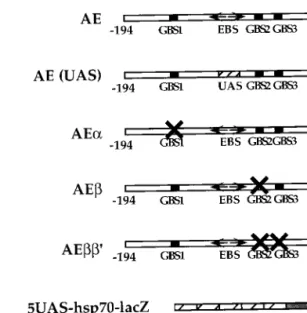
GAL4 can substitute for EcR/USP in the tissue-specific
activation of the Fbp1 promoter.
In a previous study (5), we
showed that when the Fbp1 enhancer (sequences between
po-sitions
⫺194 and ⫺68) was fused to the Fbp1 minimal
pro-moter and the lacZ reporter gene, expression of the resulting
AE construct (Fig. 1) took place exclusively in the late-third
instar larval fat body (Fig. 2A, part A).
-Galactosidase activity
was first detected in 106-h-old larvae and reached a maximum
at puparium formation (Fig. 2B). This pattern faithfully
repro-duced the transcriptional response of the endogenous Fbp1
gene to the major third-instar ecdysone pulse, as shown in
earlier studies (2) and in an RT-PCR assay (see Fig. 4A).
Replacement of the EcR/USP binding site with a UAS site
(Fig. 1) resulted in complete inactivation of the AE[UAS]
mutated construct (Fig. 2A, part C), confirming that binding of
the EcR/USP receptor is strictly required for in vivo activity of
the Fbp1 enhancer. Activation of the AE[UAS] construct by
GAL4 was further tested by using the GAL4
daG32driver in
which a GAL4 cDNA is expressed in all tissues throughout
development from the ubiquitous daughterless promoter, as
shown in a control cross with a 5UAS-hsp70-lacZ construct
(Fig. 2A, part B). When the GAL4
daG32construct was crossed
in three independent AE[UAS] transgenic lines,
-galactosi-dase activity was restored exclusively in the fat bodies of
third-instar larvae (Fig. 2A, part D). Remarkably, the timing of
GAL4-driven expression of the AE[UAS] transgene differed
significantly from that of the AE transgene. Expression was
first detected in 72-h-old larvae, after the
second-to-third-in-star molt, and subsequently increased gradually throughout the
third larval instar (Fig. 2B, black bars). The possibility could be
excluded that this dynamic profile of expression was due to
variations in the GAL4 level because expression of the
5UAS-hsp70-lacZ control construct remained constant throughout
the third instar in the same da-GAL4 context (Fig. 2B, open
bars).
Two conclusions can be drawn from these results. First, they
demonstrated that a heterologous transactivator can substitute
in vivo for the liganded EcR/USP heterodimer and activate the
Fbp1 enhancer. Second, the sequences flanking the EcR/USP
binding site are very probably targets for transcription factors
that modulate in a tissue-specific manner the activity of an
adjacent transactivator, whether the ecdysone receptor is in a
natural situation or GAL4 bound to its target site. Under this
hypothesis, the activity of these transactivating factors would
be acquired gradually during the third larval instar.
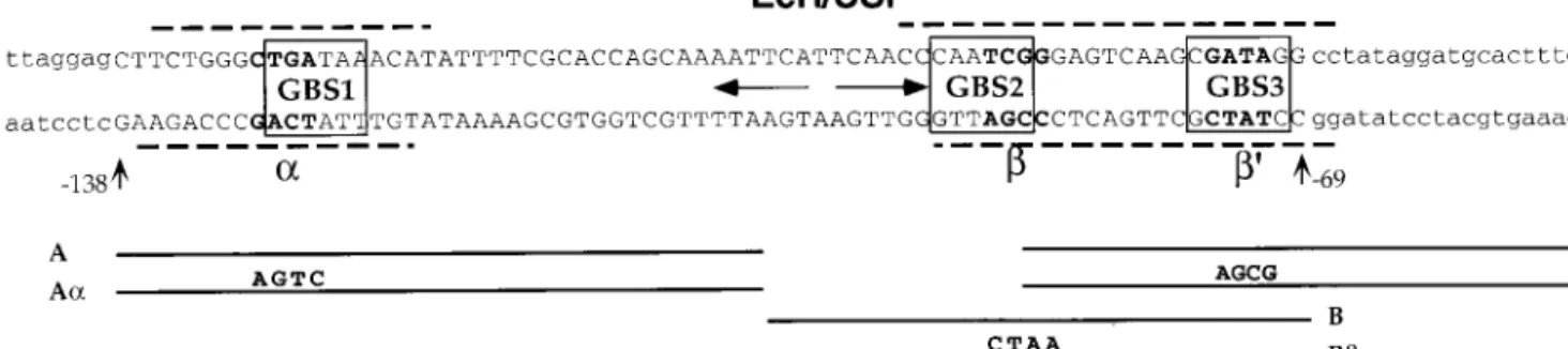
An essential GATA binding site is required for enhancer
activation.
Examination of the Fbp1 enhancer sequence
re-vealed the presence of three putative binding sites for the
GATA family of transcription factors, hereafter referred to as
GATA binding site 1 (GBS1), GBS2, and GBS3 (Fig. 3). The
individual requirement of these sites for the activity of the
enhancer was tested by mutagenesis of GBS1, GBS2, or both
GBS2 and GBS3 in the AE construct (Fig. 1). Mutation of
GBS1 completely abolished the ability of the Fbp1 enhancer to
drive lacZ expression in third-instar larvae, as tested by X-Gal
staining of dissected tissues and quantitative assay of
-galac-tosidase activity in a crude larval extract (Fig. 2A, part E; data
not shown). In contrast, neither mutation of GBS2 nor double
mutation of GBS2 and GBS3 had a significant effect on lacZ
expression (Fig. 2A, part F and G). These results indicated that
only GBS1 is crucial for the enhancer activity in animals and
prompted us to identify the factor involved.
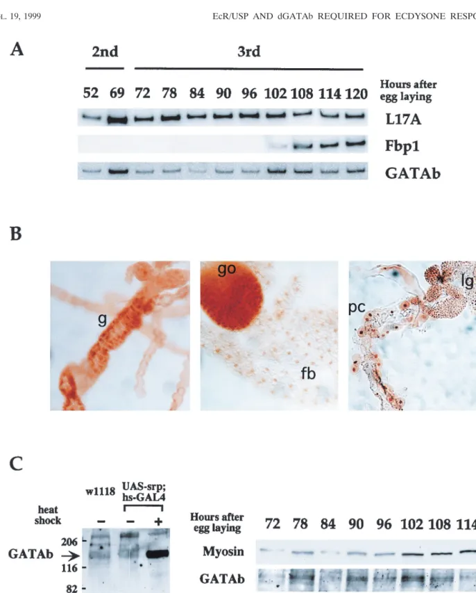
Pattern of expression of GATAb/Serpent in third-instar
lar-vae.
dGATAb/Serpent, a member of the GATA family, plays
FIG. 1. Structures of lacZ reporter transgenes. Fbp1 sequences between ⫺194 and ⫹80, including the ⫺138/⫺69 enhancer, were fused to the lacZ re-porter gene, giving rise to the AE rere-porter construct. Replacement of the EcR/ USP binding site (EBS) with the UAS site gave rise to the AE(UAS) construct. The AE␣, AE, and AE⬘ constructs are mutated at the indicated GATA sites. The 5UAS-hsp70-lacZ construct consists of five multimerized UAS sites fused to the minimal hsp70 promoter and the lacZ reporter gene.
FIG. 2. Pattern of expression conferred by the wild-type or mutated Fbp1 enhancers on the lacZ reporter transgene. (A) Late-third-instar larval tissues from transgenic lines with the indicated genotypes were dissected and histochemically stained for determination of-galactosidase activity. (B) Transgenic larvae with the indicated genotypes were synchronized at eclosion and recovered during the third larval instar at the indicated times.-Galactosidase activity was measured in extracts from whole larvae. Error bars represent the standard error of the mean.
an essential role in the development of fat body, endodermal
gut, and hematopoietic tissues during embryogenesis (1, 33, 38,
39, 41). dGATAb was also found to be expressed during the
third larval instar and involved in the transcriptional regulation
of the D. muleri and D. melanogaster Adh genes in the fat body
tissue (1). These data prompted us to consider GATAb as a
candidate for the role of a transcription factor that interacts
with the Fbp1 regulatory sequences and to establish its tissue
and temporal pattern of expression during larval stages.
Using a quantitative RT-PCR assay, we detected a constant
level of GATAb mRNA in late-second-instar larvae and
throughout the third-instar larval stage (Fig. 4A).
Immuno-staining using the #Srp anti-GATAb antibody (21) showed
that GATAb was detectable in the nuclei of fat body, gonad,
gut, lymph gland, and pericardial cells of late-third-instar
lar-vae (Fig. 4B). We detected no staining in other tissues, such as
the central nervous system, imaginal discs, or salivary glands.
In a Western blot assay using the #Srp antibody, the GATAb
protein was revealed in the fat body tissue as a double band
(Fig. 4C, left side) whose intensity was constant throughout the
third larval instar (Fig. 4C, right side). This doublet migrated
with a much lower mobility than the 102-kDa molecular mass
predicted from the sequence of the GATAb cDNA (38). A
confirmation that this doublet corresponded to GATAb
iso-forms was provided by the detection with the #Srp antibody of
an overexpressed product migrating at the same position after
heat shock induction of a UAS-GATAb cDNA construct in an
hs-GAL4 transgenic line (Fig. 4C, left side; data not shown).
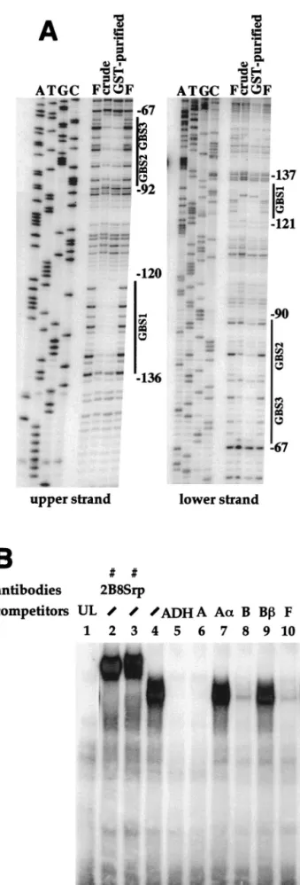
GATAb binds in vitro to the Fbp1 enhancer.
Bacterially
produced GST-GATAb protein, either in a crude extract or
affinity purified, protected two regions of the Fbp1 enhancer in
a DNase I footprint assay (Fig. 5A). One 17-bp region
ex-tended over the GBS1 site from
⫺136 to ⫺120 on the upper
strand and from
⫺137 to ⫺121 on the lower strand. A second,
larger region included tandemly arranged GBS2 and GBS3
and extended from
⫺92 to ⫺67 on the upper strand and from
⫺90 to ⫺67 on the lower strand.
Binding of in vitro-translated GATAb to a labelled ADH
probe previously shown to contain an efficient TGATAA
GATAb target site identical to GBS1 (1) gave rise to the
formation of a single retarded complex in a gel shift assay (Fig.
5B, lane 4). This complex was supershifted in the presence of
the #Srp antibody (lane 3) or the #2B8 antibody (lane 2),
which recognizes both the GATAb/Serpent and
GATAa/Pan-nier proteins (see Materials and Methods). The relative
bind-ing affinity of in vitro-translated GATAb for GBS1, GBS2, and
GBS3 was further analyzed by competition with unlabelled
oligo-nucleotides corresponding to various subregions (Fig. 3) of the
Fbp1 enhancer. Competition in the presence of oligonucleotide A
was as efficient as that in the presence of the unlabelled ADH
probe (Fig. 5B, lanes 5 and 6). As expected, this competition
was relieved by using the A␣ oligonucleotide (lane 7) carrying
a mutation destroying GBS1 (Fig. 3). Oligonucleotide B or F
also competed, although to a lesser extent, for the formation of
the GATAb retarded complex (compare lanes 8 and 10 with
lanes 5 and 6). These competitions were fully relieved in the
presence of oligonucleotides B and F⬘ carrying mutations
destroying GBS2 and GBS3, respectively (lanes 9 and 11).
Taken together, these results indicate that GATAb binds with
higher affinity to GBS1 than to GBS2 or GBS3.
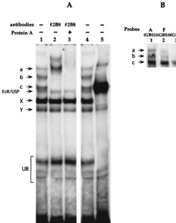
The GATAb protein in third-instar fat body nuclear extracts
binds to the Fbp1 enhancer.
When used as a radioactive probe
with nuclear extracts from hand-dissected third-instar larval fat
bodies, the Fbp1 enhancer gave rise to the formation of three
complexes a, b, and c (Fig. 6A, lanes 1 and 4), that were
supershifted in the presence of the #2B8 monoclonal antibody
alone (Fig. 6A, lane 2) and further supershifted in the presence
of #2B8 and protein A (Fig. 6A, lane 3). The #2B8
monoclo-nal antibody recognizes similar epitopes in the
GATAa/Pan-nier and GATAb/Serpent proteins. However, because GATAa/
Pannier expression is undetectable in a third-instar larval fat
body (3a), we concluded that all three complexes, a, b, and c,
involve the GATAb protein.
GBS1-containing oligonucleotide A (Fig. 6B, lane 1) and, to
a lesser extent, GBS3-containing oligonucleotide F (Fig. 6B,
lane 2) also gave rise to the formation of complexes a, b, and
c. This indicated that GBS1 and GBS3 are involved
indepen-dently in the formation of complexes a, b, and c. In contrast,
GBS2-containing oligonucleotide B gave rise to the formation
of only one retarded complex, c (Fig. 6B, lane 3). As expected,
these complexes were supershifted by the #2B8 antibody and
were not formed with any of the GBS-mutated probes, A␣, B,
or F⬘ (data not shown).
Two lines of evidence excluded the possibility that the
for-mation of distinct GATAb-containing complexes resulted from
homodimerization, as suggested by examples of GATA family
members binding to DNA as homodimers (15). First, we were
unable to detect more than one GATAb retarded complex in
gel shift assays using in vitro-translated (Fig. 5B) or bacterially
produced (not shown) GATAb. Second, a fat body nuclear
extract prepared from third-instar larvae overexpressing
GATAb gave rise to a predominant retarded band that
mi-FIG. 3. Structure of the Fbp1 enhancer. The sequence of the Fbp1 enhancer between positions⫺138 and ⫺69 relative to the Fbp1 transcription start site is in capital letters. Three GATAb binding sites (boxed; GBS1 to GBS3) were found in sequences flanking the pseudopalindromic EcR/USP binding site (horizontal arrows). GBS1 perfectly fits the (A/T)GATA(A/G) consensus sequence for a GBS (18). GBS2 (as read on the lower strand) and GBS3 do not fit this consensus but are efficient binding sites for vertebrate GATA transcription factors (32). Dashed lines indicate the extent of the GATAb footprints (see Fig. 5A) on both DNA strands. The positions and lengths of the competitor oligonucleotides used in gel shift assays are indicated in the lower part of the scheme. Positions of mutations␣, , and ⬘ are marked by bold lettering in the Fbp1 enhancer, and sequence substitutions are indicated for each mutated competitor, A␣, B, or F⬘.
grated with the same mobility as complex c (Fig. 6A, lane 5).
This retardation pattern suggested that complex c resulted
from the binding to DNA of the GATAb protein alone. If
complex a or b corresponded to homodimerization or
multim-erization of GATAb, its intensity would have been reinforced
like that of complex c.
Together, these data demonstrated that GATAb was
in-volved in the differential formation of distinct complexes on
GBS1, GBS2, and GBS3 in a site-specific manner.
GATAb is involved in the timing and tissue specificity of
Fbp1 enhancer activation.
The effect of total or partial loss of
function of GATAb on the activity of the enhancer during the
third larval instar could not be studied because all of the
GATAb mutations isolated to date are lethal to embryos (38).
FIG. 4. Expression of GATAb in third-instar larvae. (A) RT-PCR analysis. Total expression of mRNAs for GATAb, Fbp1, and the ribosomal protein L17A was analyzed at specific developmental stages by quantitative RT-PCR using specific primers. (B) Distribution of GATAb in late-third-instar tissues. Tissues were dissected from late-third-instar larvae and stained with the #Srp anti-GATAb antibody. Nuclear staining was detected in the gut (g), lymph glands (lg), pericardial cells (pc), gonads (go), and fat body (fb). No staining was detected in the other tissues (not shown). (C) Western blot analysis. GATAb-specific bands were identified with the #Srp antibody in a Western blot analysis by comparing the profiles obtained with fat bodies from w1118 or UAS-srp/hs-GAL4 larvae at 25°C with those obtained with fat bodies from UAS-srp/hs-GAL4 larvae after a 1-h heat shock at 37°C (left). On the right is the temporal profile of GATAb protein expression in isolated fat bodies as detected by Western blotting. The protein quantity loaded in each lane was estimated by detection of myosin. The values on the left are molecular sizes in kilodaltons.
We tested the effect on the AE construct of ubiquitous
overexpression of GATAb in triple transgenic AE/hs-GAL4/
UAS-srp larvae. After heat shock at various times during the
second or third larval instar followed by a 7-h recovery period,
tissues were stained for lacZ expression (Fig. 7A) and the level
of
-galactosidase activity was measured in crude extracts (Fig.
7B, black bars). As expected, in the absence of heat shock, the
AE reporter transgene was specifically expressed in the fat
body at the end of the third larval instar (Fig. 7A, top).
Quantitative measurement indicated that this expression
was detectable 108 h after egg laying and reached a maximum
10 h later, at puparium formation (Fig. 7B, open bars). In
contrast, heat shock treatment resulted in premature
induc-tion, as early as the second larval instar, of the AE reporter
transgene in the fat body (Fig. 7B). It was also ectopically
induced in the salivary glands and proventriculus (Fig. 7A,
bottom). That this deregulation was due to the overexpression
of GATAb was confirmed by the fact that the spatial and
temporal patterns of expression of the AE transgene were
unchanged in a heat-shocked AE/hs-GAL4 control line (data
not shown).
We concluded that a proper level of GATAb is crucial for
both the correct timing and tissue specificity of Fbp1
expres-sion. However, quantitative dosage of
-galactosidase activity
showed that the magnitude of the effect of GATAb
overex-pression varies during development. The significant but limited
level of AE expression detected during the second larval instar
and at the beginning of the third (Fig. 7B) markedly increased
between 78 and 108 h after egg laying. At 108 h after egg
laying, this level was about 10 times that in the absence of heat
shock. It was only at the end of the third larval instar (118 h)
that the level of AE expression in the absence of heat shock
reached a value similar to that in the presence of
overex-pressed GATAb. These results suggested that the
transcrip-tional response elicited by the increased amount of GATAb
was restricted at earlier stages by the availability of additional
factors until the end of the third larval instar. The ecdysone
receptor possesses all of the features of such a factor.
Both GATAb and the ecdysone receptor are limiting factors
in Fbp1 induction.
Two factors are known to limit the
avail-ability of the active EcR/USP receptor during the third larval
instar. First, the expression of the EcR gene increases during
the second half of the third larval instar (2). In addition, the
activity of the EcR/USP heterodimer is triggered by
ecdy-steroids, whose concentration peaks during this period of
de-velopment. We reasoned that if the effect of GATAb
overex-pression on the AE transgene was limited by the level of active
ecdysone receptor, then this dependence should be eliminated
in the case of the GAL4-activated AE[UAS] transgene, in
which the EcR/USP binding site had been replaced with a UAS
site (Fig. 1).
FIG. 5. Bacterially produced or in vitro-translated GATAb binds to the Fbp1 enhancer. (A) An Fbp1 promoter fragment 5⬘ end labelled on the upper or lower strand as indicated was incubated in the absence (lane F) or in the presence of a GST-GATAb fusion protein in crude bacterial extract (lane crude) or after GST purification (lane GST-purified). Samples were then treated with DNase I and analyzed on a sequencing gel. Sequencing reactions (lanes A, T, G, and C) performed with the free probe were run in parallel in order to locate GATAb-specific footprints (black lines). Nucleotide positions are shown on the right. (B) A gel shift assay was performed with the labelled double-stranded oligonucleo-tide ADH as a probe in the presence of unprogrammed rabbit reticulocyte lysate (lane 1, UL) or in vitro-translated GATAb protein (lanes 2 to 11). A 200-fold molar excess of competitor oligonucleotides or anti-GATAb antibodies was added as indicated (Fig. 3 shows the positions and sequences of the competitors used).
To test this hypothesis, expression of AE[UAS] was analyzed
after heat shock-induced expression of GAL4 in
triple-trans-genic AE[UAS]/hs-GAL4/UAS-srp larvae. Under these
condi-tions, one would expect GAL4 expression to produce two
si-multaneous effects. The first is direct activation of the Fbp1
enhancer resulting from the binding of GAL4 to the UAS site
in the AE[UAS] construct, as observed with the GAL4
daG32driver (Fig. 2). The second is overexpression of the UAS-srp
transgene, also resulting in activation of the Fbp1 enhancer, as
demonstrated above. Following heat shocks during the second
and third larval instars, simultaneous activation of the AE[UAS]
transgene by both GAL4 and GATAb resulted in lacZ
induc-tion in the fat body, salivary glands, and proventriculus (data
not shown). In addition, quantitative measurements indicated
that a high level of lacZ expression was obtained throughout
this period (Fig. 7C). It should be stressed that this high level
of lacZ induction in the second and early third instars required
the simultaneous overexpression of GAL4 and GATAb. It was
not obtained for either the AE[UAS] transgene upon
activa-tion by GAL4 alone (Fig. 2B) or the AE transgene upon
activation by overexpressed GATAb alone (Fig. 7B). Together,
these results strongly suggested that both GATAb and the
EcR/USP heterodimer are limiting factors in the activation of
the Fbp1 enhancer.
DISCUSSION
Tissue- and time-specific factors modulate the primary
ec-dysone response.
Understanding the mechanisms that control
the tissue-specific expression of ecdysone response genes
rep-resents a major challenge, given the fact that ecdysone
exer-cises its signaling activity on virtually all larval and imaginal
tissues at pupariation. The EcR gene encodes three different
isoforms, EcR-A, B1, and B2, that differ in the N-terminal part.
Differential expression of EcR isoforms in larval and imaginal
tissues during development and analysis of EcR
isoform-spe-cific mutants have led to the proposal that the tissue speisoform-spe-cificity
of ecdysone target genes could rely in part on selective
activa-tion by a given EcR isoform heterodimerized with USP (8, 42,
44). However, a number of genes are differentially induced in
response to ecdysone in various larval tissues where only the
EcR-B1 isoform predominates. These genes include, for
exam-ple, the Fbp1 gene in the fat body and the sgs genes in the
salivary glands (25). Hence, it is clear that differential
expres-sion of EcR isoforms is not sufficient to specify the activation
of a given ecdysone response gene in a given tissue.
Lehmann and Korge (26) showed that the transcription
fac-tor Forkhead specifies the ecdysone responsiveness of the Sgs4
gene in the salivary glands, providing the first molecular
evi-dence that a primary ecdysone response is controlled not only
by the EcR/USP heterodimer but also by other tissue-specific
transcription factors. Our study adds strong support to this
model. The replacement of the EcR/USP target site with a
UAS site in the Fbp1 enhancer allowed us to functionally
substitute the yeast transcription factor GAL4 for the ecdysone
receptor. Yet, in a genetic context where GAL4 was
ubiqui-tously expressed, the modified Fbp1 enhancer still directed
transcription specifically in the fat body. In addition, the
activ-ity of this modified enhancer was restricted to the third larval
instar. These results indicate that one or several transcription
factors modulate, both spatially and temporally, the
GAL4-driven activation of the Fbp1 enhancer and strongly suggest
that the same factors play this specific role in a natural context
where the ecdysone receptor binds to the Fbp1 enhancer.
Be-cause GAL4 and the ecdysone receptor are completely
unre-lated, it is very unlikely that these modulating factors are direct
partners of EcR/USP. This leads us to conclude that the
ge-netic response to ecdysone is controlled not only by the EcR/
USP heterodimer and the concentration of its ligand but also
by other tissue- and time-specific transcription factors targeted
to sequences flanking EcR/USP binding sites.
We have identified GATAb as belonging to this class of
factors. The Fbp1 enhancer encompasses three distinct GATAb
binding sites, but only GBS1 is functionally required in our
assay. The fact that the tandemly repeated GATAb binding
sites GBS2 and GBS3 are not essential is at odds with the
observation that pairs of GATA sites arranged in tandem have
been found to be involved in the regulation of mammalian
genes in hematopoietic cells (35) and Drosophila yolk protein
genes in ovaries (28). Nevertheless, the possibility that GBS2
and GBS3 play a functional role in the proper expression of the
endogenous Fbp1 gene is still open to question.
During the third larval instar, GATAb is expressed not only
in the fat body but also in the gonads, lymph glands, and
FIG. 6. GATAb in third-instar fat body nuclear extracts binds to the Fbp1 enhancer. (A) Binding of proteins in a nuclear extract from a late-instar fat body was analyzed by a gel shift assay using the Fbp1 enhancer (⫺138/⫺69) as a probe, in the presence or absence of 2B8 antibody and protein A, as indicated. The EcR/USP complex and faster-migrating complexes X and Y, whose identities remain unknown, have been characterized previously by using mass-prepared fat body nuclear extracts (4). Control experiments indicated that protein A alone has no effect on the retardation pattern (not shown). The nuclear extract used in lane 5 was prepared from heat-shocked hs-GAL4/5UAS-srp transgenic third-instar larvae overexpressing GATAb. Complexes a and b, as well as complexes X, Y, and EcR/USP, were markedly decreased or absent from the retardation pattern obtained with this extract, indicating that expression or stability of the factors responsible for their formation was reduced upon heat shock. Both the major retarded band and the upper minor band were supershifted with the #2B8 antibody (not shown). This minor band may correspond to the formation of GATAb dimers under conditions of high GATAb concentrations. UB, unspecific binding. (B) Oligonucleotides A, B, and F were used as radioactive probes in a gel shift assay with fat body nuclear extract. Free probes were run out from the gel.
larvae indicated that the Srp protein was ubiquitously overexpressed (data not shown). Tissues were dissected and histochemically stained for-galactosidase activity. The times indicated correspond to the dissection of larvae. The images at the top show histochemical staining of control larvae that were not heat shocked (⫺HS). Abbreviations: P, proventiculus; FB, fat body; SG, salivary glands. (B)-Galactosidase (-gal) activity in extracts from AE/hs-GAL4/UAS-srp larvae treated as described for panel A with (black bars) or without (open bars) heat shock treatment. The times indicated at the bottom in hours correspond to times after egg laying at which larvae were dissected. (C)-Galactosidase activity in extracts from heat-shocked AE[UAS]/hs-GAL4/UAS-srp larvae treated as described for panel A.
anterior midgut. This is in contrast with the restriction of Fbp1
expression to the fat body and rules out the hypothesis that
Fbp1 tissue specificity is simply determined by GATAb
trans-activation. However, ubiquitous overexpression of GATAb in
second- and third-instar larvae leads to ectopic expression of
the Fbp1-lacZ transgene in the salivary glands and the
prov-entriculus, indicating that a proper expression pattern of this
transcription factor is required to achieve the correct tissue
specificity of Fbp1 expression. The absence of ectopic
induc-tion of Fbp1-lacZ in other tissues suggests that the presence of
negative regulators and/or the lack of other transactivators
prevented the switching on of the Fbp1 enhancer by GATAb in
these tissues. Interestingly, the functional importance of GBS1
in vivo correlates with the efficient formation of
GATAb-con-taining complexes a and b in vitro. By comparison, the
forma-tion of these retarded complexes is much less efficient with
GBS3 and not detectable with GBS2. This result suggests that
the factors involved in the formation of GATAb-containing
complexes a and b may be the regulators whose activity is
necessary, in addition to that of GATAb and EcR/USP, for
activation of the Fbp1 enhancer. As we have shown that these
additional complexes do not correspond to multimers of
GATAb, two nonexclusive hypotheses can be put forward to
explain their formation. One is that they result from the
pref-erential binding of distinct GATAb isoforms to GBS1.
Al-though a single GATAb mRNA species was revealed by
North-ern blot analysis in third-instar larvae (1), the GATAb protein
is detected as a double band in a Western blot assay, indicating
that different GATAb isoforms may exist in the fat body. A
second is that cofactors associate with GATAb to form
com-plexes a and b that preferentially bind to GBS1. Yet,
overex-pression of the GATAb cDNA does not result in the
reinforce-ment of complexes a and b, suggesting that the quantity of the
factors responsible for their formation is limiting in the fat
body. Multiple interactions between GATA factors and
pro-tein partners have been described recently (17, 20, 36, 45, 48,
49). It is thus tempting to speculate that protein-protein
inter-actions with partners modulate the fat body-specific activity of
GATAb.
Dual requirement of GATAb and EcR/USP for specific
ac-tivation of the Fbp1 enhancer.
Disruption of either GATAb
binding site GBS1 or the EcR/USP binding site result in
com-plete inactivation of the Fbp1 enhancer, indicating that both
GATAb and the ecdysone receptor are strictly required for its
activity. What are the contributions of both of these factors to
the induction of Fbp1 expression?
Fbp1 is only induced at the end of the third larval instar. In
contrast, GATAb is expressed at a constant level in the fat
body from the mid-second larval instar to pupariation. Thus, it
is clear that the precise transcription timing of Fbp1 is not
controlled solely by GATAb. Consistently during GATAb
overexpression, the premature activation of the AE transgene
is gradual and remains limited during the second larval instar
and the first half of the third larval instar (Fig. 7). Thus,
although this result provides additional evidence that GATAb
is involved in Fbp1 transactivation, it also shows that other
transcription factors are required at the end of the third larval
instar for maximal induction of Fbp1. A number of findings
demonstrate that one of these factors is the ecdysone receptor
which is activated by its ligand only during the second part of
the third larval instar. First, the disruption of the EcR/USP
binding site in the Fbp1 enhancer results in its complete
inac-tivation. Second, in ecdysone-deficient mutant strains,
induc-tion of the Fbp1 gene is abolished and this inducinduc-tion can be
restored by feeding larvae with exogenous ecdysteroids (24).
Third, we show here that the AE[UAS] construct, in which the
EcR/USP target site has been replaced with a GAL4 binding
site, is prematurely induced at the beginning of the third larval
instar upon activation by constantly expressed GAL4.
However, our results indicate clearly that the active EcR/
USP receptor is not the only factor involved in the switching on
of Fbp1. The premature induction of AE[UAS] by GAL4 also
remains gradual during the third larval instar. This distinctly
suggests that, in addition to GATAb and EcR/USP, other
transcription factors whose activity increases gradually through
the third larval instar are involved in the fine tuning of the
timing of Fbp1 expression. Yet, upon simultaneous
overexpres-sion of GATAb and GAL4, the AE[UAS] transgene becomes
expressed at a constant level from the mid-second instar until
puparium formation. These data have two implications. First,
they indicate that the requirement for these other transcription
factors, whose identity remains to be determined, can be
by-passed under nonphysiological conditions. Second, they add
further evidence that GATAb and EcR/USP act together to
turn on the Fbp1 enhancer, the ecdysone receptor being the
ultimate timer for its induction.
Physical interactions between the vertebrate GATA-1 factor
and nuclear receptors for glucocorticoids (13) and estrogen (9)
have been described. The close proximity of the DNA target
sites for GATAb and the EcR/USP ecdysone receptor in the
Fbp1 enhancer makes it feasible that GATAb and EcR/USP
establish similar contacts. However, the fat body-specific
ex-pression of the AE[UAS] transgene in a GAL4 genetic context
suggests that protein-protein interactions between the
ecdy-sone receptor and GATAb are not required for the
achieve-ment of tissue specificity.
ACKNOWLEDGMENTS
We are grateful to M. D. Brennan and J. Hu for the gift of the #Srp
antibody, to P. Ramain for the gift of the #2B8 antibody, and to R.
Reuter and K.-P. Rehorn for the gift of the srp cDNA and the UAS-srp
Drosophila line. We thank R. Reuter, K.-P. Rehorn, Y. Engstro¨m, and
U.-M. Petersen for helpful discussions and for sharing information
during the course of this work. We thank F. Schweisguth and A.
Kropfinger for critical reading of the manuscript and M. Se´me´riva for
expert advice on pericardial cells and lymph glands in dissections of fat
bodies.
V. Brodu is a predoctoral fellow of the Ministe`re de la Recherche et
de l’Enseignement. This work was supported by grants to J.-A.
Lepe-sant from the Association pour la Recherche contre le Cancer (grant
6294), the Ligue Nationale Contre le Cancer, and the Centre National
de la Recherche Scientifique.
REFERENCES
1. Abel, T., A. M. Michelson, and T. Maniatis. 1993. A Drosophila GATA family member that binds to Adh regulatory sequences is expressed in the developing fat body. Development 119:623–633.
2. Andres, A. J., J. C. Fletcher, F. D. Karim, and C. S. Thummel. 1993. Molecular analysis of the initiation of insect metamorphosis: a comparative study of Drosophila ecdysteroid-regulated transcription. Dev. Biol. 160:388– 404.
3. Andres, A. J., and C. S. Thummel. 1992. Hormones, Puffs and flies: the molecular control of metamorphosis by ecdysone. Trends Genet. 8:132–138. 3a.Antoniewski, C. Unpublished data.
4. Antoniewski, C., M. Laval, A. Dahan, and J. A. Lepesant. 1994. The ecdys-one-response enhancer of the Fbp1 gene of Drosophila melanogaster is a direct target for the EcR/USP nuclear receptor. Mol. Cell. Biol. 14:4465– 4474.
5. Antoniewski, C., B. Mugat, F. Delbac, and J.-A. Lepesant. 1996. Direct repeats bind the EcR/USP receptor and mediate ecdysteroid responses in
Drosophila melanogaster. Mol. Cell. Biol. 16:2977–2986.
6. Ashburner, M. 1989. Drosophila: a laboratory handbook, p. 1331. Cold Spring Harbor Laboratory Press, Cold Spring Harbor, N.Y.
7. Barnett, S. W., K. Flynn, M. K. Webster, and S. K. Beckendorf. 1990. Noncoordinate expression of Drosophila glue genes: sgs4 is expressed at many stages and in two different tissues. Dev. Biol. 140:362–373. 8. Bender, M., F. B. Imam, W. S. Talbot, B. Ganetzky, and D. S. Hogness. 1997.
Drosophila ecdysone receptor mutations reveal functional differences among receptor isoforms. Cell 91:777–788.
9. Blobel, G. A., C. A. Sieff, and S. H. Orkin. 1995. Ligand-dependent repres-sion of the erythroid transcription factor GATA-1 by the estrogen receptor. Mol. Cell. Biol. 15:3147–3153.
10. Brand, A. H., and N. Perrimon. 1993. Targeted gene expression as a means of altering cell fates and generating dominant phenotypes. Development
118:401–415.
11. Broadus, J., J. R. McCabe, B. Endrizzi, C. S. Thummel, and C. T. Woodard. 1999. The Drosophila beta FTZ-F1 orphan nuclear receptor provides com-petence for stage-specific responses to the steroid hormone ecdysone. Mol. Cell 3:143–149.
11a.Brodu, V., and C. Antoniewski. Unpublished data.
12. Burmester, T., C. Antoniewski, and J.-A. Lepesant. Ecdysone-regulation of synthesis and processing of fat body protein 1, the larval serum protein receptor of Drosophila melanogaster. Eur. J. Biochem., in press.
13. Chang, T. J., B. M. Scher, S. Waxman, and W. Scher. 1993. Inhibition of mouse GATA-1 function by the glucocorticoid receptor: possible mechanism of steroid inhibition of erythroleukemia cell differentiation. Mol. Endocrinol.
7:528–542.
14. Coschigano, K. T., and P. C. Wensink. 1993. Sex-specific transcriptional regulation by the male and female doublesex proteins of Drosophila. Genes Dev. 7:42–54.
15. Crossley, M., M. Merika, and S. H. Orkin. 1995. Self-association of the erythroid transcription factor GATA-1 mediated by its zinc finger domains. Mol. Cell. Biol. 15:2448–2456.
16. Desplan, C., J. Theis, and P. H. O’Farrell. 1985. The drosophila develop-mental gene, engrailed, encodes a sequence-specific DNA binding activity. Nature 318:630–635.
17. Durocher, D., F. Charron, R. Warren, R. J. Schwartz, and M. Nemer. 1997. The cardiac transcription factors Nkx2-5 and GATA-4 are mutual cofactors. EMBO J. 16:5687–5696.
18. Evans, T., G. Felsenfeld, and M. Reitman. 1990. Control of globin gene transcription. Annu. Rev. Cell Biol. 6:95–124.
19. Glass, C. K., D. W. Rose, and M. G. Rosenfeld. 1997. Nuclear receptor coactivators. Curr. Op. Cell Biol. 9:222–232.
20. Haenlin, M., Y. Cubadda, F. Blondeau, P. Heitzler, Y. Lutz, P. Simpson, and
P. Ramain.1997. Transcriptional activity of Pannier is regulated negatively by heterodimerization of the GATA DNA-binding domain with a cofactor encoded by the u-shaped gene of Drosophila. Genes Dev. 11:3096–3108. 21. Hu, J. 1995. The role of DNA/protein interactions in transcription from the
larval promoter of the D. Affinidisjuncta alcohol dehydrogenase gene. Ph.D. thesis. University of Louisville, Louisville, Ky.
22. Koelle, M. R., W. S. Talbot, W. A. Segraves, M. T. Bender, P. Cherbas, and
D. S. Hogness.1991. The drosophila EcR gene encodes an ecdysone recep-tor, a new member of the steroid receptor superfamily. Cell 67:59–77. 23. Lam, G. T., C. Jiang, and C. S. Thummel. 1997. Coordination of larval and
prepupal gene expression by the DHR3 orphan receptor during Drosophila metamorphosis. Development 124:1757–1769.
24. Laval, M., F. Pourrain, J. Deutsch, and J. A. Lepesant. 1993. In vivo func-tional characterization of an ecdysone-response enhancer in the proximal upstream region of the Fbp1 gene of D. melanogaster. Mech. Dev. 44:123– 138.
25. Lehmann, M. 1996. Drosophila Sgs genes: stage and tissue specificity of hormone responsiveness. Bioessays 18:47–54.
26. Lehmann, M., and G. Korge. 1996. The fork head product directly specifies the tissue-specific hormone responsiveness of the Drosophila Sgs-4 gene. EMBO J. 15:4825–4834.
27. Lepesant, J. A., F. Maschat, J. Kejzlarova`-Lepesant, H. Benes, and C.
Yanicostas.1986. Developmental and ecdysteroid regulation of gene expres-sion in the larval fat body of Drosophila melanogaster. Arch. Insect Biochem. Physiol. s1:133–141.
28. Lossky, M., and P. C. Wensink. 1995. Regulation of Drosophila yolk protein genes by an ovary-specific GATA factor. Mol. Cell. Biol. 15:6943–6952. 29. Lucas, P. C., and D. K. Granner. 1992. Hormone response domains in gene
transcription. Annu. Rev. Biochem. 61:1131–1173.
30. Mangelsdorf, D. J., C. Thummel, M. Beato, P. Herrlich, G. Schutz, K.
Umesono, B. Blumberg, P. Kastner, M. Mark, P. Chambon, et al.1995. The nuclear receptor superfamily: the second decade. Cell 83:835–839. 31. Maschat, F., M. L. Dubertret, P. The´rond, J. M. Claverie, and J. A.
Lepe-sant.1990. Structure of the ecdysone-inducible P1 gene of Drosophila
mela-nogaster. J. Mol. Biol. 214:359–372.
32. Merika, M., and S. H. Orkin. 1993. DNA-binding specificity of GATA family transcription factors. Mol. Cell. Biol. 13:3999–4010.
33. Moore, L. A., H. T. Broihier, M. Van Doren, and R. Lehmann. 1998. Gonadal mesoderm and fat body initially follow a common developmental path in drosophila. Development 125:837–844.
34. Noselli, S., and A. Vincent. 1992. The Drosophila melanogaster ribosomal protein L17A-encoding gene. Gene 118:273–278.
35. Orkin, S. H. 1992. GATA-binding transcription factors in hematopoietic cells. Blood 80:575–581.
36. Osada, H., G. Grutz, H. Axelson, A. Forster, and T. H. Rabbitts. 1995. Association of erythroid transcription factors: complexes involving the LIM protein RBTN2 and the zinc-finger protein GATA1. Proc. Natl. Acad. Sci. USA 92:9585–9589.
37. Ramain, P., P. Heitzler, M. Haenlin, and P. Simpson. 1993. Pannier, a negative regulator of achaete and scute in Drosophila, encodes a zinc finger protein with homology to the vertebrate transcription factor GATA-1. De-velopment 119:1277–1291.
38. Rehorn, K. P., H. Thelen, A. M. Michelson, and R. Reuter. 1996. A molec-ular aspect of hematopoiesis and endoderm development common to verte-brates and Drosophila. Development 122:4023–4031.
39. Riechmann, V., K. P. Rehorn, R. Reuter, and M. Leptin. 1998. The genetic control of the distinction between fat body and gonadal mesoderm in Dro-sophila. Development 125:713–723.
40. Rubin, G. M., and A. C. Spradling. 1982. Genetic transformation of
Dro-sophila with transposable element vectors. Science 218:348–353.
41. Sam, S., W. Leise, and D. K. Hoshizaki. 1996. The serpent gene is necessary for progression through the early stages of fat-body development. Mech. Dev. 60:197–205.
42. Schubiger, M., A. A. Wade, G. E. Carney, J. W. Truman, and M. Bender. 1998. Drosophila EcR-B ecdysone receptor isoforms are required for larval molting and for neuron remodeling during metamorphosis. Development
125:2053–2062.
43. Simon, J. A., and J. T. Lis. 1987. A germline transformation analysis reveals flexibility in the organization of heat shock consensus elements. Nucleic Acids Res. 15:2971–2988.
44. Talbot, W. S., E. A. Swyryd, and D. S. Hogness. 1993. Drosophila tissues with different metamorphic responses to ecdysone express different ecdysone receptor isoforms. Cell 73:1323–1337.
45. Tevosian, S. G., A. E. Deconinck, A. B. Cantor, H. I. Rieff, Y. Fujiwara, G.
Corfas, and S. H. Orkin. 1999. FOG-2: a novel GATA-family cofactor related to multitype zinc-finger proteins Friend of GATA-1 and U-shaped. Proc. Natl. Acad. Sci. USA 96:950–955.
46. Thomas, H. E., H. G. Stunnenberg, and A. F. Stewart. 1993. Heterodimer-ization of the Drosophila ecdysone receptor with retinoid X receptor and
ultraspiracle. Nature 362:471–475.
47. Thummel, C. S. 1996. Flies on steroids—drosophila metamorphosis and the mechanisms of steroid hormone action. Trends Genet. 12:306–310. 48. Tsang, A. P., J. E. Visvader, C. A. Turner, Y. Fujiwara, C. Yu, M. J. Weiss,
M. Crossley, and S. H. Orkin.1997. FOG, a multitype zinc finger protein, acts as a cofactor for transcription factor GATA-1 in erythroid and megakaryocytic differentiation. Cell 90:109–119.
49. Wadman, I. A., H. Osada, G. G. Grutz, A. D. Agulnick, H. Westphal, A.
Forster, and T. H. Rabbitts.1997. The LIM-only protein Lmo2 is a bridging molecule assembling an erythroid, DNA-binding complex which includes the TAL1, E47, GATA-1 and Ldb1/NLI proteins. EMBO J. 16:3145–3157. 50. White, K. P., P. Hurban, T. Watanabe, and D. S. Hogness. 1997.
Coordina-tion of Drosophila metamorphosis by two ecdysone-induced nuclear recep-tors. Science 276:114–117.
51. Wodarz, A., U. Hinz, M. Engelbert, and E. Knust. 1995. Expression of crumbs confers apical character on plasma membrane domains of ectoder-mal epithelia of Drosophila. Cell 82:67–76.
52. Yao, T. P., B. M. Forman, Z. Jiang, L. Cherbas, J.-D. Chen, M. McKeown,
P. Cherbas, and R. M. Evans.1993. Functional ecdysone receptor is the product of EcR and Ultraspiracle genes. Nature 336:476–479.
53. Young, P. E., T. C. Pesacreta, and D. P. Kiehart. 1991. Dynamic changes in the distribution of cytoplasmic myosin during Drosophila embryogenesis. Development 111:1–14.