HAL Id: inserm-02913989
https://www.hal.inserm.fr/inserm-02913989
Submitted on 11 Aug 2020
HAL is a multi-disciplinary open access
archive for the deposit and dissemination of
sci-entific research documents, whether they are
pub-lished or not. The documents may come from
teaching and research institutions in France or
abroad, or from public or private research centers.
L’archive ouverte pluridisciplinaire HAL, est
destinée au dépôt et à la diffusion de documents
scientifiques de niveau recherche, publiés ou non,
émanant des établissements d’enseignement et de
recherche français ou étrangers, des laboratoires
publics ou privés.
Nodular lymphocyte predominant Hodgkin lymphoma:
a Lymphoma Study Association retrospective study
J. Lazarovici, P. Dartigues, P. Brice, L. Oberic, I. Gaillard, M.
Hunault-Berger, F. Broussais-Guillaumot, E. Gyan, S. Bologna, E.
Nicolas-Virelizier, et al.
To cite this version:
J. Lazarovici, P. Dartigues, P. Brice, L. Oberic, I. Gaillard, et al.. Nodular lymphocyte predominant
Hodgkin lymphoma: a Lymphoma Study Association retrospective study. Haematologica, Ferrata
Storti Foundation, 2015, 100 (12), pp.1579-1586. �10.3324/haematol.2015.133025�. �inserm-02913989�
Introduction
Nodular lymphocyte predominant Hodgkin lymphoma (NLPHL) is a rare entity distinct from classical Hodgkin lym-phoma (cHL), both in terms of histopathological features and clinical presentation. The disease is usually localized, with mediastinal involvement and B-symptoms being rather uncommon. NLPHL usually follows an indolent course that can last for more than 10 years, late relapses may occur, and it can transform into aggressive non-Hodgkin B-cell lym-phoma, with a reported rate of 12-23% in recent series.1-4 Due to the rarity of the disease, clinical trials evaluating a risk-adapted approach such as in cHL could not be conducted in NLPHL. Despite the issue of guidelines by the European Society for Medical Oncology5 and the National Comprehensive Cancer Network,6 questions such as the
value of watchful waiting, the benefit of early treatment with a curative intent, and the optimal modalities of immuno-chemotherapy, are still being debated. We therefore conduct-ed a retrospective study in order to describe the characteris-tics, management and outcome of adult patients with NLPHL.
Methods
We retrospectively reviewed the records of patients diagnosed with NLPHL, treated in LYSA centers in France and Belgium. Patients with a histological diagnosis of NLPHL after surgical biopsy, regardless of the date of diagnosis, and aged 18 or over, were selected for the study. Immunohistochemistry has been routinely performed since 1995, according to the REAL classification. For 85% of patients, the diagno-sis was made or confirmed by an expert hematopathologist participat-ing in the “Lymphopath” program of systematic review sponsored by
©2015 Ferrata Storti Foundation. This is an open-access paper. doi:10.3324/haematol.2015.133025 Manuscript received on July 2, 2015. Manuscript accepted on September 25, 2015.
Correspondence: christophe.ferme@free.fr
Nodular lymphocyte predominant Hodgkin lymphoma represents a distinct entity from classical Hodgkin lym-phoma. We conducted a retrospective study to investigate the management of patients with nodular lymphocyte predominant Hodgkin lymphoma. Clinical characteristics, treatment and outcome of adult patients with nodular lymphocyte predominant Hodgkin lymphoma were collected in Lymphoma Study Association centers. Progression-free survival (PFS) and overall survival (OS) were analyzed, and the competing risks formulation of a Cox regression model was used to control the effect of risk factors on relapse or death as competing events. Among 314 evaluable patients, 82.5% had early stage nodular lymphocyte predominant Hodgkin lymphoma. Initial management consisted in watchful waiting (36.3%), radiotherapy (20.1%), rituximab (8.9%), chemotherapy or immuno-chemotherapy (21.7%), combined modality treatment (12.7%), or radiotherapy plus rituximab (0.3%). With a median follow-up of 55.8 months, the 10-year PFS and OS estimates were 44.2% and 94.9%, respectively. The 4-year PFS estimates were 79.6% after radiotherapy, 77.0% after rituximab alone, 78.8% after chemotherapy or immuno-chemotherapy, and 93.9% after combined modality treatment. For the whole population, early treat-ment with chemotherapy or radiotherapy, but not rituximab alone (Hazard ratio 0.695 [0.320-1.512], P=0.3593) significantly reduced the risk of progression compared to watchful waiting (HR 0.388 [0.234-0.643], P=0.0002). Early treatment appears more beneficial compared to watchful waiting in terms of progression-free survival, but has no impact on overall survival. Radiotherapy in selected early stage nodular lymphocyte predominant Hodgkin lymphoma, and combined modality treatment, chemotherapy or immuno-chemotherapy for other patients, are the main options to treat adult patients with a curative intent.
Nodular lymphocyte predominant Hodgkin lymphoma: a Lymphoma
Study Association retrospective study
Julien Lazarovici,1 Peggy Dartigues,2Pauline Brice,3Lucie Obéric,4Isabelle Gaillard,5Mathilde Hunault-Berger,6
Florence Broussais-Guillaumot,7Emmanuel Gyan,8 Serge Bologna,9Emmanuelle Nicolas-Virelizier,10Mohamed Touati,11
Olivier Casasnovas,12Richard Delarue,13Frédérique Orsini-Piocelle,14Aspasia Stamatoullas,15Jean Gabarre,16
Luc-Matthieu Fornecker,17Thomas Gastinne,18Fréderic Peyrade,19Virginie Roland,20Emmanuel Bachy,21Marc André,22
Nicolas Mounier23and Christophe Fermé1
1Department of Medical Oncology, Gustave Roussy, Villejuif, France; 2Département de Biologie et Pathologie Médicales, Gustave
Roussy, Villejuif, France; 3Hopital Saint-Louis APHP, Université Paris Diderot, France; 4Department of Hematology, IUC Toulouse
Oncopole, France; 5Unité Hémopathies Lymphoïdes, AP-HP CHU Mondor, Créteil, France; 6Maladies du Sang, CHU Angers, France; 7Institut Paoli Calmettes, hematogy department, Marseilles, France; 8Service d'hématologie et thérapie cellulaire, Centre Hospitalier
Universitaire, Tours, France; 9Department of Hematology, CHU Nancy-Brabois, Vandoeuvre, France; 10Department of Hematology, Leon
Berard Cancer Center, Lyon, France; 11Hematology Unit, CHU Limoges, France; 12Department of Hematology, CHU Dijon, France; 13
AP-HP Hopital Necker, Paris, France; 14Service d'hématologie, centre hospitalier Annecy Genevois, Annecy, France; 15Hematology
depart-ment, Centre Henri Becquerel, Rouen, France; 16Department of Hematology, Hopital Pitié Salpétrière AP-HP, Paris, France;
17Department of Oncology and Hematology, Hôpitaux Universitaires de Strasbourg and Université de Strasbourg, France; 18University
Hospital of Nantes, France; 19Department of onco-hematology Centre Antoine Lacassagne-Comprehensive anticancer center, Nice,
France; 20Service d'hématologie clinique, Centre Hospitalier Saint Jean, Perpignan, France; 21Department of Hematology, Lyon Sud
Hospital, Pierre Benite, France; 22Hematology, CHU Dinant Godinne, UCL Namur, Yvoir, Belgium; and 23Service d'Onco-Hématologie,
CHU de Nice, France
the Institut National du Cancer, and including all current diagnos-tic criteria. We collected clinical data such as sites of the disease, Ann Arbor stage, presence of B-symptoms at diagnosis and at first relapse/progression, treatments and outcome. Patients were treat-ed outside clinical trials, without specific guidelines, neither for limited nor for advanced stages, and according to each physician’s decision and local practice. Evaluation of response was mainly based on clinical examination and CT scan.
The study received the agreement of the Commission Nationale de l’informatique et des Libertés. In accordance with the legisla-tion applicable to retrospective studies in France and Belgium, an information form was sent to all patients, and their referent physi-cian completed a non-opposition form.
Statistical methods
Follow-up started at the end of treatment and ended at the date of death or the date of the last examination. All analyses were per-formed on an intention-to-treat basis. Patient characteristics and response rates were compared using c2tests. Progression-free
sur-vival (PFS) was calculated from the date of diagnosis to the date of progression, relapse, or death from any cause. Overall survival (OS) was calculated from the date of diagnosis to the date of death from any cause. When the latter date was not reached, the date was censored at the time of the last follow-up evaluation. Survival functions were estimated by the Kaplan-Meier method and com-pared by the log-rank test. We controlled for the effects of prog-nostic factors on outcome using the competing risks formulation of a Cox regression model, which investigates the effect of the explanatory variables on different competing events, such as pro-gression, relapse, or death, during the course of the disease. All sta-tistical tests were two-sided. Analyses were performed using SAS 9.1 (SAS Institute, Cary, NC).
Results
Patients
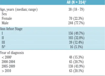
We identified 366 patients diagnosed with NLPHL between 1974 and 2012. 52 patients were excluded for reasons given in Table 1, and data on 314 patients could thus be analyzed. Patients were mostly young, 86.9% being 60 years old or less, including 59 patients between the ages of 18 and 25 (Table 1). There was a strong male predominance with a female/male ratio of 0.29. Most patients (82.5%) presented with stage I-II. Presentation was cervical, axillary or inguinal for 102 stage I patients. The mediastinum was involved in 30 patients, with half of them having early stage, and the remaining having advanced stage disease. In 11.9% of patients, stage II was subdiaphragmatic. B-symptoms were rare (4.8%, n=15), and occurred mainly in advanced stages (n=10).
Watchful waiting
Watchful waiting was defined as a period of observation of at least 3 months without any treatment. A total of 114 patients (36.3%) were in the watchful waiting group, including 35 patients who had undergone complete surgi-cal resection of the initial lesions (Table 2). In this group, early stages (67 stage I and 37 stage II) were overrepresent-ed comparoverrepresent-ed to advancoverrepresent-ed stages (9 stage III and 1 stage IV). Sixty-five patients progressed during observation, including 15 patients who had a complete resection. For the 95 evaluable patients, median PFS in the watchful waiting group was 56.4 months (range: 0.3 – 207).
At first relapse/progression, 8 patients remained in
observation, whereas 57 received treatment, which con-sisted in radiotherapy (n=21), rituximab as a single-agent (n=12), chemotherapy or immuno-chemotherapy (n=19), combined modality treatment (CMT) (n=3), rituximab plus radiotherapy (n=1), and unspecified treatment (n=1).
Frontline treatment
200 patients (63.7%) received treatment at diagnosis. Radiotherapy (RT) alone was the front-line treatment for 63 patients (20.1%) (Table 2), including 42 stage I, 20 stage II and 1 stage III. Radiotherapy was mainly delivered at a dose of 30-36 Gy (62% of patients) to cervical, supraclav-icular and/or axillary areas in 41 patients, and to inguinal and/or iliac areas in 14 patients; 2 patients underwent a subtotal nodal irradiation, and the treatment areas were not specified for 6 patients. Rituximab alone was the front-line treatment for 28 patients (8.9%). The treatment schedule consisted in 4 weekly injections of rituximab at 375 mg/m², without maintenance, with the exception of 2 patients who received 6 and 7 injections, respectively, over a period of 3 to 4 months. Chemotherapy or immuno-chemotherapy was administered to 68 patients (21.7%); anthracycline-containing regimens were used in most cases (see Table 2). CMT was the initial treatment for 40 patients (12.7%), and consisted mainly in ABVD or ABVD-like regimens followed by RT, which was delivered mainly to cervical, supraclavicular and/or axillary areas, and included the mediastinum in 10 patients (6 early stage, 4 advanced stage); 8 patients received supradiaphragmatic (mantle 6, involved-field 2), lombo-aortic and splenic irra-diation. 1 patient (0.3%) received only radiotherapy plus rituximab.
Initial management according to Ann Arbor stages is shown in Table 2. Briefly, among 259 patients with early-stage disease, 40% were in the watchful waiting group, 24% received radiotherapy, 9% rituximab, 14% chemotherapy or immuno-chemotherapy and 13% CMT. Among 55 patients with advanced stages, 18% were in the watchful waiting group, 58% received chemotherapy or immuno-chemotherapy, and 13% CMT.
J. Lazaroviciet al.
Table 1. Characteristics of the study population at diagnosis.
All (N = 314)1
Age, years (median; range) 38 (18 - 79) Sex
Female 70 (22.3%) Male 244 (77.7%) Ann Arbor Stage
I 156 (49.7%) II 103 (32.8%) III 39 (12.4%) IV2 16 (5.1%) Year of diagnosis < 20003 48 (15.3%) 2000-2004 65 (20.7%) 2005-2009 138 (43.9%) > 2010 63 (20.1%)
152 out of 366 patients were excluded for the following reasons: under 18 years of age (n=14), insufficient data (n=22), histological transformation at diagnosis (n=2), wrong diagnosis (n=6), previous history of cHL (n=3), refusal to participate in the study (n=1), or several of the previous reasons (n=4). 2Bone (n=12), liver (n=2), lung (n=1), digestive (n=1). 3Including 26 patients diagnosed in the period 1995-1999, 17 patients between 1990 and 1994, and 5 patients before 1990.
Management at first relapse/progression
A total of 117 patients experienced a relapse or progres-sion: 65 following watchful waiting and 52 after an initial treatment. Among these patients, 16.5% relapsed during the first 2 years following treatment. A minority (19 patients) did not receive any treatment following first pro-gression/relapse. In these patients, initial management had consisted in watchful waiting (n=8), radiotherapy (n=4), rituximab (n=1), chemotherapy or immuno-chemothera-py (n=3), and CMT (n=3). The management at relapse/progression was unspecified for 6 patients. The remaining 92 patients were treated as shown in Table 2. Chemotherapy or immuno-chemotherapy was the most frequent option in this setting (37 patients), including 19 patients who had not received prior therapy. 5 patients also received high-dose chemotherapy followed by autol-ogous stem-cell transplantation (ASCT), including 1 patient with a documented transformed NLPHL. 27 patients were treated with radiotherapy alone, including 21 patients who had been in the watchful waiting group. Lastly, 19 patients were treated with rituximab as a single-agent.
Efficacy and outcome
Among the 200 patients who were treated at diagnosis, evaluation of response was available for 183 patients (Table 3). The overall response rate (ORR) was 96.2%, with 166 patients achieving a complete response (CR), or complete response unconfirmed (CRu).
After initial radiotherapy, the ORR was 96.5%, with 54 patients (94.7%) achieving a CR/CRu. Relapse/progres-sion occurred in 11 out of 42 (26%) stage I patients, and 5 out of 20 (25%) stage II patients. Relapses occurred mainly outside irradiated areas, but 3 patients had disseminated relapse involving previously irradiated areas. Rituximab alone resulted in an ORR of 100%, with 24 patients (88.9%) reaching CR/CRu. Chemotherapy or immuno-chemotherapy resulted in an ORR of 95,2%, including 55
patients (87.3%) with CR/CRu. After CMT, patients expe-rienced a 94.3% ORR, including 32 patients (91.4%) with CR/CRu.
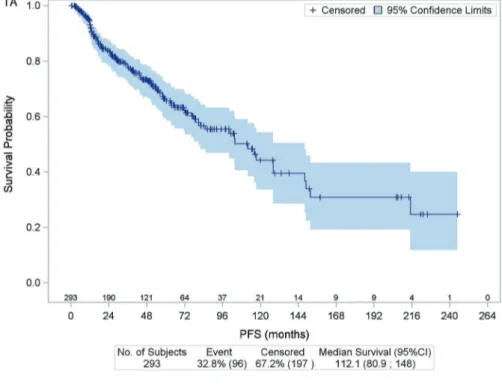
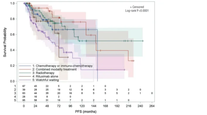
With a median follow-up of 55.8 months (range: 0.3-451.8), the 10-year PFS and OS estimates for the whole cohort were 44.2% and 94.9%, respectively (Figure 1). Ten patients died during the study period from disease pro-gression (n=1), sepsis following ASCT (n=1), secondary malignancies (n=4), traffic accident (n=2), or unknown cause (n=2). The 4-year PFS estimates according to each treatment modality was 79.6% with radiotherapy, 77.0% with rituximab alone, 78.8% with chemotherapy or immuno-chemotherapy, and 93.9% with CMT (Figure 2). Early initiation of treatment was associated with a signifi-cant reduction of the risk of progression of 65% for radio-therapy alone, 54% for chemoradio-therapy or immuno-chemotherapy, and 71% for CMT (Table 4). The risk of progression was not significantly reduced for patients treated with rituximab alone. This is consistent with the analysis of patients who had complete resection of the ini-tial lesions: median PFS was 82.0 months (range: 2.8 – 214.1 months) when surgery was followed by observa-tion, whereas it was not reached in patients who received additional therapy (P=0.0356) (Table 4).
Histological transformation
Histological transformation into aggressive B-cell lym-phoma was documented at relapse/progression in 24 patients (7.6%), including 14 patients in first, 9 patients in second and 1 patient in third relapse/progression, respec-tively. 2 patients had histological transformation to T-cell/histiocyte-rich large B-cell lymphoma, but the exact subtype was not specified in the remaining patients.
Secondary malignancies
9 patients developed secondary malignancies, including 2 adenocarcinomas of the rectum, 1 bronchopulmonary carcinoma, 1 gastric cancer, 1 angiosarcoma, 1 endometrial Table 2. Management of patients at diagnosis and first relapse/progression.
Management of patients at diagnosis
Stage I-II Stage III-IV Total
Watchful waiting 104 10 114 (36.3%)
Radiotherapy1 62 1 63 (20.1%)
Rituximab alone 24 4 28 (8.9%)
Chemotherapy or immuno-chemotherapy2 36 32 68 (21.7%)
Combined modality treatment2 33 7 40 (12.7%)
Radiotherapy plus rituximab 0 1 1 (0.3%)
Management at first relapse/progression
Stage I-II Stage III-IV Stage unknown Total
Watchful waiting 13 4 2 19 (17.0%)
Radiotherapy 19 0 8 27 (24.1%)
Rituximab alone 13 3 3 19 (17.0%)
Chemotherapy or immuno-chemotherapy3 11 19 7 37 (33.0%)
Combined modality treatment3 4 1 2 7 (6.2%)
Radiotherapy plus rituximab 1 0 1 2 (1.8%)
Management unspecified 1 0 0 1 (0.9%)
1Radiotherapy doses: 43 patients (68.2%) received 30 to 36 Gy, 7 patients (11.1%) 38 to 40 Gy, 2 patients (3.2%) 20 Gy, 3 patients (4.8%) 4 Gy. The dose of radiotherapy was unknown for 8 patients (12.7%). 2Chemotherapy (47), immuno-chemotherapy (61), including ABVD or ABVD-like regimens (76), BEACOPP (1), CHOP or CHOP-like regimens (18, all treated without radiotherapy), CVP (7), other regimens (2), unspecified (4). 3Chemotherapy alone (13), chemotherapy + rituximab (31). Chemotherapy included: ABVD or ABVD-like (14), BEACOPP (1), CHOP (15), ACVBP (5), MINE (2), DHAP or DHA-carboplatin (2), ICE (1), CVP (1), other regimens (2), unspecified (1). ABVD: doxorubicin, bleomycin, vin-blastine, dacarbazine; BEACOPP: bleomycin, etoposide, doxorubicin, cyclophosphamide, vincristine, procarbazine, prednisone; CHOP: cyclophosphamide, doxorbucin, vincristine, prednisone; CVP: cyclophosphamide, vincristine, prednisone; MOPP: mechlorethamine, vincristine, procarbazine, prednisone; DHAP: dexamethasone, cytarabine, cisplatin; ICE: ifos-famide, carboplatin, etoposide.
cancer, 1 melanoma, 1 hepatocarcinoma and 1 T-cell lym-phoma. 8 out of these 9 patients had received at least one treatment, including chemotherapy for 7 of them and radiotherapy for 5 of them (as part of CMT for 4 patients, and radiotherapy alone for 1 patient).
Risk factors and competing risks analysis
To account for competing risks between death, progres-sion/relapse, and second cancers, the regression analysis included age at diagnosis, stage, initial treatment with chemotherapy, rituximab and radiotherapy as explanatory covariates. No significant prognostic factor was identified
J. Lazaroviciet al.
Figure 1. Kaplan-Meier estimates for progression-free survival (1A) and overall survival (1B), with number of subjects at risk, and 95%
for death or for second cancers. For the whole population (stages I-IV), the competing risks analysis showed that early treatment with chemotherapy or radiotherapy sig-nificantly reduced the risk of progression versus watchful
waiting (HR 0.388 [0.234-0.643], P=0.0002), but not with rituximab alone (HR 0.695 [0.320-1.512], P=0.3593). In early stages, the risk of progression was only reduced with radiotherapy (HR 0.474 [0.277-0.812], P=0.0066).
Figure 2. Progression-free survival by management at diagnosis, with number of subjects at risk, and 95% confidence limits –all evaluable
patients.
Figure 3. Progression-free survival of 58 patients after complete surgical resection, according to additional treatment versus watchful waiting,
with number of subjects at risk, and 95% confidence limits.
+ Censored Log-rank P<0.0001
+ Censored Log-rank P<0.0356
Discussion
This study describes the clinical characteristics, manage-ment and outcome of NLPHL patients in LYSA centers, with a special focus on treatment. As reported previously, patients were mostly young, predominantly male and had early stage disease.7-9 B-symptoms were present in only 4.8% of patients, which is less frequent than the 9-10% previously reported.7-9With almost two-thirds of patients treated after 2005, and a high proportion of patients main-tained in watchful waiting after diagnosis, our study pro-vides a good opportunity to re-evaluate the role of radio-therapy and immuno-chemoradio-therapy in the treatment decision-making.
Though historically treated similarly to cHL, the individ-ualization of NLPHL as a distinct entity in the 1990’s has rendered questionable the validity of this approach. However, a risk-adapted strategy has not been developed for NLPHL and its specific management remains under debate. The new prognostic score developed by the GHSG, combining clinical and histologic features, espe-cially histopathologic variants to define risk groups, is an interesting approach.10
Our results show that initiating treatment at the time of diagnosis may improve PFS, compared with watchful waiting, which was the most frequent initial management and concerned more than one-third of our patients. Based on pediatric series,11,12the EuroNet-PHL-LP1 study recom-mends a follow-up without further treatment in children and adolescents with stage IA disease, and complete resec-tion of involved lymph nodes. However, in our study, even after complete surgical resection of local lesions, additional treatment improved PFS compared to watchful waiting. Our results do not support the recommendation of observation as a treatment option instead of RT for stage IA patients with a completely excised solitary lymph node.6 The risk of progression was reduced with radio-therapy compared to watchful waiting. However, with a 4-year PFS estimate of 79.6% for patients treated with radiotherapy alone, the benefit of radiotherapy is inferior
to the results of previously published studies,8,13,14 and relapses occurred in most cases outside the irradiated fields. Due to the low number of patients and events, a comparison between the results of radiotherapy in stage I and stage II was not possible. We conclude that radiother-apy alone is a possible option in selected early stages. This management is consistent with the international guide-lines5,6 and a recent study from the GHSG in which involved-field RT was defined as the standard of care for stage IA-NLPHL, based on an equivalent tumor control with a lower risk of long-term side effects compared to extended-field RT and CMT.15 Of note, a decline in radia-tion utilizaradia-tion for patients with NLPHL was reported in a recent large series.9Radiotherapy was the treatment for 1 out of 4 patients at first relapse/progression and remains a possible option for patients with early stages not previous-ly irradiated.
When radiotherapy alone is not indicated at diagnosis, our study shows that CMT, chemotherapy or immuno-chemotherapy, but not rituximab, result in a significant reduction of the risk of progression compared to watchful waiting. However, we could not determine which treat-ment option was the best in the different clinical presenta-tions. The excellent PFS achieved with CMT, although not significantly superior to chemotherapy or immuno-chemotherapy, suggests that a treatment similar to that of cHL might be the best choice for selected patients with early stage NLPHL. Still, the choice between RT alone and
J. Lazaroviciet al.
Table 3. Response to initial and second-line treatments.
Response to initial treatment (evaluable patients, n=200)
CR/CRu PR SD Progression Unknown
All 166 10 2 5 17
Radiotherapy 54 1 1 1 6
Rituximab alone 24 3 0 0 1
Chemotherapy or immuno-chemotherapy 55 5 1 2 5 Combined modality treatment 32 1 0 2 5
Radiotherapy plus rituximab 1 0 0 0 0
Response to second-line treatment (evaluable patients, n=92)
CR/CRu PR SD Progression Unknown
All 66 10 2 2 12
Radiotherapy 19 3 1 0 4
Rituximab alone 12 5 0 0 2
Chemotherapy or immuno-chemotherapy 27 2 1 2 5
Combined modality treatment 6 0 0 0 1
Radiotherapy plus rituximab 2 0 0 0 0
Table 4. Risk of progression after initial treatment.
Hazard ratio 95% CI P
Radiotherapy 0.345 0.196-0.610 0.0002 Rituximab alone 0.629 0.283-1.399 0.256 Chemotherapy or 0.476 0.266-0.855 0.0129 immuno-chemotherapy
Combined modality treatment 0.292 0.148-0.577 0.0004
CMT for the treatment of early stage disease remains a source of debate. Retrospective studies failed to show an improvement in disease control with CMT compared with RT alone.16,17 In contrast, a study from British Columbia reported that ABVD chemotherapy with or without RT improved outcomes compared with RT alone for limited stage NLPHL.18In our study, patients with early stages initially treated with CMT, chemotherapy or immuno-chemotherapy, achieved a good disease control, with a possible benefit over radiotherapy alone, a result that we can relate to a high rate of progression or relapse (26%) after initial radiotherapy. Chemotherapy alone has not been commonly used in adults with early stage NLPHL,18 and is the main option for patients with advanced stage.7,8,19 In this study, patients with stages III-IV who were initially treated received mainly chemotherapy or immuno-chemotherapy (71.1%), a management which is consistent with the international guidelines.5,6 An unsolved question is the choice of the chemotherapy reg-imen, in particular between a treatment similar to cHL with ABVD, or a treatment with CHOP, as in indolent NHL.20After R-CHOP therapy, favorable outcomes with a CR rate of 90% at 3.5 years have been reported, which supports the use of alkylating agents for advanced stage NLPHL.21
Rituximab therapy alone, without maintenance, yielded a high response rate, similar to previous studies.3,22 However, the 4-year PFS of only 77% suggests that ritux-imab as a single-agent is inferior to immuno-chemothera-py or CMT for long-term disease control. We cannot con-clude from our data the possible impact of the absence of maintenance on our results, which are in line with the experience of the Stanford University and the British Columbia Cancer Agency.23,24However, the benefit of rit-uximab maintenance has not been confirmed in a recent series.3The international guidelines recommend an initial treatment with6 or without rituximab alone,5 and ritux-imab could be a reasonable choice at relapse.25
The management of our patients at first relapse/progres-sion showing a low number of patients in the different
treatment groups, and the heterogeneity of reported series in terms of features of disease and therapeutic options,1,8,23 do not allow for a clear definition of the treatment of relapsed NLPHL. High-dose chemotherapy with ASCT was the salvage treatment for a few of our patients in the absence of documented histological transformation, and we cannot make a judgement on the possible benefit of ASCT in this setting.26 The short follow-up of our patients does not allow for any conclusions on risk factors, espe-cially the risk of histological transformation, as previously reported.1-4
In conclusion, our study shows that the treatment of NLPHL at diagnosis can improve PFS, compared to watch-ful waiting. The policy of watchwatch-ful waiting at diagnosis should be revisited, and discussed for carefully selected patients in order to maintain an excellent PFS and reduce the risk of late effects. In addition, a complete surgical resection of pathologic lymph nodes at diagnosis and ini-tial treatment with rituximab alone are not sufficient to improve PFS compared to watchful waiting. Radiotherapy for selected early stages, and when radiotherapy alone is not indicated, combined modality treatment, chemothera-py or immuno-chemotherachemothera-py represent the main options to treat adult patients with a curative intent. Intergroup collaborative studies with tumor banking are warranted to improve our knowledge of NLPHL, and to offer hope for patients to benefit from modern treatment and new agents.
Acknowledgments
We thank the entire staff of the Lymphoma Study Association operational organization (the Lymphoma Study Academic Research Organization [LYSARC]) and especially Sophie Pallardy and Sami Boussetta for the statistical analysis manage-ment; and David Ghez for critical review of the manuscript.
Authorship and Disclosures
Information on authorship, contributions, and financial & other disclosures was provided by the authors and is available with the online version of this article at www.haematologica.org.
References
1. Biasoli I, Stamatoullas A, Meignin V, et al. Nodular, lymphocyte-predominant Hodgkin lymphoma: a long-term study and analysis of transformation to diffuse large B-cell lymphoma in a cohort of 164 patients from the Adult Lymphoma Study Group. Cancer. 2010;116(3):631–639.
2. Al-Mansour M, Connors JM, Gascoyne RD, Skinnider B, Savage KJ. Transformation to Aggressive Lymphoma in Nodular Lymphocyte-Predominant Hodgkin’s Lymphoma. J Clin Oncol. 2010; 28(5):793–799.
3. Advani RH, Horning SJ, Hoppe RT, et al. Mature Results of a Phase II Study of Rituximab Therapy for Nodular Lymphocyte–Predominant Hodgkin Lymphoma. J Clin Oncol. 2014; 32(9):912– 918.
4. Eyre TA, Gatter K, Collins GP, Hall GW, Watson C, Hatton CS. Incidence, manage-ment and outcome of high grade transfor-mation of nodular lymphocyte predomi-nant Hodgkin lymphoma: long-term
out-comes from a 30-year experience. Am J Hematol. 2015;90(6):E103-110.
5. Eichenauer DA, Engert A, André M, et al. Hodgkin’s lymphoma: ESMO Clinical Practice Guidelines for diagnosis, treatment and follow-up. Ann Oncol. 2014;25(suppl 3):iii70–75.
6. The National Comprehensive Cancer Network. www.nccn.org
7. Diehl V, Sextro M, Franklin J, et al. Clinical Presentation, Course, and Prognostic Factors in Lymphocyte-Predominant Hodgkin’s Disease and Lymphocyte-Rich Classical Hodgkin’s Disease: Report From the European Task Force on Lymphoma Project on Lymphocyte-Predominant Hodgkin’s Disease. J Clin Oncol. 1999; 17(3):776–783.
8. Nogová L, Reineke T, Brillant C, et al. Lymphocyte-predominant and classical Hodgkin’s lymphoma: a comprehensive analysis from the German Hodgkin Study Group. J Clin Oncol. 2008;26(3):434–439. 9. Gerber NK, Atoria CL, Elkin EB, Yahalom J.
Characteristics and Outcomes of Patients With Nodular Lymphocyte-Predominant
Hodgkin Lymphoma Versus Those With Classical Hodgkin Lymphoma: A Population-Based Analysis. Int J Radiat Oncol Biol Phys. 2015;92(1):76–83. 10. Hartmann S, Eichenauer DA, Plütschow A,
et al. The prognostic impact of variant his-tology in nodular lymphocyte-predominant Hodgkin lymphoma: a report from the German Hodgkin Study Group (GHSG). Blood. 2013;122(26):4246–4252.
11. Pellegrino B, Terrier-Lacombe MJ, Oberlin O, et al. Lymphocyte-Predominant Hodgkin’s Lymphoma in Children: Therapeutic Abstention After Initial Lymph Node Resection--A Study of the French Society of Pediatric Oncology. J Clin Oncol. 2003;21(15):2948–2952.
12. Mauz-Körholz C, Gorde-Grosjean S, Hasenclever D, et al. Resection alone in 58 children with limited stage, lymphocyte-predominant Hodgkin lymphoma–experi-ence from the European network group on pediatric Hodgkin lymphoma. Cancer. 2007;110(1):179–185.
13. Wilder RB, Schlembach PJ, Jones D, et al. European Organization for Research and
Author et al.
Treatment of Cancer and Groupe d’Etude des Lymphomes de l’Adulte very favorable and favorable, lymphocyte-predominant Hodgkin disease. Cancer. 2002; 94(6):1731– 1738.
14. Wirth A, Yuen K, Barton M, et al. Long-term outcome after radiotherapy alone for lymphocyte-predominant Hodgkin lym-phoma. Cancer. 2005;104(6):1221–1229. 15. Eichenauer DA, Plütschow A, Fuchs M, et
al. Long-term course of patients with stage IA nodular lymphocyte-predominant Hodgkin lymphoma: a Report from the German Hodgkin Study Group. J Clin Oncol. 2015;33(26):2857-62.
16. Nogova L, Reineke T, Eich HT, et al. Extended field radiotherapy, combined modality treatment or involved field radio-therapy for patients with stage IA lympho-cyte-predominant Hodgkin’s lymphoma: a retrospective analysis from the German Hodgkin Study Group (GHSG). Ann Oncol. 2005;16(10):1683–1687.
17. Chen RC, Chin MS, Ng AK, et al. Early-stage, lymphocyte-predominant Hodgkin’s
lymphoma: patient outcomes from a large, single-institution series with long follow-up. J Clin Oncol. 2010; 28(1):136–141. 18. Savage KJ, Skinnider B, Al-Mansour M,
Sehn LH, Gascoyne RD, Connors JM. Treating limited-stage nodular lymphocyte predominant Hodgkin lymphoma similarly to classical Hodgkin lymphoma with ABVD may improve outcome. Blood. 2011; 118(17):4585–4590.
19. Xing KH, Connors JM, Lai A, et al. Advanced-stage nodular lymphocyte pre-dominant Hodgkin lymphoma compared with classical Hodgkin lymphoma: a matched pair outcome analysis. Blood. 2014;123(23):3567–3573.
20. Canellos GP, Mauch P. What Is the Appropriate Systemic Chemotherapy for Lymphocyte-Predominant Hodgkin’s Lymphoma? J Clin Oncol. 2010; 28(1):e8–e8. 21. Fanale MA, Lai CM, McLaughlin P, et al. Outcomes of Nodular Lymphocyte Predominant Hodgkin’s Lymphoma (NLPHL) Patients Treated with R-CHOP. Blood. 2010;ASH Meeting, Abstract 2812.
22. Eichenauer DA, Fuchs M, Pluetschow A, et al. Phase 2 study of rituximab in newly diagnosed stage IA nodular lymphocyte-predominant Hodgkin lymphoma: a report from the German Hodgkin Study Group. Blood. 2011;118(16):4363–4365.
23. Advani RH, Hoppe RT. How I treat nodular lymphocyte predominant Hodgkin lym-phoma. Blood. 2013;122(26):4182–4188. 24. Xing KH, Savage KJ. Modern Management
of Lymphocyte Predominant Hodgkin Lymphoma. Br J Haematol. 2013; 161(3):316 329.
25. Schulz H, Rehwald U, Morschhauser F, et al. Rituximab in relapsed lymphocyte-pre-dominant Hodgkin lymphoma: long-term results of a phase 2 trial by the German Hodgkin Lymphoma Study Group (GHSG). Blood. 2008;111(1):109–111.
26. Karuturi M, Hosing C, Fanale M, et al. High-Dose Chemotherapy and Autologous Stem Cell Transplantation for Nodular Lymphocyte-Predominant Hodgkin Lymphoma. Biol Blood Marrow Transplant. 2013;19(6):991-994.