HAL Id: hal-01960952
https://hal.archives-ouvertes.fr/hal-01960952
Submitted on 19 Dec 2018
HAL is a multi-disciplinary open access
archive for the deposit and dissemination of
sci-entific research documents, whether they are
pub-lished or not. The documents may come from
teaching and research institutions in France or
abroad, or from public or private research centers.
L’archive ouverte pluridisciplinaire HAL, est
destinée au dépôt et à la diffusion de documents
scientifiques de niveau recherche, publiés ou non,
émanant des établissements d’enseignement et de
recherche français ou étrangers, des laboratoires
publics ou privés.
High temperature oxidation of austenitic stainless steels:
effect of sulfur content on scale adhesion
V. Parry, E. Fedorova, C. Pascal, M. Braccini, M. Mantel, D. Oquab, D.
Monceau, Y. Wouters
To cite this version:
V. Parry, E. Fedorova, C. Pascal, M. Braccini, M. Mantel, et al.. High temperature oxidation of
austenitic stainless steels: effect of sulfur content on scale adhesion. International Symposium On High
Temperature Oxidation And Corrosion (ISHOC 2014), Jun 2014, Hakodate, Japan. �hal-01960952�
High temperature oxidation of austenitic stainless steels: effect of sulfur content on scale
adhesion.
V. Parry(1,*), E. Fedorova(2), C. Pascal (1), M.Braccini(1), M. Mantel (1,3), D.Oquab(4), D. Monceau (4), Y. Wouters(1)
(1)
SiMaP, University of Grenoble, FRANCE
(2) Polytechnic Institute of Siberian Federal University, RUSSIA (3)
UGITECH SA, Ugine, FRANCE
(4)
CIRIMAT, University of Toulouse, FRANCE e-mail: [email protected]
Two austenitic stainless steels, AISI 304L and AISI 303, containing 0.025 and 0.249 wt%S were oxidized in thermobalance at 1000°C for 50h. The chemical composition and the crystallographic structure of the oxide scales were investigated by Raman spectroscopy. Adhesion of oxide scales was tested by SEM in situ tensile tests. A correlation between the specific mass change, the chemistry, the microstructure and the adhesion properties is made and results are discussed in relation with sulphur concentration in the alloy.
1. INTRODUCTION
The life time of metallic parts submitted to high temperature oxidation is often limited by scale spallation leading to rapid oxide loss or to initiation of breakaway oxidation by iron oxides formation on chromia-forming alloys. Scale spallation is the result of a complex process where two important parameters play a role: stresses and adhesion.
A majority of industrial stainless steels contain sulfur in order to improve their forgeability but sulfur is known to be a powerful weakener of iron and to strongly segregate to metal free surfaces and to metal/oxide interfaces. Interfacial segregation of sulfur impurities is believed to cause a weakening of the normally strong interfacial bond strength of alumina and chromia scales [1]. Deleterious effects of sulfur segregation on the adherence of oxide scale grown on alumina-forming alloys have been widely studied [2] and are known to be the major cause of alumina scale failure. However,for chromia scales interfacial morphology associated with growth stresses is a more important cause of scale failure and sulfur segregation is only a secondary effect [1].
In this paper, two austenitic stainless steels AISI 304L and AISI 303, containing 0.025 and 0.249 wt% S, were oxidized at 1000°C for 50h. Thermogravimetric analysis was performed. The chemical composition and the crystallographic structure of the oxide scales were investigated by Raman spectroscopy. Adhesion of oxide scales was tested by SEM in situ tensile test. A correlation between the specific mass change, the chemistry and the adhesion properties is made and results are discussed in relation with sulphur concentration.
2. EXPERIMENTAL
Oxidation tests were performed on specimens cut by electrical discharge machining from bars of AISI 304L and AISI 303 provided by Ugitech France. Their chemical composition, displayed in Table 1, is similar except the sulfur content which is 0.25wt%for AISI 304L and 0.249 wt% for AISI 303. Before oxidation tests, samples were polished with SiC paper up to 1200 grade, cleaned in ethanol and dried in air. Isothermal oxidations were performed under synthetic air at atmospheric pressure in dynamic condition (2 mm/s).
Table 1. Chemical composition of austenitic stainless steels AISI 304L and AISI 303
Steels Ni Cr Mn Mo V Cu P Sn Co 304L 8.960 18.040 1.128 0.400 0.098 0.481 0.021 0.003 0.103 303 8.284 17.100 1.746 0.404 0.080 0.511 0.029 0.009 0.098 Steels Si Nb C S N O Ca Al Ti 304L 0.458 0.012 0.021 0.025 0.048 0.010 0.005 0.003 <0.0001 303 0.443 0.011 0.055 0.295 0.035 0.015 0.013 <0.002 <0.002
Rectangular specimens with dimension of 17 mm × 9 mm × 2 mm were used for thermogravimetric tests at 1000°C. The thermobalance used is a SETARAM™ TAG 24S with a 1 µg sensitivity. The device makes use of a double symmetrical furnace designed to compensate all signal disturbances resulting from gas flow, buoyancy and convection.
Tensile specimens, characterized by a gauge section of 2 mm width x 1 mm thick and a gauge length of 3 mm long, were isothermally oxidized in a horizontal furnace at 900°C and 10
The chemical composition and the crystallographic structure of the oxide scales were investigated by Raman spectroscopy using a Renishaw RM1000 Raman microscope.
After oxidation, in situ tensile tests were performed in a horizontal tensile mach
SEM chamber. JEOL JSM 6400 SEM was used. The procedure consists in regularly straining the sample by tensile loading while recording strength and elongation. A regular elongation rate of
spallation is observed, SEM pictures are
to a characteristic curve: spalled fraction of surface area vs. strain.
Surface SEM views after tensile tests and EDS analysis were obtained with a LEO S440 Stereoscan instrument equipped with the EDAX Genesis software.
3. RESULTS AND DISCUSSION
Isothermal kinetics of the two austenitic stainless steels oxidized for 50h at 1000°C under synthetic air are displayed in Figure 1. Experiments performed on AISI 304L were reproduced three times. Despite some significant experimental variations the parabolic rate constant is around 3.5 10-6 mg2.cm-4h
-304L and one order of magnitude higher for AISI 303: 2.9 10-5 mg2.cm-4h-1.
Assuming that the scale is only composed chromina, the equivalent oxide thickness estimated from the weight gain after 50h of oxidation is about 6 µ m for AISI 304L and 12 µ m for AISI 303.
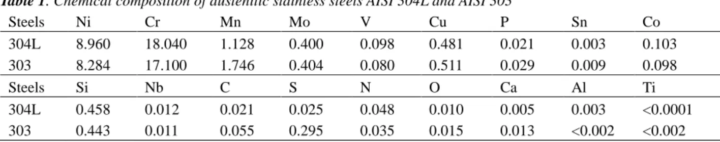
For AISI 304L, Raman spectroscopy results 2. (a), have shown that chromia Cr2
(Fe,Cr)3O4 are present in the scale [3].
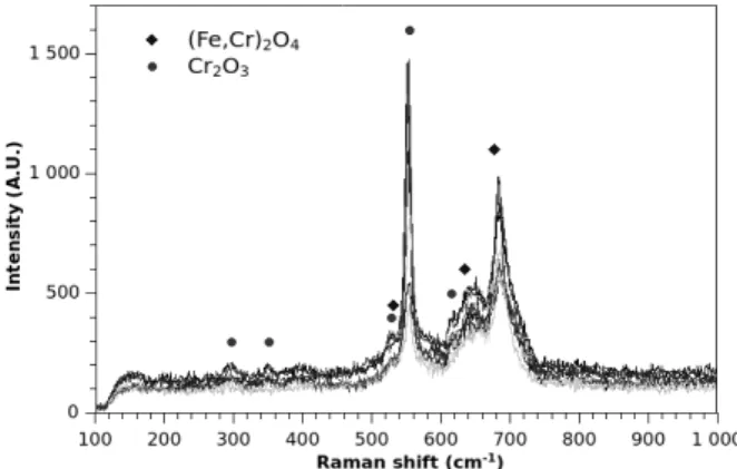
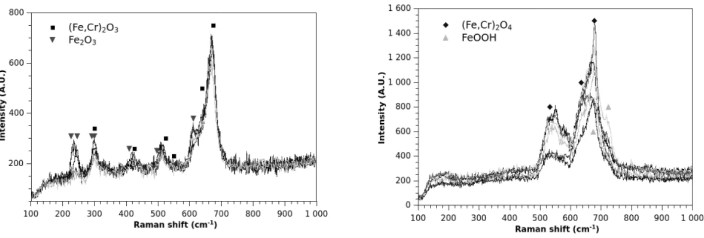
Raman spectra of the oxide scale grown on AISI 30 displayed in Fig. 2. (b). Two types of spect
depending on the analyzed zone. The iron rich oxide scale composed of hematite Cr2O3 and a solid solution of
(Fe,Cr)2O3 and of spinel type oxide (Fe,Cr)
SEM observations in BSE mode of the surface and AISI 303after oxidation for 50h at 1000°C
in Fig. 3. (a). and Fig. 3. (b). respectively. Surface morphologies of the oxides scales grown on 304L and 303 are different. For AISI 303, the scale
homogeneous and more brittle. EDS
presented) indicate that the iron content in the oxide scale is higher for AISI 303 than for AISI 304L.
Rectangular specimens with dimension of 17 mm × 9 mm × 2 mm were used for thermogravimetric tests at 1000°C. The thermobalance used is a SETARAM™ TAG 24S with a 1 µg sensitivity. The device makes use of a double symmetrical
te all signal disturbances resulting from gas flow, buoyancy and convection.
Tensile specimens, characterized by a gauge section of 2 mm width x 1 mm thick and a gauge length of 3 mm long, were isothermally oxidized in a horizontal furnace at 900°C and 1000°C.
The chemical composition and the crystallographic structure of the oxide scales were investigated by Raman spectroscopy using a Renishaw RM1000 Raman microscope.
were performed in a horizontal tensile machine specially designed to be placed in the SEM chamber. JEOL JSM 6400 SEM was used. The procedure consists in regularly straining the sample by tensile loading while recording strength and elongation. A regular elongation rate of 50 µm.min
spallation is observed, SEM pictures are periodically taken and the series of pictures is treated by image analysis, leading to a characteristic curve: spalled fraction of surface area vs. strain.
tensile tests and EDS analysis were obtained with a LEO S440 Stereoscan instrument equipped
Isothermal kinetics of the two austenitic stainless steels oxidized for 50h at 1000°C under synthetic air are displayed in Figure 1. Experiments performed on AISI 304L were significant experimental variations the parabolic rate
-1 for AISI
f magnitude higher for Assuming that the scale is only composed of de thickness estimated from the weight gain after 50h of oxidation is about 6 µ m for AISI 304L and 12
Raman spectroscopy results, displayed in Fig.
2O3 and solid solution
.
Raman spectra of the oxide scale grown on AISI 303 are Two types of spectra are recorded, on the analyzed zone. The iron rich oxide scale is and a solid solution of
(Fe,Cr)3O4[3,4].
in BSE mode of the surfaces of AISI 304L for 50h at 1000°C are displayed and Fig. 3. (b). respectively. Surface of the oxides scales grown on 304L and 303 he scale seems less EDS surface analyses (not iron content in the oxide scale is higher for AISI 303 than for AISI 304L.
Fig. 1. Isothermal oxidation kinetics of AISI 304L and AISI 303 at 1000°C under synthetic air at atmospheric pressure in dynamic conditions (2 mm.s-1)
Fig. 2. (a) Raman spectra of oxide scale grown on AISI 304L after 50h at 1000°C under synthetic air.
Rectangular specimens with dimension of 17 mm × 9 mm × 2 mm were used for thermogravimetric tests at 1000°C. The thermobalance used is a SETARAM™ TAG 24S with a 1 µg sensitivity. The device makes use of a double symmetrical
te all signal disturbances resulting from gas flow, buoyancy and convection.
Tensile specimens, characterized by a gauge section of 2 mm width x 1 mm thick and a gauge length of 3 mm long, were The chemical composition and the crystallographic structure of the oxide scales were investigated by Raman
ine specially designed to be placed in the SEM chamber. JEOL JSM 6400 SEM was used. The procedure consists in regularly straining the sample by tensile
µm.min–1 was applied. When oxide aken and the series of pictures is treated by image analysis, leading tensile tests and EDS analysis were obtained with a LEO S440 Stereoscan instrument equipped
Fig. 1. Isothermal oxidation kinetics of AISI 304L and AISI 303 at pheric pressure in dynamic
Fig. 2. (a) Raman spectra of oxide scale grown on AISI 304L after 50h at 1000°C under synthetic air.
Fig. 2. (b) Raman spectra of oxide scale grown on AISI 303 after 50h at 1000°C under synthetic air In situ tensile test were performed in SEM
stored. The behaviors of the oxide layers were quite different (see observed transverse crack initiation and growth i
cracks grew perpendicular to the tension direction, but while straight cracks propagated in the oxide scale of the 303 steel, in the 304L steel cracks looked like waves.
the two steels: this strain is about 1.6% in the 303 steel and 3.4% in the 304L steel.
Finally, spallation occurs in the oxide scale in the 303 steel, but not in the 304L steel. In the 304L
scale buckling and spallation, the transverse cracks opened, following the ductile deformation of the underneath substrate. The metal appears for a strain of 7.8%. At the end of the test, this cracks opening was so wide that we cou
at the crack root.
Fig. 3(a). Surface SEM view in BSE mode of the scale on AISI 304L before tensile test.
Fig. 3(c). Surface SEM view in BSE mode of the oxide scale on AISI 304L during tensile test (
Fig. 2. (b) Raman spectra of oxide scale grown on AISI 303 after 50h at 1000°C under synthetic air
n SEM. During the test, the oxide scale is forced to spall, releasing the elastic energy . The behaviors of the oxide layers were quite different (see Fig. 3(c) and Fig. 3(d))
observed transverse crack initiation and growth in the oxide scale, the crack patterns were different. In both case the cracks grew perpendicular to the tension direction, but while straight cracks propagated in the oxide scale of the 303 steel, in the 304L steel cracks looked like waves. The critical strains at which first cracks were observed are quite different in the two steels: this strain is about 1.6% in the 303 steel and 3.4% in the 304L steel.
in the oxide scale in the 303 steel, but not in the 304L steel. In the 304L
scale buckling and spallation, the transverse cracks opened, following the ductile deformation of the underneath substrate. The metal appears for a strain of 7.8%. At the end of the test, this cracks opening was so wide that we cou
in BSE mode of the oxide AISI 304L before tensile test.
Fig. 3(b). Surface SEM view in BSE mode of the oxide scale on AISI 304L before tensile test.
view in BSE mode of the oxide scale on AISI 304L during tensile test (28% of strain)
Fig. 3(c). Surface SEM view in BSE mode of the oxide scale on AISI 304L during tensile test (25%
Fig. 2. (b) Raman spectra of oxide scale grown on AISI 303 after 50h at 1000°C under synthetic air
the oxide scale is forced to spall, releasing the elastic energy Fig. 3(c) and Fig. 3(d)). While in both steels we n the oxide scale, the crack patterns were different. In both case the cracks grew perpendicular to the tension direction, but while straight cracks propagated in the oxide scale of the 303 steel, trains at which first cracks were observed are quite different in in the oxide scale in the 303 steel, but not in the 304L steel. In the 304L steel, instead of oxide scale buckling and spallation, the transverse cracks opened, following the ductile deformation of the underneath substrate. The metal appears for a strain of 7.8%. At the end of the test, this cracks opening was so wide that we could see the metal
Surface SEM view in BSE mode of the oxide scale on AISI 304L before tensile test.
Surface SEM view in BSE mode of the oxide scale on AISI 304L during tensile test (25% of strain)
These behaviors can be discussed in relation with sulphur concentration. Presence of impurities such as sulfur in the alloy may affect the scale microstructure. AISI 303 scale is iron rich. Iron oxides are known to be less protective [5] and their growth is ensured by cationic diffusion. The metal atoms are incorporated in the oxide at the metal/oxide interface and transferred across the interface. Since the oxide scale grows predominantly by an outward flux of cations, sulfur is believed to stay at the metal/oxide interface [1]. A relative enrichment of S occurs induced by the metal depletion leading to a lower adhesion of the oxide. On the contrary in the case of an oxide scales grown by oxygen inward diffusion, the interface moves inward and can incorporate sulfur from the alloy. The interface is the place of a chemical reaction, and sulfur is more likely to be integrated in the oxide rather than accumulated at the interface.
4. CONCLUSION
Two austenitic stainless steels, AISI 304L and AISI 303, containing 0.025 and 0.249 wt.% S were oxidized in thermobalance at 1000°C for 50h. For AISI 303, the parabolic rate constant is about one order of magnitude higher than AISI 304L. The oxide scale is thicker and brittle and richer in iron than the one obtained on AISI 304L. During tensile test, spallation occurs in the oxide scale of the AISI 303, but not in the 304L steel. In the 304L steel, instead of oxide scale buckling and spallation, the transverse cracks opened, following the ductile deformation of the underneath substrate. These results are discussed in relation with sulphur concentration. The difference in scale chemistry and microstructure may be linked with sulfur content. Iron oxides growth is ensured by cationic diffusion. The metal atoms are incorporated in the oxide at the metal/oxide interface and transferred across the interface. Since the oxide scale grows predominantly by an outward flux of cations, sulfur is believed to stay at the metal/oxide interface. A relative enrichment of S occurs induced by the metal depletion leading to a lower adhesion of the oxide.
Acknowledgements
This work has been supported by National Center for Scientific Research (CNRS, France) through the Projet International de Coopération Scientifique (PICS) Adherons!2 and Russian Foundation for Basic Research (RFBR) Ref n° 13-08-91053-CNRS_a.
References.
[1] F. H. Stott, G. C. Wood and J. Stringer, “The influence of Alloying Elements on the Development and Maintenance
of Protective Scales”, Oxidation of Metals, Vol. 44, Nos. 1/2 (1995), pp. 113-145.
[2] T. Gheno, D. Monceau, D. Oquab, Y. Cadoret, “Characterization of Sulfur Distribution in Ni-Based superalloy and
Thermal Barrier Coating After High Temperature Oxidation: A SIMS Analysis”, Oxidation of Metals, Vol. 73 (2010), pp. 95-113.
[3] J.-P. Petit, M. Mermoux, Y. Wouters, A.Galerie and C. Chemarin, “Study of the Thermal Oxidation of Fe-15 Cr by
Combined Raman and Photoelectrochemical Imaging”, Materials Science Forum, Vols. 461-464 (2004), pp. 681-688 [4] D. Oquab, D. Galy, C. Josse, A. Freulon, Y. Thebault, C. Charvillat, J. Esvan, O. Marsan; High temperature oxidation induced austenite to martensite phase transformation in austenitic 304L stainless steel at 1000°C(to be published).