Publisher’s version / Version de l'éditeur:
Vous avez des questions? Nous pouvons vous aider. Pour communiquer directement avec un auteur, consultez la première page de la revue dans laquelle son article a été publié afin de trouver ses coordonnées. Si vous n’arrivez pas à les repérer, communiquez avec nous à PublicationsArchive-ArchivesPublications@nrc-cnrc.gc.ca.
Questions? Contact the NRC Publications Archive team at
PublicationsArchive-ArchivesPublications@nrc-cnrc.gc.ca. If you wish to email the authors directly, please see the first page of the publication for their contact information.
https://publications-cnrc.canada.ca/fra/droits
L’accès à ce site Web et l’utilisation de son contenu sont assujettis aux conditions présentées dans le site LISEZ CES CONDITIONS ATTENTIVEMENT AVANT D’UTILISER CE SITE WEB.
The Journal of Hygiene, 96, 1, pp. 39-48, 1986-02
READ THESE TERMS AND CONDITIONS CAREFULLY BEFORE USING THIS WEBSITE. https://nrc-publications.canada.ca/eng/copyright
NRC Publications Archive Record / Notice des Archives des publications du CNRC :
https://nrc-publications.canada.ca/eng/view/object/?id=b8039567-d7de-4729-8200-56c70357d9d3 https://publications-cnrc.canada.ca/fra/voir/objet/?id=b8039567-d7de-4729-8200-56c70357d9d3
NRC Publications Archive
Archives des publications du CNRC
This publication could be one of several versions: author’s original, accepted manuscript or the publisher’s version. / La version de cette publication peut être l’une des suivantes : la version prépublication de l’auteur, la version acceptée du manuscrit ou la version de l’éditeur.
For the publisher’s version, please access the DOI link below./ Pour consulter la version de l’éditeur, utilisez le lien DOI ci-dessous.
https://doi.org/10.1017/S0022172400062513
Access and use of this website and the material on it are subject to the Terms and Conditions set forth at The relationship between the serogroup antigen and
lipopolysaccharide of Legionella pneumophila Conlan, J. W.; Ashworth, L. A. E.
J. Hyg., Camb. (1986), 96, 39-48 3 9 Printed in Great Britain
The relationship between the serogroup antigen and Iipopolysaccharide of Legionella pneumophila
B Y J . W. CONLAN AND L. A. E. ASHWORTH
Experimental Pathology Laboratory, PHLS Centre for Applied Microbiology and Research, Porton Down, Salisbury, Wiltshire SP4 OJG, UK
{Received 8 July 1985; accepted 30 August 1985)
SUMMARY
Serogroup-specific antigen was extracted from a number of Legionella
pneumo-phila strains and compared with phenol-water extracted Iipopolysaccharide on the
basis of gel filtration, chemical analysis, SDS-PAGE and reaction with serogroup-specific antibody in immunoblots. Serogroup serogroup-specificity is apparently borne by the 0 side-chains of the Iipopolysaccharide, which, although smooth in type, partitions in the phenol phase. For four serogroup 1 strains tested, there was no qualitative correlation between 0 side-chain length and pulmonary virulence for guinea-pigs.
INTRODUCTION
The specificity of a major heat-stable, pronase-resistant antigen provides a means of serogrouping strains of Legionella pneumophila. Antibodies to the serogroup antigen are therefore of diagnostic importance (Harrison & Taylor, 1982; Stanek el al. 1983). However, the nature of the serogroup antigen is uncertain, although results from studies of its chemical composition (Johnson et al. 1979; Wong et al. 1979; Flesher et al. 1982), its role in endotoxicity (Wong et al 1982) and its behaviour in crossed immunoelectrophoresis (Joly & Kenny, 1982; Collins
et al. 1983), suggest that it is Iipopolysaccharide (LPS).
Many of the specificities utilized in serotyping Gram-negative organisms are expressed on the O-specific polysaccharide of their LPS. This has been most studied in the salmonellae and Escherichia coli. The LPS from these organisms has been shown to display heterogeneity with respect to O-specific chain length (Palva & Makela, 1980; Goldman & Leive, 1980). In both cases there is also good correlation between possession of 0 side-chain and virulence. Other Gram-negative organisms, e.g. Neisseria gonorrhoeae (Connelly & Allen, 1983) and Bordetella pertussi& (Peppier, 1984), have been shown to possess LPS with O-specific polysaccharide of only short chain length and with little heterogeneity.
In this study we have examined the serogroup antigens from one virulent serogroup 3 strain of L. pneumophila and from four strains of serogroup 1 which differ from each other in virulence as determined by a guinea-pig model of infectior (Fitzgeorge et al. 1983; Fitzgeorge, 1985).
The purposes of this study were to determine the relationship between th( serogroup antigen of L. pneumophila and its LPS, the extent of the heterogeneity
40 J . W. CONLAN AND L. A. E . ASHWORTH
in the LPS 0 side-chain length and whether possession of 0 side-chain was a prerequisite for virulence.
MATERIALS AND METHODS
L. pneumophila strains. Serogroup 1 strains were Corby (a human isolate kindly
provided by Dr R. A. Swann, John Radcliffe Hospital, Oxford.), Corby avirulent (prepared from the above strain by multiple passage on charcoal yeast extract agar), W74/81 (isolated in this laboratory from a naturally contaminated water supply) and Philadelphia 1 (NCTC11192), also passaged repeatedly since its initial isolation. Serogroup 3 strain W166/81, isolated in this laboratory, was also from a water supply. Of the serogroup 1 strains, Corby is the most virulent for guinea-pigs by the aerosol route (Fitzgeorge, 1985), W74/81 is of intermediate virulence whilst NCTC 11192 and Corby avirulent fail to cause respiratory infection (Fitzgeorge
et al. 1983; R. B. Fitzgeorge, personal communication). The Philadelphia 1 strain
(NCTC 11192) was cultured once on charcoal yeast extract (CYE) agar (Edelstein, 1981) in this laboratory. The other strains, with the exception of Corby avirulent, had been subcultured only three times on CYE agar prior to use in this study.
Growth conditions. Starter cultures of 100 ml yeast extract broth (YEB) (Ristroph,
Hedlund & Allen, 1980) in 500 ml conical flasks were inoculated with organisms grown on CYE agar and incubated aerobically at 37 °C for 24 h on an orbital shaker. Purity was checked by Gram's stain and by the ability to grow on CYE agar but not on blood agar. Starter cultures were used to inoculate fresh YEB (5 ml into 500 ml in 2 1 flasks, 12 flasks per strain) which was incubated as above for 16 h. Organisms were harvested by centrifugation at 1500 £ for 1 h and washed once in sterile water.
For each strain the cell pellet was divided into two parts (ca. 5 g wet weight each). Half was extracted for serogroup antigen and half for LPS.
Preparation of serogroup antigen. Cells were extracted in 1 ml 0*9% (w/v)
saline per 100 mg wet weight, heated to 100 °C for 1 h with constant stirring, cooled, centrifuged at 17000 £ for 15 min and the supernatant dialysed overnight against phosphate-buffered saline (8-0gNaCl, 0-2gKCl, 1-15 g Na2HPO4, 0'2 g KH2PO4/1). Extracts were treated sequentially for 2 h at 37 °C with ribonu-clease 1, deoxyribonuribonu-clease 1 (in the presence of 0*1 M-MgCl2) and pronase (all enzymes from BDH) each at a final concentration of 0*01 mg/ml. At the end of this treatment residual enzyme activity was destroyed (100 °C for 5 min) and the extracts dialysed against Tris-HCl buffer (0-1 si, pH 8*2). Twenty-millilitre samples of each extract were gel filtered on a 2-6 x 90 cm column of Sepharose 4B at a flow rate of 10*0 ml/h. The eluate was monitored for absorbance at 280 nm and assayed for serogroup antigen by ELISA.
Preparation of LPS. Cells were extracted by the phenol-water procedure
(Westphal & Jann, 1965). For each strain, pooled aqueous phases and the phenol phase were dialysed separately for 3 days against running tap water and then against PBS. Phenol phase preparations were subjected to the same enzyme treatments and .gel filtration used for serogroup antigen.
Chemical analyses. 2-Keto-3-deoxyoctonate (KDO) was assayed by the method
of Karkhanis et al. (1978), carbohydrate by the method of Dubois et al. (1956) and protein using the Lowry assay system.
L. pneumophila LPS 41
Antisera and conjugates. Antisera to serogroup antigens 1 and 3 were obtained
by immunizing rabbits with saline extracts of strains W74/81 and W166/81, respectively. After a preliminary blood sample had been taken each rabbit was injected with 300 /il of antigen (15 fig carbohydrate) in incomplete Freund's adjuvant (Difco) distributed between two intramuscular and two dorsal subcuta-neous sites. A boost was given with the same dose after 2 weeks and blood was taken at weekly intervals for antibody assay. Sera were titrated by an indirect ELISA (see below). Two weeks after a further boost (about 12 weeks after starting the injections), the rabbits were exsanguinated under barbitone anaesthesia. High-titre sera were pooled and the IgG fraction obtained by protein A affinity chromatography; IgG was conjugated to horseradish peroxidase (HRP) according to the methods of Nakane & Kawooi (1974).
ELISA for antibody. Sera were titrated by an indirect ELISA in 96-well plates
(Dynatech M 129A) at room temperature. Wells were coated overnight with homologous saline extract (10 fig/ml carbohydrate) in 0-05 M carbonate buffer (pH 9-5), then washed. Wash solution was 0-1 % Tween 20 in PBS, which was also used for serial dilution of the test sera in a second plate. Volumes of 100 fil were transferred from each well to the corresponding well on the washed coated plate, which was incubated for 2 h with shaking. The plate was washed and sheep anti-rabbit HRP conjugate added (100/d/well at 10/ig/ml) and incubation continued for 2 h. After washing again, substrate was added and colour read at 450 nm. Substrate was 5-aminosalicylic acid (5-ASA) in water (1 mg/ml) adjusted to pH 6-0, with H2O2 added to 0007%. Reaction with heterologous antigen was
<0-4%.
SDS-PAGE. Samples were run on slab gels using the Laemmli (1970) buffer
system. The separating gel was 15% (w/v) acrylamide, 0-5% bis-acrylamide containing 4-0 M urea. Samples containing 0*5 mg/ml LPS were diluted 1:1 with double-strength sample buffer and 50 p\ applied to each track of the gel (total 12-5 fig LPS/track). Gels were run at 125 V until the bromophenol blue tracking dye had migrated to within 1-0 cm from the end of the gel. The gel was stained for LPS using a silver stain (Tsai & Frasch, 1982). A Transblot apparatus (Biorad) was used for immunoblotting the gels, and blots were visualized (Irons Ashworth & Wilton-Smith, 1983) using an HRP conjugate of antibody tc serogroup antigen.
ELISA for antigen. The wells of 96-well plates (Nunc Immunoplate 1) wen
coated overnight with 100 fd antibody to serogroup antigen at 10 fig IgG/ml ii carbonate buffer (pH 9*5), then washed as in the ELISA for antibody above. Seria twofold dilutions of test materials were made in a second plate using wash solutioi as diluent. Volumes of 100 ^1 were transferred from each well to the correspondinj well on the washed, coated plate, which was incubated for 2 h with shaking. Th plate was washed, HRP conjugate of antibody to serogroup antigen addei (100 /il/well at 10 fig IgG/ml), and incubation continued for 2 h. After washin again, substrate (5-ASA) was added and colour read at 450 nm. The activity c serogroup 1 antigen in the serogroup 3 ELISA was < 1 %, and vice versa.
42 J . W . CONLAN AND L . A. E . ASHWORTH
Table 1. Results of chemical analysis on purified saline extracted (SE) and phenol
phase (P) antigen of Legionella pneumophila
Chemical analysis of Sepharose 4B column fractions* Sample
Corby (SE) pool A Corby (SE) pool B Corby AVf (SE) pool A Corby AV (SE) pool B NCTC 11192 (SE) pool A NCTC 11192 (SE) pool B W74/81 (SE) pool A + B W166/81 (SE)pool A + B Corby (P) pool A Corby AV (P) pool A Carbohydrate (% dry wt.) 5-5 6-7 4 0 4-6 2-5 6-2 5-2 8-5 7-2 6-4 Protein (% dry wt.) 160 7-6 16-2 7-4 100 9-5 120 120 6-2 6-0 KDO (% dry wt.) 1-15 1-65 0-80 1-25 0-25 1-20 1-40 1-55 1-90 1-85 Carbohydrate: KDO 4-8:1 4-1:1 5-0:1 3-7:1 100:1 5-2:1 3-7:1 5-5:1 3-8:1 3-5:1 S. typhimurium LPS (Sigma) 53-5 6-2 7-15 7-5:1
* See fraction 1 for fraction description, t AV, Avirulent.
RESULTS
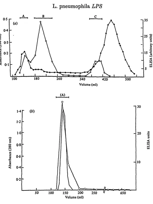
Corby-strain saline extract eluted from a Sepharose 4B column as two peaks of 280 nm absorbance and two ELISA peaks of serogroup 1 antigen (Fig. la). The elution profiles were similar for strains other than Corby. The peaks monitored by the two techniques (Fig. 1 a) were not fully coincident, indicating that much of the protein present after heating and pronase treatments was a contaminant of the serogroup antigen. The asymmetric first antigen peak (approximately 85 % of eluted antigen) is broad, suggesting either heterogeneity of a high-molecular-weight material or considerable aggregation. The second ELISA peak might represent unaggregated antigen. The elution profile of phenol phase LPS from the same column is shown in Fig. 1 b. In this case both the peak of 280 nm absorbance and the ELISA peak coincide; there is no second peak.
Fractions were pooled (A, B, C) as indicated (Fig. la), lyophilized and assayed for carbohydrate, protein and 2-keto-3-deoxyoctonate (KDO). The data from these analyses are presented in Table 1. The first ELISA peak maximum (pools B, Fig. la) contained 4-6-6-7% (w/w) carbohydrate, 1-2-1-65% (w/w) KDO and 7-6-9-5 % (w/w) protein. Whilst the first peak of 280 nm absorbance (pools A, Fig. la) contained consistently higher amounts of protein, 10-16% (w/w), the ratios of carbohydrate to KDO in pools A and B were similar to each other and to phenol phase antigen. The second antigen peak (pool C) contained insufficient material to allow detection of carbohydrate or KDO.
In contrast, aqueous phases contained little or no antigen detectable by ELISA. They might have contained lipid-A core material as KDO was present in significant amounts (data not shown).
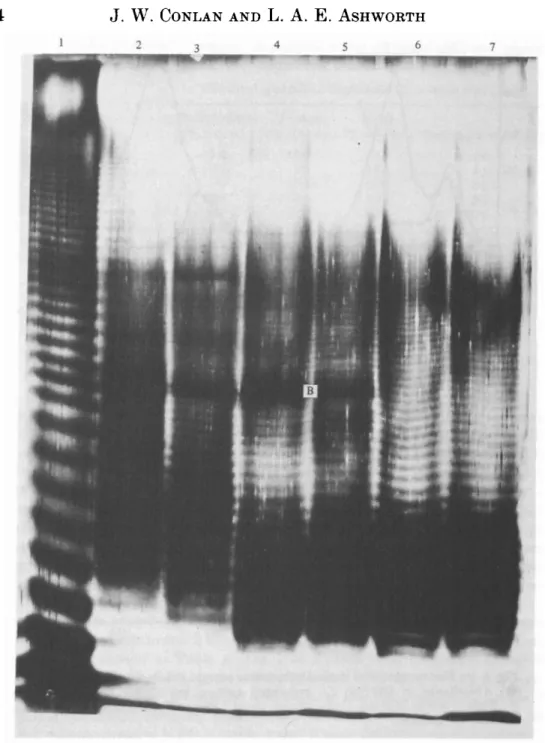
For saline extracts and for phenol-phase LPS, silver staining of SDS-PAGE gels (Fig. 2) revealed ladder-like patterns typical of smooth LPS (Tsai & Frasch, 1982), although over a slightly more restricted range compared to the LPS from
L. pneumophila LPS 43 8
I
100 180 260 340 420 Volume (ml) 500 (A) 1-4 1-2 1-0 o 0-8 8J
< 0-4 0-2 50 100 150 200 Volume (ml) 250 30 20 .5 10 650Fig. 1. (a) Elution profile of strain Corby saline extract from a Sepharose 4B column. # , Absorbance at 280 nm; Ot serogroup antigen by ELISA (arbitrary units). (6) Elution profile of strain Corby phenol phase LPS: continuous line, absorbance at 280 n m ; O , ELISA.
Salmonella typhimurium. There was a marked difference in the band spacin
between the latter and the L. pneumophila patterns.
Saline extracts of four serogroup 1 strains (e.g. Fig. 2, lane 3) and of a serogrou 3 strain (lane 2) resolved into at least 35 bands including the two more intensel staining bands (B). Phenol phase LPS (Fig. 2, lanes 6 and 7) lacked the bands'.
but otherwise resolved similarly to saline extracts. The saline extracts and phem phase LPS from both virulent and avimlent L. pneumophila gave identici
44 J . W. CONLAN AND L. A. E. ASHWORTH
Fig. 2. SDS-PAGE analysis of serogroup antigen. Lanes were loaded with 125 fig LPS. Lane 1, 5. typhimuriumLPS (Sigma); lane 2, W166/81 (SE); lane 3 W74/81 (SE); lane 4, Corby (SE); lane 5 NCTC 11192 (SE); lane 6, Corby (P); lane 7, Corby AV (P). SE, Saline extract; P, phenol phase; AV, avirulent.
L. pneumophila LPS 45 2 3
Fig. 3. Immunoblot analysis of serogroup antigen. Lane 1, W166/81 (SE); lane 2, W74/81 (SE); lane 3, Corby (P); lane 4, W74/81 aqueous phase.
46 J. W. CONLAN AND L. A. E. ASHWORTH
patterns which were absent from the stained gels of aqueous phase material (result not shown).
Samples run on gels were transferred to nitrocellulose paper and visualized as described in methods; Fig. 3 shows a developed blot representative of the results obtained. The serogroup specificity of the blot staining was confirmed by the lack of reaction of W166 saline extract. The W74/81 saline extract immunoblot had two major bands (M) which appear to transfer very efficiently and which were absent from the Corby virulent phenol phase (see also SDS-PAGE above).
DISCUSSION
Serogroup antigen from a number of strains of L. pneumophila was extracted with saline and by the phenol/water procedure of Westphal & Jann (1965). Gel exclusion chromatography on these extracts yielded serogroup antigen preparations which contained carbohydrate, KDO and protein but which otherwise were chemically undefined. Flesher et al. (1982) reported a serogroup antigen preparation from L. pneumophila containing 10 % (w/w) carbohydrate. Wong et al. (1982) gave a figure of 13 % and found KDO associated with serogroup antigen, in agreement with a previous report by Johnson et al. (1979). All these groups found that the serogroup antigen has a high lipid content, although it is not known whether this could account for the > 80% (saline extract pool B, phenol phase pool A) still uncharacterized in our preparations.
Application of the phenol/water procedure normally yields LPS in the aqueous phase. However, the results of SDS-PAGE analysis indicate that the serogroup antigen preparations are LPS of smooth type which partition in the phenol phase. This could either be due to a high lipid content or to the presence of dideoxy-sugars in the 0 side-chain increasing the hydrophobicity of the LPS. The legionella serogroup antigen has previously been found to partition in this manner (Wong
et al. 1982; Schramek, Kazar & Bazovska, 1982). SDS-PAGE analyses by other
groups (Wong et al. 1979; Flesher et al. 1982) have revealed only a single band staining as carbohydrate with periodic acid-SchifFs reagent. This procedure has been shown to be far less sensitive than the silver stain used here (Tsai & Frasch,
1982). More recently Gabay & Horwitz (1985) resolved serogroup 1L. pneumophila LPS into several bands (ca. 10) which appear to correspond to the lower region of our banding pattern. The absence of higher-molecular-weight bands may have been due either to running too little sample on the gels or to the use by these researchers of a different extraction technique to those used here, one which possibly selects only lower-molecular-weight species of LPS.
The band spacing in L. pneumophila LPS (Fig. 2) differs markedly from that of S. typhimurium. This may be due to the LPS of L. pneumophila having a much higher lipid-A content than that of S. typhimurium so that addition of 0 side-chain makes a proportionately smaller difference to the molecular weight. Alternatively the difference in band spacing could be due to a smaller 0 side-chain repeat unit in L. pneumophila.
The staining of the immunoblots demonstrates that the serogroup specificity of
L. pneumophila is in fact the 0 specificity of its LPS. The absence of stained bands
L. pneumophila LPS 47 or to a lack of sensitivity because of low antigen concentration in this region. It should now be possible to determine the chemical basis for the specificity of the serogroup antigens of L. pneumophila by analysis of the sugar compositions of the LPS 0 side-chains of different serogroups. Study of the exceptionally large lipid moiety of the LPS is also needed.
For the salmonellae and Escherichia colt, possession of 0 side-chain correlates with virulence (Roantree, 1967; Medearis, Camitta & Heath, 1968). From the similarity of SDS-PAGE findings for virulent and avirulent strains of the same serogroup, it seems unlikely that LPS is a virulence determinant for L. pneumophila. Nevertheless the LPS has been shown to elicit an unfavourable immune response
(Baskerville et al. 1983) and it may also act directly in the disease process. This work was supported by an M.R.C. project grant. We thank Drs A. Baskerville, R. B. Fitzgeorge and A. Robinson for helpful advice and discussion.
REFERENCES
BASKERVILLE, A., FITZQEOROE, R. BM CONLAN, J. W., ASHWORTH, L. A. E., GIBSON, D. H. &
MORGAN, C. P. (1983). Studies on protective immunity to aerosol challenge with Legionella
pneumophila. Zentralblatt fur Bacteriologie, Mikrobiologie und Hygiene (Abteilung 1, Originate A) 255, 150-155.
COLLINS, M. T., ESPERSEN, F., HOIBY, N., CHO, S. N., FRIIS-MOLLER, A. & REIF, J. S. (1983). Crossed immunoelectrophoretic analysis of Legionella pneumophila serogroup 1 antigens.
Infection and Immunity 35, 1428-1440.
DIEDRICH, D. L., DOMENICO, A. R. & FRALICK, J. A. (1983). Influence of urea on the resolution of lipopolysaccharide in sodium dodecylsulfate polyacrylamide gels. Journal of Microbiological
Methods 1, 245-251.
DUBOIS, M., GILLES, K. A., HAMILTON, J. K., REBERS, P. A. & SMITH, F. (1956). Colorimetric method for determination of sugars and related substances. Analytical Chemistry 28,350-356. EDELSTEIN.P. H. (1981). Improved semi-selective medium forisolation of Legionella pneumophila from contaminated clinical and environmental specimens. Journal of Clinical Microbiology 14, 298-303.
FARSHEY, C. E., KLEIN, G. C. & FEELY, J. C. (1978). Detection of antibodies to Legionnaires' disease organism by microagglutination and micro-enzyme-linked immunosorbent assay tests.
Journal of Clinical Microbiology 7, 327-331.
FrrzaEOROE, R. B., BASKERVILLE, A., BROSTER, M., HAMBLETON, P. & DENNIS, P. J. (1983). Aerosol infection of animals with strains of Legionella pneumophila of different virulence: comparison with intraperitoneal and intranasal routes of infection. Journal of Hygiene 90, 81-89.
FITZOEORGE, R. B. (1985). Effect of antibiotics on growth of L. pneumophila in guinea-pig alveolar phagocytes infected in vivo by aerosol. Journal of Infection 10, 189-193.
FLESHER, A. R., JENNINGS, H. J., LUGOWSKI, C. & KASPER, D. L. (1982). Isolation of a serogroup 1 specific antigen from Legionella pneumophila. Journal of Infectious Diseases 145, 224-233.
GABAY, J. E. & HORWITZ, M. A. (1985). Isolation and characterisation of the cytoplasmic and outer membranes of Legionella pneumophila. Journal of Experimental Medicine 161, 409-422.
GOLDMAN, R. C. & LEIVE, L. (1980). Heterogeneity of antigenic side chain length in lipopoly-saccharide from Escherichia coli 0111 and Salmonella LT2. European Journal of Biochemistry
107, 137-143.
HARRISON, T. G. & TAYLOR, A. G. (1982). A rapid microagglutination test for the diagnosis of
Legionella pneumophila (serogroup 1) infection. Journal of Clinical Pathology 35, 1028-1031.
IRONS, L. I., ASHWORTH, L. A. E. & WILTON-SMITH, P. (1983). Heterogeneity of the filamentous haemagglutinin of Bordetella pertussis studied with monoclonal antibodies. Journal of General
48 J . W . CONLAN AND L . A. E . ASHWORTH
JOHNSON, W., ELLIOT, J. A., HELMS, C. M. & RENNER, E. D. (1979). A high molecular weight antigen in Legionnaires Disease Bacterium: Isolation and partial characterisation. Annals of
Internal Medicine 90, 638-641.
JOLY, R. J. & KENNY, G. E. (1982). Antigenic analysis of Legionella pneumophila and Tatlockia
micdadei by two-dimensional immunoelectrophoresis. Infection and Immunity 35, 721-729.
KABKHANIS, Y. D., ZELTNER, J. Y., JACKSON, J. J. & CARLO, D. J. (1978). A new improved microassay to determine 2-keto-3-deoxyoctonate in lipopolysaccharide of Gram negative bacteria. Analytical Biochemistry 85, 595-601.
LAEMMLI, U. K. (1970). Cleavage of structural proteins during the assembly of the head of bacteriophage T4. Nature (London) 227, 680-685.
MEDEARIS, D. M., CAMITA, B. M. & HEATH, E. C. (1968). Cell wall composition and virulence in Escherichia coli. Journal of Experimental Medicine 128, 399-414.
NAKANE, P. K. & KAWAOI, A. (1974). Peroxidase-labelled antibody. A new method of con jugation.
Journal of Histochemistry and Cytochemistry 22, 1084-1091.
PALVA, E. T. & MAKELA, P. H. (1980). Lipopolysaccharide heterogeneity in Salmonella
typhi-murium analysed by sodium dodecylsulphate polyacrylamide gel electrophoresis. European Journal of Biochemistry 107, 137-143.
PEPPLER, M. S. (1984). Two physically and serologically distinct lipopolysaccharide profiles in strains of Bordetella pertussis and their phenotype variants. Infection and Immunity 43, 224-232.
RISTROPH, J. D., HEDLUND, K. W. & ALLEN, R. G. (1980). Liquid medium for growth of
Legionella pneumophila. Journal of Clinical Microbiology 11, 19-21.
ROANTREE, R. J. (1967). Salmonella 0 antigens and virulence. Annual Reviews of Microbiology 21, 443-466.
SCHRAMEK, S., KAZAR, J. & BAZOVSKA, S. (1982). Lipid A in Legionella pneumophila. Zentralblatt
fur Bakteriologie, Mikrobiologie und Hygiene (Abteilung 1, Originale A) 252, 401-404.
STANEK, G., HIRSCHL, A., LESSKY, E., WEWALKA, F., RUCKDESCHEL, G. & WEWALKA, G. (1983). Indirect immunofluorescent assay (IFA), microagglutination test (MA) and enzyme linked immunosorbent assay (ELISA) in diagnosis of legionellosis. Zentralblatt fixr Bakteriologie,
Mikrobiologie und Hygiene (Abteilung I, Originale A) 255, 108-114.
TSAI, C. M. & FRASCH, C. E. (1982). A sensitive silver stain for detecting lipopolysaccharides in polyacrylamide gels. Analytical Biochemistry 119, 115-119.
WESTPHAL, 0. & JANN, K. (1965). Bacterial lipopolysaccharides. Extraction with phenol-water and further applications of the procedure. Methods in Carbohydrate Chemistry, volume V (ed. R. L. Whistler), pp. 89-91.
WONO, K. H., SCHALLA, W. O., ARKO, A. J., BULLARD, J. C. & FEELY, J. C. (1979). Immuno-chemical, serologic and immunologic properties of major antigens isolated from Legionnaires' disease bacterium. Annals of Internal Medicine 90, 634-638.
WONO, K. H., SCHALL, W. 0., WONO, M. C, MCMASTER, P. R. B., FEELY, J. C. & ARKO, R. J. (1982). Biological activities of antigens from Legionella pneumophila. Seminars in Infectious